Basic Life Support for
Healthcare Providers
Provider Handbook
Basic Life Support for
Healthcare Providers
Members of the American Red Cross Scientific Advisory Council
provided guidance and review.
The American Red Cross Scientific Advisory Council is a panel of nationally recognized experts drawn from a wide variety of scientific, medical and academic disciplines. The Council provides authoritative guidance on first aid, CPR, emergency treatments, rescue practices, emergency preparedness, aquatics, disaster health, nursing, education and training.
For more information on the Scientific Advisory Council, visit http://www.redcross.org/take-a-class/scientific-advisory-council
Care steps outlined within this handbook are consistent with:
■ 2010 International Liaison Committee on Resuscitation (ILCOR)Consensus on Science and Treatment Recommendations (CoSTR)
■ 2010 American Heart Association Guidelines for CPR & ECC
About the American Red Cross:
American Red Cross
Basic Life Support
for Healthcare Providers
Handbook
The Basic Life Support for Healthcare Providers Handbook is part of the American Red Cross Basic Life Support for Healthcare Providers program. The emergency care procedures outlined in the program materials reflect the standard of knowledge and accepted emergency practices in the United States at the time this manual was published. It is the reader’s responsibility to stay informed of changes in emergency care procedures.
PLEASE READ THE FOLLOWING TERMS AND CONDITIONS (the “Terms and Conditions”) BEFORE AGREEING TO ACCESS, USE OR DOWNLOAD THE FOLLOWING AMERICAN NATIONAL RED CROSS MATERIALS. BY PURCHASING, DOWNLOADING, OR OTHERWISE USING OR ACCESSING THE MATERIALS, YOU ACKNOWLEDGE AND HEREBY AGREE TO BE LEGALLY BOUND BY BOTH THESE TERMS AND CONDITIONS AND THE AMERICAN NATIONAL RED CROSS TERMS OF USE (AVAILABLE AThttp://www.redcross.org/terms-of-use). YOU AGREE THAT THE INCLUDED COURSE MATERIALS ARE PROVIDED “AS IS” AND WITHOUT WARRANTIES OF ANY KIND, AND THAT ANY ACCESS TO OR USE OF THESE COURSE MATERIALS IS AT YOUR OWN RISK.
The following materials (including downloadable electronic materials, as applicable) including all content, graphics, images and logos, are copyrighted by, and the exclusive property of, The American National Red Cross (“Red Cross”). Unless otherwise indicated in writing by the Red Cross, the Red Cross grants you (“Recipient”) the limited right to download, print, photocopy and use the electronic materials only for use in conjunction with teaching or preparing to teach a Red Cross course by individuals or entities expressly authorized by the Red Cross, subject to the following restrictions:
• The Recipient is prohibited from creating new electronic versions of the materials.
• The Recipient is prohibited from revising, altering, adapting or modifying the materials, which includes removing, altering or covering any copyright notices, Red Cross marks, logos, or other proprietary notices placed or embedded in the materials.
• The Recipient is prohibited from creating any derivative works incorporating, in part or in whole, the content of the materials.
• The Recipient is prohibited from downloading the materials, or any part of the materials, and putting them on Recipient’s own website or any other third-party website without advance written permission of the Red Cross.
• The Recipient is prohibited from removing these Terms and Conditions in otherwise-permitted copies, and is likewise prohibited from making any additional representations or warranties relating to the materials.
Any rights not expressly granted herein are reserved by the Red Cross. The Red Cross does not permit its materials to be reproduced or published without advance written permission from the Red Cross. To request permission to reproduce or publish Red Cross materials, please submit your written request to The American National Red Cross.
Copyright © 2011, 2015 The American National Red Cross. ALL RIGHTS RESERVED.
The Red Cross emblem, American Red Cross® and the American Red Cross logo are trademarks of The American National Red Cross and protected by various national statutes.
Published by StayWell
Printed in the United States of America
Basic Life Support for Healthcare Providers Handbook iii
The care steps outlined within this handbook are consistent with the 2010 International Liaison Committee on Resuscitation (ILCOR) Consensus on Science and Treatment Recommendations (CoSTR) and the 2010 American Heart Association Guidelines for CPR & ECC.
ACKNOWLEDGMENTS
This handbook is dedicated to the thousands of employees and volunteers of the American Red Cross who contribute their time and talent to supporting and teaching lifesaving skills worldwide and to the thousands of course participants who have decided to be prepared to take action when an emergency strikes.
CONTENT DIRECTION
Jonathan L. Epstein, MEMS, NREMT-P
Senior Director, Science and Content Development American Red Cross
AMERICAN RED CROSS SCIENTIFIC ADVISORY COUNCIL
Guidance and Review of the Basic Life Support for Healthcare Providers program was provided by members of the American Red Cross Scientific Advisory Council.
The American Red Cross Scientific Advisory Council is a panel of nationally recognized experts drawn from a wide variety of scientific, medical and academic disciplines. The Council provides authoritative guidance on first aid, CPR, emergency treatments, rescue practices, emergency preparedness, aquatics, disaster health, nursing, education and training.
For more information on the Scientific Advisory Council, visit http://www.redcross.org/take-a-class/ scientific-advisory-council
Members of the Scientific Advisory Council at the time of publication include:
Leadership
David Markenson, MD, MBA, FCCM, FAAP, FACEP, EMT-P
Chair
Chief Medical Officer, Sky Ridge Medical Center
Linda Quan, MD
Vice Chair
Pediatric Emergency Physician, Seattle Children’s Hospital
Professor of Pediatrics, University of Washington School of Medicine
Resuscitation Sub-Council Richard N. Bradley, MD
Resuscitation Sub-Council Chair
Associate Professor of Emergency Medicine, University of Texas Medical School at Houston
Division Chief of Emergency Medical Services and Disaster Medicine, University of Texas Medical School at Houston
Michael G. Millin, MD, MPH, FACEP
Resuscitation Sub-Council Vice Chair
Assistant Professor of Emergency Medicine, Johns Hopkins University School of Medicine Medical Director, BWI Airport Fire and Rescue
Department
Wendell E. Jones, MD, MBA, CPE, FACP
Chief Medical Officer, Veterans Integrated Service Network 17
Assistant Professor, Internal Medicine, University of Texas Southwestern
Siobán Kennedy, MA, ACP, CQIA
Manager of Paramedic Practice, Sunnybrook Centre for Prehospital Medicine
Stamatios Lerakis, MD, PhD, FAHA, FACC, FASE, FASNC, FCCP
Professor of Medicine (Cardiology), Radiology and Imaging Sciences, Emory University School of Medicine
Director of Cardiac MRI and Interventional Echocardiography, Emory University Hospital Adjunct Professor of Biomedical Engineering,
Emory University and Georgia Institute of Technology
Ira Nemeth, MD
Assistant Professor in the Department of Medicine, Baylor College of Medicine Director of EMS and Disaster Medicine, Baylor
College of Medicine
Assistant Medical Director, Ben Taub General Hospital’s Emergency Department
Joseph W. Rossano, MD
Assistant Professor of Pediatrics/Cardiology, University of Pennsylvania and Children’s Hospital of Philadelphia
Joan Elizabeth Shook, MD, FAAP, FACEP
Professor of Pediatrics, Baylor College of Medicine Pediatric Emergency Medicine Section
Nursing and Caregiving Sub-Council Jean Johnson, PhD, RN, FAAN
Nursing and Caregiving Sub-Council Chair
Dean and Professor, George Washington University School of Nursing
Christy Blackstone, MSW, LCSW
Licensed Clinical Social Worker and Caregiver Support Coordinator, Alexandria Veterans Affairs Health Care System
Barbara J. Burgel, RN, ANP, PhD, FAAN
Professor of Clinical Nursing and Adult Nurse Practitioner, University of California, San Francisco, School of Nursing Occupational and Environmental Health Nursing Graduate Program
Susan L. Carlson, MSN, APRN, ACNS-BC, GNP-BC, FNGNA
Nurse Practitioner, South Texas Veterans Healthcare System Neurology Department
Marie O. Etienne, DNP, ARNP, PLNC
Professor and Faculty Service-Learning Coordinator, Miami Dade College School of Nursing
Susan M. Heidrich, PhD, RN
Nurse Scientist, Middleton Memorial Veterans Administration Hospital (Madison, WI) Helen Denne Shulte Emeritus Professor,
University of Wisconsin –Madison School of Nursing
John P. Hirdes, MD
Professor and Ontario Home Care Research and Knowledge Exchange Chair, University of Waterloo School of Public Health and Health Systems
Senior Canadian Fellow and Board Member, interRAI
Deanna Colburn Hostler, DPT, CCS (ABD)
Clinical Assistant Professor of Physical Therapy, University at Buffalo, State University of New York
Carla M. Tozer, MSN, APN/CPN, ACHPH, ANP-BC, GNP-BC
Visiting Nursing Practice Specialist/Visiting Clinical Instructor, University of Illinois at Chicago College of Nursing
Tener Goodwin Veenema, PhD, MPH, MS, FNAP, FAAN
Associate Professor and Pediatric Emergency Nurse Practitioner, Johns Hopkins School of Nursing
President, CEO of Tener Consulting Group, LLC
First Aid Sub-Council
Andrew MacPherson, MD, CCFP-EM
First Aid Sub-Council Chair
Emergency Physician, Victoria, BC Medical Consultant, British Columbia
Emergency Health Services
L. Kristian Arnold, MD, MPH, FACEP
Chief Medical Officer, ArLac Health Services
Medical Director, Boston Police Department Occupational Medicine Unit
David C. Berry, PhD, ATC, EMT-B
Assistant Professor and Coordinator of Athletic Training Clinical Education, Weber State University
Adelita Gonzales Cantu, PhD, RN
Basic Life Support for Healthcare Providers Handbook v
Sarita A. Chung, MD
Director of Disaster Preparedness, Boston Children’s Hospital Division of Emergency Medicine
Jeffrey H. Fox, PhD
Regional Chair of Disaster Mental Services, American Red Cross Northeast New York Region
Robin M. Ikeda, MD, MPH, USPHS
Deputy Director for Noncommunicable Diseases, Injury and Environmental Health, Centers for Disease Control and Prevention (CDC)
Lewis J. Kaplan, MD, FACS, FCCM, FCCP
Associate Professor, University of Pennsylvania Perelman School of Medicine Division of Traumatology, Surgical Critical Care Director of the SICU, Philadelphia VA Medical
Center
Deborah C. Mandell, VMD, ACVECC
Adjunct Associate Professor, Emergency and Critical Care Medicine, Veterinary Hospital of the University of Pennsylvania
National American Red Cross Pet Care Advisor
Edward McManus, MD
Infection Disease Specialist, St. Claire’s Health System
Jeffrey L. Pellegrino, PhD, WEMT-B/FF, EMS-I
EMS-Instructor and EMT/Firefighter, City of Hudson (OH)
Strategic Initiatives and Assessment of Undergraduate Studies, Kent State University
Tod Schimelpfenig
Curriculum Director, NOLS Wilderness Medicine Institute
S. Robert Seitz, M.Ed, RN, NREMT-P
Assistant Professor, University of Pittsburgh’s School of Health and Rehabilitation Sciences Emergency Medicine Program
Eunice (Nici) Singletary, MD FACEP
Associate Professor of Emergency Medicine, University of Virginia
Jeffery S. Upperman, MD
Associate Professor of Surgery, University of Southern California
Attending Surgeon and Director of Trauma Program, Children’s Hospital of Los Angeles
Aquatics Sub-Council
Peter G. Wernicki, MD, FAAOS
Aquatics Sub-Council Chair
Associate Clinical Professor of Orthopedic Surgery, University of Florida Medical School
Medical Advisor, U.S. Lifesaving Association Chair, International Life Saving Federation’s
Medical Committee
Angela K. Beale, PhD
Assistant Professor, Department of Health Studies, Physical Education and Human Performance Science, Adelphi University
Peter R. Chambers, PhD, DO
Chair of Emergency Medicine, Mayo Clinic Health System/LaCrosse
Medical Director, Great Lakes Region of the United States Lifesaving Association
Roy Fielding, MS, LGIT, WSIT
Senior Lecturer of Department of Kinesiology, University of North Carolina at Charlotte Vice Chair, Centers for Disease Control and
Preventions’ Model Aquatic Health Code Technical Committee on Bather Supervision and Lifeguarding
Vice Chair, Technical Committee on Recirculation and Filtering
Louise Kublick
Aquatics Operations Manager, Holland Bloorview Kids Rehabilitation Hospital (Toronto, ON)
Stephen J. Langendorfer, PhD
Professor of Exercise Science and Interim Director, Bowling Green State University School of Human Movement, Sport, and Leisure Studies
Teresa (Terri) Lees, MS
Aquatic Supervisor, North Kansas City Community Center
Aquatic Coordinator, Wichita State University Heskett Center for Campus Recreation
Linda Quan, MD, FAAP
Pediatric Emergency Physician, Seattle Children’s Hospital
Professor of Pediatrics, University of Washington School of Medicine
William Dominic Ramos, MS, PhD
Associate Professor, Indiana University School of Public Health-Bloomington
Preparedness and Disaster Health Sub-Council
James A. Judge, II, EMT-P, CEM, BPA
Preparedness and Disaster Health Sub-Council Chair
Emergency Management Director, Volusia County (FL)
Judith K. Bass, PhD, MPH
Assistant Professor, Johns Hopkins Bloomberg School of Public Health Department of Mental Health
Faculty Member, Johns Hopkins Bloomberg School of Public Health Center for Refugee and Disaster Response (CRDR)
Richard Bissell, PhD, MS, MA
Professor, University of Maryland, Baltimore County Emergency Health Services Graduate Program Director, University of
Maryland, Baltimore County Emergency Health Services
Frederick (Skip) M. Burkle, Jr., MD, MPH, DTM, FAAP, FACEP
Senior Fellow and Scientist, Harvard School of Public Health Harvard Humanitarian Initiative
Senior International Public Policy Scholar, Woodrow Wilson Center for International Scholars
Senior Associate Faculty Member, Johns Hopkins University Medical Institutes Department of International Health
Steven Jensen, PhD
Advisor and Lecturer in Emergency
Management, California State University at Long Beach
Thomas D. Kirsch, MD, MPH, FACEP
Director, Center for Refugee and Disaster Response
Associate Professor, Johns Hopkins Bloomberg School of Public Health, School of Medicine and Whiting School of Engineering
John R. Lindsay, MCP
Assistant Professor, Brandon University Applied Disaster and Emergency Studies Department
Rebecca S. Noe, MN, MPH, FNP
Epidemiologist, Centers for Disease Control and Prevention
Project Officer, American Red Cross–CDC Disaster Mortality and Shelter Morbidity Surveillance Systems
Scott C. Somers, PhD, EMT-P
Member, Phoenix AZ Fire Department Hazardous Materials Specialist, FEMA Urban
Search and Rescue
Erika S. Voss, CBCP, MBCI
Senior Business Continuity Manager, Microsoft Interactive Entertainment Business
SPECIAL THANKS
We would like to extend our gratitude to the Fairfax County Fire & Rescue Department and the
Fairfax County Fire & Rescue Academy without whose support we could not have successfully piloted and produced video for this program.
Basic Life Support for Healthcare Providers Handbook vii
Table of Contents
SECTION 1: BASIC LIFE SUPPORT
1
Introduction
2
Basic Life Support
3
Arriving on Scene
4
Scene Size-Up
5
Using Your Senses
5
Initial Impression
6
Primary Assessment of the Unresponsive Adult Patient
6
Level of Consciousness (LOC)
6
Airway
7
Simultaneous Breathing and Pulse Check
8
Primary Assessment Results
8
Providing CPR/AED for Adults
12
Compressions
13
Ventilations
13
Mouth-to-Mouth Ventilations
14
Pocket Mask Ventilations
14
Bag-Valve-Mask Resuscitator
15
Special Considerations: Advanced Airways
16
Stopping CPR
16
Automated External Defibrillators
17
Using an AED
18
AED Safety
19
One-Rescuer and Two-Rescuer CPR—Adult
21
One-Rescuer CPR
21
Two-Rescuer CPR
21
High-Performance CPR
22
Chest Compression Fraction (CCF)
23
Integration of More Advanced Personnel
23
Crew Resource Management
24
Providing CPR/AED for Children and Infants
25
Pediatric Considerations
26
Age
26
Basic Life Support for Healthcare Providers Handbook ix
Additional Resources
27
CPR/AED Differences Between Children and Adults
27
Airway
27
Compressions
29
Compressions-to-Ventilations Ratio
29
AEDs
29
CPR/AED Differences for Infants
31
Primary Assessment Variations: Infant
31
Airway
31
Compressions
32
AEDs
33
Providing Care for an Obstructed Airway
36
Obstructed Airway
37
Caring for an Adult and Child
37
Caring for an Infant
38
SECTION 2: SKILL SHEETS
39
CPR/AED—Adult
40
CPR/AED—Child
42
CPR/AED—Infant
44
SECTION 3: ADDITIONAL TOPICS
47
Key Skills
48
Critical Thinking
48
Problem Solving
48
Communication
49
Teamwork
50
The Emergency Medical Services System
51
Legal Considerations
51
Standard Precautions
53
APPENDIX
55
Basic Life Support Differences: Adult, Child and Infant
55
INDEX
57
PHOTOGRAPHY CREDITS
Page 2: © iStockphoto.com/kali9
Page 5 (top): Image © marcelozippo, 2015. Used under license from Shutterstock.com Page 5 (bottom): Image © MegaPixel, 2015. Used under license from Shutterstock.com Page 10: Image © Robert Kneschke, 2015. Used under license from Shutterstock.com Page 12: © iStockphoto.com/mkurtbas
Page 20: Image © Jaimie Duplass, 2015. Used under license from Shutterstock.com Page 25: Image © GelpiJM, 2015. Used under license from Shutterstock.com
Image © SamuelBorgesPhotography, 2015. Used under license from Shutterstock.com Image © richyrichimages, 2015. Used under license from Shutterstock.com
Basic Life Support for Healthcare Providers Handbook 1
Section 1:
Basic Life Support
Introduction
Basic Life Support for Healthcare Providers Handbook 3
Basic Life Support
Basic Life Support (BLS) refers to the care healthcare providers and public safety professionals provide to patients who are experiencing respiratory arrest, cardiac arrest or airway obstruction. BLS includes psychomotor skills for performing high-quality cardiopulmonary resuscitation (CPR), using an automated external defibrillator (AED) and relieving an obstructed airway for patients of all ages. BLS also focuses on the integration of the following key skills to help rescuers achieve optimal patient outcomes:
Critical thinking: clear and rational thinking based on facts presented and the learner’s experience and expertise
Problem solving: identifying solutions to issues that arise using readily available resources
Communication: a closed-loop process involving a sender, message and receiver
Team dynamics: integration and coordination of all team members working together toward a common goal
For more information about these key skills, see Section 3: Additional Topics, page 45.
The technical content within Basic Life Support for Healthcare Providers Handbook is consistent with the most current science and treatment recommendations from the 2010 International Liaison Committee on Resuscitation (ILCOR), Consensus on Science and Treatment Recommendations (CoSTR), the 2010 American Heart Association Guidelines for CPR and ECC, and the American Red Cross Scientific Advisory Council (SAC), a panel of nationally recognized experts in fields that include emergency medicine, emergency medical services (EMS), nursing, occupational health, sports medicine, school and public health, aquatics, emergency preparedness and disaster mobilization. More information on the science of the course content can be found at the following websites:
www.ilcor.org
http://www.redcross.org/take-a-class/scientific-advisory-council
Arriving on Scene
Basic Life Support for Healthcare Providers Handbook 5
Scene Size-Up
As a healthcare or public safety professional, you have a duty to respond in an emergency. Your actions during emergency situations are often critical and may
determine whether a seriously ill or injured patient survives. To learn more about your duty to respond and legal considerations, see Section 3: Additional Topics.
When called to emergencies, you must keep in mind a few critical steps for your safety, the safety of your team, as well as the patient and bystanders. As part of your duty to respond, you must size up the scene to determine if the situation is safe, how many patients are involved and the nature of the illness/mechanism of injury; gather an initial impression; and call for additional resources including any additional equipment and providers as needed.
Using Your Senses
Recognizing an emergency requires you to size up the scene using your senses such as hearing, sight and smell to acquire a complete picture of the situation. Using your senses can give you clues to what happened and any potential dangers that may exist such as the smell of gas or the sound of a downed electrical wire sparking on the roadway. It takes more than just a quick look around to
appropriately size up the scene. Safety is paramount. Before you can help an ill or injured patient, make sure that the scene is safe for you and any bystanders, and gather an initial impression of the situation. Check the scene and try to answer these questions:
Is it safe?
- Check for anything unsafe, such as traffic, fire, escaping steam, downed electrical lines, smoke, extreme weather or even overly emotional bystanders that could become a threat.
- Are you wearing appropriate personal protection equipment (PPE) and following standard precautions for the situation? For more information about PPE, see Section 3, Additional Topics.
Is immediate danger involved?
- Do not move an ill or seriously injured patient unless there is an immediate danger, such as fire, flood or poisonous gas; you have to reach another patient who may have a more serious illness or injury; or you need to move the ill or injured patient to give proper care and you are able to do so without putting yourself in harm’s way.
- If you must move the patient, do it as quickly and carefully as possible with your available resources.
What happened? What is the nature of the illness or mechanism of injury?
- Look for clues to what may have caused the emergency and how the patient became ill or injured, for example, a fallen ladder, broken glass or a spilled bottle of medication.
- Critically think about the situation and ask yourself if what you see makes sense. Are there other less obvious explanations to explain the current situation? For example, a single vehicle has crashed. There is minimal damage but the patient is slumped over the wheel. Is this a traumatic situation or could this crash have been caused by a medical emergency while the patient was driving?
- Quickly ask bystanders what happened and use the information in determining what happened.
- Keep in mind that an ill or injured patient may have moved themselves or been moved before you arrived.
How many patients are involved?
- Never assume there is just one patient.
- Ask bystanders if anyone else was involved in the incident. - Take a complete 360-degree view of the scene.
Is anyone else available to help?
- Are there additional resources such as an advanced life support unit or code team available to respond?
- Do you need any additional equipment brought to the scene such as an AED or a stretcher?
What is your initial impression?
- Look for signs and symptoms that indicate a life-threatening emergency.
Initial Impression
Before you reach the patient, continue to use your senses to obtain an initial impression about the illness or injury and identify what may be wrong. The information you gather helps to determine your immediate course of action. Does the patient look sick? Is he or she awake or moving? Look for signs that may indicate a life-threatening emergency such as unconsciousness, abnormal skin color or threatening bleeding. If you see life-threatening bleeding, use any available resources to control the hemorrhage including a tourniquet if one is available and you are trained.
Primary Assessment of the
Unresponsive Adult Patient
After completing the scene size-up and determining that it is safe to approach the patient, you need to conduct a primary assessment. This assessment involves three major areas: assessing the level of consciousness, breathing and circulation.
Level of Consciousness (LOC)
Basic Life Support for Healthcare Providers Handbook 7
patient is responsive, obtain the patient’s consent, reassure him or her and try to find out what happened. For more information about consent, see Section 3: Additional Topics.
If the person is silent and not moving, he or she may be unresponsive. To check for responsiveness, tap the patient on the shoulder and shout, “Are you okay?” Use the person’s name if you know it. Speak loudly. In addition, use the pneumonic AVPU to help you determine the patient’s level of consciousness. See AVPU below for more information. Remember that a response to verbal or painful stimuli may be subtle, such as some slight patient movement or momentary eye opening that occurs as you speak to the patient or apply a painful stimulus such as a tap to the shoulder.
AVPU
Alert—fully awake, but may still be confused
Verbal—responds to verbal stimuli
Painful—responds to painful stimuli
Unresponsive—does not respond
If the patient is not awake, alert and oriented or does not respond, summon additional resources if needed and if you have not already done so.
Airway
Once you have assessed the patient’s level of consciousness, evaluate the patient’s airway. Remember, if the patient is alert and talking, the airway is open. For a patient who is unresponsive, make sure that he or she is in a supine (face-up) position to effectively evaluate the airway. If the patient is face-down, you must roll the patient onto his or her back, taking care not to create or worsen an injury.
If the patient is unresponsive and his or her airway is not open, you need to open the airway. Two methods may be used:
Head-tilt/chin-lift technique
Modified jaw-thrust maneuver, if a head, neck or spinal injury is suspected
Head-tilt/chin-lift technique
To perform the head-tilt/chin lift technique on an adult:
Press down on the forehead while pulling up on the bony part of the chin with two to three fingers of the other hand.
For adults, tilt the head past a neutral position to open the airway while avoiding hyperextension of the neck.
Modifi ed jaw-thrust maneuver
The modifi ed jaw-thrust maneuver is used to open the airway when a patient is
suspected of having a head, neck or spinal injury. To perform this maneuver on an adult, kneel above the patient’s head and:
Put one hand on each side of the patient’s head with the thumbs near the corners of the mouth pointed toward the chin, using the elbows for support.
Slide the fi ngers into position under the angles of the patient’s jawbone without moving the head or neck.
Thrust the jaw upward without moving the head or neck to lift the jaw and open the airway.
Simultaneous Breathing and Pulse Check
Once the airway is open, simultaneously check for breathing and a carotid pulse, for at least 5 but no more than 10 seconds.
When checking for breathing, look to see if the patient’s chest rises and falls, listen for escaping air and feel for it against the side of your cheek. Normal breathing is quiet, regular and effortless. Isolated or infrequent gasping in the absence of other breathing in a patient who is unresponsive may be agonal breaths. See Agonal Breaths for more information.
Agonal Breaths
Agonal breaths are isolated or infrequent gasping that occurs in the absence of normal breathing in an unconscious patient. These breaths can occur after the heart has stopped beating and are considered a sign of cardiac arrest. Agonal breaths are NOT normal breathing. If the patient is demonstrating agonal breaths, you need to care for the patient as if he or she is not breathing at all.
When checking the pulse on an adult patient, palpate the carotid artery by sliding two fi ngers into the groove of the patient’s neck, being careful not to reach across the neck and obstruct the airway. As an alternative, you may check the femoral artery for a pulse by palpating the area between the hip and groin. This is particularly useful when there are multiple team members caring for the patient simultaneously and access to the carotid artery is obscured.
Primary Assessment Results
Basic Life Support for Healthcare Providers Handbook 9
Respiratory arrest
If the patient is not breathing but has a definitive pulse, the patient is in respiratory arrest. To care for a patient experiencing respiratory arrest, you must give ventilations.
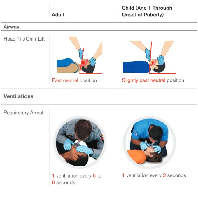
Giving ventilations is a technique to supply oxygen to a patient who is in respiratory arrest. Give 1 ventilation every 5 to 6 seconds for an adult patient, with each ventilation lasting about 1 second and making the chest rise. See pages 13–15 for more
information about how to give ventilations.
When giving ventilations, it is critical to avoid overventilation and hyperventilation of a patient by giving ventilations at a rate and volume greater than recommended; that is, more than 1 ventilation every 5 to 6 seconds or for longer than 1 second each.
Science Note
In addition to causing gastric distension and possible emesis,
hyperventilation leads to increased intrathoracic pressure and a subsequent
decrease in coronary filling and coronary perfusion pressures by putting
pressure on the vena cava. This most commonly occurs when patients are
being ventilated in respiratory arrest or when an advanced airway is placed
during cardiac arrest.
Once you begin giving ventilations, you must continue until:
The patient begins to breathe on his or her own.
Another trained rescuer takes over.
The patient has no pulse, in which case you should begin CPR or use an AED if one is available and ready to use.
The scene becomes unsafe.
Cardiac arrest
If there is no breathing, no pulse and the patient is unresponsive, the patient is in cardiac arrest. Cardiac arrest is a life-threatening situation in which the electrical and/or mechanical system of the heart malfunctions resulting in complete cessation of the heart’s ability to function and circulate blood efficiently.
Remember: Cardiac arrest is different from myocardial infarction; however, a myocardial infarction can lead to cardiac arrest. See Myocardial Infarction on the next page for more information.
The key to the patient’s survival is ensuring the Cardiac Chain of Survival. Following the links in the Cardiac Chain of Survival gives a patient in cardiac arrest the greatest chance of survival. See Cardiac Chain of Survival on the next page for more information.
Myocardial Infarction
A myocardial infarction (MI) or heart attack refers to the necrosis (death) of heart tissue as a result of a loss of oxygenated blood. The sooner the signs and symptoms are recognized and treated, the lower the risk of morbidity and mortality. Even patients who have had a myocardial infarction may not recognize the signs because each myocardial infarction may present differently.
Signs and Symptoms of MI
Chest discomfort or pain that is severe, lasts longer than 3 to 5 minutes, goes away and comes back, or persists even during rest
Discomfort, pressure or pain that is persistent and ranges from discomfort to an unbearable crushing sensation in the chest, possibly spreading to the shoulder, arm, neck, jaw, stomach or back, and usually not relieved by resting, changing position or taking medication
Pain that comes and goes (such as angina pectoris)
Difficulty breathing, such as at a faster rate than normal or noisy breathing
Pale or ashen skin, especially around the face
Sweating, especially on the face
Dizziness or light-headedness
Possible loss of consciousness
Nausea or vomiting
Although women may experience the most common signs and symptoms, such as chest pain or discomfort, they may also experience common atypical warning signs, such as:
Shortness of breath.
Nausea or vomiting.
Stomach, back or jaw pain.
Unexplained fatigue or malaise.
These warning signs may occur with or without chest pain. When women do
Basic Life Support for Healthcare Providers Handbook 11
Cardiac Chain of Survival
Adult Cardiac Chain of Survival
The Cardiac Chain of Survival for adults consists of fi ve links:
Recognition of cardiac arrest and activation of the emergency response system
Early CPR to keep oxygen-rich blood fl owing and to help delay brain damage and death
Early defi brillation with an automated external defi brillator (AED) to help restore an effective heart rhythm and signifi cantly increase the patient’s chance for survival
Advanced life support using advanced medical personnel who can provide the proper tools and medication needed to continue the lifesaving care
Integrated post-cardiac arrest care to optimize ventilation and oxygenation and treat hypertension immediately after the return of spontaneous circulation
Pediatric Cardiac Chain of Survival
The pediatric Cardiac Chain of Survival is similar to the adult Cardiac Chain of Survival. The fi ve links include the following:
Prevention of arrest
Early, high-quality CPR
Rapid activation of the EMS system or response team to get help on the way quickly—no matter the patient’s age
Effective, advanced life support
Integrated post-cardiac arrest care
When you determine that a patient is in cardiac arrest (unresponsive, no normal breathing and no defi nitive pulse), you need to begin cardiopulmonary resuscitation (CPR) that starts with the immediate delivery of chest compressions followed by ventilations.
Providing CPR/AED
for Adults
Basic Life Support for Healthcare Providers Handbook 13
Compressions
One component of CPR is chest compressions. To ensure optimal patient outcomes, high-quality CPR must be performed. You can ensure high-quality CPR by providing high-quality chest compressions, making sure that the:
Patient is on a firm, flat surface to allow for adequate compression. In a
non-healthcare setting this would typically be on the floor or ground, while in a healthcare setting this may be on a
stretcher or bed with a CPR board or CPR feature applied.
The chest is exposed to ensure proper hand placement and the ability to visualize chest recoil.
Hands are correctly positioned with the heel of one hand in the center of the chest on the lower half of sternum with the other hand on top. Most rescuers find that interlacing their fingers makes it easier to provide compressions while keeping the fingers off the chest.
Arms are as straight as possible, with the shoulders directly over the hands to promote effective compressions. Locking elbows will help maintain straight arms.
Compressions are given at the correct rate of at least 100 per minute to a maximum of 120 per minute, and at the proper depth of at least 2 inches for an adult to promote adequate circulation.
The chest must be allowed to fully recoil between each compression to allow blood to flow back into the heart following the compression.
For adult patients, CPR consists of 30 chest compressions followed by 2 ventilations.
Ventilations
Ventilations supply oxygen to a patient who is not breathing. They may be given via several methods including:
Mouth-to-mouth.
Pocket mask.
Bag-valve-mask (BVM) resuscitator.
During adult CPR, you give 2 ventilations that last approximately 1 second each and make the chest rise.
Mouth-to-Mouth Ventilations
If a pocket mask or BVM are not available, you may need to provide mouth-to-mouth ventilations:
Open the airway past a neutral position using the head-tilt/chin-lift technique.
Pinch the nose shut and make a complete seal over the patient’s mouth with your mouth.
Give ventilations by blowing into the patient’s mouth. Ventilations should be given one at a time. Take a break between breaths by breaking the seal slightly between ventilations and then taking a breath before re-sealing over the mouth.
When giving ventilations, if the chest does not
rise after the
first breath, reopen the airway, make a seal and try a second breath.
If the breath is not successful, move directly back to compressions and
check the airway for an obstruction before attempting subsequent
ventilations. If an obstruction is found, remove it and attempt ventilations.
However,
NEVER perform a blind finger sweep
.
With mouth-to-mouth ventilations, the patient
receives a
concentration of oxygen at approximately 16 percent compared to
the oxygen concentration of ambient air at approximately 20 percent.
Giving individual ventilations can help maintain this oxygen concentration
level. However, if you do not break the seal and take a breath between
ventilations, the second ventilation may contain an oxygen concentration
of 0 percent with a high concentration of carbon dioxide (CO
2)
.If you are otherwise unable to make a complete seal over a patient’s mouth, you may need to use mouth-to-nose ventilations:
With the head tilted back, close the mouth by pushing on the chin.
Seal your mouth around the patient’s nose and breathe into the nose.
If possible, open the patient’s mouth between ventilations to allow air to escape.
Pocket Mask Ventilations
Basic Life Support for Healthcare Providers Handbook 15
To use a pocket mask:
Assemble the mask and valve.
Open the airway past the neutral position using the head-tilt/chin-lift technique from the patient’s side when alone.
Place the mask over the mouth and nose of the patient starting from the bridge of the nose, then place the bottom of the mask below the mouth to the chin (the mask should not extend past the chin).
Seal the mask by placing the “webbing” between your index finger and thumb on the top of the mask above the valve while placing your remaining fingers on the side of the patient’s face. With your other hand (the hand closest to the patient’s chest), place your thumb along the base of the mask while placing your bent index finger under the patient’s chin, lifting the face into the mask.
When using a pocket mask, make sure to use one that matches the size of the patient; for example, use an adult pocket mask for an adult patient, but an infant pocket mask for an infant. Also, ensure that you position and seal the mask properly before blowing into the mask.
Bag-Valve-Mask Resuscitator
A bag-valve-mask (BVM) resuscitator is a handheld device used to ventilate patients and administer higher concentrations of oxygen than a pocket mask. While often used by a single rescuer, evidence shows that two rescuers are needed to effectively operate a BVM. One rescuer opens and maintains the airway and ensures the BVM mask seal, while the second rescuer delivers ventilations by squeezing the bag slowly with both hands at the correct intervals to the point of creating chest rise.
To use a BVM:
Assemble the BVM as needed.
Open the airway past neutral position while positioned at the top of the patient’s head (cephalic position).
Use an E-C hand position (first rescuer):
- Place both hands around the mask, forming an E with the last three fingers on each hand and a C with the thumb and index finger around both sides of the mask.
- Seal the mask completely around the patient’s mouth and nose by lifting the jaw into the mask while maintaining an open airway.
Provide ventilations (second rescuer):
- Depress the bag about halfway to deliver between 400 to 700 milliliters of volume to make the chest rise.
- Give smooth and effortless ventilations that last about 1 second.
BVMs can hold greater than 1000 milliliters
of volume and
should never be completely deflated when providing ventilations.
Doing so could lead to overventilation and hyperventilation. Also, pay close
attention to any increasing difficulty when providing bag-valve-mask
ventilation. This difficulty may indicate an increase in intrathoracic pressure,
inadequate airway opening or other complications. Be sure to share this
information with the team for corrective actions.
Special Considerations: Advanced Airways
When a patient has an advanced airway such as a supraglottic airway device or an endotracheal tube, CPR must be performed a little differently. At a minimum, two rescuers must be present. One rescuer gives 1 ventilation every 6 to 8 seconds, which is about 8 to 10 ventilations per minute. At the same time, the second rescuer continues giving compressions at a rate of 100 to 120 compressions per minute. There is no pause between compressions or ventilations and rescuers do not use the 30 compressions to 2 ventilations ratio. This process is a continuous cycle of compressions and ventilations with no interruption.
As in any resuscitation situation, it is essential not to hyperventilate the patient. That is because, during cardiac arrest, the body’s metabolic demand for oxygen is decreased. With each ventilation, intrathoracic pressure increases which causes a decrease in atrial/ ventricular filling and a reduction in coronary perfusion pressures. Hyperventilation further increases the intrathoracic pressure, which in turn further decreases atrial/ventricular filling and reduces coronary perfusion pressures.
It is common during resuscitation to
accidently hyperventilate a
patient due to the emotional response of caring for a patient in
cardiac arrest. You should be constantly aware of the ventilations being
provided to the patient and supply any corrective feedback as needed.
Stopping CPR
Once started, continue CPR with 30 compressions followed by 2 ventilations (1 cycle = 30:2) until:
You see signs of return of spontaneous circulation (ROSC) such as patient movement or breathing. See Recovery Positions on the next page for more information.
An AED is ready to analyze the patient’s heart rhythm.
Basic Life Support for Healthcare Providers Handbook 17
You are presented with a valid do not resuscitate (DNR) order.
You are alone and too exhausted to continue.
The scene becomes unsafe.
Recovery Positions
While not generally used in a healthcare setting, it is important to understand how and when to use a recovery position, especially when you are alone with a patient. In most cases while you are with the patient, you would leave an unconscious patient who is breathing and has no head, neck or spinal injury in a supine (face-up) position and maintain the airway. You could also use the recovery or side-lying position. The modified H.A.IN.E.S. recovery position
is used for situations in which the patient is suspected of having a head, neck or spinal injury; the rescuer is alone and must leave the patient; or the rescuer is unable to maintain an open and clear airway because of fluid or vomit. To place a patient in the
modified H.A.IN.E.S. recovery position, do the following:
Kneel at the side of the patient and roll the patient toward the rescuer.
Place the top leg on the other with both knees in a bent position.
Align the arm on top with the upper body. If the patient is an infant, follow these steps:
Carefully position the infant face-down along the forearm.
Support the infant’s head and neck with your other hand while keeping the infant’s mouth and nose clear.
Keep the head and neck slightly lower than the chest.
Automated External Defibrillators
Automated external defibrillators (AEDs) are portable electronic devices that automatically analyze the patient’s heart rhythm and can provide defibrillation, an electrical shock that may help the heart re-establish a perfusing rhythm.
When a patient experiences a cardiac arrest, an AED should be applied as soon as one is readily available. AEDs deliver defibrillation(s) to patients in cardiac
arrest with two specific dysrhythmias: ventricular fibrillation (V-fib) and ventricular tachycardia (V-tach). By using an AED early, the patient’s chances of survival are greatly increased.
Science Note
For each minute CPR and defibrillation are delayed, a patient’s
chance for survival is reduced by 7 to 10 percent.
If CPR is in progress, continue CPR until the AED is turned on, the AED pads are applied and the AED is ready to analyze the heart rhythm. If you are alone and an AED is available, you should use it once you have determined the patient is in cardiac arrest.
Using an AED
For an AED to be effective, you MUST use it properly by doing the following:
Turn it on first.
Make sure the patient’s chest is clearly exposed and dry.
- Remove any medication patches with a gloved hand. - If necessary, remove or cut any
undergarments that may be in the way. The pads need to be adhered to the skin for the shock to be delivered to the heart.
Apply the appropriate-sized pads for the patient’s age in the proper location on the bare chest.
- Use adult pads for adults and children over the age of 8 years or over 55 pounds.
- Place one pad on the upper right chest
below the right clavicle to the right of the sternum; place the other pad on the left side of the chest on the mid-axillary line a few inches below the left armpit.
Plug in the connector, and push the analyze button, if necessary. (Most AEDs available today have their pads pre-connected and will automatically analyze once the pads are applied to the chest. Make sure you understand how the AED within your organization operates.)
Tell everyone to “clear” while the AED is analyzing to ensure accurate analysis. Ensure no one is touching the patient during the analysis or shock.
When “clear” is announced, have the rescuer performing the compressions stop compressions and hover a few inches above the chest, but remain in position to resume compressions immediately after a shock is delivered or the AED advises that a shock is not indicated.
Observe the AED analysis and prepare for a shock to be delivered if advised.
- Ensure that everyone is clear of the patient before the shock is delivered.
- Remember that the AED delivers an electrical current that could injure anyone in contact with the patient.
Basic Life Support for Healthcare Providers Handbook 19
Deliver the shock by pressing the shock button, if indicated.
After the shock is delivered, immediately start compressions and perform about 2 minutes of CPR (about 5 cycles of 30:2) until the AED prompts that it is
reanalyzing, the patient shows signs of return of spontaneous circulation (ROSC), or you are instructed by the team leader or more advanced personnel to stop.
Do not wait for the AED to prompt to begin CPR after a shock or no shock advised message.
Science Note
Some AEDs allow for compressions post-analysis while charging.
Rescuers may perform compressions from the time the shock advised
prompt is noted through the time that the prompt to clear occurs, just
prior to depressing the shock button. Be sure to follow the manufacturer’s
recommendations and your local protocols and practices.
AED Safety
In some situations, such as when you are around water or the patient is on a metal surface, you may question whether or not it is safe to use an AED. The answer is yes. AEDs are very safe and built for almost any environment.
As long as the ill or injured patient is not actually in water, you can use an AED near water and in light rain or snow. Light rain, mist or snow does not generally pose a concern for AED operation. However, take steps to make sure that the patient is as dry as possible, is sheltered from the rain, is not lying in a pool or puddle of water and his or her chest is completely dry before attaching the pads. Also make sure that you and other rescuers are not in contact with water when operating the AED. Moreover, avoid getting the AED or AED pads wet if possible. Do not delay defibrillation when taking steps to create a dry environment. The same is true for metal surfaces. Just make sure that the pads are not touching the metal surface.
It is also safe to use AEDs on patients who have pacemakers, other implantable cardioverter defibrillators or metal body piercings. To maintain safety, avoid placing the AED pads directly over these items. Position the pads so that they are at least an inch away, just to be safe.
Some patients may be wearing a medication patch. Medication patches on the chest can create a hazard or interfere with analysis and defibrillation when AED pads are applied on top of them. If this is the case, act swiftly and remove the patch with a gloved hand and wipe away any of the remaining medication from the skin. Then, make sure the chest is dry and apply the pads.
For an AED to work properly, it is important that the pads are attached securely to the patient’s chest. However, some patients have excessive chest hair that may cause problems with AED pad-to-skin contact. If the chest hair is excessive (typically on the right upper chest), quickly shave the right upper chest area before applying the AED pads. See Do’s and Don’ts for AED Use for more information.
Do’s and Don’ts for AED Use
Follow these general precautions when using an AED.
Do’s
Before shocking a patient with an AED, do make sure that no one is touching or is in contact with the patient or any resuscitation equipment.
Do use an AED if a patient is experiencing cardiac arrest as a result of traumatic injuries. Follow local protocols or practice.
Do use an AED for a patient who is pregnant. Defibrillation shocks transfer no significant electrical current to the fetus. The mother’s survival is paramount to the infant’s survival. Follow local protocols and medical direction.
Don’ts
Do not use alcohol to wipe the patient’s chest dry. Alcohol is flammable.
Do not touch the patient while the AED is analyzing. Touching or moving the patient may affect analysis.
Do not touch the patient while the device is
defibrillating. You or someone else could be shocked.
Do not defibrillate someone when around flammable or combustible materials, such as gasoline or free-flowing oxygen.
For AEDs to perform properly and safely, they must be maintained as with any medical device. AEDs require minimal maintenance, but rescuers should be familiar with the various visual and audible prompts to warn of malfunctions or a low battery. To maintain the AED:
Know the manufacturer’s recommendations for maintenance, because many manufacturers require that they be contacted for service.
Periodically check equipment.
Have a fully charged backup battery, when available, that is properly sealed and unexpired, and also have correct AED pads available.
Basic Life Support for Healthcare Providers Handbook 21
One-Rescuer and Two-Rescuer
CPR—Adult
When performing CPR on an adult, certain components are the same regardless of the number of rescuers present. These are highlighted in Table 1-1.
Table 1-1
One- and Two-Rescuer Adult CPR
One-Rescuer CPR Two-Rescuer CPR
Hand Position Hands centered on lower half of sternum
Hands centered on lower half of sternum
Rate At least 100 but no more than 120 per minute
At least 100 but no more than 120 per minute
Depth At least 2 inches At least 2 inches
Compressions: Ventilations
30:2 30:2
One-Rescuer CPR
When performing one-rescuer CPR on an adult patient, the lone rescuer is responsible for conducting the scene size-up and the primary assessment and performing all the steps of CPR including the use of the AED, if available. CPR can be exhausting, and attempts should be made to find additional resources as early as possible during the scene size-up.
Two-Rescuer CPR
When two rescuers are available, Rescuer 1, considered the team leader, performs the scene size-up and primary assessment, and begins the process of providing CPR, starting with chest compressions. Meanwhile, Rescuer 2 calls for additional resources and gets/prepares the AED, if available. Rescuer 1 continues to provide high-quality CPR with 30 compressions to 2 ventilations until Rescuer 2 is ready to assist and/or the AED is ready to analyze.
When the AED is ready to analyze, Rescuer 1 should move to the patient’s head, and Rescuer 2 should prepare to provide chest compressions and get into the hovering position. The rescuers will continue the cycle of chest compressions and ventilations, switching positions about every 2 minutes, when the AED prompts to analyze or when
the rescuer performing compressions begins to fatigue. Rescuers call for a position change by using an agreed-upon term at the end of the last compression cycle. The rescuer providing compressions should count out loud and raise the volume of his or her voice as he or she nears the end of each cycle (… 21 … 22 … 23 … 24 … 25 … 26 … 27 … 28 … 29 … 30). The rescuer at the chest will move to give ventilations while the rescuer at the head will move to the chest to provide compressions.
In a healthcare setting, often there will be more than 2 rescuers. It is the responsibility of the team leader to orchestrate movements between rescuers to ensure no one rescuer becomes fatigued and that all critical areas are addressed: compressions, ventilations and AED. For example, additional rescuers may be assimilated into roles of compressor or ventilator, allowing the team leader to monitor performance and ensure that high-quality CPR is maintained. Additionally, if a BVM is available, ideally it is prepared by a third rescuer positioned at the top of the head and used upon completion of a cycle of chest compressions, with the first rescuer squeezing the bag while the third rescuer maintains an open airway and seals the mask.
High-Performance CPR
High-performance CPR refers to providing high-quality chest compressions as part of a well-organized team response to a cardiac arrest. Coordinated, efficient, effective teamwork is essential to minimize the time spent not in contact with the chest to improve patient outcomes.
Think about all of the activities performed during a resuscitation. For example:
AED pads are applied.
AED must charge.
Mask or BVM may need to be repositioned.
Airway may need to be reopened.
Other personnel arrive on scene.
Rescuers switch positions.
Advanced airway may need to be inserted.
Pulse checks may be done, but unnecessarily.
All of these activities could affect your ability to maintain contact with the patient’s chest.
Science Note
Basic Life Support for Healthcare Providers Handbook 23
Chest Compression Fraction
Chest compression fraction, or CCF, is the term used to denote the time that chest compressions are performed. It represents the fraction of time spent performing compressions, that is, the time that the rescuers are in contact with the patient’s chest, divided by the total time of the resuscitation, beginning with the arrival on scene until the return of spontaneous circulation or ROSC. Expert consensus identifies a CCF of at least 80 percent to promote optimal outcomes.
To achieve the best CCF percentage, a coordinated team approach is needed, with each member assuming pre-assigned roles, anticipating the next action steps for yourself and other team members. This coordinated team approach also includes integrating and assimilating additional personnel, such as paramedics or a code team, who arrive on scene.
To further your understanding of high-performance CPR, consider the example of an automotive racing team. Each crew member has a specific role when the race car arrives in the pit area. They are supervised by a leader, who keeps the crew on task and gets the race car back on the track. The quality, efficiency and swiftness of the crew’s actions can ultimately affect the outcome of how the race car performs. The same is true for the CPR pit crew. All crew members have specific roles during a resuscitation. Based on available resources, potential roles include the following:
Team leader
Compressor
Rescuer managing the airway
Rescuer providing ventilations
Rescuer managing the AED
Recorder
Keep in mind that there are no national protocols in place for high-performance CPR. How you function within a team setting, including how additional personnel assimilate into the team, may vary depending on your local protocols or practice.
Integration of More Advanced Personnel
During resuscitation, numerous people may be involved in providing care to the patient. Rescuers must work together as a team in a coordinated effort to achieve the best outcomes for the patient. Characteristics of effective teamwork include well-defined roles and responsibilities; clear, closed-loop communication; and respectful treatment of others.
Coordination becomes even more important when more advanced personnel such as an advanced life support team or code team arrives on the scene. This coordination of all involved is necessary to:
Ensure that all individuals involved work as a team to help promote the best outcome for the patient.
Promote effective perfusion to the vital organs.
Minimize interruptions of chest compressions, which have been shown to improve survival.
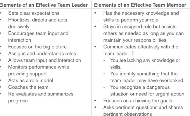
Ultimately, it is the team leader who is responsible for this coordination. When more advanced personnel arrive on scene, it is the team leader who communicates with advanced personnel, providing them with a report of the patient’s status and events. The team leader also sets clear expectations, prioritizes, directs, acts decisively, encourages team input and interaction and focuses on the big picture.
Crew Resource Management
During resuscitation, crew resource management helps to promote effective and efficient teamwork. Crew resource management is a communication process that centers around the team leader, who coordinates the actions and activities of team members so that the team functions effectively and efficiently. For example, when new individuals arrive on the scene or when team members switch roles
during an emergency, it is the team leader who is responsible for coordinating these activities.
During resuscitation, the team leader directs and coordinates all the working elements, including team members, activities and actions, as well as equipment, to focus on providing high-quality CPR, the goal of any resuscitation effort.
Crew resource management also guides team
members to directly and effectively communicate to a team leader about dangerous or time-critical decisions. It was developed as a result of several airline disasters as a way to prevent future incidents. Crew resource management has been shown to help avoid medical errors in healthcare.
Basic Life Support for Healthcare Providers Handbook 25
Providing CPR/AED
for Children and
Infants
W
hile the differences in care for infants and children may appear
subtle, it is important to understand them in order to achieve the best
possible outcomes.
Pediatric Considerations
Children are not small adults. Therefore, they need to be cared for differently in an emergency including using equipment such as a pocket mask or BVM designed specifi cally for the size and age of the child.
Age
So how is a child defi ned as it relates to providing care? See When Is a Child a Child? for more information.
When Is a Child a Child?
In most instances, determining whether to treat a child as a child or as an adult has been based on age. Typically, an adult is defi ned as someone about the age of 12 (adolescent) or older; someone between the ages of 1 and 12 has been considered to be a child for CPR care; and an infant is someone younger than 1 year of age. However,
for the purposes of this course, a child is defi ned as the age of 1 to the onset of puberty as evidenced by breast development in girls and underarm hair development in boys. An infant is considered under the age of 1 year.
Consent
Another factor to consider when caring for children and infants is consent. Legally, adults who are awake and alert can consent to treatment; if they are not alert, consent is implied. However, for most infants and children up to the age of 17 years, you must obtain consent from the child’s parent or legal guardian if they are present regardless of the child’s level of consciousness.
Basic Life Support for Healthcare Providers Handbook 27
Additional Resources
While it is rare in the professional setting to be alone with a child or infant, there is a slight change of when you should call for additional resources when you are alone. After determining that an adult is unresponsive and you are alone, you should immediately call for additional resources and get an AED. With children, it is more important to provide about 2 minutes of CPR before leaving them to call for help or get an AED unless the arrest is witnessed and believed to be cardiac in origin.
Science Note
Most child-related cardiac arrests occur as a result of a hypoxic
event such as an exacerbation of asthma, an airway obstruction or a
drowning. As such, ventilations and appropriate oxygenation are important
for a successful resuscitation. In these situations, laryngeal spasm may
occur, making passive ventilation during chest compressions minimal or
nonexistent. Therefore, it is critical to correct the oxygenation problem by
providing high-quality CPR prior to leaving the child or infant.
Note:
Based on local protocols or practice, it is permissible to provide
two ventilations prior to initiating CPR after the primary assessment if
a hypoxic event is suspected.
CPR/AED Differences Between
Children and Adults
When performing CPR on a child, there are some subtle differences in technique. These differences include opening the airway, compression depth, the ratio of compressions to ventilations depending on the number of rescuers, and AED pads and pad placement.
Airway
To open the airway of a child, you would use the same head-tilt/chin-lift technique as an adult. However, you would only tilt the head slightly past a neutral position, avoiding any hyperextension or flexion in the neck. Table 1-2 illustrates airway and ventilation differences for an adult and child.
Table 1-2
Airway and Ventilation Differences: Adult and Child
Adult
Child (Age 1 Through Onset of Puberty)
Airway
Head-Tilt/Chin-Lift
Past neutral position Slightly past neutral position
Ventilations
Respiratory Arrest
1 ventilation every 5 to
6 seconds
Basic Life Support for Healthcare Providers Handbook 29
Compressions
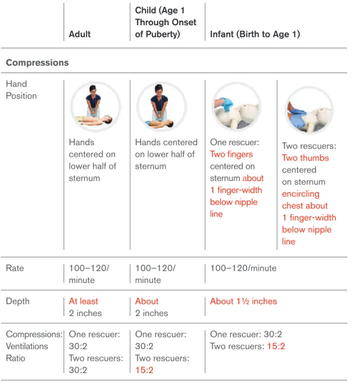
The positioning and manner of providing compressions to a child are also very similar to an adult. Place your hands in the center of the chest on the lower half of the sternum and compress at a rate between 100 to 120 per minute. However, the depth of compression is different. For a child, compress the chest only ABOUT 2 inches, instead of at least 2 inches as you would for an adult.
Compressions-to-Ventilations Ratio
When you are the only rescuer, the ratio of compressions to ventilations for a child is the same as for an adult, that is, 30 compressions to 2 ventilations (30:2). However, in two-rescuer situations, this ratio changes to 15 compressions to 2 ventilations (15:2).
AEDs
AEDs work the same way regardless of the patient’s age, but there are differences in the pads used for children as well as the pad placement based on the size of the child. For children over the age of 8 years and weighing more than 55 pounds, you would continue to use adult AED pads, placing them in the same location as for an adult—one pad to the right of the sternum and below the right clavicle, with the other pad on the left side of the chest on the mid-axillary line a few inches below the left armpit. However, for children 8 years of age or younger or weighing less than 55 pounds, use pediatric AED pads if available. Be aware that some AEDs use a switch or key instead of changing pads, so follow the directions from the AED manufacturer on how to care for pediatric patients with their device.
At no time should the AED pads touch each other when applied. If it appears that the AED pads would touch each other based on the size of the child’s chest, use an anterior and posterior pad placement as an alternative. Apply one pad to the center of the child’s chest on the sternum and one pad to the child’s back between the scapulae. Table 1-3 summarizes the differences for CPR and AED for adults and children.
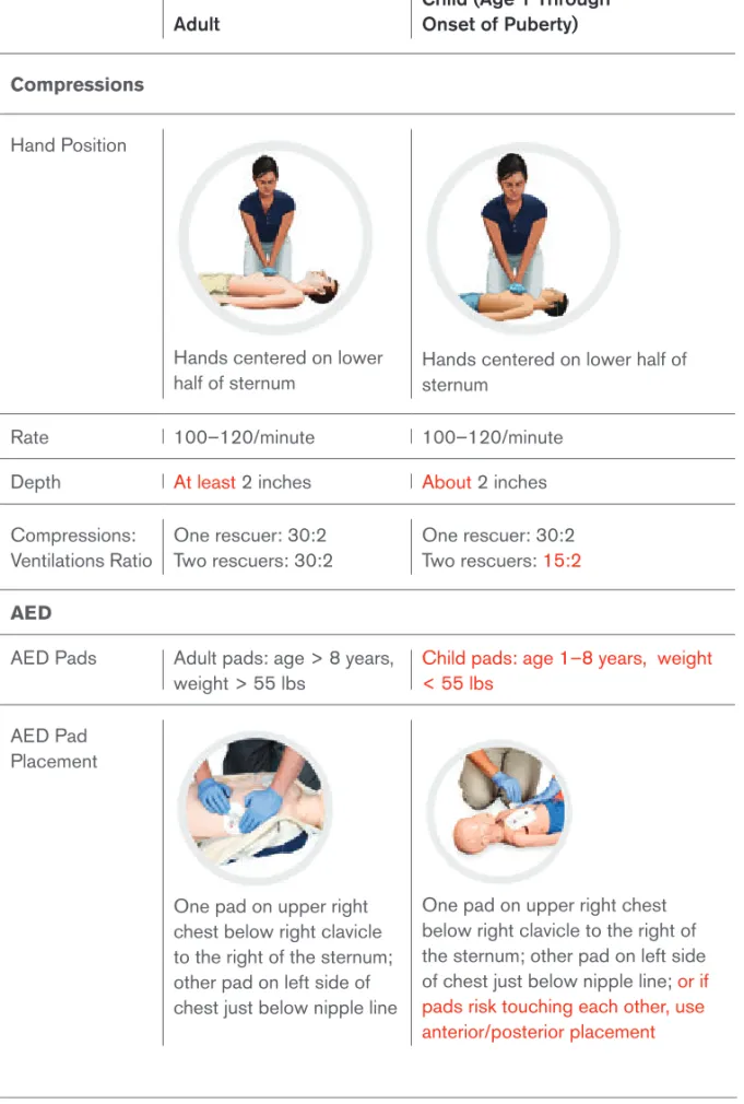
Table 1-3
CPR/AED Differences: Adult and Child
Adult
Child (Age 1 Through Onset of Puberty)
Compressions
Hand Position
Hands centered on lower
half of sternum Hands centered on lower half of sternum
Rate 100–120/minute 100–120/minute
Depth At least 2 inches About2 inches Compressions:
Ventilations Ratio
One rescuer: 30:2 Two rescuers: 30:2
One rescuer: 30:2 Two rescuers: 15:2
AED
AED Pads Adult pads: age > 8 years, weight > 55 lbs
Child pads: age 1–8 years, weight < 55 lbs
AED Pad Placement
One pad on upper right chest below right clavicle to the right of the sternum; other pad on left side of chest just below nipple line