How Ligands Illuminate GPCR Molecular Pharmacology
Daniel Wacker,1Raymond C. Stevens,2and Bryan L. Roth1,*
1Department of Pharmacology and Division of Chemical Biology and Medicinal Chemistry, University of North Carolina at Chapel Hill School
of Medicine, Chapel Hill, NC 27514, USA
2Departments of Biological Sciences and Chemistry, Bridge Institute, University of Southern California, Los Angeles, CA 90089, USA
*Correspondence:[email protected]
http://dx.doi.org/10.1016/j.cell.2017.07.009
G protein-coupled receptors (GPCRs), which are modulated by a variety of endogenous and
syn-thetic ligands, represent the largest family of druggable targets in the human genome. Recent
structural and molecular studies have both transformed and expanded classical concepts of
receptor pharmacology and have begun to illuminate the distinct mechanisms by which
structur-ally, chemicstructur-ally, and functionally diverse ligands modulate GPCR function. These molecular
insights into ligand engagement and action have enabled new computational methods and
accelerated the discovery of novel ligands and tool compounds, especially for understudied
and orphan GPCRs. These advances promise to streamline the development of GPCR-targeted
medications.
at GPCRs are transforming molecular pharmacology and drug discovery.
Importance of GPCRs for Physiology, Disease, and Therapeutics
As of 2017, between 20%–30% of FDA-approved medications target GPCRs (Rask-Andersen et al., 2011). The popularity of GPCRs as drug targets is predominantly due to their physiolog-ical relevance, as GPCRs are expressed in most of the body’s tissues, are involved in cellular communication, and participate in virtually all aspects of human physiology via GPCR-mediated signal transduction, as well as their druggability, as GPCRs possess binding pockets with beneficial physiochemical proper-ties that lend to the design of drug-like small molecules (Mason et al., 2012). Particularly prominent therapeutic applications involving GPCRs (see Figure 2A) include opioid analgesics (mopioid receptor; OPRM1 receptor agonists), antihistamines (HRH1-histamine antagonists), anticholinergics (CHRM antago-nists), typical and atypical antipsychotics (D2 dopamine receptor antagonists; DRD2), antimigraine drugs (5-HT1D serotonergic agonists; HTR1D),b2-agonists for asthma (ADBAR2), and anti-hypertensives (targetinga1adrenergic and angiotensin II recep-tors; ADAR1, ATGR1).
In addition to being therapeutic targets for drug discovery, GPCR variants are occasionally implicated in disease pro-cesses. Indeed, dozens of monogenic diseases have been linked to constitutively activating mutations (CAMs), which augment GPCR constitutive activity. These include such condi-tions as congenital stationary night blindness caused by rhodopsin CAMs (McAlear et al., 2010), uveal melanoma by CAMs of the cysteinyl leukotriene receptor 2 (Moore et al., 2016), and many others (Smit et al., 2007). Additionally, loss-of-function GPCR mutations are occasionally linked to human disease. V2-vasopressin receptor mutations, for example, are associated with nephrogenic diabetes insipidus (Pan et al., Introduction
G protein-coupled receptors (GPCRs) are seven-transmem-brane integral membrane proteins that typically translate extracellularstimulationintointracellularsignals.GPCR activa-tion is usually mediated by agonist binding, which stabilizes receptor conformations that recruit and ultimately activate intracellular transducers. GPCR agonist ligands are physically and chemically diverse and can include: photons; ions (H+, Zn2+, Ca2+, etc.); odorants; tastants; vitamins (e.g., niacin, vitaminA1aldehyde, etc.); peptidicand non-peptidergic hor-mones (estrogen, angiotensin, etc.); proteins (e.g., chemo-kines); neurotransmitters (dopamine, serotonin, etc.); natural products (morphine, salvinorin A, etc.); a large number of intermediary metabolites (ATP, ADP, fatty acids, bile acids, etc.); and products from human commensal bacteria (see AllenandRoth[2011]and RothandKroeze[2015]for reviews). Intracellularly, GPCR activation is translated into various signals mediated via heterotrimeric G proteins, arrestins ( Lut-trelletal.,1999),kinases(Benovicetal.,1989),ionchannels, and various scaffolding proteins (Brownetal.,2003) (Figure1). In this context, arrestins function to abrogate G protein-mediated signaling (Lohse et al., 1990), as scaffolds for GPCR internalization via clathrin-coated vesicles (Goodman et al., 1996), and as scaffolds for signaling (Luttrell et al., 1999) (Figure1).
1992), and drugs which rescue the misfolded phenotype of V2-vasopressin receptor mutants via action as pharmacological chaperones have been proposed as therapeutics (Morello et al., 2000).
GPCRs as Deleterious Off-Targets in Therapeutic Drug Discovery
GPCRs are also frequent medication ‘‘off-targets’’ that display unpredicted interactions, which can result in unanticipated therapeutic (Roth et al., 2004) or life-threatening side effects (Rothman et al., 2000). The most infamous example is likely the anti-obesity drug fenfluramine, which was withdrawn because of association with valvular heart disease in many individuals (Allen and Roth, 2011; Roth, 2007). Years after fenfluramine’s widely publicized withdrawal from the world-wide market and legal damages totaling more than $10 billion, it was discovered that the compound’s metabolite—norfenfluramine—activated cardiac 5-HT2B serotonin receptors, leading eventually to valvular heart disease (Rothman et al., 2000; Roth, 2007). Since then, several more drugs have been withdrawn due to similar 5-HT2B-mediated valvular heart disease complications (Roth, 2007; Allen and Roth, 2011).
As GPCRs represent frequent off-targets for drugs that target kinases and other non-GPCR molecular targets, compounds are typically profiled against large numbers of cloned GPCRs prior to clinical trials in humans (Allen and Roth, 2011). Importantly, the potent actions of sorafinib and many other approved and inves-tigational kinase inhibitors on serotonergic, purinergic, and other GPCRs have been discovered via GPCR profiling (Elkins et al., 2016). Identification of potentially important off-target actions of drugs through GPCRs is also facilitated by large databases of drug-target information, including ChEMBL (https://www. ebi.ac.uk/chembl/), PubChem (https://pubchem.ncbi.nlm.nih. gov/), PHAROS (https://pharos.nih.gov/idg/index), and the Ki Database (https://kidbdev.med.unc.edu/databases/kidb.php). These cheminformatic datasets also have been useful for the
in silico prediction and in vitro and in vivo confirmation of GPCRs as relevant and important drug off-targets (Keiser et al., 2009).
Understudied and Orphan GPCRs and Their Therapeutic Potential
Although there are more than 350 GPCR-targeted FDA approved drugs, they target only a small sector of the universe of poten-tially druggable GPCRs (Roth and Kroeze, 2015; Rask-Andersen et al., 2011) (Figure 2A). Approximately 100 human GPCRs are currently active targets for late-stage preclinical development, and a total of nearly 400 small molecules are being actively inves-tigated as therapeutics (Lafferty-Whyte et al., 2017). Current drug development, though, is geared mainly toward those GPCRs with extensive validation as potential therapeutic targets (Lafferty-Whyte et al., 2017). Conversely, only a few orphan or understudied GPCRs, ‘‘oGPCRs’’—as defined by (1) their comparatively low number of publications, (2) their low number of annotated small molecules interactors, or (3) absence of their known endogenous ligands (Roth and Kroeze, 2015)—are currently being investigated for therapeutic drug discovery. Significantly, the Adhesion, Tastant, and Frizzled families of re-ceptors are reported to have no annotated small drug-like mole-cules in clinical testing (Lafferty-Whyte et al., 2017).
The lack of drug development programs targeting oGPCRs stems mainly from risk aversion, as little is known regarding oGPCR’s physiological roles and druggability. Although knockout studies and replacement with chemogenetic mutant GPCRs, such as DREADDs (designer receptors exclusively acti-vated by designer drugs) (Roth, 2016), or chimeric opsins for optogenetic studies, such as OptoXRs (Airan et al., 2009), repre-sent strategies to identify the basic physiological roles of oGPCRs, the biggest bottleneck remains the lack of chemical tool compounds to reliably characterize receptor function in vitro and in vivo. Yet, oGPCRs have emerged as important re-ceptors for natural products and synthetic drugs; drugs targeting oGPCRs have been used as tools to illuminate fundamental
Figure 1. Different Ligand-Stabilized GPCR Conformations Cause Binding and Activa-tion of Distinct Signal Transducers, in-cluding G Proteins and Arrestins
Left: crystal structure ofb2AR (light blue cartoon) coupled to Gas (blue), Gb(orange), Gg (green) heterotrimer (PDB: 3SN6 [Rasmussen et al.,
2011b]) illustrates G protein-mediated signaling.
Figure 2. GPCRome-wide Targets of Approved and Marketed Medications and How Ligands Uncover Unknown GPCR Phys-iology toward Potential Therapeutic Applica-tions
(A) Sphere size corresponds to number of approved drugs for highlighted therapeutic GPCR target with antagonists, agonists, and negative allosteric mod-ulators shown in red, green, and blue, respectively. Phylogenetic tree of the GPCRome highlights the small fraction of GPCRs that are currently targeted by approved medications.
biological processes and therapeutic approaches mediated by oGPCRs (Figure 2B). For instance, the naturally occurring teratogen cyclopamine (Cooper et al., 1998) facilitated identifica-tion of the smoothened receptor (SMO) as a hedgehog signaling pathway modulator and target for cancer chemotherapy (Rudin et al., 2009). Similarly, the discovery that the hallucinogen salvi-norin A from the sageSalvia divinorumis a selectivek-opioid receptor agonist validated this receptor as a target for psychoto-mimetic compounds (Roth et al., 2002). Several benzodiaze-pines were found to also activate GPR68, suggesting that some of the side effects of these anti-anxiety medications could be mediated by this receptor (Huang et al., 2015b). Additionally, the discovery of amphetamine actions at the TAAR1 trace amine receptor (Bunzow et al., 2001) identified TAAR1 as a potential target for neuropsychiatric diseases. The endogenous TAAR1 agonists known as thyronamines (Scanlan et al., 2004) revealed trace amine receptors as potential mediators of metabolic, ther-mogenic, and neurologic processes. Clearly, expanding our understanding of GPCR on- and off-target pharmacology is important for both successful drug discovery, as well as for illu-minating basic biological and chemical processes in health and disease. As exemplified by oGPCRs, however, we are still far from a comprehensive molecular and physiological understand-ing of GPCR biology.
Next, we review how recent molecular insights from crystal structures have transformed classical receptor pharmacology and facilitated our understanding of the mechanisms by which li-gands modulate GPCR function. We further highlight the utility of ligands in identifying and characterizing the physiological roles of poorly understood GPCRs, and we provide an overview of cur-rent advances to computationally leverage molecular insight toward identifying novel GPCR ligands.
Toward a Structure-Based Understanding of GPCR-Ligand Pharmacology
Key Pharmacological Concepts
Historically, the concepts of agonism and antagonism arose from the observations of drug actions on isolated organs. One example is highlighted by the ordered and regular agonist activ-ity of acetylcholine, which is antagonized by atropine (Clark, 1926). Observations like these led to the initial concepts that agonists either induce or stabilize an ‘‘active’’ state of a ‘‘recep-tor,’’ while antagonists have no effect on their own but block agonist access to this receptor. Once GPCRs were cloned and expressed in vitro, it was observed that GPCRs also possessed variable degrees of basal or constitutive activity and could popu-late active signaling states in the absence of ligands. With a few notable exceptions (e.g., adhesion-, thrombin-, and some viral receptors, like the KHSV-related receptor), GPCRs typically require exogenous agonists to stabilize fully active states for maximal signaling. Endogenous agonists such as neurotransmit-ters or hormones usually, but not invariably, maximally activate their cognate GPCRs and are considered full agonists (see Box 1); agonists which do not induce 100% activation are defined as partial agonists. It is important to note that partial agonists can appear as full agonists when receptor reserve is present and that a full response of the system can be elicited, even when not all receptors are occupied. Antagonists, on the
other hand, are compounds (either naturally occurring or syn-thetic) that block agonists activity; antagonists are classified as inverse agonists (antagonists that decrease constitutive activity) or neutral antagonists (antagonists that inhibit agonist effects but do not interfere with constitutive activity;Box 1, for further details and seeRoth [2016]).
Agonists, partial agonists, and antagonists interact with the so-called orthosteric site, which represents the binding site through which endogenous agonists activate the GPCR. Some GPCRs, most notably the adhesion- (Hamann et al., 2015) and protease-activated receptors (Coughlin, 2000), lack classical endogenous agonists, although following proteolysis, an N-ter-minal fragment occupies the orthosteric site.
Additionally, GPCRs may also be modulated allosterically by molecules that bind at a site that is distinct from the orthosteric site. Generally, allosteric modulators are classified as negative allosteric modulators or positive allosteric modulators (NAMs and PAMs, respectively) (Christopoulos et al., 2014) (Box 1). Allo-steric modulators do not directly interact with the orthoAllo-steric site but modulate the function of orthosteric ligands in a negative (NAM) or positive (PAM) way. Molecules that interact with both the orthosteric and allosteric sites are defined as bitopic ligands and may be either agonists or antagonists. Allosteric modulators can be endogenous, as in the case of the nearly universal allo-steric modulators sodium (which is a NAM [Fenalti et al., 2014]) and cholesterol (which could function as a NAM or PAM [Katritch et al., 2013]), or exogenous natural products or synthetic com-pounds (Kenakin and Boselli, 1989).
GPCR ligands also often display functional selectivity or biased signaling (Box 1; [Urban et al., 2007]), a process by which ligands will direct or bias the signaling toward one pathway or another (Figure 1). Related to this, although GPCRs were initially classified based on the presumed main G protein with which they interact (e.g., Gs-, Gi-, Gq-, and G12/13-coupled [Simon et al., 1991]), the schema was abandoned due to observations that GPCRs can couple to multiple G proteins (Asano et al., 1984). Many theoretical models have arisen to explain the agonist, allosteric, and antagonist actions and biased signaling, including the highly useful, albeit phenomenological, operational model (Box 1) of Black and Leff (1983). These simplified models continue to be useful for quantifying and predicting drug actions at GPCRs (Kenakin et al., 2012). With the discovery that GPCRs require G proteins for activation, more detailed models have arisen, including the so-called ternary (De Lean et al., 1980) and extended ternary complex models (Samama et al., 1993), as well as other models incorporating arrestin signaling (Roth, 2016) (Figure 3). The simple ternary and extended ternary com-plex models are called ‘‘ternary’’ because they have three mem-bers: receptor (R), ligand (L), and heterotrimeric G protein (G). Extended versions incorporating other effectors, like arrestins (Figure 3), are becoming validated by more mechanistic ap-proaches which incorporate structure-based insights into ligand pharmacology.
states can spontaneously interact with G protein or arrestin transducers to yield signaling complexes in the absence of agonist (R*E with E = G protein, Arrestin, or other transducers) (Figure 3). Indeed, this constitutive activity has been amply docu-mented in both recombinant (Burns et al., 1997) and endogenous (Arvanitakis et al., 1997) expression systems. These models also predict that antagonists do not simply ‘‘antagonize’’ GPCRs, but that they stabilize inactive states (R0L) and thereby inhibit consti-tutive activity by acting as inverse agonists. The models further predict that so-called neutral antagonists are likely to be rare, as any ligand will bias the ensemble of spontaneously arising conformations at least to some extent. The predictions that antagonists are actually inverse agonists have been extensively validated in recombinant systems in vitro (Chidiac et al., 1994) and in vivo (Dillon et al., 2011). These models also predict that the active states stabilized by agonist (R*L) might differ confor-mationally from ternary signaling complexes (R*GL; R*AL). These predictions have been validated by biochemical studies demon-strating that G protein binding allosterically enhances agonist binding affinity (Cerione et al., 1984).
The Impact of Molecular Insights into GPCR-Ligand Interactions Illuminate Both Empirically Based and Classical Concepts of Receptor Pharmacology
Historically, most of the initial functional concepts describing binding interactions were based on phenomenological observa-tions from ligand-binding and signaling studies done in recombi-nant or endogenous systems. It wasn’t until technological advances enabled the study of ligand-receptor interactions by X-ray crystallography, NMR, and other biophysical assays that high-resolution insights enabled the molecular characterization of these distinct states (Figure 3). Initial biochemical studies sug-gested that GPCR activation involves helical movements ( Far-rens et al., 1996); since then, crystallographic and now cryoelec-tron microscopy (cryo-EM) structures are greatly enhancing our understanding of the molecular characteristics that define distinct GPCR conformations and signaling states. Most GPCRs have been crystallized in apparently inactive conformations (R0L), bound to antagonists or agonists (Tesmer, 2016). These inactive-state structures highlight distinct features of inactive GPCRs, such as binding of the endogenous NAM sodium (Fenalti et al., 2014; Liu et al., 2012b) and the closed ‘‘ionic Box 1. Key Pharmacologic Concepts
CONSTITUTIVE ACTIVITY
Receptor-mediated signaling in the absence of ligand due to sponta-neous population of active receptor states
FULL AGONISTS
Ligands that elicit maximum signal at the interrogated pathway (endog-enous ligands are, per definition full agonists),
PARTIAL AGONISTS
Ligands that elicit activity below maximum level
INVERSE AGONISTS
Ligands that inhibit constitutive receptor activity
NEUTRAL ANTAGONISTS
Ligands that bind the receptor but do not affect constitutive receptor activity
RECEPTOR RESERVE
Receptors not coupled to the system resulting in maximum signal by activation of only a fraction of total receptors
ORTHOSTERIC SITE
Binding pocket accommodating endogenous receptor ligand
ALLOSTERIC SITE
Pocket distinct from the orthosteric site that can modulate ligand bind-ing and receptor activity
PAMS AND NAMS
PAMs increase and NAMs decrease a receptor’s activity in response to an orthosteric ligand, while binding at a site distinct from the orthos-teric site
Box 1.Continued
BITOPIC LIGANDS
Ligands that possess both orthosteric and allosteric moieties
FUNCTIONAL SELECTIVITY/BIASED SIGNALING
Ability of ligands to impart different degrees of activation in distinct pathways downstream of the receptor
OPERATIONAL MODEL
lock’’ (Staus et al., 2016). Intermediate active states in the absence of bound effector (R*L) have been described for the 5-HT1Bserotonin (Wang et al., 2013), the A2Aadenosine (A2AAR) (Allen et al., 2011), and neurotensin (White et al., 2012) receptors. Structures of the 5-HT2Bserotonin receptor bound to the agonists ergotamine (Wacker et al., 2013) and lysergic acid diethylamide (LSD) (Wacker et al., 2017) have identified features of an arrestin-biased intermediate state (R*L). They particularly include ‘‘active-like’’ conformations of selected ‘‘trigger’’ motifs, includ-ing the ‘‘PIF’’ and ‘‘NpxxY’’ motifs (Figure 3; seeVenkatakrishnan et al. [2013]andWacker et al. [2017]for details), which are struc-tural elements found in many GPCRs that are critical for receptor activation. Active-state receptor structures stabilized by nano-bodies have been particularly helpful in characterizing structural hallmarks of GPCR activation (Huang et al., 2015a; Kruse et al., 2013; Rasmussen et al., 2011a). Crystal structures have also
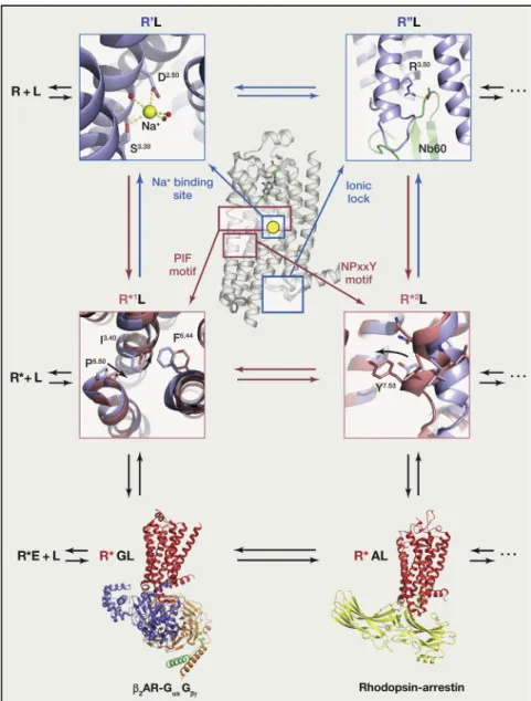
Figure 3. Molecular and Structural Pharma-cology Extend the Ternary Complex Model for Quantitative Description of Drug Action at GPCRs
Several ligand-bound inactive receptor states (R0L, R00L, etc.) and ligand-bound active receptor states (R*1
L, R*2
L, etc.), as well as ternary complex structures of ligand- and effector-bound active receptor states (R*GL, R*AL) (PDB: 3SN6 [
Ras-mussen et al., 2011b]; PDB: 4ZWJ [Kang et al.,
2015]), have been structurally characterized by X-ray crystallography. Distinct conformational characteristics such as the sodium-binding site of the A2AAR (PDB: 4EIY [Liu et al., 2012b]), the ionic lock of a nanobody stabilizedb2AR (PDB: 5JQH [Staus et al., 2016]), the PIF motif of the 5-HT2B receptor (PDB: 4IB4 [Wacker et al., 2013], PDB: 3NY8 [Wacker et al., 2010]), and the NPxxY motif of a nanobody-bound b2AR (PDB: 3NY8 [Wacker et al., 2010], PDB: 3P0G [Rasmussen et al., 2011a]) highlight diverse GPCR activation states.
been determined for ternary signaling complexes, such as that of b2AR with agonist and hetereotrimeric G protein (R*GL) (Rasmussen et al., 2011b) and rhodopsin with arrestin (R*AL) (Kang et al., 2015) (Figure 3). Recent cryo-EM structures of R*GL states (Liang et al., 2017; Zhang et al., 2017) have confirmed some of the key features of theb2AR-G protein complex. Finally, although not associated with a high-resolution struc-ture, a recent report indicates that under some circumstances, a ‘‘megaplex’’ of GPCR, heterotrimeric G protein, and ar-restin may exist as a functioning signaling entity (Thomsen et al., 2016), mediating endosomally based GPCR signaling. Thus, several conformational ensembles predicted by these models have finally been revealed structurally and greatly improve our molecular understanding of GPCR activation and regulation.
the extracellular domains of the mGluRs (Kunishima et al., 2000), the adenosine-bound A2AAR (Lebon et al., 2011), the neu-rotensin-bound NT-1 neurotensin receptor (White et al., 2012), the endothelin-bound endothelin ETBreceptor (Shihoya et al., 2016), and adrenaline-bound b2AR (Ring et al., 2013). Since rhodopsin is an exception, as it is covalently bound to its endog-enous ligand, the first structural evidence for a more general orthosteric binding site shared among many class A GPCRs came from the structures of A2AAR and b2AR bound to their diffusible endogenous ligands (Lebon et al., 2011; Ring et al., 2013). For peptidergic GPCRs—based on structures of NT-1 neurotensin receptor in complex with neurotensin and the d-opioid receptor in complex with a modified peptide agonist— the orthosteric site likely overlaps with the shared class A orthos-teric site and extends to include extensive contacts with the extracellular loops. Recent cryo-EM studies showed that endog-enous peptides of several class B receptors occupy a similar
Figure 4. Crystal Structures of Different GPCR-Ligand Complexes Highlight the Diverse Locations of Ligand-Binding Sites
Ligands are shown as stick models with transparent surfaces, and receptors are shown in cartoon representation in light blue. Complexes show retinal-bound RHO, LY2119620-bound CHRM2, sodium-bound ADORA2A, ergotamine-bound HTR2B, CCR2-RA-[R]-bound CCR2, MK-0893-bound GCGR, CP-376395-bound CRHR1, and BPTU-CP-376395-bound P2RY1.
evidence for GPCR-G protein precoupling (RG; [Noblesetal., 2005]).Fromthepreviouslydiscussedobservations,itisclear that multiple intermediate conformational states exist for GPCRs (aloneandincomplexwiththeireffectors);elucidatingthefull complementoftheseeffectorswillbeimportantforfutureefforts to design drugs to stabilize unique conformations and signaling intermediates.
MultipleModesofLigandRecognition:Orthosteric, Bitopic,andAllostericGPCR-LigandInteractions
orthosteric site as in class A receptors but appear to prefer an even larger and more extended binding pocket (Liang et al., 2017; Zhang et al., 2017).
Frequently, the orthosteric binding site of class A GPCRs re-sides in the middle of the seven-transmembrane helical bundle, located between the extracellular loops and the middle plane of the membrane. Orthosteric sites exhibit different shapes and chemostatic makeup depending on the nature of ligand. For instance, more lipophilic ligands, such as cannabinoids, likely enter the receptor from the membrane, and the hydrophobic binding site is thus covered with residues from the extracellular loops to provide a barrier to the hydrophilic extracellular space (Hua et al., 2016). Conversely, peptide ligands—particularly for class B receptors—are often large and possess considerable flexibility (O’Connor et al., 2015) and require an orthosteric site open to the extracellular space (Liang et al., 2017; Zhang et al., 2017). Interestingly, while the nature of orthosteric sites is critical to accommodate the ligands’ chemostatic and steric properties, entry to the binding site through a network of receptor-specific residues can present the largest energetic barrier to compound binding (Dror et al., 2011). This finding is particularly important for the design of receptor selective ligands and the control of re-ceptor binding and dissociation rates.
Several studies have also provided critical structural insights into ligand-binding sites that are structurally distinct from the or-thosteric pocket as observed in class A GPCRs (Figure 4). These include the M2 muscarinic receptor bound to a PAM located in a vestibule above the orthosteric agonist binding site (Kruse et al., 2013) and the corticotropin-releasing factor 1 receptor bound to a presumed allosteric antagonist situated deep within the cyto-plasmic portion of the helical bundle (Hollenstein et al., 2013). Allosteric ligands have also been found bound to the putative intracellular binding site of the C-terminal tail of the Gasubunit in the chemokine receptors CCR9 (Oswald et al., 2016), and ex-tra-helical binding sites in the glucagon (Jazayeri et al., 2016) and purinergic P2Y1 receptor (Zhang et al., 2015) (Figure 4). Lastly, general allosteric modulators of GPCR function include choles-terol, which binds to different extra-helical binding sites in different receptors (Gimpl, 2016), and sodium, which is bound in a highly conserved pocket in the center of the helical bundle below the orthosteric site (Katritch et al., 2014) (Figure 4).
Surprisingly, and despite decades of work exploring GPCR allosteric modulation (Kenakin and Boselli, 1989; Lanzafame et al., 1997), currently approved drugs mainly target orthosteric sites (Figure 2A). Only a handful of FDA-approved GPCR allo-steric modulators exist: cinacalcet, a NAM for the Calcium-sensing receptor (CASR); maraviroc, a CCR5 chemokine recep-tor NAM (CCR5); and the Smoothened receprecep-tor (SMO) NAMs sonedigib and vismodegib. Despite these limited numbers, targeting GPCRs allosterically remains a promising approach for therapeutic drug development (Changeux and Christopoulos, 2016), as allosteric modulators are often more selective for their targets, and allosterically modulating the actions of endog-enous ligands provides substantial therapeutic benefits (Aitken et al., 2009).
One important reason for the lack of allosteric therapeutics could be that it is challenging to design compounds with suffi-cient efficacy, as for most GPCRs, only the orthosteric pocket
has evolved to govern receptor modulation. It is important to note that, while endogenous ions, lipids, adaptor proteins, and effectors modulate GPCR function (van der Westhuizen et al., 2015), their interacting surfaces rarely possess the physiochem-ical properties necessary for the structure-informed design of synthetic modulators. Other reasons for the lack of available allo-steric modulators may include the lack of tool compounds, as well as difficulties associated with developing suitable assays to test for allosteric modulation. The increasing abundance of crystal structures in combination with computational methods should greatly facilitate the identification and characterization of potential allosteric sites and patches, which in turn could greatly accelerate the targeted design of allosteric modulators. Biased Signaling and Kinetics as Drivers of
Ligand-Encoded Activities
GPCR signaling has both contextual and kinetic aspects, and these can be exploited—ultimately, from structural ap-proaches—to fine tune signaling for basic science and therapeu-tic applications. As classically illustrated for b2AR, G protein signaling generally occurs rapidly within a second or so of agonist administration, while it typically lasts only a few minutes. Signal termination occurs via desensitization through receptor phosphorylation, arrestin binding, and internalization (Lefkowitz and Shenoy, 2005). By contrast, arrestin binding and G pro-tein-independent arrestinergic signaling events, such as MAP ki-nase activation, typically occur on the minute to hour timescale (Lefkowitz and Shenoy, 2005). The delineation of these two signaling pathways has led to a major reconceptualization of how GPCRs mediate their actions in both normal physiology and disease and has ushered in a new era of GPCR drug discov-ery to identify agonists biased for either G protein (White et al., 2015) or arrestin (Allen et al., 2011) signaling with the promise of improved therapeutic properties and reduced side effects (DeWire and Violin, 2011). It should be noted, however, that iden-tification and characterization of GPCR effectors remains an active area of investigation (Paek et al., 2017) and that there are likely many more unidentified intracellular proteins that interact with signaling complexes and modulate GPCR signaling (Paek et al., 2017).
From a mechanistic perspective, it remains largely unclear how receptors mediate biased signaling, although recent struc-tures and biophysical studies have begun to clarify this issue. For instance, the structure of the arrestin-biased drug ergotamine bound 5-HT2Bserotonin receptor (Wacker et al., 2013) revealed how ergotamine stabilizes a distinct receptor conformation in which motifs that are essential for arrestin-biased signaling (e.g., NPxxY) are activated, while others associated with G pro-tein signaling (e.g., DRY or PIF) remain in the inactive state. NMR (Liu et al., 2012a) and fluorescent spectroscopy studies (Rahmeh et al., 2012) highlight how ligands selectively engage distinct mo-tifs to stabilize GPCR conformations that are more conducive to accommodating one effector over the other.
that yielded nanomolar potency inverse agonists (Kolb et al., 2009). One of the resulting compounds, when crystallized, revealed a novel b2AR ligand-binding conformation (Wacker et al., 2010).
Given this and other successes in structure-guided discovery of new GPCR ligands, one might ask whether it might be possible to use molecular models in lieu of experimentally deter-mined structures as templates for docking in other receptors. An early example of this approach with the D3 dopamine receptor (Carlsson et al., 2011) featured 3.3 million compounds docked initially against a homology model of the D3 dopamine receptor that usedb2AR as a template. Several nanomolar potency antag-onists were discovered. Docking was also performed using the D3 receptor X-ray structure as a template (Chien et al., 2010) and, surprisingly, a distinct set of nanomolar potency antago-nists was identified. What emerged from this exercise was the observation that the slightly different conformations sampled by the crystal structure and the models of this receptor yielded chemically distinct sets of active molecules.
An exciting extension of this overall approach—and one which may prove to provide a template going forward—is the serial structure-guided and docking-based optimization of active mol-ecules into potential therapeutic entities. In one instance, three million commercially available compounds from the ZINC data-base (Irwin and Shoichet, 2005) were docked against an inactive conformation of themopioid receptor (Manglik et al., 2016), the molecular receptor for morphine. Initial lead compounds from the docking campaign were experimentally tested, and commer-cially available analogs were purchased to obtain ligands of higher affinity. Active compounds were then optimized through modest medicinal chemistry, guided by their docking pose, structural considerations, and experimental pharmacology. This process ultimately yielded PZM-21, a selective, high-affinity mopioid agonist with modest G protein bias that was ultimately found to be analgesic with fewer side effects compared to morphine (Manglik et al., 2016).
Structure-Inspired Discovery of Chemical Probes for oGPCRs
That homology models can be used to identify novel chemical matter using virtual ligand screening has proven to be particu-larly useful for oGPCRs, for which conventional ligand screening campaigns have not yielded useful probes. For instance, selec-tive allosteric modulators (both NAMs and PAMs) were identified for the oGPCRs GPR68 and GPR65 and further optimized by a combination of physical screening and in silico docking (Huang et al., 2015b). One GPR68 PAM, ogerin, was demonstrated to have on-target activity in vivo by modulating conditioned fear responses in mice. A similar approach has recently been used to identify small-molecule ligands for the oGPCRs GPR171 (Wardman et al., 2016) and MRGPRX2 (Lansu et al., 2017), and these compounds were subsequently used to further characterize the receptor’s role in feeding behavior and itch, respectively.
The successful use of homology models, however, largely de-pends on the accuracy of the models. The analysis of docking results is a process that considers previous biochemical and pharmacological information regarding ligand-receptor interac-tions as validated from crystal structures. Accordingly, the kinetics modulate patterns of biased signaling (KleinHerenbrink
etal.,2016).Takentogether,thesefindingshighlightacrucial kinetic component to functional selectivity at the level of com-poundassociation,stabilizationofdistinctreceptor conforma-tions, and intracellular signal progression, which, together, appear to dramatically influence cellular responses.
Thedevelopmentandidentificationofdrugswithdifferential patterns of biased signaling represent major areas of investiga-tion,asthesechemicaltoolshelptodelineatethedownstream signaling network of GPCRs and are particularly useful to explore thephysiologicalroleofoGPCRs.Thusforinstance,the halluci-nogen LSD (Wackeretal.,2017) appears to display bias toward b-arrestin signaling (Box1, Figure1), while the synthetic opioids TRV-130(DeWireetal.,2013)andPZM-21(Mangliketal.,2016) are biased toward G protein signaling (Box1, Figure1). Allosteric modulators have also manifested biased potentiation in in-stances in which a single GPCR may interact with multiple G pro-teins,asinthecaseoftheproton-sensingGPCRGPR68(Huang etal.,2015b). Due to the differential expression of key signal transducers, signal transduction—and thus, signaling bias— willvarybetweencelltypes(Ursetal.,2016).Exploitingsignaling bias for drug discovery promises to yield much-improved com-pounds that specifically target therapeutic pathways while avoiding pathologic events downstream of the same receptor (Allenetal.,2011;ViolinandLefkowitz,2007).
ExploitingGPCR-LigandStructuresforDrugDiscovery With the increasing number of GPCR structures, we can antici-pate the generation of new chemical tools for the study of GPCRs by providing platforms for the structure-guided design ofimprovedtherapeutics.SinceGPCRsareroutinelycrystallized in complex with ligands, structure-guided drug design is a prom-isingalternativetoclassicalmethodsofliganddiscovery,which employ cycles of medicinal chemistry-based modification of ex-istingscaffolds.However,althoughtruestructure-guideddrug discoveryandoptimizationhavebeenroutinelydoneforprotein kinase inhibitors for more than a decade (Nobleetal.,2004), the successfulexamplesforGPCRsarerare.Forexample, struc-ture-based drug design for A2AAR yielded new 1,2,4-triazine de-rivatives that were shown cystallographically to adopt novel binding modes (Congreveetal.,2012). Similarly, new mGluR5 metabotropicglutamatereceptorNAMswereinitiallydiscovered via a combination of fragment-based screening and medicinal chemistry,andtheirbindingmodessubsequentlywereidentified crystallographically (Christopheretal.,2015). It is important to note that, while this kind of structure-guided drug discovery andoptimizationisrelativelyroutineforotherdrugtargets,where several leading compounds are serially crystallized with the mo-lecular target and derivatives synthesized based on insights gained from the structures, routine crystallization of GPCRs re-mainshighlychallenging.
Structure-BasedVirtualDiscoveryofNovelChemotypes forGPCRs
description of key residues accessible for ligand binding in class A GPCRs (Gloriam et al., 2009) or the definition of so-called pro-tein-ligand interaction fingerprints (reviewed inVass et al. [2016]) not only greatly enhances the identification of relevant mole-cules, but also highlights the importance of using tool com-pounds to study GPCRs interactions and function.
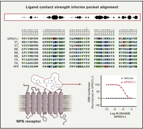
An alternative approach, dubbed ‘‘pickpocketing,’’ analyzes ligand-residue contact strength to identify potential ligand-bind-ing similarity between non-homologous receptors. This method allowed the authors of a recent paper to identify new chemo-types for the oGPCR GPR37L by ‘‘stealing’’ ligands from orexin and neuropeptide receptors that possess sufficient chemical matter and exhibit substantial contact-strength similar-ity with GPR37L (Ngo et al., 2017) (Figure 5). Taken together, these recent successes indicate that homology-model-based approaches can be powerful and useful approaches for discovering agonists, antagonists, and allosteric modulators of oGPCRs.
Conclusions and Future Directions
As is clear, the past 10 years have witnessed a renaissance in GPCR research catalyzed by structural insights into the most basic aspects of GPCR ligand binding and signaling. Concepts of ligand recognition previously considered to be theoretical— such as allosteric modulation (Kenakin and Boselli, 1989)—are now firmly validated from structural and functional perspectives. Additionally, we are beginning to get hints of how different ligands might stabilize distinct conformational ensembles, lead-ing to a variety of active, inactive, and biased states as was pre-dicted many years ago (Urban et al., 2007). Finally, the plethora
Figure 5. Computational Approaches Generate Novel Tool Compounds for GPCRs
As shown here, the ‘‘pickpocketing’’ approach identifies GPCRs with potentially related ligand-binding properties according to ligand contact strength informed pocket alignment. Similar ligand-binding properties between the Neuropep-tide S receptor (NPS) and the orphan GPCR GPR37L1 identified the NPS receptor ligand SHA68 as a novel ligand to interrogate GPR37L1 function. Data schematized according to Ngo et al. (2017).
of structures has provided computational biologists with tremendous opportunities for both structure-guided and -inspired drug discovery.
Although these advances are breath-taking when considered from a historical perspective, huge gaps remain in our un-derstanding of GPCR structure, function, signaling, and pharmacology. Clearly, our understanding of GPCR functional selectivity from a mechanistic, molecular perspective is inadequate and remains phenomenological. Thus, for instance, there are no structures of GPCRs with G proteins in complex with G-protein-biased agonists, nor are there structures of GPCRs with ar-restin-biased ligands in complex with arrestin.
Additionally, how biased ligands might stabilize distinct states is unknown, and approaches to discover and develop such li-gands is discovery based rather than mechanistically based. Consider, for instance, that although the structures of more than 40 GPCRs have been solved by X-ray crystallography, in most cases, this has been achieved with only one ligand and only in an inactive state. For some receptors, including b2AR and A2AAR, multiple ligand complexes are available, but only minimal plasticity within the binding pocket is evident. In our recent studies on 5-HT2B receptors, however, we observed extensive plasticity within the binding pocket of a GPCR when examining structures stabilized by similar compounds (Wacker et al., 2017). From the foregoing, we predict that depending upon the particular GPCR and ligands that are examined, a range of conformational rearrangements within the binding site will be observed. Further, it is clear that regions of GPCRs normally considered to be ‘‘undruggable’’ (e.g., intracellular loops, inter-faces, etc.) (Oswald et al., 2016) are sites of action for current and potential therapeutics. Thus, understanding and predicting how GPCR binding sites change in complex with specific ligands and how these alterations ultimately lead to differential signaling and physiological outcomes remain major grand challenges for structural biologists and molecular pharmacologists.
Benovic, J.L., DeBlasi, A., Stone, W.C., Caron, M.G., and Lefkowitz, R.J. (1989). Beta-adrenergic receptor kinase: primary structure delineates a
multi-gene family. Science246, 235–240.
Black, J.W., and Leff, P. (1983). Operational models of pharmacological ago-nism. Proc. R. Soc. Lond. B Biol. Sci.220, 141–162.
Brown, A.J., Goldsworthy, S.M., Barnes, A.A., Eilert, M.M., Tcheang, L., Dan-iels, D., Muir, A.I., Wigglesworth, M.J., Kinghorn, I., Fraser, N.J., et al. (2003). The Orphan G protein-coupled receptors GPR41 and GPR43 are activated
by propionate and other short chain carboxylic acids. J. Biol. Chem.278,
11312–11319.
Bunzow, J.R., Sonders, M.S., Arttamangkul, S., Harrison, L.M., Zhang, G., Quigley, D.I., Darland, T., Suchland, K.L., Pasumamula, S., Kennedy, J.L., et al. (2001). Amphetamine, 3,4-methylenedioxymethamphetamine, lysergic acid diethylamide, and metabolites of the catecholamine neurotransmitters
are agonists of a rat trace amine receptor. Mol. Pharmacol.60, 1181–1188.
Burns, C.M., Chu, H., Rueter, S.M., Hutchinson, L.K., Canton, H., Sanders-Bush, E., and Emeson, R.B. (1997). Regulation of serotonin-2C receptor
G-protein coupling by RNA editing. Nature387, 303–308.
Carlsson, J., Coleman, R.G., Setola, V., Irwin, J.J., Fan, H., Schlessinger, A., Sali, A., Roth, B.L., and Shoichet, B.K. (2011). Ligand discovery from a
dopa-mine D3 receptor homology model and crystal structure. Nat. Chem. Biol.7,
769–778.
Cerione, R.A., Codina, J., Benovic, J.L., Lefkowitz, R.J., Birnbaumer, L., and Caron, M.G. (1984). The mammalian beta 2-adrenergic receptor: reconstitu-tion of funcreconstitu-tional interacreconstitu-tions between pure receptor and pure stimulatory nucleotide binding protein of the adenylate cyclase system. Biochemistry
23, 4519–4525.
Changeux, J.P., and Christopoulos, A. (2016). Allosteric Modulation as a
Uni-fying Mechanism for Receptor Function and Regulation. Cell166, 1084–1102.
Chidiac, P., Hebert, T.E., Valiquette, M., Dennis, M., and Bouvier, M. (1994).
Inverse agonist activity of beta-adrenergic antagonists. Mol. Pharmacol.45,
490–499.
Chien, E.Y., Liu, W., Zhao, Q., Katritch, V., Han, G.W., Hanson, M.A., Shi, L., Newman, A.H., Javitch, J.A., Cherezov, V., and Stevens, R.C. (2010). Structure of the human dopamine D3 receptor in complex with a D2/D3 selective
antag-onist. Science330, 1091–1095.
Christopher, J.A., Aves, S.J., Bennett, K.A., Dore´, A.S., Errey, J.C., Jazayeri, A., Marshall, F.H., Okrasa, K., Serrano-Vega, M.J., Tehan, B.G., et al. (2015). Fragment and Structure-Based Drug Discovery for a Class C GPCR: Discovery of the mGlu5 Negative Allosteric Modulator HTL14242 (3-Chloro-5-[6-(5-fluo-ropyridin-2-yl)pyrimidin-4-yl]benzonitrile). J. Med. Chem.58, 6653–6664.
Christopoulos, A., Changeux, J.P., Catterall, W.A., Fabbro, D., Burris, T.P., Ci-dlowski, J.A., Olsen, R.W., Peters, J.A., Neubig, R.R., Pin, J.P., et al. (2014). International Union of Basic and Clinical Pharmacology. XC. multisite pharma-cology: recommendations for the nomenclature of receptor allosterism and allosteric ligands. Pharmacol. Rev.66, 918–947.
Clark, A.J. (1926). The antagonism of acetyl choline by atropine. J. Physiol.61, 547–556.
Congreve, M., Andrews, S.P., Dore´, A.S., Hollenstein, K., Hurrell, E., Lang-mead, C.J., Mason, J.S., Ng, I.W., Tehan, B., Zhukov, A., et al. (2012). Discov-ery of 1,2,4-triazine derivatives as adenosine A(2A) antagonists using structure
based drug design. J. Med. Chem.55, 1898–1903.
Cooper, M.K., Porter, J.A., Young, K.E., and Beachy, P.A. (1998).
Teratogen-mediated inhibition of target tissue response to Shh signaling. Science280,
1603–1607.
Coughlin, S.R. (2000). Thrombin signalling and protease-activated receptors.
Nature407, 258–264.
De Lean, A., Stadel, J.M., and Lefkowitz, R.J. (1980). A ternary complex model explains the agonist-specific binding properties of the adenylate
cyclase-coupled beta-adrenergic receptor. J. Biol. Chem.255, 7108–7117.
DeWire, S.M., and Violin, J.D. (2011). Biased ligands for better cardiovascular
drugs: dissecting G-protein-coupled receptor pharmacology. Circ. Res.109,
205–216. and how they might be useful as therapeutic targets. As was
recentlyemphasized,formorethanhalfofthedruggableGPCRs in the human genome, little useful information is available regardingtheirrolesinnormalphysiology,muchlesstheirutility as therapeutic targets (Roth and Kroeze, 2015). Additionally, many GPCRs remain ‘‘orphan’’ by having no bona fide endoge-nousagonistsidentified(RothandKroeze,2015).Whileclassical knockout studies and/or chemo- and optogenetic approaches mayprovidesomeinsightintooGPCRfunction,itistool com-pounds that are desperately required to delineate their physio-logicalroles, characterizetheirfunction ona molecularlevel, and perhaps thereby identify novel therapeutic targets. Applica-tion of technologies such as virtual ligand screening and a variety of computational approaches to build ligands de novo, both enabled by molecular GPCR studies, are already beginning to yield valuable chemical tools. With the implementation of machine learning technology to better predict compound activity inacomplexbiologicalsystem,thespeedandsuccessrateof computational screening is more than likely to drastically in-crease over the coming years. Based on an ever-increasing number of GPCR structures, computational approaches are thus poised to augment or even replace manual high-throughput drugscreening while accelerating thegeneration of newtool compounds to study GPCR function or develop drug design platforms. Ligands, synthetic or natural, are the single most powerful tool for elucidating GPCR mechanisms and physi-ology—datathatwillnotonlyhelptobetterunderstandthesingle largest class of membrane proteins, but likely also translate into bettertherapies.
ACKNOWLEDGMENTS
ThisreviewwassupportedbygrantsfromtheNationalInstituteofHealth (RO1MH112205; UO1MH104974;U19MH82441; and PO1DA035764) and theMichaelHookerDistinguishedProfessorshiptoBLR.
REFERENCES
Airan,R.D.,Thompson,K.R.,Fenno,L.E.,Bernstein,H.,andDeisseroth,K.
(2009).Temporallypreciseinvivocontrolofintracellularsignalling.Nature
458,1025–1029.
Aitken,M.,Berndt,E.R.,andCutler,D.M.(2009).Prescriptiondrugspending
trendsintheUnitedStates:lookingbeyondtheturningpoint.HealthAff.
(Mill-wood)28,w151–w160.
Alexander,S.P.,Davenport,A.P.,Kelly,E.,Marrion,N.,Peters,J.A.,Benson,
H.E.,Faccenda,E.,Pawson,A.J.,Sharman,J.L.,Southan,C.,andDavies,
J.A.;CGTPCollaborators(2015).TheConciseGuidetoPHARMACOLOGY
2015/16:Gprotein-coupledreceptors.Br.J.Pharmacol.172,5744–5869.
Allen,J.A.,andRoth,B.L.(2011).Strategiestodiscoverunexpectedtargetsfor
drugsactiveatGprotein-coupledreceptors.Annu.Rev.Pharmacol.Toxicol.
51,117–144.
Allen,J.A.,Yost,J.M.,Setola,V.,Chen,X.,Sassano,M.F.,Chen,M.,Peterson, S.,Yadav,P.N.,Huang,X.P.,Feng,B.,etal.(2011).Discoveryof
b-arrestin-biaseddopamineD2ligandsforprobingsignaltransductionpathways
essen-tialforantipsychoticefficacy.Proc.Natl.Acad.Sci.USA108,18488–18493.
Arvanitakis,L.,Geras-Raaka,E.,Varma,A.,Gershengorn,M.C.,and
Cesar-man,E.(1997).HumanherpesvirusKSHVencodesaconstitutivelyactive
G-protein-coupledreceptorlinkedtocellproliferation.Nature385,347–350.
Asano,T.,Katada,T.,Gilman,A.G.,andRoss,E.M.(1984).Activationofthe
DeWire, S.M., Yamashita, D.S., Rominger, D.H., Liu, G., Cowan, C.L., Graczyk, T.M., Chen, X.T., Pitis, P.M., Gotchev, D., Yuan, C., et al. (2013). A G
protein-biased ligand at them-opioid receptor is potently analgesic with reduced
gastrointestinal and respiratory dysfunction compared with morphine.
J. Pharmacol. Exp. Ther.344, 708–717.
Dillon, G.M., Lubbers, L.S., Ferguson, M.T., Lao, J.Z., Huang, R.R., Xiao, J.C., Fong, T.M., Hale, J.J., Rupprecht, K., Miao, S., et al. (2011). MK-7128, a novel CB1 receptor inverse agonist, improves scopolamine-induced learning and
memory deficits in mice. Behav. Pharmacol.22, 91–100.
Dror, R.O., Pan, A.C., Arlow, D.H., Borhani, D.W., Maragakis, P., Shan, Y., Xu, H., and Shaw, D.E. (2011). Pathway and mechanism of drug binding to
G-pro-tein-coupled receptors. Proc. Natl. Acad. Sci. USA108, 13118–13123.
Elkins, J.M., Fedele, V., Szklarz, M., Abdul Azeez, K.R., Salah, E., Mikolajczyk, J., Romanov, S., Sepetov, N., Huang, X.P., Roth, B.L., et al. (2016). Compre-hensive characterization of the Published Kinase Inhibitor Set. Nat. Biotechnol.
34, 95–103.
Farrens, D.L., Altenbach, C., Yang, K., Hubbell, W.L., and Khorana, H.G. (1996). Requirement of rigid-body motion of transmembrane helices for light
activation of rhodopsin. Science274, 768–770.
Fenalti, G., Giguere, P.M., Katritch, V., Huang, X.P., Thompson, A.A., Chere-zov, V., Roth, B.L., and Stevens, R.C. (2014). Molecular control ofd-opioid re-ceptor signalling. Nature506, 191–196.
Fredriksson, R., Lagerstro¨m, M.C., Lundin, L.G., and Schio¨th, H.B. (2003). The G-protein-coupled receptors in the human genome form five main families. Phylogenetic analysis, paralogon groups, and fingerprints. Mol. Pharmacol.
63, 1256–1272.
Gimpl, G. (2016). Interaction of G protein coupled receptors and cholesterol.
Chem. Phys. Lipids199, 61–73.
Gloriam, D.E., Foord, S.M., Blaney, F.E., and Garland, S.L. (2009). Definition of the G protein-coupled receptor transmembrane bundle binding pocket and
calculation of receptor similarities for drug design. J. Med. Chem. 52,
4429–4442.
Goodman, O.B., Jr., Krupnick, J.G., Santini, F., Gurevich, V.V., Penn, R.B., Gagnon, A.W., Keen, J.H., and Benovic, J.L. (1996). Beta-arrestin acts as a clathrin adaptor in endocytosis of the beta2-adrenergic receptor. Nature
383, 447–450.
Hamann, J., Aust, G., Arac¸, D., Engel, F.B., Formstone, C., Fredriksson, R., Hall, R.A., Harty, B.L., Kirchhoff, C., Knapp, B., et al. (2015). International Union of Basic and Clinical Pharmacology. XCIV. Adhesion G protein-coupled
recep-tors. Pharmacol. Rev.67, 338–367.
Hollenstein, K., Kean, J., Bortolato, A., Cheng, R.K., Dore´, A.S., Jazayeri, A., Cooke, R.M., Weir, M., and Marshall, F.H. (2013). Structure of class B GPCR corticotropin-releasing factor receptor 1. Nature499, 438–443.
Hua, T., Vemuri, K., Pu, M., Qu, L., Han, G.W., Wu, Y., Zhao, S., Shui, W., Li, S., Korde, A., Laprairie, R.B., Stahl, E.L., Ho, J.H., Zvonok, N., Zhou, H., Kufareva, I., Wu, B., Zhao, Q., Hanson, M.A., Bohn, L.M., Makriyannis, A., Stevens, R.C., and Liu, Z.J. (2016). Crystal Structure of the Human Cannabinoid Receptor
CB1. Cell167, 750–762 e714.
Huang, W., Manglik, A., Venkatakrishnan, A.J., Laeremans, T., Feinberg, E.N., Sanborn, A.L., Kato, H.E., Livingston, K.E., Thorsen, T.S., Kling, R.C., et al.
(2015a). Structural insights intom-opioid receptor activation. Nature524,
315–321.
Huang, X.P., Karpiak, J., Kroeze, W.K., Zhu, H., Chen, X., Moy, S.S., Saddoris, K.A., Nikolova, V.D., Farrell, M.S., Wang, S., et al. (2015b). Allosteric ligands for
the pharmacologically dark receptors GPR68 and GPR65. Nature 527,
477–483.
Irwin, J.J., and Shoichet, B.K. (2005). ZINC–a free database of commercially
available compounds for virtual screening. J. Chem. Inf. Model.45, 177–182.
Jazayeri, A., Dore´, A.S., Lamb, D., Krishnamurthy, H., Southall, S.M., Baig, A.H., Bortolato, A., Koglin, M., Robertson, N.J., Errey, J.C., et al. (2016).
Ex-tra-helical binding site of a glucagon receptor antagonist. Nature 533,
274–277.
Kang, Y., Zhou, X.E., Gao, X., He, Y., Liu, W., Ishchenko, A., Barty, A., White, T.A., Yefanov, O., Han, G.W., et al. (2015). Crystal structure of rhodopsin
bound to arrestin by femtosecond X-ray laser. Nature523, 561–567.
Katritch, V., Cherezov, V., and Stevens, R.C. (2013). Structure-function of the
G protein-coupled receptor superfamily. Annu. Rev. Pharmacol. Toxicol.53,
531–556.
Katritch, V., Fenalti, G., Abola, E.E., Roth, B.L., Cherezov, V., and Stevens, R.C. (2014). Allosteric sodium in class A GPCR signaling. Trends Biochem. Sci.39, 233–244.
Keiser, M.J., Setola, V., Irwin, J.J., Laggner, C., Abbas, A.I., Hufeisen, S.J., Jensen, N.H., Kuijer, M.B., Matos, R.C., Tran, T.B., et al. (2009). Predicting
new molecular targets for known drugs. Nature462, 175–181.
Kenakin, T., and Boselli, C. (1989). Pharmacologic discrimination between re-ceptor heterogeneity and allosteric interaction: resultant analysis of gallamine and pirenzepine antagonism of muscarinic responses in rat trachea. J.
Phar-macol. Exp. Ther.250, 944–952.
Kenakin, T., Watson, C., Muniz-Medina, V., Christopoulos, A., and Novick, S. (2012). A simple method for quantifying functional selectivity and agonist bias.
ACS Chem. Neurosci.3, 193–203.
Klein Herenbrink, C., Sykes, D.A., Donthamsetti, P., Canals, M., Coudrat, T., Shonberg, J., Scammells, P.J., Capuano, B., Sexton, P.M., Charlton, S.J., et al. (2016). The role of kinetic context in apparent biased agonism at GPCRs.
Nat. Commun.7, 10842.
Kolb, P., Rosenbaum, D.M., Irwin, J.J., Fung, J.J., Kobilka, B.K., and Shoichet, B.K. (2009). Structure-based discovery of beta2-adrenergic receptor ligands.
Proc. Natl. Acad. Sci. USA106, 6843–6848.
Kruse, A.C., Ring, A.M., Manglik, A., Hu, J., Hu, K., Eitel, K., Hu¨bner, H., Pardon, E., Valant, C., Sexton, P.M., et al. (2013). Activation and allosteric
modulation of a muscarinic acetylcholine receptor. Nature504, 101–106.
Kunishima, N., Shimada, Y., Tsuji, Y., Sato, T., Yamamoto, M., Kumasaka, T., Nakanishi, S., Jingami, H., and Morikawa, K. (2000). Structural basis of
gluta-mate recognition by a dimeric metabotropic glutagluta-mate receptor. Nature407,
971–977.
Lafferty-Whyte, K., Mormeneo, D., and Del Fresno Marimon, M. (2017). Trial watch: Opportunities and challenges of the 2016 target landscape. Nat. Rev.
Drug Discov.16, 10–11.
Lansu, K., Karpiak, J., Liu, J., Huang, X.P., McCorvy, J.D., Kroeze, W.K., Che, T., Nagase, H., Carroll, F.I., Jin, J., et al. (2017). In silico design of novel
probes for the atypical opioid receptor MRGPRX2. Nat. Chem. Biol.13,
529–536.
Lanzafame, A., Christopoulos, A., and Mitchelson, F. (1997). Three allosteric modulators act at a common site, distinct from that of competitive antagonists,
at muscarinic acetylcholine M2 receptors. J. Pharmacol. Exp. Ther. 282,
278–285.
Lebon, G., Warne, T., Edwards, P.C., Bennett, K., Langmead, C.J., Leslie, A.G., and Tate, C.G. (2011). Agonist-bound adenosine A2A receptor structures
reveal common features of GPCR activation. Nature474, 521–525.
Lefkowitz, R.J., and Shenoy, S.K. (2005). Transduction of receptor signals by
beta-arrestins. Science308, 512–517.
Liang, Y.L., Khoshouei, M., Radjainia, M., Zhang, Y., Glukhova, A., Tarrasch, J., Thal, D.M., Furness, S.G.B., Christopoulos, G., Coudrat, T., et al. (2017). Phase-plate cryo-EM structure of a class B GPCR-G-protein complex. Nature
546, 118–123.
Liu, J.J., Horst, R., Katritch, V., Stevens, R.C., and Wu¨thrich, K. (2012a). Biased
signaling pathways inb2-adrenergic receptor characterized by 19F-NMR.
Sci-ence335, 1106–1110.
Liu, W., Chun, E., Thompson, A.A., Chubukov, P., Xu, F., Katritch, V., Han, G.W., Roth, C.B., Heitman, L.H., IJzerman, A.P., et al. (2012b). Structural basis
for allosteric regulation of GPCRs by sodium ions. Science337, 232–236.
Lohse, M.J., Benovic, J.L., Codina, J., Caron, M.G., and Lefkowitz, R.J. (1990). beta-Arrestin: a protein that regulates beta-adrenergic receptor function.
structure of theb2 adrenergic receptor-Gs protein complex. Nature 477, 549–555.
Ring, A.M., Manglik, A., Kruse, A.C., Enos, M.D., Weis, W.I., Garcia, K.C., and Kobilka, B.K. (2013). Adrenaline-activated structure ofb2-adrenoceptor
stabi-lized by an engineered nanobody. Nature502, 575–579.
Roth, B.L. (2007). Drugs and valvular heart disease. N. Engl. J. Med.356, 6–9.
Roth, B.L. (2016). DREADDs for Neuroscientists. Neuron89, 683–694.
Roth, B.L., and Kroeze, W.K. (2015). Integrated Approaches for Genome-wide Interrogation of the Druggable Non-olfactory G Protein-coupled Receptor
Superfamily. J. Biol. Chem.290, 19471–19477.
Roth, B.L., Baner, K., Westkaemper, R., Siebert, D., Rice, K.C., Steinberg, S., Ernsberger, P., and Rothman, R.B. (2002). Salvinorin A: a potent naturally occurring nonnitrogenous kappa opioid selective agonist. Proc. Natl. Acad.
Sci. USA99, 11934–11939.
Roth, B.L., Sheffler, D.J., and Kroeze, W.K. (2004). Magic shotguns versus magic bullets: selectively non-selective drugs for mood disorders and
schizo-phrenia. Nat. Rev. Drug Discov.3, 353–359.
Rothman, R.B., Baumann, M.H., Savage, J.E., Rauser, L., McBride, A., Hu-feisen, S.J., and Roth, B.L. (2000). Evidence for possible involvement of 5-HT(2B) receptors in the cardiac valvulopathy associated with fenfluramine
and other serotonergic medications. Circulation102, 2836–2841.
Rudin, C.M., Hann, C.L., Laterra, J., Yauch, R.L., Callahan, C.A., Fu, L., Hol-comb, T., Stinson, J., Gould, S.E., Coleman, B., et al. (2009). Treatment of me-dulloblastoma with hedgehog pathway inhibitor GDC-0449. N. Engl. J. Med.
361, 1173–1178.
Samama, P., Cotecchia, S., Costa, T., and Lefkowitz, R.J. (1993). A mutation-induced activated state of the beta 2-adrenergic receptor. Extending the
ternary complex model. J. Biol. Chem.268, 4625–4636.
Scanlan, T.S., Suchland, K.L., Hart, M.E., Chiellini, G., Huang, Y., Kruzich, P.J., Frascarelli, S., Crossley, D.A., Bunzow, J.R., Ronca-Testoni, S., et al. (2004). 3-Iodothyronamine is an endogenous and rapid-acting derivative of thyroid
hormone. Nat. Med.10, 638–642.
Shihoya, W., Nishizawa, T., Okuta, A., Tani, K., Dohmae, N., Fujiyoshi, Y., Nur-eki, O., and Doi, T. (2016). Activation mechanism of endothelin ETB receptor by
endothelin-1. Nature537, 363–368.
Simon, M.I., Strathmann, M.P., and Gautam, N. (1991). Diversity of G proteins
in signal transduction. Science252, 802–808.
Smit, M.J., Vischer, H.F., Bakker, R.A., Jongejan, A., Timmerman, H., Pardo, L., and Leurs, R. (2007). Pharmacogenomic and structural analysis of consti-tutive g protein-coupled receptor activity. Annu. Rev. Pharmacol. Toxicol.
47, 53–87.
Staus, D.P., Strachan, R.T., Manglik, A., Pani, B., Kahsai, A.W., Kim, T.H., Wingler, L.M., Ahn, S., Chatterjee, A., Masoudi, A., et al. (2016). Allosteric nanobodies reveal the dynamic range and diverse mechanisms of
G-pro-tein-coupled receptor activation. Nature535, 448–452.
Tesmer, J.J. (2016). Hitchhiking on the heptahelical highway: structure
and function of 7TM receptor complexes. Nat. Rev. Mol. Cell Biol. 17,
439–450.
Thomsen, A.R., Plouffe, B., Cahill, T.J., 3rd, Shukla, A.K., Tarrasch, J.T., Dosey, A.M., Kahsai, A.W., Strachan, R.T., Pani, B., Mahoney, J.P., et al. (2016). GPCR-G Protein-b-Arrestin Super-Complex Mediates Sustained G Protein Signaling. Cell166, 907–919.
Urban, J.D., Clarke, W.P., von Zastrow, M., Nichols, D.E., Kobilka, B., Wein-stein, H., Javitch, J.A., Roth, B.L., Christopoulos, A., Sexton, P.M., et al. (2007). Functional selectivity and classical concepts of quantitative
pharma-cology. J. Pharmacol. Exp. Ther.320, 1–13.
Urs, N.M., Gee, S.M., Pack, T.F., McCorvy, J.D., Evron, T., Snyder, J.C., Yang, X., Rodriguiz, R.M., Borrelli, E., Wetsel, W.C., et al. (2016). Distinct cortical and
striatal actions of ab-arrestin-biased dopamine D2 receptor ligand reveal
unique antipsychotic-like properties. Proc. Natl. Acad. Sci. USA 113,
E8178–E8186.
Luttrell,L.M.,Ferguson,S.S.,Daaka,Y.,Miller,W.E.,Maudsley, S.,Della
Rocca,G.J.,Lin,F.,Kawakatsu,H.,Owada,K.,Luttrell,D.K.,etal.(1999).
Beta-arrestin-dependentformationofbeta2adrenergicreceptor-Srcprotein
kinasecomplexes.Science283,655–661.
Manglik,A.,Kim,T.H.,Masureel,M.,Altenbach,C.,Yang,Z.,Hilger,D.,Lerch, M.T.,Kobilka,T.S.,Thian,F.S.,Hubbell,W.L.,etal.(2015).StructuralInsights
intotheDynamicProcessofb2-AdrenergicReceptorSignaling.Cell161,
1101–1111.
Manglik,A.,Lin,H.,Aryal,D.K.,McCorvy,J.D.,Dengler,D.,Corder,G.,Levit, A.,Kling,R.C.,Bernat,V.,Hu¨bner,H.,etal.(2016).Structure-baseddiscovery ofopioidanalgesicswithreducedsideeffects.Nature537,185–190.
Mason,J.S.,Bortolato,A.,Congreve,M.,andMarshall,F.H.(2012).New
in-sightsfromstructuralbiologyintothedruggabilityofGprotein-coupled
recep-tors.TrendsPharmacol.Sci.33,249–260.
McAlear,S.D.,Kraft,T.W.,andGross,A.K.(2010).1rhodopsinmutationsin
congenitalnightblindness.Adv.Exp.Med.Biol.664,263–272.
Moore,A.R.,Ceraudo,E.,Sher,J.J.,Guan,Y.,Shoushtari,A.N.,Chang,M.T., Zhang,J.Q.,Walczak,E.G.,Kazmi,M.A.,Taylor,B.S.,etal.(2016).Recurrent
activatingmutationsofG-protein-coupledreceptorCYSLTR2inuveal
mela-noma.Nat.Genet.48,675–680.
Morello,J.P.,Salahpour,A.,Laperrie`re,A.,Bernier,V.,Arthus,M.F.,Lonergan,
M.,Peta¨ja¨-Repo,U.,Angers,S.,Morin,D.,Bichet,D.G.,andBouvier,M.
(2000).Pharmacologicalchaperonesrescuecell-surfaceexpressionand
func-tion ofmisfolded V2 vasopressin receptormutants. J. Clin.Invest. 105,
887–895.
Ngo,T.,Ilatovskiy,A.V.,Stewart,A.G.,Coleman,J.L.,McRobb,F.M.,Riek,
R.P.,Graham,R.M.,Abagyan,R.,Kufareva,I.,andSmith,N.J.(2017).Orphan
receptorliganddiscoverybypickpocketingpharmacologicalneighbors.Nat.
Chem.Biol.13,235–242.
Noble,M.E.,Endicott,J.A.,andJohnson,L.N.(2004).Proteinkinaseinhibitors:
insightsintodrugdesignfromstructure.Science303,1800–1805.
Nobles,M.,Benians,A.,andTinker,A.(2005).HeterotrimericGproteins pre-couplewithGprotein-coupledreceptorsinlivingcells.Proc.Natl.Acad.Sci.
USA102,18706–18711.
O’Connor,C.,White,K.L.,Doncescu,N.,Didenko,T.,Roth,B.L.,Czaplicki,G., Stevens,R.C.,Wu¨thrich,K.,andMilon,A.(2015).NMRstructureanddynamics
oftheagonistdynorphinpeptideboundtothehumankappaopioidreceptor.
Proc.Natl.Acad.Sci.USA112,11852–11857.
Oswald,C.,Rappas,M.,Kean,J.,Dore´,A.S.,Errey,J.C.,Bennett,K., Deflor-ian,F.,Christopher,J.A.,Jazayeri,A.,Mason,J.S.,etal.(2016).Intracellular
allostericantagonismoftheCCR9receptor.Nature540,462–465.
Paek,J.,Kalocsay,M.,Staus,D.P.,Wingler,L.,Pascolutti,R.,Paulo,J.A.,
Gygi, S.P., andKruse, A.C. (2017). Multidimensional Tracking of GPCR
SignalingviaPeroxidase-CatalyzedProximityLabeling.Cell169,338–349.
Palczewski,K.,Kumasaka,T.,Hori,T.,Behnke,C.A.,Motoshima,H.,Fox,
B.A.,LeTrong,I.,Teller,D.C.,Okada,T.,Stenkamp,R.E.,etal.(2000).Crystal
structureofrhodopsin:AGprotein-coupledreceptor.Science289,739–745.
Pan,Y.,Metzenberg,A.,Das,S.,Jing,B.,andGitschier,J.(1992).Mutationsin
theV2vasopressinreceptorgeneareassociatedwithX-linkednephrogenic
diabetesinsipidus.Nat.Genet.2,103–106.
Rahmeh,R.,Damian,M.,Cottet,M.,Orcel, H.,Mendre,C., Durroux,T.,
Sharma,K.S.,Durand,G.,Pucci,B.,Trinquet,E.,etal.(2012).Structural
in-sightsintobiasedGprotein-coupledreceptorsignalingrevealedby
fluores-cencespectroscopy.Proc.Natl.Acad.Sci.USA109,6733–6738.
Rask-Andersen,M.,Alme´n,M.S.,andSchio¨th,H.B.(2011). Trendsinthe
exploitationofnoveldrugtargets.Nat.Rev.DrugDiscov.10,579–590.
Rasmussen,S.G.,Choi,H.J.,Fung,J.J.,Pardon,E.,Casarosa,P.,Chae,P.S.,
Devree,B.T.,Rosenbaum,D.M.,Thian, F.S.,Kobilka,T.S.,etal.(2011a).
Structureofananobody-stabilized activestateof theb(2)adrenoceptor.
Nature469,175–180.
Rasmussen,S.G.,DeVree,B.T.,Zou,Y.,Kruse,A.C.,Chung,K.Y.,Kobilka,
van der Westhuizen, E.T., Valant, C., Sexton, P.M., and Christopoulos, A. (2015). Endogenous allosteric modulators of G protein-coupled receptors.
J. Pharmacol. Exp. Ther.353, 246–260.
Vass, M., Kooistra, A.J., Ritschel, T., Leurs, R., de Esch, I.J., and de Graaf, C. (2016). Molecular interaction fingerprint approaches for GPCR drug discovery.
Curr. Opin. Pharmacol.30, 59–68.
Venkatakrishnan, A.J., Deupi, X., Lebon, G., Tate, C.G., Schertler, G.F., and Babu, M.M. (2013). Molecular signatures of G-protein-coupled receptors.
Nature494, 185–194.
Violin, J.D., and Lefkowitz, R.J. (2007). Beta-arrestin-biased ligands at
seven-transmembrane receptors. Trends Pharmacol. Sci.28, 416–422.
Wacker, D., Fenalti, G., Brown, M.A., Katritch, V., Abagyan, R., Cherezov, V., and Stevens, R.C. (2010). Conserved binding mode of human beta2 adren-ergic receptor inverse agonists and antagonist revealed by X-ray
crystallog-raphy. J. Am. Chem. Soc.132, 11443–11445.
Wacker, D., Wang, C., Katritch, V., Han, G.W., Huang, X.P., Vardy, E., McCorvy, J.D., Jiang, Y., Chu, M., Siu, F.Y., et al. (2013). Structural features for functional selectivity at serotonin receptors. Science340, 615–619.
Wacker, D., Wang, S., McCorvy, J.D., Betz, R.M., Venkatakrishnan, A.J., Levit, A., Lansu, K., Schools, Z.L., Che, T., Nichols, D.E., et al. (2017). Crystal
struc-ture of an LSD-bound human serotonin receptor. Cell168, 377–389.
Wang, C., Jiang, Y., Ma, J., Wu, H., Wacker, D., Katritch, V., Han, G.W., Liu, W., Huang, X.P., Vardy, E., et al. (2013). Structural basis for molecular recognition
at serotonin receptors. Science340, 610–614.
Wardman, J.H., Gomes, I., Bobeck, E.N., Stockert, J.A., Kapoor, A., Bi-signano, P., Gupta, A., Mezei, M., Kumar, S., Filizola, M., and Devi, L.A. (2016). Identification of a small-molecule ligand that activates the neuropep-tide receptor GPR171 and increases food intake. Sci. Signal.9, ra55.
White, J.F., Noinaj, N., Shibata, Y., Love, J., Kloss, B., Xu, F., Gvozdenovic-Jeremic, J., Shah, P., Shiloach, J., Tate, C.G., and Grisshammer, R. (2012).
Structure of the agonist-bound neurotensin receptor. Nature490, 508–513.
White, K.L., Robinson, J.E., Zhu, H., DiBerto, J.F., Polepally, P.R., Zjawiony, J.K., Nichols, D.E., Malanga, C.J., and Roth, B.L. (2015). The G protein-biased k-opioid receptor agonist RB-64 is analgesic with a unique spectrum of activ-ities in vivo. J. Pharmacol. Exp. Ther.352, 98–109.
Zhang, D., Gao, Z.G., Zhang, K., Kiselev, E., Crane, S., Wang, J., Paoletta, S., Yi, C., Ma, L., Zhang, W., et al. (2015). Two disparate ligand-binding sites in the
human P2Y1 receptor. Nature520, 317–321.
Zhang, Y., Sun, B., Feng, D., Hu, H., Chu, M., Qu, Q., Tarrasch, J.T., Li, S., Sun Kobilka, T., Kobilka, B.K., and Skiniotis, G. (2017). Cryo-EM structure of the