constitutive ones include more precise control of expression and potential assessment of reversibility of expression. Targeted inacti-vation and actiinacti-vation of neurotransmitter related genes, as well as those for other neuronally expressed genes, has allowed for more precise elucidation of the function of neurotransmitters and cir-cuits within the CNS of the intact organism. Other approaches that involve selective expression of toxins within defi ned neural circuits have also been informative (Drago et al., 1998; Martin et al., 2002; Yu et al., 2004; Nakashiba et al., 2008). These methods, however, are not without their limitations. Non-inducible systems are often associated with confounding developmental issues, inducible sys-tems are slow to turn on and off, eliminating expression of an endogenous gene or introduction of a constitutively active isoform can have confounding effects on a particular cell’s physiology with respect to its role in neuronal function, and toxins can damage or kill the neurons they are expressed in.
Recently, transgenic technologies have been used to create novel systems able to primarily infl uence neuronal activity states. These systems rely upon targeted expression of particular recep-tor proteins that, when activated, can inhibit or enhance neuronal activity. These approaches are more systems-based, allowing for functional analysis of distinct neuronal circuits as a whole, rather than examination of the role of a particular gene/protein within a neuron. One strategy has been to use potassium channels to inhibit neuronal activity. Both targeted expression of modifi ed constitutively open potassium channels in Drosophila (Nitabach et al., 2002), and targeted expression of the inward rectifying Kir2.1 INTRODUCTION
The perturbation of normal biological processes has been one of the primary strategies employed to investigate the role of specifi c genes, proteins, tissues, and neural circuits in the intact organism or system. A primary methodology long used to modify neuronal activity and function has been with pharmacological tools. The dis-covery and utilization of agonist and antagonist ligands selective for specifi c neurotransmitter receptors and related signaling pathways and processes has allowed researchers to probe the role of select neuronal circuits in CNS function with tremendous power. Even so, there are certain limitations associated with pharmacological treatments, especially in vivo, that include off target affi nities and undesired side-effects effects of drugs, and inability to effectively target a drug to act only in a restricted subset of tissues or neuro-nal circuits normally expressing the target. A more recent method to study neuronal function has involved the creation and use of transgenic animals. Initially it was possible to only knock-out a gene or knock-in a modifi ed gene systemically. Modifi ed genes usually represented hypo- or hyperactive versions, or particular splice isoforms. Subsequent developments both in insect and mammalian models allowed for the creation of expression systems where, through the use of a bipartite genetic system, transgene expression can be induced in defi ned tissues, and conditionally at defi ned time points by the administration of a drug, hormone, or temperature shift (Brand and Perrimon, 1993; Wells and Carter, 2001; McGuire et al., 2004; Aiba and Nakao, 2007; Gaveriaux-Ruff and Kieffer, 2007). Advantages of inducible genetic systems over
Engineered G-protein coupled receptors are powerful tools
to investigate biological processes and behaviors
Charles D. Nichols1* and Bryan L. Roth2*
1 Department of Pharmacology and Therapeutics, Louisiana State University Health Sciences Center, New Orleans, LA, USA 2 Department of Pharmacology, University of North Carolina, Chapel Hill, NC, USA
Understanding how discreet tissues and neuronal circuits function in relation to the whole organism to regulate physiological processes and behaviors is a fundamental goal of modern biological science. Powerful and important new tools in this discovery process are modifi ed G-protein coupled receptors (GPCRs) known as ‘Receptors Activated Solely by Synthetic Ligands (RASSLs),’ and ‘Designer Receptors Exclusively Activated by a Designer Drug (DREADDs).’ Collectively, these are GPCRs modifi ed either through rational design (RASSLs) or directed molecular evolution (DREADDs), that do not respond to native ligand, but functionally respond only to synthetic ligands. Importantly, the utility of these receptors is not limited to examination of the role of GPCR-coupled effector signal transduction pathways. Due to the near ubiquitous expression of GPCRs throughout an organism, this technology, combined with whole animal transgenics to selectively target expression, has the ability to regulate activity of discreet tissues and neuronal circuits through effector pathway modulation to study function and behavior throughout the organism. Advantages over other systems currently used to modify
in vivo function include the ability to rapidly, selectively and reversibly manipulate defi ned signal transduction pathways both in short term and long term studies, and no need for specialized equipment due to convenient systemic treatment with activating ligand.
Keywords: G-protein coupled receptors, Receptors Activated Solely by Synthetic Ligands, Designer Receptors Exclusively Activated by a Designer Drug, signal transduction, muscarinic receptor, opioid receptor, serotonin receptor, transgenic Edited by:
William Wisden, Imperial College, UK
Reviewed by:
Cornelius Gross, European Molecular Biology Laboratory, Italy
William Wisden, Imperial College, UK
*Correspondence:
Charles D. Nichols, Department of Pharmacology and Experimental Therapeutics, LSU Health Sciences Center, 5258 MEB, 1901 Perdido St., New Orleans, LA 70112, USA. e-mail: [email protected]; Bryan L. Roth, Department of Pharmacology, University of North Carolina, 4072 Genetic Medicine, CB # 7365, Chapel Hill, NC 27599-7365, USA.
potassium channel (Yu et al., 2004) in mice, have been found to be effective in silencing neuronal activity. To modulate neuronal activ-ity, a system has been developed to target expression of GABAA chlo-ride channels that respond to the allosteric modulator zolpidem within a transgenic mouse model insensitive to zolpidem (Wulff et al., 2007). Diffi culties with these approaches include potential developmental effects of constitutively expressed channels, and temporal induction methods of expression can take days or weeks to become effective.
A very interesting approach to perform targeted modifi cation of neuronal activity has involved the use of light to activate ion channels and proteins. One method utilizes targeted expression of a light-activated non-selective cationic channel protein iso-lated from unicellular green algae, channelrhodopsin, to excite neuronal activity. (Nagel et al., 2003, 2005; Schroll et al., 2006; Lin et al., 2009). Whereas this is a very powerful method for rap-idly activating neurons, channelrhodopsins are useful only for short-term neuronal modulation because they are hindered by rapid desensitization. Another light-based method that is used to promote neuronal inhibition uses lentiviral mediated expres-sion of the light-activated halorhodopsin chloride pump from the microorganism Natronomonas pharaonis to hyperpolarize neurons in the CNS (Tonnesen et al., 2009). The most recent of these opti-cal techniques are a collection of chimeras between rhodopsin and β2 and α1 adrenergic G- protein coupled receptors (GPCRs). Lentiviral mediated expression of these optoXR proteins in CNS tissues was found to modify fi ring rates of neurons in slice culture upon exposure to light consistent with the effects of neuronal Gαs or Gαq signal pathway activation to enhance or decrease fi ring respectively (Airan et al., 2009). Advantages of these optic tech-niques include very rapid activation, however, limitations include poor penetration of light into whole organisms and the need of specialized equipment including light sources and fi ber optics. Furthermore, lentiviral methods involve surgical procedures and the effects are usually transient.
Other studies have employed transgenic expression of certain wild type GPCRs to induce neuronal silencing. For example, selec-tive targeted restoration of 5-HT1A serotonin receptor expression within a 5-HT1A receptor knockout background followed by con-venient administration of receptor selective agonist has been found to be effective (Tsetsenis et al., 2007) in mice. Similarly, selective
targeting of different Drosophila 5-HT receptors to defi ned tissues has also had some success to modulate tissue function (Kerr et al., 2004). Whereas these methods utilizing native GPCRs are a promis-ing and powerful avenue, care must be taken with respect to data interpretation due to potential confounding effects of endogenous neurotransmitters, and some of these models require specialized genetic backgrounds. Another approach involves heterologous tar-geted expression of the Drosophila melanogaster allostatin peptide GPCR within the mouse CNS followed by application of allostatin, an insect peptide hormone not normally found in mammals, to induce neuronal silencing in the CNS (Tan et al., 2006; Wehr et al., 2009). Peptides, however, have limited use because of their low solu-bility and must therefore be directly applied to tissues of interest. A new approach to study biological function and behaviors that circumvents many of the disadvantages of other technologies has been developed that combines the tissue specifi city of transgenics with the rapid and reversible effects of pharmacological agents. The methodology involves targeted expression of GPCRs that have been modifi ed to respond only to non-endogenous chemicals to rapidly and reversibly modulate effector pathway activity in defi ned tissues and neural circuits. Importantly, these receptors can be used to produce both short-term and long-term modulation of activity, and only require simple and convenient systemic administration of ligand by feeding or peripheral injection. Invasive procedures including stereotactic injection, and specialized equipment like fi ber optics and light sources are not required. These modifi ed G-protein receptors receptors have been termed: ‘Receptors Activated Solely by Synthetic Ligands (RASSLs)’ and ‘Designer Receptors Exclusively Activated by a Designer Drug (DREADDs)’ (Table 1).
GPCRs
G-protein coupled receptors are 7-α-helical transmembrane pro-teins that transduce and amplify extracellular signals to multiple pathways inside the cell. GPCRs are the most widespread receptor class throughout the organism, and modulate not only cellular processes directly, but also the function of other receptor families, and are the primary mechanism of communication between cells (Kroeze et al., 2003; Armbruster and Roth, 2005). The physiological processes that GPCRs modulate are diverse and include neuro-transmission, development, cardiovascular function, gut motility, and odor detection, among others. About 80% of hormones and
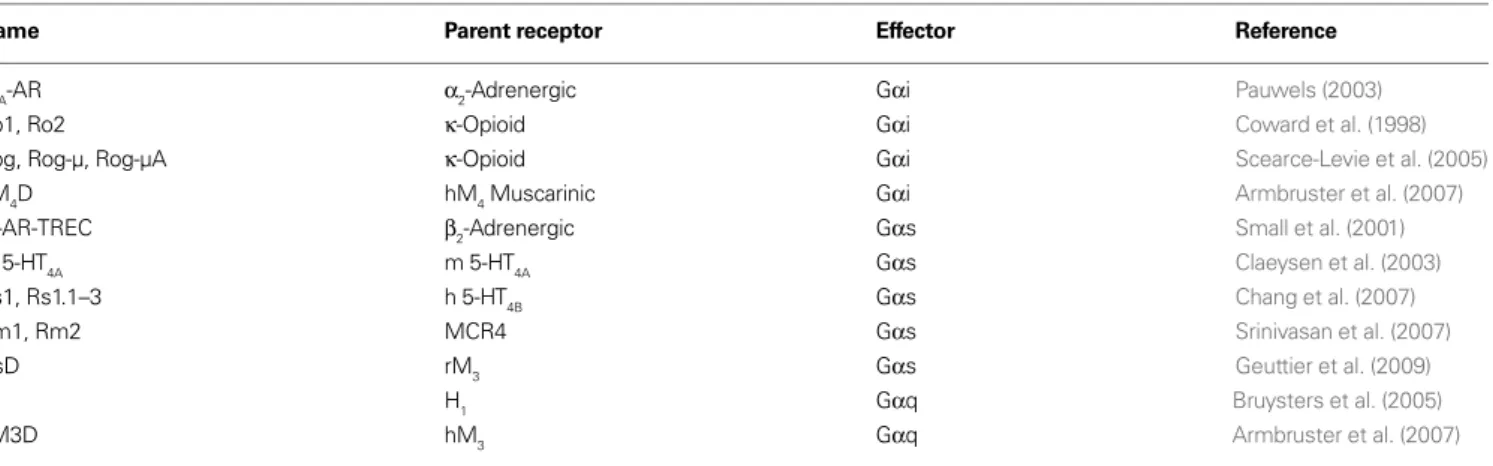
Table 1 | The major RASSL and DREADD receptors grouped according to the primary G-protein activated. The parent receptor of each is listed.
Name Parent receptor Effector Reference
α2A-AR α2-Adrenergic Gαi Pauwels (2003)
Ro1, Ro2 κ-Opioid Gαi Coward et al. (1998)
Rog, Rog-µ, Rog-µA κ-Opioid Gαi Scearce-Levie et al. (2005)
hM4D hM4 Muscarinic Gαi Armbruster et al. (2007)
β2-AR-TREC β2-Adrenergic Gαs Small et al. (2001)
m 5-HT
4A m 5-HT4A Gαs Claeysen et al. (2003)
Rs1, Rs1.1–3 h 5-HT4B Gαs Chang et al. (2007)
Rm1, Rm2 MCR4 Gαs Srinivasan et al. (2007)
GsD rM3 Gαs Geuttier et al. (2009)
H
1 H1 Gαq Bruysters et al. (2005)
neurotransmitters involved in signal transduction are thought to act through GPCRs. Signifi cantly, >50% of current drugs on the market target GPCR function as a major mechanism of therapeutics (Roth et al., 2004; Strachan et al., 2006). Thus, GPCRs remain the most popular family of targets used in drug discovery.
G-protein coupled receptors are functionally coupled to het-erotrimeric G-proteins at the intracellular loops and C-terminus of the GPCR. Heterotrimeric G proteins are comprised of a Gα subunit and a dimeric Gβγ subunit, and are grouped into four classes: Gαs, Gαi/o, Gαq/11, and Gα12/13 (Gilman, 1987). Whereas Gαs stimulates the production of cAMP through activation of ade-nylate cyclase, Gαi/o produces the opposite effect, and reduces lev-els of intracellular cAMP through inhibition of adenylate cyclase. The Gαq subunit stimulates phospholipase-Cβ, which catalyzes the production of phosphoinositides and the release of intracel-lular calcium, among other processes. Gα12/13 interacts with a number of effectors including RhoGEFs to modulate cell growth and cytoskeleton structure (Kelly et al., 2007).
Because of the widespread expression of GPCRs (Regard et al., 2008), and the variety of effector pathways that they can couple to (Urban et al., 2007), targeted expression of modifi ed GPCRs is an ideal tool to utilize to probe not only the role of how specifi c signal transduction pathways infl uence cellular function, but to also probe the role of specifi c tissues and neuronal circuits underly-ing physiological and behavioral processes. The primary RASSL/ DREADD strategy is to engineer a collection of modifi ed GPCRs, each coupling to different primary effector pathways, that respond to synthetic or non-endogenous compounds.
RASSLs
The fi rst published report on the creation of a modifi ed GPCR to respond to a non-endogenous ligand was by Strader et al. (1991). In the process of exploring the nature of ligand-receptor interactions for the β2-adrenergic receptor, a single point mutant (S113A) was found to eliminate binding of endogenous ligands while simul-taneously conferring activity for a class of compounds with little affi nity for the wild type receptor (Strader et al., 1991). Whereas the potencies of these synthetic compounds was relatively low at the modifi ed β2AR, this work nevertheless demonstrated that GPCRs could be modifi ed to lose native ligand recognition while maintain-ing affi nity for synthetic ligands. The Strader et al. (1991) study was thus an important proof-of-concept study for this approach.
The next major development was the creation of modifi ed human kappa-opioid receptors with dramatically reduced affi nity for the natural peptide ligands (1000-fold reduction), but retained affi nity for small molecule drugs like bremazocine and spiradoline (Coward et al., 1998). Two receptor variants were created: Ro1 and Ro2. Ro1 is a chimeric receptor of the κ-opioid receptor contain-ing the second extracellular loop of the delta opioid receptor, and Ro2 is essentially the Ro1 receptor with an additional mutation at the top of the sixth transmembrane helix. The overall design of the modifi ed receptors arose from previous studies of chimeras between κ and δ receptors indicating that the second extracellu-lar loop contains a major determinant for binding of a κ selec-tive ligand, dynorphin. What was achieved by this manipulation was a Gαi-coupled κ-opioid receptor with signifi cantly reduced binding to κ-specifi c peptides, but retained affi nity for κ-selective
small molecule synthetic ligands whose binding regions were not determined by the second extracellular loop region (Coward et al., 1998). These receptors were termed RASSLs, and when expressed in rat fi broblast cells found to be functional for inducing prolifera-tion, a process known to be stimulated by Gαi signaling, with the synthetic ligand spiradoline (Coward et al., 1998).
Many studies using the Ro1 RASSLs were subsequently pub-lished utilizing this receptor as a tool to explore the effects of activating the Gαi effector pathway in various tissues as defi ned by specifi c transgene targeting (Conklin et al., 2008; Pei et al., 2008). The fi rst transgenic mouse study detailed the conditional expression and activation of the Ro1 receptor in mouse heart, liver, and brain (Redfern et al., 1999). Activation of Gαi signal-ing in the heart with the κ-opioid receptor agonist spiradoline produced signifi cant and dose dependent bradycardia with a
∼50% reduction in heart rate in less than a minute after drug administration (Redfern et al., 1999). In a subsequent study, it was found that the induction of expression of Ro1 in the heart induces cardiomyopathy in the absence of spiradoline (Redfern et al., 2000). Partially inhibiting expression of Ro1, treatment with the κOR antagonist nor-binaltomorphine, and treatment with pertussis toxin, restored normal function demonstrating that the heart defects were indeed due to excessive Gαi signal-ing from the Ro1 receptor in the absence of ligand stimulation (Redfern et al., 2000). Signifi cantly, these experiments indicate that there is a level of basal constitutive activity associated with the Ro1 receptor that is able to infl uence physiological processes in the absence of pharmacological activation. Nevertheless, this system has proven valuable as a new model for the study of dilated cardiomyopathy (Baker et al., 2001; McCloskey et al., 2008). The inducible doxycycline-responding Ro1 mouse strain was also uti-lized by Sweger et al. (2007) to investigate the role of astrocytes in mouse brain. They created a transgenic κOR knockout mouse with conditional expression of Ro1 in astrocytes induced by dis-continuation of feeding doxycycline and found that even in the absence of the κOR agonist spiradoline the mice developed severe hydrocephalus (Sweger et al., 2007) through mechanisms regulat-ing CSF production, defi nregulat-ing a new role for Gαi in astrocytes. Importantly, these results independently confi rmed that the Ro1 receptor is associated with a certain physiologically relevant level of constitutive activity.
An interesting study using the doxycycline-responding Ro1 transgenic mice created by Redfen and colleagues was to examine the nature of the sweet and umami taste. Here, Ro1 was expressed in taste buds under the control of the T1R2 receptor promoter in T1R2 null mice. These mice responded to the κ-selective agonist spiradoline as control mice did to sweet taste, indicating that Gαi signaling through the T1R2 receptor is responsible for perception of sweet taste (Zhao et al., 2003).
Following the success of the initial Ro1 receptor, attempts were made to modify it. First, the Rog (RASSL opioid green) receptor was created by fusing GFP to the N-terminus of Ro1 to visualize receptor localization in the living cell (Scearce-Levie et al., 2005). After validating its ability to internalize upon agonist stimulation, signaling, and RASSL function, subsequent modifi cations included mutation of the four C-terminal phosphorylation sites to alanine (Rog-A), replacement of the entire C-terminus with the µ-opioid receptor (Rog-µ), and mutation of fi ve C-terminal serine and glutamic acid residues of the Rog-µ receptor to alanine (Rog-µA) (Scearce-Levie et al., 2005). The loss of C-terminal phosphoryla-tion sites in the Rog-A resulted in a signifi cant reducphosphoryla-tion in ago-nist induced internalization and a resistance to desensitization, however maximal cAMP inhibition was unaffected (Scearce-Levie et al., 2005). The Rog-µ receptor demonstrated increased agonist induced receptor internalization, as was predicted based upon the ability of the µ receptor to more readily internalize than the δ recep-tor (Scearce-Levie et al., 2005). Whereas the Rog-µA receptor was predicted to be resistant to agonist induced internalization as the Rog-A receptor was, it surprisingly demonstrated constitutive inter-nalization (Scearce-Levie et al., 2005). Addition of an antagonist, nor-BNI, was found to rescue cell surface expression (Scearce-Levie et al., 2005). Another physiological effect, adenylate cyclase superac-tivation, was examined in these receptors. Long-term activation of Gαi signaling pathways can lead to an increase in adenylate cyclase activity and an enhanced response to forskolin stimulation over baseline conditions (Watts and Neve, 2005). Neither the Rog-µ nor-Rog-µA receptors showed this effect after overnight treatment with spiradoline followed by forskolin stimulation (Scearce-Levie et al., 2005). The authors speculated that it is not constitutive activity of Gαi from the Ro1 receptors that is responsible for cardiomyopathy observed in their previous studies, but rather an increase in Gαs signaling through adenylate cyclase superactivation that produces the phenotype (Scearce-Levie et al., 2005). It is possible that these newly modifi ed Ro1-related RASSLs will address this issue as well as others.
Through a series of structure/function studies, another modifi ed Gαi coupled receptor, the α2A-adrenergic receptor, was demon-strated to have RASSL-like properties. Based upon previous work predicting the importance of two conserved serine residues within the putative transmembrane binding site the serine at position 200, and the serine at position 204 were each mutated to alanine and the resulting variants pharmacologically characterized. The result-ing receptors had reduced (S200A), to negligent (S204A) affi nity and activity for the native ligand, but high affi nity and activity for certain classes of drug including synthetic imidazoline derivatives (Pauwels and Colpaert, 2000). Interestingly, several α2A-adrenergic receptor antagonists, including atipamezole and SKF86466, retained antagonist properties at the S200A variant but demonstrated
signifi cant partial agonist activity at the S204A receptor (Pauwels and Colpaert, 2000). Signifi cantly, these studies demonstrated that aspects of functional selectivity can be potentially engineered in to a RASSL where the response of a receptor to a particular ligand is not only lost or retained in mutant RASSL variants, but can be fundamentally altered to a different response.
There have been three independent RASSL families developed to probe Gαs signaling. The fi rst was an attempt to modify the
β2-adrenergic receptor as a tool for use in gene therapies and create a ‘modifi ed therapeutic receptor–effector complex’. In the human
β2-adrenergic receptor, glutamine at position 27 was mutated to glutamate to reduce ligand-induced receptor internalization, aspar-tate at position 113 was muaspar-tated to serine to alter ligand bind-ing properties, 15 potential phosphorylation sites were changed to alanine, and the entire open reading frame of the rat Gαs was fused to the C-terminus coding region (Small et al., 2001). This modifi ed receptor complex was found to be unresponsive to cata-cholamines, but responsive to a single tested ligand L158870 at micromolar concentrations (Small et al., 2001). Due to the low potency of the modifi ed receptor complex to the synthetic ligand its utility in vivo is likely limited.
The second approach at developing a Gαs RASSL involved modifying the serotonin 5-HT4A receptor. Key amino acids in the native ligand binding pocket were deduced based upon the pub-lished crystal structure of the β-adrenergic receptor to identify Asp100 in TM3, which was predicted to interact with the amine in the
indole ring of serotonin. Mutation of this Asp to Ala in the mouse 5-HT4A receptor signifi cantly reduced binding as well as activation of the receptor by serotonin and other tryptamines with respect to adenylate cyclase activation, while maintaining affi nity and activity for other ligands without a protonated amine in the core structure (Claeysen et al., 2003). Interestingly, a number of antagonists at the native receptor were found to have agonist activity with respect to adenylate cyclase at the modifi ed receptor (Rs1) (Claeysen et al., 2003). This may allow for selective activation of the modifi ed recep-tor in animal models, while keeping native receprecep-tors inactive, some-thing not possible with the Ro1 receptors. In a subsequent study, conditional expression of the Rs1 receptor in transgenic mouse osteoblasts resulted in increased bone mass in the absence of acti-vating ligand, presumably by constitutive Gαs signaling from the Rs1 receptor (Hsiao et al., 2008). These bone- enhancing effects were observed to be different from a mouse expressing a constitu-tively activated form of the Gαs-coupled PPR receptor, indicating that additional factors are differentially recruited to the receptors to mediate physiological effects.
certain classes of ligand predominantly activate Gαs, while other classes, represented by zacopride, also induce coupling to Gαq. Surprisingly, the ability of zacopride to induce coupling to Gαq was lost in the two mutant 5-HT4B RASSLs (Chang et al., 2007).
In an attempt to create a predominantly Gαq-coupled RASSL, the intracellular loops of the human Rs1 were replaced with those from the Gαq-coupled human 5-HT2C receptor to create Rs1-5-HT2C (Rs1.1). In this process, 12 different chimeras were created and tested that each had different splicing junctions and combinations of intracellular loops. It was found that replacement of the second or third intracellular loops eliminated both Gαs and Gαq activity, and only replacement of the C-terminus was able to increase coupling to Gαq (Chang et al., 2007). This new recep-tor, Rs1-C-5-HT2C, however, still had signifi cant constitutive and ligand-induced Gαs activity. Following similar techniques utilizing intracellular domains from the human 5-HT1A receptor, attempts were made to engineer a Gαi-couple RASSL from Rs1. Replacement of the third intracellular loop was found to abolish Gαs and Gαq activity and to confer Gαi activity to the Rs1-i3-5-HT1A (Rs1.3) receptor (Chang et al., 2007). The potency of this effect, however, was weak thus limiting the utility of this receptor. Importantly, these studies with the Rs1 series of receptors demonstrated that it was possible to continue to modify a RASSL to alter and refi ne physiological properties to something more desirable as a tool for in vivo use.
The third approach to develop a Gαs coupled RASSL involved modifying the melanocortin-4 receptor (MCR4). An extensive review of previous work examining the location and functional result of individual mutations in the MCR4 led to the identifi cation of fi ve candidate mutations for further study as potential RASSLs (Srinivasan et al., 2007). Of these, two were designated RASSLs: L106P-MCR4 (Rm1), and D122A-MCR4 (Rm2). The native RCM4 has signifi cant constitutive basal activity associated with Gαs signaling, and although Rm1 has a 30% reduction in activity, Rm2 has a nearly twofold increase in basal activity over the native receptor (Srinivasan et al., 2007). Importantly, neither Rm1 nor-Rm2 respond to native melanocortin peptides, but do respond to nanomolar concentrations of the synthetic MCR4 selective ligand THIQ with respect to measured cAMP release (Srinivasan et al., 2007).
The third major class of G-protein effector is Gαq, and one attempt has been made to generate a RASSL coupled to this path-way. Based upon previous structure function studies examining ligand binding properties, the phenylalanine at position 435 of the human H1 receptor was mutated to an alanine and the resulting receptor characterized (Bruysters et al., 2005). Rather than assess Gαq coupling by measuring PI turnover or calcium mobilization, a luciferase reporter assay for NFkB activity was used to measure activity. This was based upon previous work that demonstrated direct functional coupling of Gαq/11 to NFkB activation (Bakker et al., 2001). Therefore, RASSL activity from this receptor can only be reliably extended to activation of the NFkB signaling pathway. Interestingly, whereas the F453A H1 receptor variant maintained affi nity for histamine, affi nity and potency at NFkB activation increased up to 1000-fold for certain synthetic ligands (Bruysters et al., 2005). In the case of ClPheHA, potency was increased from an EC50 of ∼1 mM at the native receptor to ∼1 nM at the F453A
variant. Rather than engineering loss of affi nity and potency for native ligand while retaining those properties for a synthetic lig-and, greatly enhancing affi nity and potency for a synthetic ligand through receptor modifi cation was demonstrated here to be a new strategy to develop a RASSL. The F453A H1 receptor has negligent basal constitutive activity with respect to NFkB signaling, however, additional pathways coupled to Gαq have yet to be investigated, as well as the utility of this receptor to study in vivo processes.
Of signifi cant concern with nearly all RASSLs created thus far is the presence of physiologically relevant basal constitutive activity with the primary Gα effector protein. Whereas this may effectively allow functional analysis of an effector pathway under certain con-ditions, constitutive activity present from embryogenesis has the potential to seriously confound results examining acute function. Another considerable shortcoming of the conventional RASSLs is that they by and large utilize synthetic ligands that have signifi cant affi nity and potency for the endogenous wild type receptor as well as potential off-target actions. Therefore, ligands used to stimulate RASSLs in vivo could induce behaviors and physiological processes from native receptors and confound results. Nonetheless, based on the large number of important papers, conventional RASSLs continue to be widely used with great success.
DREADDs
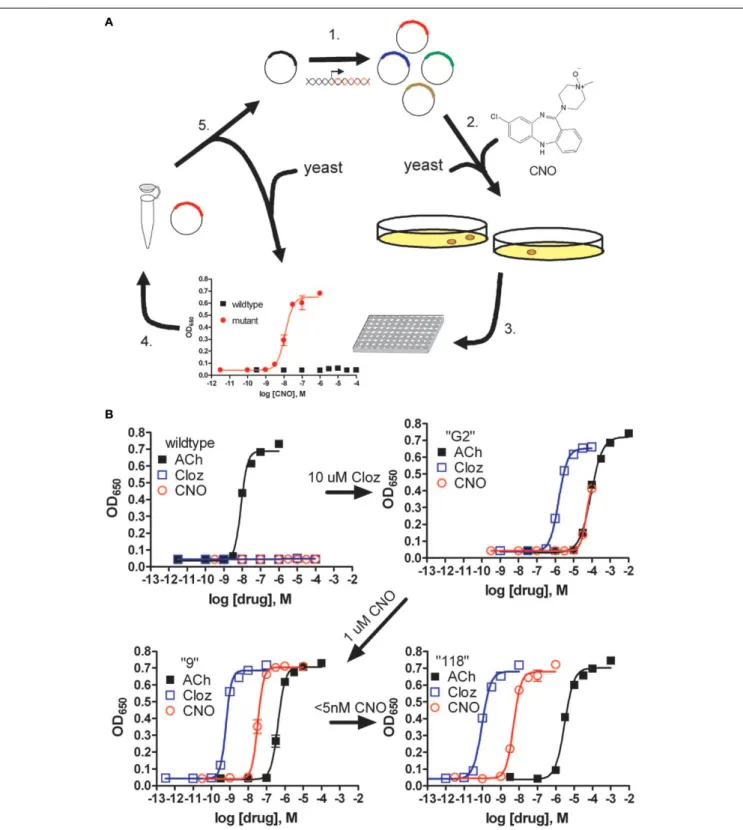
FIGURE 1 | Pharmacological profi les of an rM3Δi3 receptor mutant selected during directed molecular evolution for CNO responsiveness. (A)
Experimental design for directed evolution of mammalian GPCRs in yeast to create DREADDs. (1) Libraries of randomly mutated rM3Δi3 receptors were produced by mutagenic PCR; (2) yeast-expressing mutant receptors activated by synthetic ligands (e.g., CNO) were selected for by growth on nutrient defi cient medium; (3) mutants were verifi ed by secondary liquid growth assays in 96-well plates; (4) plasmid DNA was isolated from yeast; (5) clones were retransformed into yeast to pharmacologically profi le mutants by liquid growth assays, and those
at position 149 mutated to cystine, and alanine at position 239 mutated to glycine (Armbruster et al., 2007). Importantly, not only was CNO able to activate Gαq signaling and PI turnover, but CNO was also shown to activate MAPK signaling through interactions with β-arrestin, indicating that coupling to multiple effector path-ways was preserved in the DREADD receptor. Mutation of the con-served tyrosine and glycine residues in the other human muscarinic receptors resulted in the creation of hM2D and hM4D DREADD receptors coupled to Gαi/o, and hM1D coupled to Gαq. Additional functionality of the hM4D was determined by demonstrating that stimulation of the receptor with CNO, but not with ACh, activated inwardly rectifying potassium channels (GIRKs) in both transfected HEK cells and transfected hippocampal neurons (Armbruster et al., 2007). These results indicate that the hM4D receptor has potential as a tool for in vivo neuronal silencing.
To develop the fi nal Gαs coupled DREADD (GsD), we modifi ed the rat M3D receptor by replacing the second and third intracellular loops with the corresponding loops from the Gαs-coupled turkey
β1-adrenergic receptor (Geuttier et al., 2009). Transgenic mouse
lines expressing the hM3D and GsD receptors in pancreatic beta-cells have been made that show stimulation of either receptor has
signifi cant effects on beta-cell function including aspects of glucose tolerance and insulin release (Geuttier et al., 2009). Additional transgenic mouse studies with all three DREADD receptors are currently being performed to analyze the role of G-protein sig-naling in a variety of tissues (Figure 2). We have demonstrated remote control of neuronal activity in mice expressing the hM3D receptor in hippocampus (Alexander et al., 2009). Administration of CNO to these transgenic mice led to both increases in hippoc-ampal neuronal activity as well as behavioral modifi cations includ-ing increased locomotor activity and seizures in a dose dependent fashion (Alexander et al., 2009). In slice cultures, the increases in neuronal activity from a single pulse of CNO returned to baseline levels in about 60 min. The locomotor effects of systemic CNO, however, did not return to baseline levels for about 9 h (Alexander et al., 2009). Importantly, these data indicate that this system is reversible with in vitro cellular effects of CNO extinguishing more rapidly than the in vivo behavioral effects.
We have recently created transgenic Drosophila melanogaster expressing the Gαs-coupled hM4D, Gαi-coupled rM3D-βar, and Gαq-coupled hM1D receptors under the control of the bipartite GAL4/UAS system. The GAL4/UAS system is a genetic method to
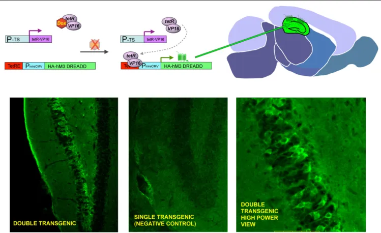
FIGURE 2 | General strategy to achieve inducible, tissue-specifi c expression of DREADDs. The upper panel shows the basic strategy to generate tissue specifi c expression of DREADDs in mouse brain. Double transgenic animals are made where one element contains a tissue specifi c promotor (P-TS) driving expression of the tetR-VP16 fusion protein, and the other contains the tetracycline response element (TetRE), followed by a minimal CMV promoter driving expression of HA-tagged DREADDs. Whereas both transgenic elements are present in every cell throughout the body, expression of tetR-VP19 is only in
express transgenes selectively in defi ned tissues (Brand and Perrimon, 1993). In preliminary studies we have targeted DREADD expression to distinct neural circuits and have found that behaviors are dramati-cally altered when adult fl ies expressing DREADDS are fed CNO (unpublished results). Importantly, wild type and parental strains maintained on food containing CNO (at least to 10 mM) exhibit no overt abnormal developmental or behavioral effects, indicating that the effects of CNO on the transgenic fl ies expressing DREADDs are indeed due to DREADD activation. Given the conservation of biological processes between mammalian systems and the power of the fl y as a genetic model (Nichols, 2006), studies performed in the fl y are likely to be informative to both insect and mammalian models with respect to elucidation of signal transduction pathways and molecular processes underlying neuronal function as they relate to behaviors. Furthermore, this system is anticipated to be useful in the fl y to probe the role of signal transduction pathways and discreet tissues in developmental processes.
Aside from using DREADDs to directly probe the role of individual signal transduction pathways in physiological processes and the role of specifi c tissues and circuits in behaviors, the hM4D receptor has recently proven to be a valuable tool in understanding mechanisms of allosteric modulation in GPCRs. A known allosteric modulator of the M4 receptor, LY2033298, was shown to act cooperativity with the orthosteric binding site to restore affi nity and functionality of acetyl-choline to the orthosteric site of the hM4D receptor (Nawaratne et al., 2008). These muscarinic DREADD receptors, and potentially other similarly modifi ed receptors, may represent novel tools to screen for allosteric modulators that act solely through interactions at dis-tinct allosteric sites and have cooperativity with the orthosteric site. Compounds that would be able to restore affi nity and activity to the native ligand would therefore potentially represent novel therapeutics that enhance the ability of native ligand to activate its receptor.
SUMMARY
An exciting and powerful method of probing the role of G-protein effector signaling, as well as the function of discreet tissues and neural circuits in mediating physiological processes and behaviors has recently been developed and refi ned. The fi rst generation of engi-neered GPCRs, RASSLs, were primarily rationally designed based upon structure/function studies to eliminate native ligand binding, while maintaining affi nity for synthetic ligands. These receptors have been used to generate conditionally expressing transgenic mice, where they have been used to study how G-protein effector pathways affect various processes including heart function, bone growth, and brain development. They have also been used to defi ne the function of other GPCRs as well as particular tissues, as they were used to elu-cidate the receptor and effector pathway underlying the perception of sweet taste. Advantages that this system offers are the ability to very rapidly turn on and off signaling pathways by simply administering a synthetic ligand. Importantly, because GPCRs and their associated effector pathways are ubiquitous throughout an organism, studies are not limited to examining the native role of the particular receptor a RASSL is derived from, but can be used to defi ne the function of entire tissues and neural circuits in a more systems-based approach. These fi rst generation RASSLs, unfortunately, are frequently associ-ated with physiologically relevant basal constitutive activity, as well as response to synthetic ligands that also target the endogenous wild
type receptors. Whereas these properties may be advantageous for some studies, they likely present certain limitations and challenges for widespread use.
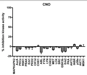
The next generation of engineered GPCRs, DREADDs, created through a process of directed molecular evolution overcome many of the limitations present in the fi rst generation RASSLs. DREADDS have negligent basal constitutive activity associated with them, and are activated by synthetic ligands with no appreciable affi nity for any of the known receptorome. Additionally, CNO has minimal activity at a variety of relevant kinases (Figure 3). Transgenic expression models in both mouse and fl y have validated their functionality to probe both physiological processes and complex behaviors. Given the advantages of the DREADD receptors over the RASSLs, there are certain caveats that must be considered for them, as well as for the RASSLs, involving expression levels.
Under most normal transgenic conditions, expression levels are likely high, such that even under non-activated conditions the stoichiometric balance between receptor and precoupled effector may be perturbed such that the normal function of the tissue and circuit may be affected. Furthermore, the presence of high levels of activated receptor may induce activation of effector pathways not normally functionally coupled to the particular GPCR, con-founding results. To achieve the most relevant data for a particular system, expression levels should be determined, and manipulated if possible, with those having expression closest to naturally occurring GPCRs used for experimentation. Nevertheless, both RASSLs and DREADDs present a very effective tool to elucidate biological func-tion and behaviors. Advantages over other current systems include the specifi city of transgenic targeting, the convenience of systemic administration of small molecule drugs with no other appreciable biological targets within the organism, rapidity and reversibility of effect, and no need for specialized equipment.
Lin, J. Y., Lin, M. Z., Steinbach, P., and Tsien, R. Y. (2009). Characterization of engineered channelrhodopsin variants with improved properties and kinetics. Biophys. J. 96, 1803–1814.
Martin, J. R., Keller, A., and Sweeney, S. T. (2002). Targeted expression of tetanus toxin: a new tool to study the neuro-biology of behavior. Adv. Genet. 47, 1–47.
McCloskey, D. T., Turcato, S., Wang, G. Y., Turnbull, L., Zhu, B. Q., Bambino, T., Nguyen, A. P., Lovett, D. H., Nissenson, R. A., Karliner, J. S., and Baker, A. J. (2008). Expression of a Gi-coupled receptor in the heart causes impaired Ca2+ handling, myofi lament
injury, and dilated cardiomyopathy. Am. J. Physiol. Heart Circ. Physiol. 294, H205–H212.
McGuire, S. E., Roman, G., and Davis, R. L. (2004). Gene expression systems in Drosophila: a synthesis of time and space. Trends Genet. 20, 384–391. Nagel, G., Szellas, T., Huhn, W., Kateriya, S.,
Adeishvili, N., Berthold, P., Ollig, D., Hegemann, P., and Bamberg, E. (2003). Channelrhodopsin-2, a directly light-gated cation-selective membrane channel. Proc. Natl. Acad. Sci. U.S.A. 100, 13940–13945. Nagel, G., Szellas, T., Kateriya, S.,
Adeishv ili, N., He gemann, P., a n d B a m b e r g , E . ( 2 0 0 5 ) . Channelrhodopsins: directly light-gated cation channels. Biochem. Soc. Trans. 33, 863–866.
Nakashiba, T., Young, J. Z., McHugh, T. J., Buhl, D. L., and Tonegawa, S. (2008). Transgenic inhibition of synaptic transmission reveals role of CA3 out-put in hippocampal learning. Science 319, 1260–1264.
Nawaratne, V., Leach, K., Suratman, N., Loiacono, R. E., Felder, C. C., Armbruster, B. N., Roth, B. L., Sexton, P. M., and Christopoulos, A. (2008). New insights into the function of M4 muscarinic acetylcholine receptors gained using a novel allosteric modula-tor and a DREADD (designer recepmodula-tor exclusively activated by a designer drug). Mol. Pharmacol. 74, 1119–1131. Nichols, C. D. (2006). Drosophila
mela-nogaster neurobiology, neurophar-macology, and how the fl y can inform central nervous system drug discovery. Pharmacol. Ther. 112, 677–700. Nitabach, M. N., Blau, J., and Holmes, T. C.
(2002). Electrical silencing of Drosophila pacemaker neurons stops the free-running circadian clock. Cell 109, 485–495.
Pauwels, P. J., and Colpaert, F. C. (2000). Disparate ligand-mediated Ca(2) responses by wild-type, mutant Ser(200)Ala and Ser(204)Ala alpha(2A)-adrenoceptor: G(alpha15) fusion proteins: evidence for multiple
REFERENCES
Aiba, A., and Nakao, H. (2007). Conditional mutant mice using tetracycline-controlled gene expres-sion system in the brain. Neurosci. Res. 58, 113–117.
Airan, R. D., Thompson, K. R., Fenno, L. E., Bernstein, H., and Deisseroth, K. (2009). Temporally precise in vivo control of intracellular signalling. Nature 458, 1025–1029.
Alexander, G. M., Rogan, S. C., Abbas, A. I., Armbruster, B. N., Pei, Y., Allen, J. A., Nonneman, R. J., Hartmann, J., M o y, S . S . , N i c o l e l i s , M . A . , McNamara, J. O., and Roth, B. L. (2009). Remote control of neuronal activity in transgenic mice expressing evolved G protein-coupled receptors. Neuron 63, 27–39.
Armbruster, B. N., Li, X., Pausch, M. H., Herlitze, S., and Roth, B. L. (2007). Evolving the lock to fit the key to create a family of G protein-coupled receptors potently activated by an inert ligand. Proc. Natl. Acad. Sci. U.S.A. 104, 5163–5168.
Armbruster, B. N., and Roth, B. L. (2005). Mining the receptorome. J. Biol. Chem. 280, 5129–5132.
Baker, A. J., Redfern, C. H., Harwood, M. D., Simpson, P. C., and Conklin, B. R. (2001). Abnormal contraction caused by expression of G(i)-coupled receptor in transgenic model of dilated cardio-myopathy. Am. J. Physiol. Heart Circ. Physiol. 280, H1653–H1659. Bakker, R. A., Schoonus, S. B., Smit, M. J.,
Timmerman, H., and Leurs, R. (2001). Histamine H(1)-receptor activation of nuclear factor-kappa B: roles for G beta gamma- and G alpha(q/11)-subunits in constitutive and agonist-mediated signaling. Mol. Pharmacol. 60, 1133–1142.
Brand, A. H., and Perrimon, N. (1993). Targeted gene expression as a means of altering cell fates and generating dominant phenotypes. Development 118, 401–415.
Bruysters, M., Jongejan, A., Akdemir, A., Bakker, R. A., and Leurs, R. (2005). A G(q/11)-coupled mutant histamine H(1) receptor F435A activated solely by synthetic ligands (RASSL). J. Biol. Chem. 280, 34741–34746.
Chang, W. C., Ng, J. K., Nguyen, T., Pellissier, L., Claeysen, S., Hsiao, E. C., and Conklin, B. R. (2007). Modifying ligand-induced and constitutive sig-naling of the human 5-HT4 receptor. PLoS ONE 2, e1317. doi: 10.1371/jour-nal.pone.0001317.
Claeysen, S., Joubert, L., Sebben, M., Bockaert, J., and Dumuis, A. (2003). A single mutation in the 5-HT4 receptor (5-HT4-R D100(3.32)A) generates a Gs-coupled receptor activated exclusively by synthetic
ligands (RASSL). J. Biol. Chem. 278, 699–702.
Conklin, B. R., Hsiao, E. C., Claeysen, S., D u mu i s , A . , Sr i n iv a s a n , S . , Forsayeth, J. R., Guettier, J. M., Chang, W. C., Pei, Y., McCarthy, K. D., Nissenson, R. A., Wess, J., Bockaert, J., and Roth, B. L. (2008). Engineering GPCR signaling pathways with RASSLs. Nat. Methods 5, 673–678. Coward, P., Wada, H. G., Falk, M. S.,
Chan, S. D., Meng, F., Akil, H., and Conklin, B. R. (1998). Controlling signaling with a specifi cally designed Gi-coupled receptor. Proc. Natl. Acad. Sci. U.S.A. 95, 352–357.
Drago, J., Padungchaichot, P., Wong, J. Y., Lawrence, A. J., McManus, J. F., Sumarsono, S. H., Natoli, A. L., Lakso, M., Wreford, N., Westphal, H., Kola, I., and Finkelstein, D. I. (1998). Targeted expression of a toxin gene to D1 dopamine receptor neurons by cre-mediated site-specifi c recombination. J. Neurosci. 18, 9845–9857. Erlenbach, I., Kostenis, E., Schmidt, C.,
Hamdan, F. F., Pausch, M. H., and Wess, J. (2001). Functional expres-sion of M(1), M(3) and M(5) mus-carinic acetylcholine receptors in yeast. J. Neurochem. 77, 1327–1337. Gaveriaux-Ruff, C., and Kieffer, B. L.
(2007). Conditional gene targeting in the mouse nervous system: insights into brain function and diseases. Pharmacol. Ther. 113, 619–634. Geuttier, J. M., Gautam, D., Scarselli, M.,
Ruiz de Azula, I., Li, J. H., Rosemond, E., Ma, X., Gonzales, F., Armbruster, B., Lu, H., Roth, B. L., and Wess, J. (2009). A chemical-genetic approach to study G protein regulation of β cell function in vivo. Proc. Natl. Acad. Sci. U.S.A. (in press).
Gilman, A. G. (1987). G proteins: trans-ducers of receptor-generated signals. Annu. Rev. Biochem. 56, 615–649. Hsiao, E. C., Boudignon, B. M.,
Chang, W. C., Bencsik, M., Peng, J., N g u y e n , T. D. , Ma n a l a c , C . , Halloran, B. P., Conklin, B. R., and Nissenson, R. A. (2008). Osteoblast expression of an engineered Gs-coupled receptor dramatically increases bone mass. Proc. Natl. Acad. Sci. U.S.A. 105, 1209–1214. Kelly, P., Casey, P. J., and Meigs, T. E.
(2007). Biologic functions of the G12 subfamily of heterotrimeric g proteins: growth, migration, and metastasis. Biochemistry 46, 6677–6687. Kerr, M., Davies, S. A., and Dow, J. A.
(2004). Cell-specific manipulation of second messengers; a toolbox for integrative physiology in Drosophila. Curr. Biol. 14, 1468–1474.
Kroeze, W. K., Sheffl er, D. J., and Roth, B. L. (2003). G-protein-coupled receptors at a glance. J. Cell. Sci. 116, 4867–4869.
ligand-activation binding sites. Br. J. Pharmacol. 130, 1505–1512. Pauwels, P. J. (2003). Unravelling multiple
ligand-activation binding sites using RASSL receptors. Trends Pharmacol. Sci. 24, 504–507.
Pei, Y., Rogan, S. C., Yan, F., and Roth, B. L. (2008). Engineered GPCRs as tools to modulate signal transduction. Physiology (Bethesda) 23, 313–321. Peng, J., Bencsik, M., Louie, A., Lu, W.,
Millard, S., Nguyen, P., Burghardt, A., Majumdar, S., Wronski, T. J., Halloran, B., Conklin, B. R., and Nissenson, R. A. (2008). Conditional expression of a Gi-coupled receptor in osteoblasts results in trabecular osteo-penia. Endocrinology 149, 1329–1337. Redfern, C. H., Coward, P., Degtyarev, M. Y., Lee, E. K., Kwa, A. T., Hennighausen, L., Bujard, H., Fishman, G. I., and Conklin, B. R. (1999). Conditional expression and signaling of a specifi cally designed Gi-coupled receptor in transgenic mice. Nat. Biotechnol. 17, 165–169. Redfern, C. H., Degtyarev, M. Y., Kwa, A. T.,
Salomonis, N., Cotte, N., Nanevicz, T., Fidelman, N., Desai, K., Vranizan, K., Lee, E. K., Coward, P., Shah, N., Warrington, J. A., Fishman, G. I., Bernstein, D., Baker, A. J., and Conklin, B. R. (2000). Conditional expression of a Gi-coupled receptor causes ventricular conduction delay and a lethal cardiomyopathy. Proc. Natl. Acad. Sci. U.S.A. 97, 4826–4831. Regard, J. B., Sato, I. T., and Coughlin, S. R. (2008). Anatomical profiling of G protein-coupled receptor expres-sion. Cell 135, 561–571.
Roth, B. L., Sheffl er, D. J., and Kroeze, W. K. (2004). Magic shotguns versus magic bullets: selectively non-selective drugs for mood disorders and schizophrenia. Nat. Rev. Drug Discov. 3, 353–359. Sakamoto, A., Chen, M., Nakamura, T.,
Xie, T., Karsenty, G., and Weinstein, L. S. (2005). Defi ciency of the G-protein alpha-subunit G(s)alpha in osteob-lasts leads to differential effects on trabecular and cortical bone. J. Biol. Chem. 280, 21369–21375.
Scearce-Levie, K., Lieberman, M. D., Elliott, H. H., and Conklin, B. R. (2005). Engineered G protein coupled receptors reveal independent regula-tion of internalizaregula-tion, desensitizaregula-tion and acute signaling. BMC Biol. 3, 3. Schroll, C., Riemensperger, T., Bucher, D.,
Ehmer, J., Voller, T., Erbguth, K., Gerber, B., Hendel, T., Nagel, G., Buchner, E., and Fiala, A. (2006). Light-induced activation of distinct modulatory neurons triggers appeti-tive or aversive learning in Drosophila larvae. Curr. Biol. 16, 1741–1747. Small, K. M., Brown, K. M., Forbes, S. L.,
Ryba, N. J., and Zuker, C. S. (2003). The receptors for mammalian sweet and umami taste. Cell 115, 255–266.
Conflict of Interest Statement: The authors declare that the research was conducted in the absence of any com-mercial or financial relationships that could be construed as a potential confl ict of interest.
Received: 29 May 2009; paper pend-ing published: 08 July 2009; accepted: 12 September 2009; published online: 23 October 2009.
Citation: Nichols CD and Roth BL (2009) Engineered G-protein coupled receptors are powerful tools to inves-tigate biological processes and behav-iors. Front. Mol. Neurosci. 2:16. doi: 10.3389/neuro.02.016.2009
Copyright © 2009 Nichols and Roth. This is an open-access article subject to an exclu-sive license agreement between the authors and the Frontiers Research Foundation, which permits unrestricted use, distribu-tion, and reproduction in any medium, provided the original authors and source are credited.
engineer a receptor–effector complex for gene therapy. J. Biol. Chem. 276, 31596–31601.
Srinivasan, S., Santiago, P., Lubrano, C., Vaisse, C., and Conklin, B. R. (2007). Engineering the melanocortin-4 receptor to control constitutive and ligand-mediated GS signaling in vivo. PLoS ONE 2, e668. doi: 10.1371/ journal. pone.0000668.
Strachan, R. T., Ferrara, G., and Roth, B. L. (2006). Screening the receptorome: an effi cient approach for drug discovery and target validation. Drug Discov. Today 11, 708–716.
Strader, C. D., Gaffney, T., Sugg, E. E., Candelore, M. R., Keys, R., Patchett, A. A., and Dixon, R. A. (1991). Allele-specifi c activation of genetically engineered receptors. J. Biol. Chem. 266, 5–8. Sweger, E. J., Casper, K. B., Scearce-Levie, K.,
Conklin, B. R., and McCarthy, K. D. (2007). Development of hydrocepha-lus in mice expressing the G(i)-coupled GPCR Ro1 RASSL receptor in astro-cytes. J. Neurosci. 27, 2309–2317. Tan, E. M., Yamaguchi, Y., Horwitz, G D.,
Gosgnach, S., Lein, E. S., Goulding, M., Albright, T. D., and Callaway, E. M.
(2006). Selective and quickly reversible inactivation of mammalian neurons in vivo using the Drosophila allatostatin receptor. Neuron 51, 157–170. Tonnesen, J., Sorensen, A. T., Deisseroth, K.,
Lundberg, C., and Kokaia, M. (2009). Optogenetic control of epileptiform activity. Proc. Natl. Acad. Sci. U.S.A. 106, 12162–12167.
Tsetsenis, T., Ma, X. H., Lo Iacono, L., Beck, S. G., and Gross, C. (2007). Suppression of conditioning to ambiguous cues by pharmacogenetic inhibition of the den-tate gyrus. Nat. Neurosci. 10, 896–902. Urban, J. D., Clarke, W. P., von
Zastrow, M., Nichols, D. E., Kobilka, B., Weinstein, H., Javitch, J. A., Roth, B. L., Christopoulos, A., Sexton, P. M., Miller, K. J., Spedding, M., and Mailman, R. B. (2007). Functional selectivity and classical concepts of quantitative pharmacology. J. Pharmacol. Exp. Ther. 320, 1. Watts, V. J., and Neve, K. A. (2005).
Sensitization of adenylate cyclase by Galpha i/o-coupled receptors. Pharmacol. Ther. 106, 405–421. Wehr, M., Hostick, U., Kyweriga, M.,
Tan, A., Weible, A. P., Wu, H., Wu, W.,
Callaway, E. M., and Kentros, C. (2009). Transgenic silencing of neurons in the mammalian brain by expression of the allatostatin receptor (AlstR). J. Neurophysiol. 2009; 0: 00480.2009v1 [Epub ahead of print].
Wells, T., and Carter, D. A. (2001). Genetic engineering of neural function in transgenic rodents: towards a compre-hensive strategy? J. Neurosci. Methods 108, 111–130.
Wulff, P., Goetz, T., Leppa, E., Linden, A. M., Renzi, M., Swinny, J. D., Vekovischeva, O. Y., Sieghart, W., Somogyi, P., Korpi, E. R., Farrant, M., and Wisden, W. (2007). From syn-apse to behavior: rapid modula-tion of defi ned neuronal types with engineered GABAA receptors. Nat. Neurosci. 10, 923–929.
Yu, C. R., Power, J., Barnea, G., O’Donnell, S., Brow n, H. E., Osborne, J., Axel, R., and Gogos, J. A. (2004). Spontaneous neural activity is required for the establishment and maintenance of the olfactory sensory map. Neuron 42, 553–566.