R E S E A R C H
Open Access
Factors associated with change in exacerbation
frequency in COPD
Gavin C Donaldson
1*, Hanna Müllerova
2, Nicholas Locantore
3, John R Hurst
4, Peter MA Calverley
5, Jorgen Vestbo
6,7,
Antonio Anzueto
8and Jadwiga A Wedzicha
1Abstract
Background:Patients with chronic obstructive pulmonary disease (COPD) can be categorized as having frequent (FE) or infrequent (IE) exacerbations depending on whether they respectively experience two or more, or one or zero exacerbations per year. Although most patients do not change category from year to year, some will, and the factors associated with this behaviour have not been examined.
Methods:1832 patients completing two year follow-up in the Evaluation of COPD Longitudinally to Identify Predictive Surrogate End-points (ECLIPSE) study were examined at baseline and then yearly. Exacerbations were defined by health care utilisation. Patient characteristics compared between those patients who did or did not change exacerbation category from year 1 to year 2.
Findings:Between years 1 and 2, 221 patients (17%) changed from IE to FE and 210 patients (39%) from FE to IE. More severe disease was associated with changing from IE to FE and less severe disease from FE to IE. Over the preceding year, small falls in FEV1and 6-minute walking distance were associated with changing from IE to FE, and small falls in platelet count associated with changing from FE to IE.
Conclusion:No parameter clearly predicts an imminent change in exacerbation frequency category. Trial registration:SCO104960, clinicaltrials.gov identifier NCT00292552
Keywords:COPD, Exacerbations, Exacerbation frequency, Exacerbation phenotype
Background
The natural history of chronic obstructive pulmonary disease (COPD) is punctuated by exacerbations defined as episodes of acute symptomatic deterioration that may warrant a change in treatment [1,2]. Frequent exacerba-tions are associated with reduced quality of life [3], increased airway inflammation [4], accelerated lung func tion decline [5] and increase mortality [6]. Patients with frequent exacerbations make more visits to their medical practitioner, require more medication, experience more hospital admissions [7] and consequently impose a greater financial burden on health services [8,9].
The number of exacerbations a patient experiences over a year is very strongly associated with their fre-quency in the previous year [3,7]. This stability in
exacerbation frequency means that patient recall of events in the previous year can be used to categorize patients as having frequent (FE) or infrequent (IE) exa-cerbations during the subsequent year [7,10]. These cat-egories are now recognised as important COPD patient phenotypes that influence the clinical management of the patient [11]. Although exacerbation frequency is relatively stable in most patients, some year on year vari-ation exists and the factors associated with any change in category have not yet been investigated. Therefore we
analysed the data from the “Evaluation of COPD
lon-gitudinally to identify predictive surrogate endpoints
(ECLIPSE)” study to determine whether patients kept or
changed their IE or FE category. Previously, three groups of 0, 1 and 2+ exacerbations per a year have been de-scribed [7] but interpretation of the nine potential tran-sitions would be complex and less clinically relevant. In this study, we have also examined a slightly smaller group of 1832 patients who completed two years of * Correspondence:g.donaldson@ucl.ac.uk
1
Centre for Respiratory Medicine, UCL Medical School, Royal Free Campus, Rowland Hill Street Hampstead, London NW3 2PF, UK
Full list of author information is available at the end of the article
observation rather than the 2138 patients who were en-rolled into the ECLIPSE study [7] since the exacerbation frequency measured over both years 1 and 2 was needed to identify patients who did or did not changed IE and FE categories.
Additionally we tried to establish whether specific fac-tors were associated with a change in exacerbation fre-quency as such data would be useful in identifying patients with an increased risk of a worsening exacerba-tion frequency and potentially suggest ways to prevent this clinically important change. Some of the results of this study have been previously reported in the form of an abstract [12].
Methods
Study design and patients
All patients provided written informed consent and the study was approved by the relevant ethics and review boards ([13]; see online supplement). Eligible patients were aged between 40 and 75 years, with a 10 or more pack-year smoking history, a post-bronchodilator forced
expiratory volume in 1 second (FEV1) of less than 80%
predicted from age, height and sex [14] and a ratio of FEV1to forced vital capacity (FVC) of 0.7 or less.
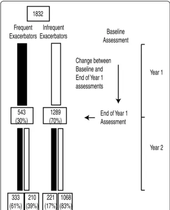
All patients completing at least 2 years of observation are included in this study. Patients underwent compre-hensive medical reviews at baseline and then at the end of each 12 months of observation (see Figure 1). Data recorded at the 3, 6 and 18 month visits have not been analysed in this study as data on some patient charac-teristics were not available at these time points. Patients underwent standard spirometry testing after the
admin-istration of 400 μg of inhaled albuterol, completed a
modified Medical Research Council dyspnoea scale (mMRC) and a St. Georges Respiratory Questionnaire (SGRQ). Co-morbidities were assessed from questions in
the American Thoracic Society–Division of Lung
Dis-eases (ATS-DLD) questionnaire and symptoms of de-pression by the Center for Epidemiologic Studies of Depression (CES-D) questionnaire. A venous blood sam-ple was taken and a medical history including start/stop dates of medications and smoking history recorded. The patients underwent a six minute walking test (6MWD) according to ATS guidelines [15], with distance expressed as a percentage of predicted [16] which was used in calcu-lating the patient’s BODE index [17]. Patients also under-went computed tomographic (CT) scanning, at baseline and year 1, to evaluate the severity of emphysema as the percentage of low attenuation areas (LAA%) [18].
Blood samples were dispatched immediately after each visit for a full-blood count, and serum and plasma
samples stored at −80°C until assayed for fibrinogen,
creactive protein (CRP), surfactant protein D (SP-D) and pulmonary activation regulated chemokine (PARC) [19].
Any samples with values below the lower limit of quanti-fication were assigned a value that was half of the assay lower limit.
Exacerbations were defined by health care utilization requiring the prescription of antibiotics and/or oral cor-ticosteroids by the patient’s primary care clinician or study personnel, or hospitalisation for an exacerbation. To ensure capture of these events patients were asked at clinic visits and during monthly telephone calls about visits to health care professionals and change in medica-tions. The end of an exacerbation was based on investi-gator judgement, and no new exacerbation could start before resolution of an existing exacerbation. Frequent exacerbations were defined as 2 or more per year, and infrequent exacerbations as 1 or zero per year.
Statistical analysis
Patients were categorized according to their exacerba-tion frequency assessed over year 1 of follow-up; whe-ther they changed category was assessed using year 2 of follow-up. No data on how many exacerbations the pa-tient recalled experiencing in year prior to recruitment was used in this analysis. Cross-sectional patient cha-racteristics obtained at the year 1 assessment were ex-amined in relation to those who did or did not change category, rather than the baseline assessment, as this
was closest in time to the point of category change.
“Longitudinal” characteristics were calculated as the difference between baseline and the year 1 assessment -that is, over the year preceding the point of change in exacerbation frequency (see Figure 1).
Means and standard deviations are reported for the continuous variables, with frequencies reported for the categorical variables. For tests between exacerbation fre-quency groups, an analysis of variance (ANOVA) for continuous variables or chi-square test for categorical variables was used. Due to skewed distributions, blood and plasma biomarkers were summarized using medians and inter-quartile ranges (IQR). Comparisons of their distributions across exacerbation frequency groups used a Kruskal-Wallis test.
Results
Change in exacerbation category
1832 patients completed at least 2 years of follow-up and had data available for this analysis.
543 of the 1832 patients (30%) had frequent exa-cerbations and 1289 (70%) infrequent exaexa-cerbations at the end of year 1 of follow-up. Between years 1 and 2, 431 of the 1832 patients (24%) changed category, 221 (17%) of the IE patients changed category to FE com-pared to 210 (39%) of the FE patients who changed to IE (P<0.001).
Table 1 shows the demographic characteristics of all 1832 patients recorded at the year 1 assessment, and after division into infrequent (IE) and frequent exacer-bator (FE) groups by their observed exacerbation fre-quency over year 1.
Change in exacerbation frequency
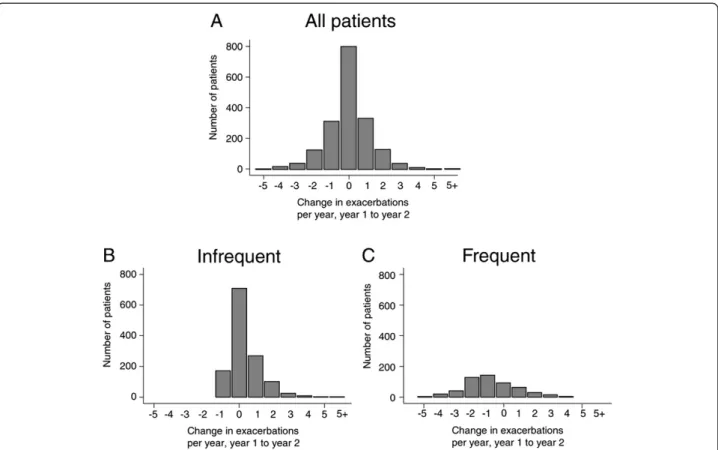
Figure 2A shows a histogram for the change in exacerba-tion frequency between years 1 and 2 of the 1832 patients. 44% of the patients had an identical exacerbation fre-quency in years 1 and 2, and for those patients whose ex-acerbation frequency did change, the magnitude and direct of the changes were evenly distributed around zero. This was consistent with the finding that the number of exacerbations per patient per year (PPPY) was 1.17 in both year 1 and year 2. However, when sub-divided into IE and FE groups, exacerbation frequency was seen to increased from 0.35 PPPY in year 1 to 0.70 PPPY in year 2, while in the FE group, it fell from 3.10 PPPY in year 1 to 2.30 PPPY in year 2. Figures 2B and 2C show that the distribution of these changes was not symmetrical. In the IE group, the distribution was skewed because exacerbation frequency could not fall by more than 1 exacerbation per year. In the FE group, without such restriction, there was a more even distribution but still many more patients in the−1 and−2 category than in the +1 or +2 category.
Patient characteristics associated with IE to FE, and FE to IE category changes
Table 2 shows the cross-sectional characteristics of patients at the year 1 assessment, divided into those who did or did not change exacerbation category be-tween years 1 and 2. Patients who changed from IE to FE had worse, and those changing from FE to IE better
baseline post-bronchodilator FEV1, FEV1% predicted,
mMRC, SGRQ-C, BODE, anxiety and hospitalised exa-cerbations per patient per year than those who re-mained in the respective categories. However, some patient characteristics were unique to the direction of change. Patients changing from IE to FE were more
likely to be female, have a lower BMI, poorer FEV1/
FVC ratio, less diabetes, more symptoms of depression and more emphysema (LAA%), but these factors were not different in those who remained FE compared to those who changed from FE to IE. Similarly, for the FE to IE transition, a greater 6MWD and lower systemic inflammation (CRP, Fibrinogen, PARC) were significant factors not seen in IE patients changing to FE.
A total of 116 patients (6.4%) either started or stopped any maintenance treatment for COPD, i.e. LAMA, LABA, ICS and ICS/LABA combination between base-line and year 1 of follow-up; ninety-five patients (5.2%) initiated maintenance treatment and 21 (1.2%) stopped maintenance treatment.
Change in patients characteristics over year 1
Table 3 shows the“longitudinal”changes in patient char-acteristics from baseline to the Year 1 assessment that took place before the category transitions. Falls in plate-let count were significantly associated with a future
category change from FE to IE whereas; falls in FEV1
and in 6MWD were significantly associated with chan-ging from IE to FE. Table 3 also reports differences in the number of exacerbations either hospitalised and/or treated with oral corticosteroids. These two charac-teristics were assessed over the same time periods as the exacerbation categories and cannot therefore be consi-dered as prospective markers. The patients who changed from IE in year 1 to FE in year 2 had 0.57 hospitalized exacerbation per patient year compared to 0.07 per pa-tient year in IE papa-tients who did not change category. Similarly, there were only 0.14 hospitalised exacerbations per patient year in those changing from FE to IE com-pared to 0.68 in those who remained FE. These results were consistent with the data on oral corticosteroid treated exacerbations.
Discussion
to identify a parameter that might prospectively alert physicians to an imminent change in category but no single parameter emerged. Those variables that did change in the year before IE patients became frequent
exacerbators (falls in FEV1 and 6-minute walking
dis-tance) or FE patients switched to IE (fall in platelet count) were too small in magnitude relative to the over-all variability to provide a useful predictive tool. We did observe more hospitalized exacerbations in those chan-ging from IE to FE, and fewer hospitalized exacerbations
in those changing from FE to IE, compared to those pa-tients who remained in their category. Since, the number of hospitalized exacerbation over year 1 partly deter-mined how the patients were categorized; this parameter could not be considered sufficiently independent or pro-spective for predicting a change in category. However, it is possible that the presence, or absence, of exacerba-tions severe enough to warrant hospital admission indi-cates a transition in the trajectory of the disease. Our results highlight the multi-facetted nature of the disease Table 1 Patient characteristics at the year 1 assessment: data is presented for all patients and sub-divided by
exacerbation category in year 1 of follow-up
All COPD (n=1832) FE (n=543) IE (n=1289) p-value FE v IE
Age (years) 64 (7) 64 (7) 64 (7) 0.751
Females (%) 35% 41% 32% <0.001
BMI (kg/m2) 27 (6) 26 (6) 27 (6) 0.006
Active smoker (%) 36% 33% 37% 0.113
*FEV1 (L) 1.37 (0.55) 1.14 (0.45) 1.47 (0.55) <0.001
*FEV1 %predicted 49 (16) 43 (16) 52 (16) <0.001
*FEV1/FVC % 45 (12) 41 (12) 46 (12) <0.001
6-minute walk distance (m) 385 (123) 350 (119) 399 (122) <0.001
%predicted 6MWD 74 (24) 69 (23) 77 (23) <0.001
Blood oxygen saturation (%) 94.7 (2.9) 94.2 (3.1) 94.9 (2.8) <0.001
Emphysema LAA% (−950HU) 18 (12) 20 (13) 17 (12) <0.001
mMRC Dyspnoea Score 1.7 (1.1) 2.0 (1.1) 1.5 (1.0) <0.001
SGRQ-C Total Score 47 (21) 57 (18) 43 (21) <0.001
BODE Index 3.0 (2.1) 3.9 (2.2) 2.7 (2.0) <0.001
White blood cells (10^9/L) 7.6 (2.2) 8.1 (2.4) 7.4 (2.0) <0.001
Platelets (10^9/L) 266 (71) 276 (75) 261 (69) 0.002
Fibrinogen (mg/dL) 436 [383–504] 468 [405–535] 426 [376–492] <0.001
CRP (mg/dL) 3.3 [1.5-7.3] 4.1 [1.8-9.1] 3.1 [1.4-6.6] <0.001
SP-D (ng/mL) 119 [87–163] 115 [86–159] 121 [87–166] 0.294
PARC/CCL-18 (ng/mL) 112 [87–145] 119 [92–154] 111 [85–141] 0.001
Neutrophils (10^9/L) 5.0 (1.9) 5.4 (2.2) 4.8 (1.8) <0.001
Treated with ICSL % 71% 81% 66% 0.159
Treated with LAMA % 63% 68% 60% <0.001
Treated with ICS % 17% 15% 17% 0.336
Treated with LABA % 14% 14% 15% 0.813
Treated with Oxygen % 10% 16% 6% <0.001
PPPY COPD Hosp. Year 1 0.20 0.55 0.05 <0.001
PPPY OCS only exbs Y1 0.13 0.35 0.04 <0.001
Diabetes 10% 8% 10% 0.287
Cardiovascular (excl. HTN) 33% 32% 33% 0.868
Reflux/heartburn 26% 33% 23% <0.001
Anxiety 16% 21% 14% <0.001
Depression symptoms 25% 33% 21% <0.001
and suggests that all patients are at risk of developing the frequent exacerbator phenotype.
Across the whole study population, exacerbation fre-quency was unchanged in year 2 compared to year 1. This agrees with another observational study that found no change in exacerbation frequency over 6 years of follow-up albeit with a symptomatic definition of exacer-bation [20]. Aaron and colleagues have reported with transitional regression models that patients experience a significant acceleration in the rate of exacerbations over just one year [21]. It might be that changes in inter-exacerbation intervals provide a more sensitive measure of changes in exacerbation frequency over time, but this metric lacks the simplicity required for clinical assess-ment. We observed that 44% of patients had no change in exacerbation frequency between years 1 and 2. In the remaining 56% of patients, the changes in frequency had a bell-shaped distribution with an approximately equal
number of rises and falls. This could simply be “
reg-ression to the mean” but there will also be some random variability in the number of viruses and bacteria circulating in the community which are thought to trigger the majority of COPD exacerbations [22,23].
The rise in exacerbation frequency that we observed in the IE group could involve four factors. Firstly, our definition of IE as 0 or 1 exacerbation per a year meant that falls in
exacerbation frequency were restricted to a maximum of−1 per year, but there was no limit to any rise in fre-quency, and the maximum increase we observed was +10 per year. Secondly, COPD progresses and higher exacerba-tion frequencies are more likely to be reported in more se-vere patients [24]. We found that the patients who changed from IE to FE were generally more severe than those who stayed in the IE group over a wide range of pa-tient characteristics. Thirdly, some papa-tient characteristics fell over the year preceding the change from IE to FE. An acute deterioration is occasionally observed in clinical practice but the underlying reasons for the change are not totally recognised. The physiological markers that wors-ened relative to the normal reduction observed in those
patients who did not change category were FEV1 and
6MWD. These are important indicators of COPD severity,
with rapid decline in both FEV1[25] and 6MWD [26]
as-sociated with increased mortality. Unfortunately, the
mag-nitude of the falls in FEV1 and 6MWD were small
are unreported [3]. Unreported exacerbations are generally similar to reported exacerbations in symptoms and spiro-metric changes [27] and contribute to a poorer quality of life [28,29] but there is much heterogeneity of reporting behaviour and even some patients with severe airflow ob-struction will not report exacerbations [30].
We observed that a significant falls in platelet count occurred over the year proceeding patients changing from the FE to IE group but the standard deviation of these changes was too large to be of clinical utility. Platelet count has been found to be elevated in COPD compared to healthy controls [31] and increases during lower respiratory tract infection [32]. Platelets are not
only involved in haemostasis but produce a broad array of inflammatory mediators and factors that alter innate and adaptive immunity [33,34]. We speculate that a reduced platelet count is indicative of reduced airway inflammation or improvement in immunity to infection thus reducing susceptibility to future exacerbation. There is also emerging evidence that statins, which reduce platelet-activation [35], have the potential to reduce ex-acerbation frequency in COPD [36].
The relatively large proportion of patients changing from FE to IE could also be explained by the method by which exacerbation frequency was assessed. A number of patients could be experiencing three exacerbations Table 2 Patient characteristics at year 1 assessment split by exacerbation phenotype over study years 1 and 2
Year 1 Category Year 1 IE (n=1289) Year 1 FE (n=543)
Year 2 Category Stayed as IE
(n=1068)
Change to FE (n=221)
p-value Stayed as FE (n=333)
Change to IE (n=210)
p-value
Characteristics at Year 1 Assessment
Age (years) 64 (7) 64 (7) 0.496 64 (7) 65 (7) 0.508
Females (%) 31% 41% 0.002 42% 40% 0.561
BMI (kg/m2) 27 (6) 26 (5) 0.007 26 (6) 26 (5) 0.918
Active smoker (%) 37% 35% 0.646 33% 32% 0.776
*FEV1 (L) 1.52 (0.56) 1.23 (0.45) <0.001 1.09 (0.41) 1.23 (0.50) <0.001
*FEV1 %predicted 53 (16) 45 (15) <0.001 41 (15) 46 (16) 0.001
*FEV1/FVC % 47 (12) 43 (11) <0.001 41 (11) 43 (12) 0.098
6-minute walk distance (m) 402 (124) 388 (114) 0.125 339 (125) 368 (107) 0.006
%predicted 6MWD 77 (23) 76 (22) 0.480 66 (24) 73 (21) 0.002
Blood oxygen saturation (%) 95.0 (2.8) 94.4 (2.7) 0.012 93.9 (3.2) 94.5 (2.7) 0.031
Emphysema LAA% (−950HU) 16 (12) 20 (13) <0.001 21 (13) 19 (13) 0.126
mMRC Dyspnoea Score 1.5 (1.0) 1.7 (1.1) 0.004 2.1 (1.0) 1.8 (1.1) <0.001
SGRQ Total Score 41 (18) 47 (19) <0.001 57 (16) 50 (16) <0.001
BODE Index 2.5 (1.9) 3.3 (2.0) <0.001 4.2 (2.2) 3.4 (2.2) <0.001
White blood cells (10^9/L) 7.4 (2.0) 7.5 (2.2) 0.926 8.2 (2.5) 7.9 (2.1) 0.082
Neutrophils, (10^9/L) 4.8 (1.7) 4.9 (2.1) 0.601 5.6 (2.4) 5.2 (1.9) 0.084
Platelets (10^9/L) 261 (70) 263 (69) 0.713 281 (77) 268 (70) 0.055
Fibrinogen (mg/dL) 423 [376–490] 438 [380–502] 0.092 480 [416–543] 444 [388–508] <0.001
CRP (mg/dL) 3.0 [1.4-6.5] 3.4 [1.6-7.6] 0.071 4.7 [2.1-10.4] 3.1 [1.6-6.9] 0.002
SP-D (ng/mL) 121 [86–167] 125 [89–162] 0.625 116 [85–162] 114 [88–157] 0.651
PARC/CCL-18 (ng/mL) 111 [86–141] 110 [83–141] 0.943 121 [94–162] 113 [87–145] 0.027
PPPY COPD Hosp. Year 1 0.04 0.12 <0.001 0.65 0.38 0.004
PPPY OCS only exbs Y1 0.04 0.05 0.218 0.38 0.29 0.218
Diabetes 11% 6% 0.024 8% 9% 0.689
Cardiovascular (excluding HTN)
33% 33% 0.904 29% 37% 0.072
Reflux/heartburn 23% 23% 0.890 32% 33% 0.888
Anxiety 13% 20% 0.003 24% 16% 0.022
Depression symptoms 20% 29% 0.004 36% 28% 0.058
over a two year period, with for example, two exacerba-tions in year 1 and then one exacerbation in year 2. They would initially be categorized as FE but change to IE in the following year. Another possibility is that that patients who normally experience two exacerbations per year change to having 3 in one year and 1 in the next year, be-cause they have an exacerbation just before, rather than just after, the division between years. Another explanation is that participation in an observational study reduces anx-iety and/or improving adherence to treatment with a con-sequential fall in exacerbation frequency.
There are a number of limitations concerning the major findings that should be mentioned. A practice 6MWD test was not performed and approximately 6% of the results might have differed if this rehearsal had this been under-taken [37]. The declines observed in FEV1in the Eclipse
study [38] were modest but in-line with a recent
interven-tional study [39]. Measurements of FEV1 and blood cell
counts may have affected by exacerbations that took lon-ger than 30 days to resolved, by the development of co-morbidities or initiation of pulmonary rehabilitation. The
analysis was based on change in the total number of exac-erbations and we did not examine the data by change in the number of hospitalized or oral corticosteroid treated exacerbations.
Amongst the strengths of this study was the follow-up of a large number of COPD patients with systematic mea-surements on a wide variety of patient characteristics. The patients were recruited worldwide from primary and sec-ondary care and thus the results are generalizable to cli-nical practice in COPD. We did not feel that changes in oral or inhaled medication were responsible for a change in exacerbation phenotype. Our reasons were the very small proportion of patients involved and the impossibility to determining whether any change in treatment was in response to a rise in exacerbation frequency or to pre-empt an increase.
Conclusion
Exacerbation frequency has been shown to be an import-ant phenotype in COPD. A few patients may shift from one category to another. There is no clear explanation or Table 3 Change in COPD patient characteristics between baseline and year 1 assessment
Year 1 Category Year 1 IE (n=1289) Year 1 FE (n=543)
Year 2 Category Stayed as IE (n=1068) Change to FE (n=221) p-value Stayed as FE (n=333) Change to IE (n=210) p-value
Change in patient measures (year 1 assessment–baseline)
BMI (kg/m2) 0.0 (1.4) 0.0 (1.1) 0.750 −0.1 (1.4) −0.1 (1.6) 0.589
FEV1 (mL) 14 (233) −46 (184) <0.001 −32 (180) −22 (217) 0.543
FEV1 % predicted 0.4 (7.7) −1.6 (6.9) <0.001 −1.1 (6.4) −0.9 (7.8) 0.690
FEV1/FVC % −0.1 (5.8) −0.4 (5.2) 0.534 −0.3 (6.0) −0.9 (6.0) 0.250
*Changed smoking status 6% 9% 0.053 6% 9% 0.153
*Re-Started smoking (%) 5% 9% 0.051 4% 4% 0.726
*Stopped smoking (%) 7% 10% 0.448 9% 17% 0.125
6-min walk distance (m) 10 (90) −5 (85) 0.036 −6 (94) −1 (84) 0.568
Blood oxygen saturation (%) 0.0 (2.9) −0.1 (3.0) 0.478 −0.3 (3.3) −0.2 (2.8) 0.713
mMRC Dyspnoea Score 0.0 (0.9) 0.0 (1.0) 0.682 0.1 (1.0) 0.0 (1.0) 0.691
SGRQ Total Score –2.0 (10.6) –1.9 (12.1) 0.879 −0.1 (9.9) −0.1 (10.2) 0.986
BODE Index 0.0 (1.2) 0.1 (1.3) 0.138 0.2 (1.5) 0.1 (1.3) 0.424
White blood cells (10^9/L) −0.3 (1.6) −0.2 (1.9) 0.606 0.0 (2.3) −0.2 (2.1) 0.289
Neutrophils (10^9/L) −0.2 (1.5) −0.2 (1.9) 0.894 0.2 (2.3) −0.1 (2.0) 0.232
Platelets (10^9/L) −7 (47) −11 (41) 0.238 −5 (51) −16 (49) 0.014
Fibrinogen (mg/dL) −6 [−51, 36] 0 [−38, 41] 0.151 5 [−41,58] −3 [−52, 38] 0.109
CRP (mg/dL) 0.1 [−1.3, 1.5] 0.0 [−1.8, 2.1] 0.999 0.3 [−1.4,3.0] −0.2 [−2.4, 1.9] 0.083
SP-D (ng/mL) 0 [−18, 20] 0 [−23, 26] 0.654 −1 [−24, 24] 0 [−21, 21] 0.806
PARC/CCL-18 (ng/mL) 6 [−6, 21] 6 [−9, 23] 0.628 9 [−6, 27] 6 [−7, 21] 0.114
Change between the number of events in year 1 and in year 2
PPPY COPD Hosp. Year 2 0.07 0.57 <0.001 0.68 0.14 <0.001
PPPY OCS only exbs Y2 0.04 0.25 <0.001 0.37 0.06 <0.001
P-values are comparisons between those who stayed in or changed exacerbation category. Mean (SD) or Median [25% - 75% percentile].
marker apart from the fact that patients on the borderline between infrequent and frequent exacerbations are more likely to change category. Thus, physicians need to con-sider that all patients are at risk during the course of the disease of becoming a frequent exacerbator.
Abbreviations
BMI:Body Mass Index; ICSL: Inhaled corticosteroid and long-acting b2-agonist combination; LAMA: Long acting muscarinic antb2-agonist; ICS: Inhaled corticosteroid; LABA: Long-acting b2-agonist; OCS: Oral corticosteroid; HTN: Hypertension; HU: Hounsfield units; Hosp: Hospitalized exacerbation; PPPY: Per patient per year.
Competing interests
GlaxoSmithKline funded the ECLIPSE study. The authors conceived and planned the current analyses and were responsible for decisions with regard to publication. The study sponsor did not place any restrictions with regard to statements made in the final paper. GD has had support from GSK to attend Eclipse meetings; HM and NL are employees of GSK; JH has had support from GSK to attend Eclipse meetings and received payments for lectures; PC had had funding from GSK for the Eclipse study; meetings; membership of the scientific committees of TORCH, ECLIPSE and SUMMIT and for lectures; JV has received funding from GSK for consultancy for the COPD phase 2&3 program and for lectures, his wife previously worked for GSK; AA has received funding from GSK for consultancy, honoraria and lectures; JW has had funding from GSK for lectures, grants and board membership. The authors have all received funding from other pharmaceutical companies.
Authors’contributions
Study idea and design: HM, GD, JW. Statistical Analysis: HM, NL.
Interpretation of results: GC, HM, NL, JH, PC, JV, AA, JW. Manuscript drafting/ revision: GC, HM, JH, PC, JV, AA, JW. All authors read and approved the final manuscript.
Acknowledgments
This work was funded by GlaxoSmithKline. The authors acknowledge the principle investigators, steering and scientific committee of the ECLIPSE study. The authors would also like to thank the patients participating in the ECLIPSE study.
Author details
1
Centre for Respiratory Medicine, UCL Medical School, Royal Free Campus, Rowland Hill Street Hampstead, London NW3 2PF, UK.2GlaxoSmithKline R&D, Building 9, Iron Bridge Road, Stockley Park West, Uxbridge, Middlesex UB11 1BT, UK.3GlaxoSmithKline, Research Triangle Park, NC, USA.4Centre of Inflammation and Tissue Repair, University College London, London, UK. 5School of Ageing and Chronic Disease, University Hospital Aintree, Lower
Lane, Liverpool L9 7AL, UK.6Department of Respiratory Medicine, Odense University Hospital and University of Southern Denmark, Odense, Denmark. 7
Respiratory Research Group, School of Translational Medicine, Manchester Academic Health Sciences Centre, University of Manchester, Manchester, UK. 8
Pulmonary Section, University of Texas Health Science Center, and South Texas Veterans Health Care System, San Antonio, TX, USA.
Received: 25 April 2013 Accepted: 25 July 2013 Published: 30 July 2013
References
1. Wedzicha JA, Seemungal TA:COPD exacerbations: defining their cause and prevention.Lancet2007,370:786–796.
2. Wedzicha JA, Donaldson GC:Exacerbations of chronic obstructive pulmonary disease.Respir Care2003,48:1204–1213.
3. Seemungal TA, Donaldson GC, Paul EA, Bestall JC, Jeffries DJ, Wedzicha JA: Effect of exacerbation on quality of life in patients with chronic obstructive pulmonary disease.Am J Respir Crit Care Med1998, 157:1418–1422.
4. Bhowmik A, Seemungal TA, Sapsford RJ, Wedzicha JA:Relation of sputum inflammatory markers to symptoms and lung function changes in COPD exacerbations.Thorax2000,55:114–120.
5. Donaldson G, Seemungal TA, Bhowmik A, Wedzicha JA:Relationship between exacerbation frequency and lung function decline in chronic obstructive pulmonary disease.Thorax2002,57:847–852.
6. Soler-Cataluna JJ, Martinez-Garcia MA:Roman Sanchez P, Salcedo E, Navarro M, Ochando R: Severe acute exacerbations and mortality in patients with chronic obstructive pulmonary disease.Thorax2005, 60:925–931.
7. Hurst JR, Vestbo J, Anzueto A, Locantore N, Mullerova H, Tal-Singer R, Miller B, Lomas DA, Agusti A, Macnee W,et al:Susceptibility to exacerbation in chronic obstructive pulmonary disease.N Engl J Med2010,
363:1128–1138.
8. McGuire A, Irwin DE, Fenn P, Gray A, Anderson P, Lovering A, MacGowan A: The excess cost of acute exacerbations of chronic bronchitis in patients aged 45 and older in England and Wales.Value in health: the journal of the International Society for Pharmacoeconomics and Outcomes Research
2001,4:370–375.
9. Borg S, Ericsson A, Wedzicha J, Gulsvik A, Lundback B, Donaldson GC, Sullivan SD:A computer simulation model of the natural history and economic impact of chronic obstructive pulmonary disease.Value Health
2004,7:153–167.
10. Quint JK, Donaldson GC, Hurst JR, Goldring JJ, Seemungal TR, Wedzicha JA: Predictive accuracy of patient-reported exacerbation frequency in COPD. The European respiratory journal: official journal of the European Society for Clinical Respiratory Physiology2011,37:501–507.
11. Vestbo J, Hurd SS, Agusti AG, Jones PW, Vogelmeier C, Anzueto A, Barnes PJ, Fabbri LM, Martinez FJ, Nishimura M,et al:Global Strategy for the Diagnosis, Management and Prevention of Chronic Obstructive Pulmonary Disease, GOLD Executive Summary.Am J Respir Crit Care Med
2013,187:347–365.
12. Donaldson GC, Mullerova H, Locantore NW, Hurst JR, Vestbo J, Anzueto A, Wedzicha JA:Factors Associated With Change In COPD Exacerbation Frequency Phenotype.J Respir Crit Care Med2011,183:A5362.
13. Vestbo J, Anderson W, Coxson HO, Crim C, Dawber F, Edwards L, Hagan G, Knobil K, Lomas DA, MacNee W,et al:Evaluation of COPD Longitudinally to Identify Predictive Surrogate End-points (ECLIPSE).The European respiratory journal: official journal of the European Society for Clinical Respiratory Physiology2008,31:869–873.
14. Quanjer PH, Tammeling GJ, Cotes JE, Pedersen OF, Peslin R, Yernault JC: Lung volumes and forced ventilatory flows. Report Working Party Standardization of Lung Function Tests, European Community for Steel and Coal. Official Statement of the European Respiratory Society. Eur Respir J Suppl1993,16:5–40.
15. Spruit MA, Watkins ML, Edwards LD, Vestbo J, Calverley PM, Pinto-Plata V, Celli BR, Tal-Singer R, Wouters EF:Determinants of poor 6-min walking distance in patients with COPD: the ECLIPSE cohort.Respir Med2010, 104:849–857.
16. Enright PL, Sherrill DL:Reference equations for the six-minute walk in healthy adults.Am J Respir Crit Care Med1998,158:1384–1387. 17. Celli BR, Cote CG, Marin JM, Casanova C, Montes De Oca M, Mendez RA,
Pinto Plata V, Cabral HJ:The body-mass index, airflow obstruction, dyspnea, and exercise capacity index in chronic obstructive pulmonary disease.N Engl J Med2004,350:1005–1012.
18. Gietema HA, Muller NL, Fauerbach PV, Sharma S, Edwards LD, Camp PG, Coxson HO:Quantifying the extent of emphysema: factors associated with radiologists’estimations and quantitative indices of emphysema severity using the ECLIPSE cohort.Acad Radiol2011,18:661–671. 19. Dickens JA, Miller BE, Edwards LD, Silverman EK, Lomas DA, Tal-Singer R:
COPD association and repeatability of blood biomarkers in the ECLIPSE cohort.Respir Res2011,12:146.
20. Donaldson GC, Seemungal TA, Patel IS, Lloyd-Owen SJ, Wilkinson TM, Wedzicha JA:Longitudinal changes in the nature, severity and frequency of COPD exacerbations.Eur Respir J2003,22:931–936.
21. Aaron SD, Ramsay T, Vandemheen K, Whitmore GA:A threshold regression model for recurrent exacerbations in chronic obstructive pulmonary disease.J Clin Epidemiol2010,63:1324–1331.
22. Wilkinson TMA, Hurst JR, Perera WR, Wilks M, Donaldson GC, Wedzicha JA: Effect of interactions between lower airway bacterial and rhinoviral infection in exacerbations of COPD.Chest2006,129:317–324.
and stable chronic obstructive pulmonary disease.Am J Respir Crit Care Med2001,164:1618–1623.
24. Hoogendoorn M, Feenstra TL, Hoogenveen RT, Al M, Molken MR: Association between lung function and exacerbation frequency in patients with COPD.Int J Chron Obstruct Pulmon Dis2010,5:435–444. 25. Mannino DM, Davis KJ:Lung function decline and outcomes in an elderly
population.Thorax2006,61:472–477.
26. Pinto-Plata VM, Cote C, Cabral H, Taylor J, Celli BR:The 6-min walk distance: change over time and value as a predictor of survival in severe COPD.The European respiratory journal: official journal of the European Society for Clinical Respiratory Physiology2004,23:28–33.
27. Seemungal TA, Donaldson GC, Bhowmik A, Jeffries DJ, Wedzicha JA: Time course and recovery of exacerbations in patients with chronic obstructive pulmonary disease.Am J Respir Crit Care Med2000, 161:1608–1613.
28. Wilkinson TM, Donaldson GC, Hurst JR, Seemungal TA, Wedzicha JA: Early therapy improves outcomes of exacerbations of chronic obstructive pulmonary disease.Am J Respir Crit Care Med2004, 169:1298–1303.
29. Xu W, Collet JP, Shapiro S, Lin Y, Yang T, Wang C, Bourbeau J:Negative impacts of unreported COPD exacerbations on health-related quality of life at 1 year.The European respiratory journal: official journal of the European Society for Clinical Respiratory Physiology2010,35:1022–1030. 30. Agusti A, Calverley PM, Celli B, Coxson HO, Edwards LD, Lomas DA, MacNee
W, Miller BE, Rennard S, Silverman EK,et al:Characterisation of COPD heterogeneity in the ECLIPSE cohort.Respir Res2010,11:122. 31. Biljak VR, Pancirov D, Cepelak I, Popovic-Grle S, Stjepanovic G, Grubisic TZ:
Platelet count, mean platelet volume and smoking status in stable chronic obstructive pulmonary disease.Platelets2011,22:466–470. 32. Vlacha V, Feketea G:Thrombocytosis in pediatric patients is associated
with severe lower respiratory tract inflammation.Arch Med Res2006, 37:755–759.
33. Pitchford SC:Novel uses for anti-platelet agents as anti-inflammatory drugs.Br J Pharmacol2007,152:987–1002.
34. Ferroni P, Vazzana N, Riondino S, Cuccurullo C, Guadagni F, Davi G:Platelet Function in Health and Disease: from Molecular Mechanisms, Redox Considerations to Novel Therapeutic Opportunities.Antioxid Redox Signal
2012,17:1447–1485.
35. Tsai NW, Lin TK, Chang WN, Jan CR, Huang CR, Chen SD, Cheng KY, Chiang YF, Wang HC, Yang TM,et al:Statin pre-treatment is associated with lower platelet activity and favorable outcome in patients with acute non-cardio-embolic ischemic stroke.Crit Care2011,15:R163. 36. Huang CC, Chan WL, Chen YC, Chen TJ, Chou KT, Lin SJ, Chen JW, Leu HB:
Statin Use and Hospitalization in Patients with Chronic Obstructive Pulmonary Disease: A Nationwide Population-Based Cohort Study in Taiwan.Clin Ther2011,33:1365–1370.
37. Hernandes NA, Wouters EF, Meijer K, Annegarn J, Pitta F, Spruit MA: Reproducibility of 6-minute walking test in patients with COPD. The European respiratory journal: official journal of the European Society for Clinical Respiratory Physiology2011,38:261–267.
38. Vestbo J, Edwards LD, Scanlon PD, Yates JC, Agusti A, Bakke P, Calverley PM, Celli B, Coxson HO, Crim C,et al:Changes in forced expiratory volume in 1 second over time in COPD.N Engl J Med2011,365:1184–1192. 39. Tashkin DP, Celli B, Senn S, Burkhart D, Kesten S, Menjoge S, Decramer M:A
4-year trial of tiotropium in chronic obstructive pulmonary disease. N Engl J Med2008,359:1543–1554.
doi:10.1186/1465-9921-14-79
Cite this article as:Donaldsonet al.:Factors associated with change in exacerbation frequency in COPD.Respiratory Research201314:79.
Submit your next manuscript to BioMed Central and take full advantage of:
• Convenient online submission
• Thorough peer review
• No space constraints or color figure charges
• Immediate publication on acceptance
• Inclusion in PubMed, CAS, Scopus and Google Scholar
• Research which is freely available for redistribution