ORIGINAL ARTICLE Iran J Allergy Asthma Immunol April 2019; 18(2):182-189.
Gelatinases Increase in Bleomycin-induced Systemic Sclerosis Mouse Model
Fatemeh Vafashoar1, Kazem Mousavizadeh2, Hadi Poormoghim3, Abbas Tavasoli4, Tahereh Musavi Shabestari5, Sayed Ali Javadmoosavi6, and Nazanin Mojtabavi1,7
1 Department of Immunology, Iran University of Medical Sciences, Tehran, Iran 2 Department of Molecular Medicine, Faculty of Advanced Technologies in Medicine,
Iran University of Medical Sciences, Tehran, Iran
3 Scleroderma Study Group, Firoozgar Hospital, Iran University of Medical Sciences, Tehran, Iran 4 Department of Pathobiology, Faculty of Veterinary Medicine, University of Tehran, Tehran, Iran
5
Institute of Immunology and Infectious Diseases, Antimicrobial Resistance Research Center, Iran University of Medical Sciences, Tehran, Iran
6 Air Pollution Research Centre, School of Public Health, Iran University of Medical Sciences, Tehran, Iran 7 Cellular and Molecular Research Center, Iran University of Medical Sciences, Tehran, Iran
Received: 2 March 2018; Received in revised form: 29 July 2018; Accepted: 30 July 2018
ABSTRACT
Systemic sclerosis is a fibrotic autoimmune disease in which aberrant remodeling of the extracellular
matrix in organs disturbs their functionalities. The aim of this study was to investigate the expression of
gelatinases on systemic sclerosis. Consequently, a mouse model of systemic sclerosis was employed and the
gelatinolytic activity of gelatinases was evaluated on the fibrotic tissues of this model.
Two groups of ten mice were considered in this work: a group of systemic sclerosis model and control
group. For the generation of systemic sclerosis model, mice received bleomycin, while the control group was
subjected to phosphate buffered saline (PBS) reception. Mice were tested for fibrosis by using trichrome
staining, hydroxyproline measurement and α-SMA detection in tissue sections. Additionally, the gelatinolytic
activity of matrix metalloproteinase 2 and matrix metalloproteinase 9 were measured using gelatin zymography
in lungs and skin tissue homogenates.
The obtained results indicated that subcutaneous injection of bleomycin-induced fibrosis in skin and lung
tissues of mice. Pro and active forms of matrix methaloproteinase 9 were increased in fibrotic lung tissues
(p<0.05 and
p<0.01, respectively), while, the gelatinolytic activity of MMP2 was unaffected in these tissues.
Additionally, in skin tissues of bleomycin-treated animals, both pro and active forms of MMP9 and MMP2
were increased (p<0.05).
Pro and active forms of gelatinases increase differently in skin and lung tissues of bleomycin-induced
scleroderma.
Keywords:
Bleomycin; Gelatinase; Matrix metalloproteinase 2; Matrix metalloproteinase 9; Systemic
scleroderma
Corresponding Author: Nazanin Mojtabavi, PhD;
Department of Immunology, Iran University of Medical Sciences,
INTRODUCTION
Systemic sclerosis (SSc) is a fatal multisystem autoimmune disease in which tissue homeostasis is disrupted. Dysregulation of the immune system and
production of auto antibodies cause vascular
endothelial and alveolar epithelial cell injury; consequently, the fibrotic process is initiated in tissues. Cellular injury and aberrant healing lead to accumulation of extracellular matrix (ECM) proteins, especially collagen, in different organs.1 Excessive deposition of ECM results in tissue fibrosis which
compromises tissue architecture and organ
functions.2The amount of ECM proteins in tissues is
controlled through the balance between their
production and degradation by matrix
metalloproteinases (MMPs).2
Matrix metalloproteinases are a family of 25 zinc-dependent proteinases which are capable of degrading all extracellular matrix components. Most of MMPs are secreted as inactive zymogens and activated by proteolytic remodeling.3 At steady state, these proteins are not constitutively expressed; however, the exogenous stimuli such as various cytokines, growth factors, inflammation and cell to cell contact can induce their expression.4,5 Additionally, due to central roles of MMPs in morphogenesis, wound healing, tissue repair and remodeling, their activity is rigorously regulated.6,7 Dysregulation of MMP activities may lead to various pathological diseases such as inflammation, arthritis, cancer, and fibrosis.8-10
MMP2 (gelatinase A) and MMP9 (gelatinase B), gelatinases, are the two well-known enzymes from MMPs family. It is generally accepted that MMP2 and MMP9 have considerable impacts on the degradation of fibrillar collagens after their initial cleavage by collagenases.3,11 Also Investigations demonstrate that these two gelatinases have direct collagenolytic activities and the ability to cleave several other ECM proteins such as elastin, type IV collagen, and latent TGF-β.3 Accordingly, gelatinases have an important function in ECM homeostasis and pathogenesis of fibrotic diseases. In an animal model of idiopathic pulmonary fibrosis (IPF) the activity of gelatinases increases in lung tissues. It is indicated that the type of gelatinases and their expressing cells are different in the early and late phase of disease episodes.12,13 These studies proposed that MMP2 and MMP9 are involved in the establishment of fibrosis in the early phase of
disease but in latephase, they may be involved in tissue repair process.12,13
Animal models are inevitable tools for
understanding the pathogenesis of human disease. To the best of our knowledge, no study has investigated the alterations of gelatinases in tissues of an animal model of systemic sclerosis. Due to the immense impact of gelatinases in fibrotic disease, we induced a mouse model of systemic sclerosis with bleomycin (BLM) and evaluate the gelatinolytic activity of gelatinases in fibrotic murine tissues. Subcutaneous injection of bleomycin for 4 weeks described by Yamamoto et al, induces a mouse model of
scleroderma.14In this model, skin fibrosis is
accompanied by lung fibrosis, inflammation, and production of auto antibodies, which closely resemble
human SSc.14
MATERIALS AND METHODS
Animal Model of Systemic Sclerosis
Tissue Harvesting, Histology, and Immunohistochemistry
On day 29 under deep anesthesia, the chest was opened and lungs were removed. The skin in bleomycin or PBS injection site was excised. The left lungs and half of the excised injured skins were fixed overnight in 10% neutral formalin. Paraffin-embedded 4 μm tissue sections from lung and skin were stained with Hematoxylin-eosin and Masson’s trichrome.
In microscopic examination, fibrosis was detected by the tissue content of collagen which was stained blue by trichrome staining. The dermis thickness was defined as the distance between the epidermal-dermal junction and the dermal-subcutaneous fat junction.15 After image acquisition from trichrome sections under the light microscope at 100X magnification, the thickness was measured by ImageJ software (Fiji- Win64) in 5 different skin field of each specimen.15 Tissue microscopic evaluations were performed by two independent observers.
The appearance of myofibroblasts is an essential step in tissue repair and fibrosis formation process. Alpha-smooth muscle actin (α-SMA) is a useful marker for the identification of myofibroblasts. For detection of α –SMA, 5μm sections from injured skin and lung were deparaffinized and rehydrated. After the antigen was retrieved with fresh sodium citrate buffer, sections were blocked with H2O2 and bovine serum albumin. Slides were incubated with conjugated primary
antibody α-SMA-HRP (1:500, Santa Cruz
Biotechnology, USA) overnight at 4˚C. The chromogen diaminobenzidine tetra hydrochloride (DAB, Sigma-Aldrich, St. Louis, MO, USA) was added until sufficient color developed. Finally, sections were counterstained with hematoxylin. For quantification, α- SMA positive cells were counted in all microscopic fields of skin tissue sections. In lung tissues, percent of α- SMA positive areas were calculated by imageJ software in images of 10 random microscopic fields, then the mean percent of α-SMA positive areas was determined for each specimen.
Hydroxyproline Assay
Collagen deposition was estimated as a marker for fibrosis by hydroxyproline measurement. After tissue excision, two upper lobes from right lungs and the one-fourth of the excised skins were immediately snap frozen and stored at -80˚C until the process of hydroxyproline assay. Hydroxyproline was measured
with hydroxyproline assay kit (Sigma Aldrich, St. Louis, MO, USA) according to manufacturer protocol.
Gelatin Zymography
Gelatin zymography was performed according to the Toth and Fridman protocol to detect semi quantitatively pro and active levels of MMP2 and
MMP9.16 MMP2 and MMP9, due to their gelatinolytic
activity, hydrolysegelatin, that was embedded in the polyacrylamide gel in the gelatin zymographic method. After coomassie staining, areas of degradation are visible as clear bands against a darkly blue stained background.
For gelatin zymography, lower lobes of right lungs
and other one-fourth of excised skins were
homogenized in ice-cold homogenization buffer (25 mM Tris–HCl PH 7.5, 100 mM NaCl, 1% Triton X -100 and Protease inhibitor) and centrifuged at 10,000×g for 10 min at +4°C. The supernatants were stored at −40°C until further analysis.
Total protein concentrations were measured with the bicinchoninic acid assay (Pars Tous, Iran). For each sample, 20 μg of total protein was mixed with
non-reducing sample buffer and electrophoretically
(constant voltage125) separated in SDS polyacrylamide (10%) gel containing 1 mg/ml gelatin. Gels were then treated with 2.5% Triton X-100 for 45 min at room temperature to remove SDS. Zymograms were subsequently incubated 16 hours at 37 °C in developing buffer (50 mM Tris-HCl, pH 7.8, 0.2 M NaCl, 5 mM CaCl2, and 0.02% v/v Triton –X 100). The gels were stained with 0.5% Coomassie Blue R-250 and destained in 10% acetic acid and 30% ethanol in dH2O. Human recombinant MMP9 (R&D) protein was used as positive control. Prestained protein marker (SMOBIO 9-245 kDa) was used as molecular weight standard. For densitometric analysis of bands,imageJ software was used.
Statistics
Statistical analysis was performed using Graph Pad Prism (version 8). Unpaired t-test was used for data analysis. All data are expressed as mean±SEM.
Differences were considered significant when
RESULTS
Bleomycin-induces Fibrosis in Skin and Lung Tissues of Mice
Bleomycin injection caused fibrosis in skin and lung tissues of mice as we described in our previous work.15 Fibrosis was accompanied with an increase in
dermis thickness and hydroxyproline amount in skin and lung tissues of mice.15
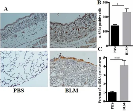
Bleomycin injection enhanced the expression of α-SMA in skin and lung tissues of mice compared with control group (p<0.05 in the skin and p<0.001 in the lung) (Figure 1A, B, C).
Figure 1. Bleomycin increases the expression of α-SMA in skin and lung tissues of bleomycin-induced systemic sclerosis mouse model. (A) Immunohistochemistry staining of α-SMA in skin and lung tissues of control group and bleomycin-induced systemic sclerosis mouse model group. Original magnification was 100X for skin (top) and 200X for lung slides (below). (B) Quantitative expression of α-SMA in skin tissues of control group and bleomycin-induced systemic sclerosis mouse model group. (C) Quantitative expression of α-SMA in lung tissues of control group and bleomycin-induced systemic sclerosis mouse model group. Results are presented as Mean±SEM. N=9-10 mice in each group. Data was analysed with unpaired t-test. BLM=Bleomycin group, PBS=Control group, α-SMA=Alpha-smooth muscle actin, p=probability value, ***=p<0.001, *=p<0.05
MMP9 Increases in Bleomycin-induced Lung Fibrosis
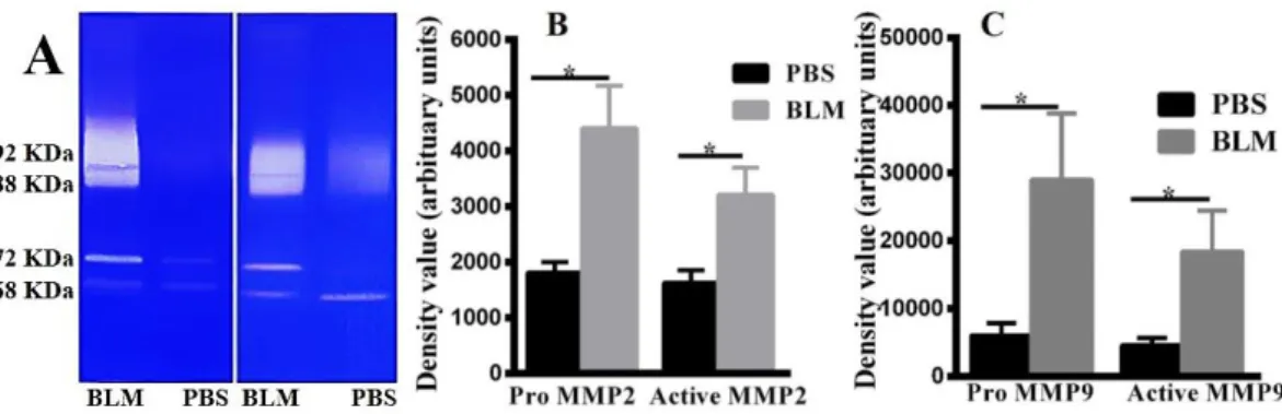
Zymographic analysis from lung tissue lysates of control group showed little or no bands of pro MMP9 and pro MMP2. Bleomycin administration significantly induced the expression of MMP9 bands in zymograms (Figure 2A).Administration of bleomycin increased the expression of active and pro MMP2 in a non-significant manner (p>0.05) (Figure 2B). The mean expression of active MMP9 in the bleomycin-treated group was 2.9 fold higher than the control group (26503±5658 vs
9121±1850, p<0.01) (Figure 2C). Additionally, the expression of pro MMP9 in BLM group was
significantly higher than the control group
(16660±5405 vs 4476±1058, p<0.05) (Figure 2C).
MMP9 and MMP2 Increase in Bleomycin-induced Skin Fibrosis
Analysis of gelatin zymographic gels demonstrated that MMP2 was the main gelatinase in skin tissues. In most normal skin lysates, MMP9 did not show any
augmented gelatinase activity in zymograms (Figure 3A). Expression of pro and active forms of MMP2 in skin zymograms of bleomycin received mice was intensified compared with control group (respectively
4411±758.8 vs 1813±192.0, 3213±488.2 vs
1632±224.5, p<0.05) (Figure 3B). Pro and active MMP9 increased in skin tissues of bleomycin received mice compared with the control group (respectively
29043±9779 vs 6044±1813, 18438±6016 vs
4590±1084, p<0.05) (Figure 3C).
Figure 2. MMP9 increases in lung tissues of bleomycin-induced systemic sclerosis mouse model. (A) Gelatin zymography of lung tissue lysate. 92 KDa=Pro MMP9, 88 KDa=Active MMP9, 72 KDa=Pro MMP2, 68 KDa=Active MMP2. (B) Density values of MMP9 in lung tissues. (C) Density values of MMP2 in lung tissues. Results are presented as Mean±SEM. N=9-10 mice in each group. Data was analysed with unpaired t-test. BLM=Bleomycin group, PBS=Control group, p=probability value, ns=p>0.05, ** =p<0.01, * =p<0.05
Figure 3. Gelatinases increase in skin tissues of bleomycin-induced systemic sclerosis mouse model. (A) Gelatin zymography of skin tissue lysate. 92 KDa=Pro MMP9, 88 KDa=Active MMP9, 72 KDa=Pro MMP2, 68 KDa=Active MMP2. (B) Density values of MMP9 in skin tissues. (C) Density values of MMP2 in skin tissues. Results are presented as Mean±SEM. N=9-10 mice in each group. Data was analysed using unpaired t-test. BLM=Bleomycin group, PBS=Control group, p=probability value,*=p<0.05
DISCUSSION
The obtained experimental results demonstrate that gelatinases increase in the lungs and skin tissues of a
mouse model of systemic sclerosis.
Systemic sclerosis (SSC) is a multisystem fibrotic autoimmune disease. Cell injury and aberrantly sustained production of cytokines, growth factors, and
angiogenic factors in SSc result in a deregulated tissue repair. These deregulated repairs in a tissue tilt its
homeostasis toward hyperplasia and excessive
accumulation of extracellular matrix (ECM).1
Excessive deposition of ECM components in tissues of an organ leads to fibrosis which destroys the operations of the organ.1 Due to the importance of ECM in the maintenance of organ function, regulation of its homeostasis is strictly critical. Studies emphasize that MMPs and their endogenous inhibitors represent a fundamental effect in this maintenance and their alterations can play a crucial role in the pathogenesis
of fibrotic diseases.2Additionally, MMPs have a
significant task in immunity and repair particularly in the procedures like cell migration, leukocyte activation, antimicrobial defense, and chemokine processing.17 As stated previously, the general function of MMPs is the degradation of the ECM proteins. It is argued that this functionality diminishes in fibrotic organs; however, considering the other operations of MMPs, this notion may be inaccurate.18 Accordingly, animal models and in vitro studies indicate pro and anti-fibrotic effects for MMPs. MMP3 and MMP7 in the mouse model of idiopathic pulmonary fibrosis (IPF) show pro-fibrotic function. Overproduction of MMP3 leads to fibrosis,
as a result, bleomycin-induced fibrosis was
dramatically reduced in MMP3–/– and MMP7−/−
mice.19,20
Over expression of MMP2 and MMP9 was confirmed in IPF patients, their expressions were seen in myofibroblasts, neutrophils, and epithelial cells.21,22 Also, alveolar macrophages of these patients expressed excessive amounts of MMP9 which was reduced by
immunosuppressive therapy.23 The increment of
gelatinases causes the disruption of the basement membrane and the invasion of fibroblast to the alveolar spaces which in turn benefits the fibrotic process.21,22 In SSc patients with interstitial lung disease (SSc- ILD) amount and activity of MMP9 is increased in
bronchoalveolar lavages compared with
bronchoalveolar lavages of SSc patients without ILD and healthy controls.24 Even though, there are no data available concerning the expression of these two gelatinases in tissues of SSc-ILD patients. Our results showed overexpression of pro and active form of MMP9 in fibrotic lung tissue and these are in accordance to results obtained from bronchoalveolar lavages of SSc-ILD patients and also compatible with results obtained from IPF patients.
In bleomycin-induced IPF, the expression of MMP9 is elevated especially in the early phase of disease induction. In this phase, MMP9 contributes to disruption of the alveolar epithelial cell basement membrane and progression of fibrosis. In the late stages, the gelatinolytic activities of the latent and active forms of MMP2 are prominent, and MMP2 is localized to the regenerated alveolar epithelial cells. MMP2, especially its active form, possibly plays a role in alveolar epithelial cell regeneration.12,13 In our study MMP9 was elevated but the increment of MMP2 was not seen, this event may be due to the use of different delivery protocols of bleomycin. In IPF model direct instillation of bleomycin occurred one time through trachea but in subcutaneous injection delivery of bleomycin occurs for 28 days period.25 In our model, chronic injury exists and repair does not occur until the injection is stopped, therefore increment of MMP2 for repairing was not seen in this model. Necessarily, further investigation would be done to evaluate the gelatinase activity after the induction of fibrosis and with the initiation of repair in a mouse model of scleroderma.
Despite the assumed function of MMP9 in lung fibrosis, the MMP9 null mice developed fibrosis similar to that of wild-type after bleomycin administration.26 By contrast, over expression of MMP9 in transgenic mice decreased bleomycin-induced fibrosis.27 The discrepancy between the results reported by different groups can be partly due to the timing of pathological assessment. Fibrosis process initiates with injury, inflammation, repair and finally resolution, the latter being was disrupted in the fibrotic disease. It is assumed that if MMPs are expressed in the early phase of disease induction, they have a pro-fibrotic effect but in late stages, they have an anti-fibrotic effect.18 Our study was done in the late stage of disease, therefore overexpression of MMP9 in fibrotic lung tissues may have an anti-fibrotic and protective effect against the bleomycin-induced fibrosis.
level of MMP9.30,31 In contrast to lung tissue (IPF patient), the expression of MMP9 in the skin of diffuse cutaneous SSc patients are low, but this expression does not alter in limited cutaneous disease.32 In keeping with results of other studies, our results indicate that in all samples of control skin, the gelatinolytic activity of MMP2 exists but the amount of MMP9 is low in normal skin tissues. We detected the increment of MMP2 in fibrotic skins but our obtained MMP9 data was different from results achieved from SSc patients, this may be due to the different origin of samples. However, it is obscure whether the overexpression of MMPs is a defense reaction of the body against the fibrogenic effect of bleomycin or this overproduction has synergy with the fibrogenic effect of bleomycin. There is still much unknown concerning the action of gelatinases in SSc based on the complexity in their functions, regulation and the lack of animal studies. Therefore further animal studies are required.
In this study, we could not evaluate the activity of gelatinases and their inhibitors (TIMPs) in fibrotic tissues before and after the disease induction. Also, it is necessary to mention that gelatinase zymography is a semiquantitative method therefore further techniques such as ELISA are required to reveal the real level of gelatinases.16
In conclusion, according to other studies, our study shows that MMPs activity is different and specific in various organs. The major gelatinase in normal lung tissue is MMP-9 while in skin tissue the MMP2 is highly expressed. In fibrotic lung and skin tissues, amount of gelatinases increased differently.
ACKNOWLEDGEMENTS
This work was partly supported by Grant Number 94013026723 from Iran University of Medical Sciences.
All animal works were done according to approval Number IR.IUMS.FMD.REC1394.25723.
REFERENCES
1. Ho YY, Lagares D, Tager AM, Kapoor M. Fibrosis—a lethal component of systemic sclerosis. Nat Rev Rheumatol 2014; 10(7):390-402.
2. Cox TR, Erler JT. Remodeling and homeostasis of the extracellular matrix: implications for fibrotic diseases and cancer. Dis Model Mech 2011; 4(2):165-78.
3. Nagase H, Visse R, Murphy G. Structure and function of matrix metalloproteinases and TIMPs. Cardiovasc Res 2006; 69(3):562-73.
4. Galis ZS, Muszynski M, Sukhova GK, Simon-Morrissey E, Libby P. Enhanced expression of vascular matrix metalloproteinases induced in vitro by cytokines and in regions of human atherosclerotic lesions. Ann N Y Acad Sci 1994; 748(1):501-7.
5. Liang KC, Lee CW, Lin WN, Lin CC, Wu CB, Luo SF, et al. Interleukin‐1β induces MMP‐9 expression via p42/p44 MAPK, p38 MAPK, JNK, and nuclear factor‐κB signaling pathways in human tracheal smooth muscle cells. J Cell Physiol 2007; 211(3):759-70.
6. Vu TH, Werb Z. Matrix metalloproteinases: effectors of development and normal physiology. Genes Dev 2000; 14(17):2123-33.
7. Gaffney J, Solomonov I, Zehorai E, Sagi I. Multilevel regulation of matrix metalloproteinases in tissue homeostasis indicates their molecular specificity in vivo. Matrix Biol 2015; 44:191-9.
8. Hajari Taheri F, Seyedolmohadesin M, Bayat M, Mahdavi M,Y azdi MH, Eslamifar A, et al. The effect of Candida albicans systemic infection on matrix metalloproteinases in breast cancer bearing BALB/c mice. Iran J Allergy Asthma Immunol 2013; 12(1):81-5. 9. Khorramizadeh MR, Hosseinzadeh S, Safavifar F, Saadat
F, Aalizadeh N, Falak R, et al. Interaction of CpG-oligodeoxynucleotides with Toll like receptor 9 induces apoptosis and modulates metaloproteinase-2 activity in human intestinal epithelium. Iran J Allergy Asthma Immunol 2007; 6(3):107-14.
10. Hadler-Olsen E, Fadnes B, Sylte I, Uhlin-Hansen L, Winberg JO. Regulation of matrix metalloproteinase activity in health and disease. FEBS J 2011; 278(1):28-45.
11. Aimes RT, QuigleyJP. Matrix Metalloproteinase-2 Is an Interstitial Collagenase Inhibitor-Free Enzyme Catalyzes the Cleavage of Collagen Fibrils and Soluble Native Type I Collagen Generating the Specific ¾-and ¼-Length Fragments. J Biol Chem 1995; 270(11):5872-6.
12. Kim JY, Choeng HC, Ahn C, Cho SH. Early and late changes of MMP-2 and MMP-9 in bleomycin-induced pulmonary fibrosis. Yonsei Med J 2009; 50(1):68-77. 13. Yaguchi T, Fukuda Y, Ishizaki M, Yamanaka N.
Immunohistochemical and gelatin zymography studies for matrix metalloproteinases in bleomycin‐induced pulmonary fibrosis. Pathol Int 1998; 48(12):954-63. 14. Yamamoto T, Takagawa S, Katayama I, Yamazaki
sclerotic skin. I: Local injections of bleomycin induce sclerotic skin mimicking scleroderma. J Invest Dermatol 1999; 112(4):456-62.
15. vafashoar F, Poormoghim H, Mousavizadeh K, Mousavi Shabestari T, Tavasoli A, Javadmoosavi SA, et al., The role of progesterone in cellular apoptosis of skin and lung in a bleomycin-injured mouse model. Iran J Allergy Asthma Immunol 2019; 18(1):100-7.
16. Toth M, Fridman R. Assessment of gelatinases (MMP-2 and MMP-9 by gelatin zymography. Methods Mol Med2001;57:163-74.
17. Page-McCaw A, Ewald AJ, Werb Z. Matrix metalloproteinases and the regulation of tissue remodelling. Nat Rev Mol Cell Biol 2007; 8(3):221-33. 18. Giannandrea M, Parks WC. Diverse functions of matrix
metalloproteinases during fibrosis. Dis Model Mech 2014; 7(2):193-203.
19. Manicone AM, Huizar I, and McGuire JK. Matrilysin (matrix metalloproteinase-7) regulates anti-inflammatory and antifibrotic pulmonary dendritic cells that express CD103 (αEβ7-integrin). The American Journal Pathology 2009; 175(6):2319-31.
20. Yamashita CM, Dolgonos L, Zemans RL, Young SK, Robertson J, Briones N, et al. Matrix metalloproteinase 3 is a mediator of pulmonary fibrosis. Am J Pathol2011; 179(4):1733-45.
21. Hayashi T, Stetler-Stevenson WG, Fleming MV, Fishback N, Koss MN, Liotta LA, et al. Immunohistochemical study of metalloproteinases and their tissue inhibitors in the lungs of patients with diffuse alveolar damage and idiopathic pulmonary fibrosis. Am J Pathol 1996; 149(4):1241-56.
22. Selman M, Ruiz V, Cabrera S, Segura L, Ramírez R, Barrios R, et al. TIMP-1,-2,-3, and-4 in idiopathic pulmonary fibrosis. A prevailing nondegradative lung microenvironment? Am J PhysiolLungCell Mol Physiol 2000; 279(3):L562-L574.
23. Lemjabbar H, Gosset P, Lechapt-Zalcman E, Franco-Montoya ML, WallaertB, Harf A, et al. Overexpression of alveolar macrophage gelatinase B (MMP-9) in patients with idiopathic pulmonary fibrosis: effects of steroid and immunosuppressive treatment. Am J Respir Cell Mol Biol 1999;20(5):903-13.
24. Andersen GN, Nilsson K, Pourazar J, Hackett TL, Kazzam E, Blomberg A, et al. Bronchoalveolar matrix metalloproteinase 9 relates to restrictive lung function impairment in systemic sclerosis. Respir Med 2007;101(10):2199-2206.
25. Lam AP, et al. Distinct patterns of pulmonary injury and
fibrosis induced by intratracheal and subcutaneous bleomycin in the mouse: Relevance for distinct forms of human lung fibrosis.in: Lorenzo Robertson editor.Cystic and Idiopathic Pulmonary Fibrosis: Risk Factors, Management and Long-Term Health Outcomes. Nova Science Publishers, Inc. 2016: 127-52.
26. Betsuyaku T, Fukuda Y, Parks WC, Shipley JM, Senior RM. Gelatinase B is required for alveolar bronchiolization after intratracheal bleomycin. Am J Pathol 2000; 157(2):525-35.
27. Cabrera S, Gaxiola M, Arreola JL, Ramírez R, Jara P, D'Armiento J, et al. Overexpression of MMP9 in macrophages attenuates pulmonary fibrosis induced by bleomycin. Int J Biochem Cell Biol 2007; 39(12):2324-38.
28. Solli AI, Fadnes B, Winberg JO, Uhlin-Hansen L, Hadler-Olsen E. Tissue-and cell-specific co-localization of intracellular gelatinolytic activity and matrix metalloproteinase 2. J Histochem Cytochem 2013; 61(6):444-61.
29. Fakhoury H, Hillarby M, and. Weiss J. Increased gelatinase activity in systemic sclerosis dermal fibroblast cultures with unaltered gelatinase A mRNA expression. J Dermatol Sci 2002; 29(1):62-9.
30. Kim WU, Min SY, Cho ML, Hong KH, Shin YJ, Park SH, et al. Elevated matrix metalloproteinase-9 in patients with systemic sclerosis. Arthritis Res Ther 2004; 7(1):R71-9.
31. Yazawa N, Kikuchi K, Ihn H, Fujimoto M, Kubo M, Tamaki T, et al. Serum levels of tissue inhibitor of metalloproteinases 2 in patients with systemic sclerosis. J Am Acad Dermatol 2000; 42(1):70-5.