Address for correspondence
Zeinab A. Salem
E-mail: [email protected]
Funding sources
None declared
Conflict of interest
None declared
Received on October 14, 2017 Reviewed on November 17, 2017 Accepted on December 27, 2017
Abstract
Background. Sofosbuvir is a nucleotide compound that has proved to be among the most potent orally available antiviral treatments. Infrequent but serious adverse events have been reported with the use of oral nucleos(t)ide analogues.
Objectives. Investigating sofosbuvir-induced alterations in the rat mandibular alveolar process. Material and methods. In the study, 30 adult male albino rats were used and divided randomly into fol-lowing groups: group 1, group 2 and group 3 (10 rats per group). Group 1 served as a control, group 2 re-ceived sofosbuvir through oral gavage at a dose of 40 mg/kg/day for 6 weeks, and group 3 was similar to group 2 but received sofosbuvir for 12 weeks. The animals were sacrificed at the end of the experiment. The mandibles were dissected and examined histologically as well as by scanning electron microscope (SEM) and energy dispersive X-ray unit (EDX).
Results. Histologically, group 1 showed normal alveolar process. In group 2, histopathological changes oc-curred as bone trabeculae demonstrated obvious Howship’s lacuna of osteoclasts. In group 3, bone trabe-culae exhibited multiple degenerated areas as well as apparent vacuolization. Scanning electron microsco-pic examination revealed smooth alveolar bone architecture in group 1. On the other hand, groups 2 and 3 demonstrated irregular bone architecture with formation of multiple pores. EDX analysis demonstrated the highest calcium concentration in the control group, while the lowest was found in group 3. Statistical ana-lysis of the EDX results revealed a statistically significant difference among the studied groups as the p-va-lue was <0.01.
Conclusions. It has been concluded that sofosbuvir induced apparent alterations in the rats’ alveolar bone. This effect was exaggerated in a longer period of drug administration. The sofosbuvir-induced alterations might be attributed mainly to mitochondrial toxicity. The effect had been clearly shown histologically and morphologically as well as in bone mineral (calcium) content.
Key words: hepatitis C virus, alveolar bone, sofosbuvir, nucleos(t)ide analogues, scanning microscopy Słowa kluczowe: wirus zapalenia wątroby typu C, kość wyrostka zębodołowego, sofosbuwir, analogi nukle-ozydów, mikroskopia skaningowa
DOI
10.17219/dmp/81592
Copyright
© 2018 by Wroclaw Medical University and Polish Dental Society
This is an article distributed under the terms of the Creative Commons Attribution Non-Commercial License (http://creativecommons.org/licenses/by-nc-nd/4.0/)
Alveolar process changes associated with administration
of nucleos(t)ide analogue (sofosbuvir) in rat model
Zmiany wyrostka zębodołowego związane z zastosowaniem
analogu nukleozydów (sofosbuwiru) w badaniu u szczurów
Dina Badawy Elsayed Farag
A–D,F, Dalia Abdel-Hameed El-Baz
A,C,D,F, Zeinab Amin Salem
A,B,E,FDepartment of Oral Biology, Faculty of Dentistry, Cairo University, Egypt
A – research concept and design; B – collection and/or assembly of data; C – data analysis and interpretation; D – writing the article; E – critical revision of the article; F – final approval of the article
Introduction
Nucleos(t)ide inhibitors are analogues of the natu-rally occurring polymerase substrates. They form the basis of antiviral therapy for a number of chronic viral diseases such as herpes simplex virus (HSV), human immunodeficiency virus (HIV), and hepatitis B virus (HBV).1,2 Nucleos(t)ides analogues resemble
physiologi-cal nucleos(t)ides in terms of uptake and metabolism, and are incorporated into newly synthesized DNA. The in-corporation of these compounds into DNA may induce either the termination of chain elongation or the accumu-lation of mutations in viral offspring. Some of these drugs also inhibit key enzymes involved in the generation of the purine and pyrimidine nucleotides and RNA synthesis, and directly activate caspase cascade. All of these effects may lead to cell death.3,4
Infrequent but serious adverse events have been re-ported with the use of oral nucleos(t)ide analogues. All nucleos(t)ide analogues have a “black box warning” regarding potential mitochondrial toxicity in their prod-uct labeling. The differences in analogue toxicity might result from a combination of cellular uptake, organellar transport, metabolic activation, incorporation and re-moval or degradation from the system.5
Sofosbuvir, acyclovir and tenofovir are nucleotide com-pounds proved to be among the most potent orally avail-able antiviral treatments. These drugs exhibit high ef-ficacy and a wide therapeutic index, with demonstrated utility in a number of chronic viral infections.6
Considering the published data regarding the adverse events associated with the use of oral nucleos(t)ide ana-logues, the present study was undertaken to evaluate the possible effects of sofosbuvir – a nucleo(s)tide ana-logue – on the mandibular alveolar process in albino rats through light microscopy, scanning electron microscopic examination and energy dispersive X-ray analysis (EDX).
Material and methods
Ethical statement
All experiments were conducted in the animal house of the Faculty of Medicine, Cairo University in Egipt ac-cording to the recommendations and approval of the Eth-ics Committee on animal experimentation of the Faculty of Oral and Dental Medicine, Cairo University in Egipt.
Experimental design
A total of 30 rats were used in the current study, weighing approx. 200 g. The animals were kept in a 12-hour light/dark cycle, at a temperature of 22°C ±2°C with relative humidity 50% ±20%. The animals were fed
on standard chow pellets and tap water ad libitum for the entire test period. The rats were randomly assigned into 3 groups of 10 rats each. Group 1 served as a con-trol and the rats received distilled water through oral ga-vage. Group 2 received sofosbuvir (Gratisovir® tablets, Pharco Pharmaceuticals, Alexandria, Egypt) dissolved in distilled water (8 mg/mL) through oral gavage at a dose of 40 mg/kg/day for 6 weeks.7 Group 3 received
sofosbu-vir dissolved in distilled water (8 mg/mL) through oral gavage at a dose of 40 mg/kg/day for 12 weeks.7
All ani-mals were sacrificed by ketamine overdose and the man-dibles were dissected. The right side of each mandible was used for light microscopic examination, forming a total of 30 bone segments (10 specimens from each group). The other 30 bone segments (10 specimens from each group) of the left sides of the mandibles were used for scanning electron microscopic (SEM) examination as well as analysis of the constituent element (calcium concentration) using an energy dispersive X-ray unit at-tached to the SEM.
Light microscopic examination
All the specimens were fixed in 10% neutral formalin for 48 h, washed, soaked in 10% ethylene diamine tetra-acetic acid (EDTA) for decalcification for 4 weeks, and then rinsed in distilled water. The specimens were dehy-drated in ascending grades of alcohol and embedded in paraffin. Mesiodistal sectioning of the right side of the jaw was carried out. Histological sections were prepared of 5 μm thickness. The sections were subjected to hema-toxylin and eosin staining according to the conventional method. Histopathological examination was performed using light microscope.
Scanning electron microscopic
examination
The specimens were fixed in 2.5% glutaraldehyde in phosphate buffer (pH 7.4) for 6 h. The specimens were then dehydrated in ascending grades of ethanol for 10 min at each passage and 20 min twice in absolute etha-nol, subjected to critical point drying according to stan-dard procedures for SEM processing, and left to dry in air at room temperature for 3 days. The specimens were then mounted on scanning electron microscope stubs and studied with a Quanta 250 Field Emission Gun (FEG) SEM (Thermo Fisher Scientific, Waltham, USA).
Energy dispersive X-ray analysis
The quantitative composition of calcium in the stud-ied samples was determined by EDX spot measurement, EDX line scan and element mapping. The EDX analysis unit works as an integrated feature of the Quanta 250 FEG SEM.
Statistical analysis
All the data obtained from the EDX analysis was statisti-cally evaluated. This data was represented as mean values ±SD, which was then statistically compared among the 3 studied groups. One-way analysis of variance (ANOVA) test for more than 2 independent samples was used, fol-lowed by Tukey’s post hoc test for pair wise comparison. The results were expressed in the form of p-values that were considered significant when the probability value was ≤ 0.05 and highly significant when the probability val-ue was ≤ 0.01. All statistical calculations were performed using following computer programs: Microsoft Excel 2007 and Statistical Package for the Social Science (SPSS) (SPSS Inc., IBM Corp., Armonk, USA) v. 15 for Microsoft Windows.
Results
Histological results
Group 1 (control group) showed normal alveolar bone architecture. The spongiosa consisted of interconnecting bone trabeculae containing osteocytes filling their lacu-nae. The bone trabeculae enclosed marrow cavities that were intensely lined with osteoblasts and filled with fibro-cellular tissue (Fig. 1).
Group 2 demonstrated bone trabeculae with randomly distributed osteocytes as well as scalloped reversal lines. Howship’s lacuna of osteoclasts were also clearly observed (Fig. 2).
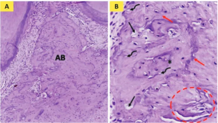
Group 3 exhibited bone trabeculae with multiple de-generated areas as well as apparent vacuolization. The teocytes were irregularly arranged. Moreover, some os-teocyte lacunae were seen empty. Multiple reversal lines were clearly demonstrated (Fig. 3).
Scanning electron microscopic results
The SEM images of the control group showed normal smooth alveolar bone architecture (Fig. 4).
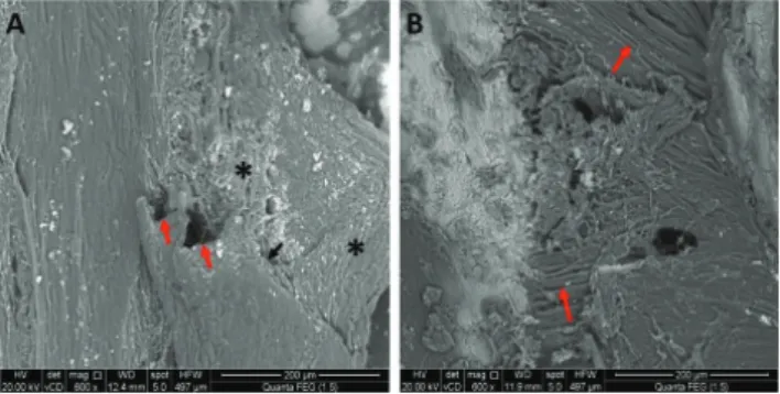
SEM examination of group 2 demonstrated alterations of normal bone architecture. The bone surface revealed roughness as well as pore formation. Peeling off of the bone surface with some broken trabeculae was also observed
Fig. 1. Photomicrograph of group 1 (control): (A) showing normal interdental alveolar bone (AB) consisting of interconnecting bone trabeculae (bt), osteocytes fi lling their lacunae (arrows) and marrow spaces (asterisks) (A: H&E ×100, B: H&E ×400)
Fig. 2. Photomicrograph of group 2: (A) showing interdental alveolar bone (AB), (B) showing bone trabeculae (bt) containing reversal lines (black arrows) and Howship’s lacuna (red arrows) (A: H&E ×100, B: H&E ×400)
Fig. 3. Photomicrograph of group 3: (A) showing interdental alveolar bone (AB), (B) showing bone trabeculae with multiple vacuoles (black arrows) and degenerated areas (red circle), multiple reversal lines (red arrows) and empty osteocytes lacunae (curved black arrows) (A: H&E ×100, B: H&E ×400)
Fig. 4. Scanning electron microscopic photomicrograph of group 1 (control) showing normal smooth alveolar bone architecture (asterisks) (SEM ×600)
in some areas. Other specimens displayed areas of disin-tegrated bone architecture with apparent mineralized col-lagen bundles. These colcol-lagen bundles seemed relatively well-organized (Fig. 5).
SEM examination of group 3 showed a rough porous eroded bone surface with irregular disorganized bone architecture. The mineralized collagen fibers were seen forming a meshwork. A crack along the bone surface was also observed (Fig. 6).
Energy-dispersive X-ray analysis
The EDX elemental analysis demonstrated the peak of the chemical element – calcium (Ca) concentration – found in the samples in each group. The calcium con-tent in the alveolar bone was evaluated in weight percent (wt%). EDX quantifies the relative contribution of Ca ele-ment to 100%. The mean values ±SD for total weight per-centage of calcium in groups 1, 2 and 3 were calculated (Table 1).
Mean total weight percentage of calcium was highest in group 1 while the lowest was demonstrated in group 3. ANOVA test demonstrated a statistically significant dif-ference among the 3 groups (p-value ≤ 0.01). Post-hoc multiple comparisons between 2 groups revealed that the difference in the mean value was statistically significant between groups 1 and 2, groups 1 and 3, and groups 2 and 3 (p-value ≤ 0.01) (Table 1).
Discussion
Nucleotide inhibitors (NIs) have proved great promise as direct-acting antivirals with broad genotype coverage, lack of preexisting alternatives with reduced susceptibil-ity, a high barrier to resistance, and the ability to produce potent and durable antiviral responses.8
The hepatitis C virus (HCV) infects approx. 3 million individuals in the United States and 180 million world-wide per year.9 NIs of HCV RNA synthesis served as
alter-nate substrates and inhibitors of the viral RNA-dependent RNA polymerase HCV nonstructural protein 5B (NS5B). The binding of NIs to the highly conserved NS5B active site resulted in activity that maintained across genotypes and substantial loss of viral fitness upon the infrequent development of resistance mutations.10
Sofosbuvir is an oral NI of the HCV NS5B RNA depen-dent RNA polymerase enzyme, and the mediator demon-strated this activity across all genotypes of HCV.11
Although sofosbuvir showed a good secure profile in clinical trials, common side effects have been reported with sofosbuvir treatment in humans. These included headache, insomnia, fatigue, nausea, dizziness, pruritis, upper respi-ratory tract infections, rash, back pain, grade 1 anemia, and grade 4 lymphopenia.12 Thus, we conducted this study to
investigate the possible effects of sofosbuvir on alveolar bone histology and surface structure in a rat model.
In the current study, the sofosbuvir dose was calculated so that it was equivalent to the clinically administered dose. The usual total human adult dose of sofosbuvir per day is 400 mg.13 According to Shin et al., this dose was equivalent
to 8 mg per day for a rat weighing about 200 g.7
The alterations in the alveolar process were evaluated in the ongoing research following administration of sofos-buvir for 6 and 12 weeks in groups 2 and 3, respectively.
Fig. 5. Scanning electron microscopic photomicrograph of group 2 showing: (A) rough bone architecture (asterisks), peeling off of the bone surface (black arrow) and broken trabeculae (red arrows); (B) areas of disintegrated bone architecture with apparent mineralized collagen bundles (red arrows) (SEM A&B ×600)
Fig. 6. Scanning electron microscopic photomicrograph of group 3 showing eroded disorganized bone architecture (asterisks), mesh work of mineralized collagen fi bers (red arrows) and a crack along the bone surface (white arrow) (SEM ×600)
Table 1. Comparison of mean values (mean ±SD) of calcium weight percentage in the studied groups
Studied groups Ca weight [%]
Group 1 85.79 ±0.3725
Group 2 73.468 ±0.3182a
Group 3 69.638 ±0.3669a,b
p-value* ≤0.01
* ANOVA test; Tukey’s post hoc test; a statistically significant with group 1
These chosen time intervals were based on data from clinical trials reporting that the recommended treatment course duration with sofosbuvir 400 mg once daily is 12 weeks.14 However the treatment duration was reported
to be guided by on-treatment response and it might be extended to 24 weeks.15
The histological results of the present study revealed histopathological changes in the rat alveolar process in both groups 2 and 3 as compared to group 1. In group 2, the bone trabeculae exhibited irregularly arranged osteo-cytes. Multiple Howship’s lacunae were also clearly seen denoting osteoclastic activity. Moreover, group 3 showed more pronounced changes as compared to group 2, where multiple vacuoles as well as degenerated areas in bone trabeculae were observed, indicating deteriorated bone structure. Empty osteocyte lacunae were observed, which indicate degeneration of osteocytes. Areas of post osteo-clastic activity were reflected by the presence of multiple reversal lines in both groups 2 and 3.
Osteocytes in the current study displayed various changes in both group 2 and 3. These changes suggested bone pathology, as it was reported that osteocytes rep-resent the key responder to various stimuli that regulate bone formation and remodeling as well as one of the key endocrine regulators of bone metabolism.16
The scanning electron microscopic results in the re-search provided good support for the histological findings. In group 2, the alveolar process bone architecture was rough. Moreover, group 3 showed a porous eroded bone surface with irregular disorganized bone architecture.
The relative content of calcium is critical for main-taining mineral homeostasis and bone metabolism.17
Ca content was considered a suitable biomarker for the assessment of bone health.18 EDX is a sensitive
qualita-tive and semiquantitaqualita-tive technique for the assessment of the mineral content variations in calcified tissue.19 The
EDX results in the herein study support the SEM find-ings. The analysis of the Ca concentration (%) revealed the highest concentration in the control group while the lowest was found in group 3. Moreover, statistical analysis of the EDX results revealed a statistically significant dif-ference among the studied groups.
It would appear from the reported results that there were obvious alterations in the alveolar bone accompa-nying sofosbuvir administration. The exact mechanism behind sofosbuvir’s effect on the alveolar process is not clear but it may be attributed to the fact that sofosbuvir is a member of the nucleos(t)ide analogues.
Early generations of NIs were reported to cause toxic-ity. All HCV NIs are ribonucleotide analogues and, there-fore, are more likely to attack host RNA polymerases than DNA polymerases.20
The mechanisms for toxicity of ribonucleotide analogues were not well characterized but they could be attributed to mitochondrial toxicity. NIs that were incorporated by the mitochondrial RNA polymerase (POLRMT) inhibited
mi-tochondrial protein synthesis and showed a corresponding decrease in mitochondrial oxygen consumption in cells.21
It has been reported that the use of sofosbuvir for 5 weeks resulted in swollen mitochondria with apparent cristolysis in the rat visual cerebral cortex.22
Mitochondrial dysfunction was reported to be the main factor in the formation of excess reactive oxygen spe-cies (ROS). The imbalance between these ROS and natural antioxidants created the conditions for oxidative stress.23
In bone tissues, recent studies have demonstrated that ROS generation is a key modulator of bone cell function and that oxidative status influences the pathophysiology of mineralized tissues. ROS can adversely affect bone ho-meostasis so that a proresorptive environment is favored. It was reported that ROS function as a signal mediator in osteoclast differentiation.24,25
Cadmium (Cd) has been shown to induce caspase-independent apoptosis through a mitochondria–ROS pathway.26 Zhao et al. investigated the effects of ROS in
response to cadmium exposure on osteoblasts in rats.27
They clarified that Cd had direct cytotoxic effect on os-teoblasts, which mediated by caspases and mitogen acti-vated protein kinase (MAPK) pathways.
A common dermatologic side effect reported with so-fosbuvir was pruritis and rash.28 Yonova has reported
that pruritis was found to be caused by some chemi-cal substance including histamine.29 Another proposed
cofactor that might be attributed to the alveolar bone histopathological changes demonstrated in the current study is the effect of histamine. Histamine was reported to be involved during the early phases of strong osteo-clast resorption.30
Conclusions
It has been concluded that sofosbuvir induced appar-ent alterations in rats’ alveolar bone. This effect was ex-aggerated upon a longer period of drug administration. The sofosbuvir-induced alterations might be attributed mainly to mitochondrial toxicity. The effect has been clearly shown histologically and morphologically as well as in bone mineral (calcium) content.
Further clinical and experimental studies are recom-mended to evaluate the effect of using sofosbuvir on vari-ous oral tissues.
References
1. Gerber L, Welzel TM, Zeuzem S. New therapeutic strategies in HCV polymerase inhibitors. Liver Int. 2013;33:85–92.
2. De Clercq E. A 40 year journey in search of selective antiviral che-motherapy. Annu Rev Pharmacol Toxicol. 2011;51:1–24.
3. Galmarini CM, Mackey JR, Dumontet C. Nucleoside analogues: Mechanisms of drug resistance and reversal strategies. Leukemia. 2001;15:875–890.
4. Lam AM, Espiritu C, Bansal S, et al. Genotype and subtype profiling of PSI-7977 as a nucleotide inhibitor of hepatitis C virus. Antimicrob Agents Chemother. 2012;56:3359–3368.
5. Johnson AA, Ray AS, Hanes J, et al. Toxicity of antiviral nucleoside analogs and the human mitochondrial DNA polymerase. J Biol Chem. 2001;276:40847–40857.
6. Mc Quaid T, Savini C, Seyedkazemi S. Sofosbuvir, a significant para-digm change in HCV treatment. J Clin Transl Hepatol. 2015;3:27–35. 7. Shin JW, Seol IC, Son CG. Interpretation of animal dose and human
equivalent dose for drug development. J Korean Oriental Med. 2010;31:1–7.
8. Sofia MJ. Nucleotide prodrugs for HCV therapy. Antivir Chem Che-mother. 2011;22:23–49.
9. Gower E, Estes C, Blach S, Razavi-Shearer K, Razavi H. Global epide-miology and genotype distribution of the hepatitis C virus infec-tion. J Hepatol. 2014;61:45–57.
10. Le Pogam S, Seshaadri A, Kosaka A, et al. Existence of hepatitis C virus NS5B variants naturally resistant to non-nucleoside, but not to nucleoside, polymerase inhibitors among untreated patients. J Antimicrob Chemother. 2008;61:1205–1216.
11. Stedman C. Sofosbuvir, a NS5B polymerase inhibitor in the treat-ment of hepatitis C: A review of its clinical potential. Therap Adv Gatroenterol. 2014;7:131–140.
12. Bhatia HK, Singh H, Grewal N, Natt NK. Sofosbuvir: A novel treat-ment option for chronic hepatitis C infection. J Pharmacol Pharma-cother. 2014;5:278–284.
13. Gentile I, Borgia F, Buonomo AR, Castaldo G, Borgia G. A novel promising therapeutic option against hepatitis C virus: An oral nucleotide NS5B polymerase inhibitor sofosbuvir. Curr Med Chem. 2013;20:3733–3742.
14. Lawitz E, Mangia A, Wyles D, et al. Sofosbuvir for previously untreat-ed chronic hepatitis C infection. N Engl J Med. 2013;368:1878–1887. 15. Zeuzem S, Dusheiko GM, Salupere R, et al. Sofosbuvir + ribavirin for
12 or 24 weeks for patients with HCV genotype 2 or 3: the VALENCE trial [abstract 1085]. In: Program and abstracts of the 64th Annual Meeting of the American Association for the Study of Liver Diseases. Washington, DC: 2013.
16. Neve A, Corrado A, Cantatore FP. Osteocytes: Central conductors of bone biology in normal and pathological conditions. Acta Physiol (Oxf). 2012;204:317–330.
17. Shapiro R, Heaney RP. Co-dependence of calcium and phospho-rus for growth and bone development under conditions of vary-ing deficiency. Bone. 2003;32:532–540.
18. Coats AM, Zioupos P, Aspden RM. Material properties of subchon-dral bone from patients with osteoporosis or osteoarthritis by microindentation testing and electron probe microanalysis. Calcif Tissue Int. 2003;73:66–71.
19. Kourkoumelis N, Balatsoukas I, Tzaphlidou M. Ca/P concentration ratio at different sites of normal and osteoporotic rabbit bones evaluated by Auger and energy dispersive X-ray spectroscopy. J Biol Phys. 2012;38:279–291.
20. Feng JY, Xu Y, Barauskas O, et al. Role of mitochondrial RNA poly-merase in the toxicity of nucleotide inhibitors of hepatitis C virus. Antimicrob Agents Chemother. 2015;60:806–817.
21. Arnold JJ, Sharma SD, Feng JY, et al. Sensitivity of mitochondri-al transcription and resistance of RNA polymerase II dependent nuclear transcription to antiviral ribonucleosides. PLoS Pathog. 2012;8:e1003030.
22. Issa NM, El-Sherif NM. Light and electronic histological studies to the effect of Sofosbuvir on the visual cerebral cortex of adult male albino rat. J Am Sci. 2017;13:79–87.
23. Betteridge DJ. What is oxidative stress? Metabolism. 2000;49:3–8. 24. Wauquier F, Leotoing L, Coxam V, Guicheux J, Wittrant Y.
Oxi-dative stress in bone remodeling and disease. Trends Mol Med. 2009;15:468–477.
25. Moon H, Kim SE, Yun YP, et al. Simvastatin inhibits osteoclast dif-ferentiation by scavenging reactive oxygen species. Exp Mol Med. 2011;43:605–612.
26. Shih CM, Ko WC, Wu JS, et al. Mediating of Caspase independent apoptosis by cadmium through the mitochondria-ROS pathway in MRC-5 fibroblasts. J Cell Biochem. 2004;91:384–397.
27. Zhao H, Liu W, Wang Y, et al. Cadmium induces apoptosis in prima-ry rat osteoblasts through Caspase and mitogen-activated protein kinase pathways. J Vet Sci. 2015;16:297–306.
28. Bhatia HK, Singh H, Grewal N, Natt NK. Sofosbuvir: A novel treat-ment option for chronic hepatitis C infection. J Pharmacol Pharma-cother. 2014;5:278–284.
29. Yonova D. Pruritis in certain internal diseases. Hippokratia. 2007;11:67–71.
30. Lesclous P, Schramm F, Gallina S, Baroukh B, Guez D, Saffar JL. Hista-mine mediates osteoclastic resorption only during the acute phase of bone loss in ovariectomized rats. Exp Physiol. 2006;91:561–570.