Original Article
Effect of pH and Lidocaine on the Compressive Strength of Calcium
Enriched Mixture Cement
Sobhnamayan Fa, Adl ARb, Farzaneh Zc, Sadat Shojaee Na
aDepartment of Endodontics, School of Dentistry, Shiraz Universal of Medical Sciences, Shiraz, Iran
bDepartment of Endodontics and Biomaterials Research Center, School of Dentistry, Shiraz University of Medical Sciences, Shiraz, Iran
cUndergraduate Student, Student Research Committee, Shiraz Universal of Medical Sciences, Shiraz, Iran
ARTICLE INFO Abstract
Article History: Received 5 August 2015 Accepted 21 November 2015
Statement of Problem: The pH of the human abscess has been measured as low as 5.0. This low pH could potentially inhibit setting reactions, affect
adhesion, or increase the solubility of root end filling materials hence affect the compressive strength. Moreover, root end filling materials might expose or even mix with lidocaine HCL during periapical surgery.
Objectives: The aim of this in vitro study was to evaluate the effect of acidic pH
and lidocaine on the compressive strength of calcium-enriched mixture (CEM).
Materials and Methods: CEM was mixed according to the manufacturer’s instructions or with lidocaine (L), and condensed into 6 × 4 mm split moulds. The samples were exposed to phosphate buffered saline (PBS) at pH 5 or 7.4 for 7 or 28 days. Cylindrical blocks of CEM (total number = 120 and 15 for
each group) were subjected to compressive strength test using a universal testing machine. Data were analysed using three-factor analysis of variance (ANOVA).
Results: Regardless of pH and time, significant differences were not found between lidocaine groups and the groups that were mixed according to the manufacturer’s instruction (p = 0.083). For both mixing agents, regardless of time, there were no significant differences between the two pH levels (p = 0.157). Regardless of the material and pH, there was a significant increase in the compressive strength from days 7 to 28 (p < 0.001).
Conclusions: Mixtures with lidocaine and exposure to an acidic environment
had no adverse effects on the compressive strength of CEM Cement. Key words:
Calcium-enriched Mixture
Compressive Strength
Lidocaine
pH
Corresponding Author: Alireza Adl,
Department of Endodontics and Biomaterials Research Center, School of Dentistry, Shiraz University of Medical Sciences, Shiraz, Iran Email: adla@sums.ac.ir
Tel: +98-9171005071
Cite this article as: Sobhnamayan F, Adl AR, Farzaneh Z, Sadat Shojaee N. Effect of pH and Lidocaine on the Compressive Strength of Calcium Enriched Mixture Cement. J Dent Biomater, 2015;2(4):118-123.
Introduction
Root end surgery is considered to be the final option for cases in which non-surgical endodontic retreatment
has failed or conventional root canal therapyis not possible [1]. In this method, after root resection and root end cavity preparation, a reparative material is applied to fill the root end cavity. Root end fillings are
inevitably exposed to the inflamed tissue or even anaesthetic solutions used for bleeding control. The pH of inflamed tissue has been estimated to be as low as 5.0, and the pH of lidocaine HCL as an anaesthetic solution is estimated to be 4.2 [2]. The low pH of the environment or the anaesthetic solution may potentially affect the physical and chemical properties of root end filling materials. Acidic environments adversely affect the hydration behaviour of mineral trioxide aggregates (MTA) [3,4]. Low pH also reduces the surface hardness [5], and bond strength [6] of this material.
Calcium enriched mixture (CEM) (Bionique Dent, Tehran, Iran) is a tooth-coloured, water-based cement that has been introduced recently. This new cement is composed of different calcium compounds, such as calcium hydroxide, calcium oxide, calcium phosphate, calcium sulphate, calcium silicate and calcium carbonate [7]. High concentrations of water-soluble calcium and phosphate cause the immediate formation of hydroxy apatite during and after setting [8]. CEM cement is compatible for handling and setting in an aqueous environment [8]. It has been shown that this cement exhibits excellent sealing properties [9] and biocompatiblity [10]. The cytotoxicity of this cement is comparable to MTA [11]. Because of these characteristics, CEM is used for management of internal and external resorption [12], furcalperforation [13], and pulpotomy of primary and permanent molars [14,15]. In a recent study on CEM, lower pH values in highly acidic environments (pH = 4.4) adversely affected the
force needed for the displacement of this cement, while in higher pH values (6.4) the bond-strength was not affected [16].
Given the uncertain characteristic of CEM cement when exposed to different environments, and the fact that during periapical surgery, CEM might be exposed or even mix with lidocaine HCL. The purpose of this study was to evaluate the compressive strength of CEM cement when mixed with its specific liquid or lidocaine and exposed to an acidic or neutral environment.
Materials and Methods
Twenty-four Custom-made, two-part split Plexiglass moulds were used in this experiment. Each mould had five holes with internal diameters of 4 mm and heights of 6 mm. The moulds were randomly allocated into eight groups, according to the mixing agent, pH, andtime period (Table 1).
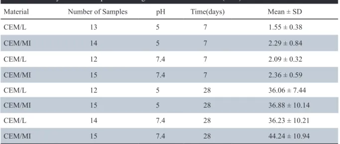
For half of the groups, CEM was mixed with its specific liquid according to the manufacturer’s instructions (CEM/MI), and for the other half, 2% lidocaine HCL (DarouPakhsh, Tehran, Iran) withepinephrine (1:80000) was substituted for the specific liquid (CEM/L). The experimental CEM mixtures were introduced incrementally into the moulds by amalgam carrier. After gentle packing and compacting with condensers, excess material was removed with wet cotton pellets. A glass slab was secured to one end of each split mould. The moulds were then wrapped into wet pieces of gauze Table 1: Summary of mean compressive strength and standard deviation (MPa)
Material Number of Samples pH Time(days) Mean ± SD
CEM/L 13 5 7 1.55 ± 0.38
CEM/MI 14 5 7 2.29 ± 0.84
CEM/L 12 7.4 7 2.09 ± 0.32
CEM/MI 15 7.4 7 2.36 ± 0.59
CEM/L 12 5 28 36.06 ± 7.44
CEM/MI 15 5 28 36.88 ± 10.14
CEM/L 14 7.4 28 36.23 ± 10.21
saturated with PBS titrated to pH 5.0 or 7.4, and kept in an incubator at 37 °C for 24 hours, to ensure preliminary set. Next, the moulds were wrapped into new wet pieces of gauze saturated with the same solutions for a total of seven or 28 days. On each testing day, the moulds were split and CEM blocks were removed carefully by applying a light force, taking care not to damage the CEM samples. After removal, the blocks were evaluated for voids or cracks.
To test for compressive strength, the CEM blocks were placed lengthwise between the platens of a universal testing machine (Zwick/Roell Z020 Zwick, CombH & Co, Germany) and compressed using a cross-head speed of 1 mm/min [6]. The load at fracture was recorded and plotted on a graph in megapascals (MPa). Differences between the groups were statistically analysed using three-factor analysis of variance (ANOVA). The Statistical Package for Social Sciences, version 16 (SPSS Inc.,
Chicago, IL, USA) was used to analysed the data. Results
Some CEM blocks, mainly from the lidocaine groups, fractured during the removal of the sample from the moulds. The number of samples, mean compressive strengths and standard deviations of the eight experimental groups, are presented in Table 1. The highest and the lowest compressive strength values were recorded in the CEM/MI in the pH 5/day 28 group, and CEM/L in the pH 5/day 7 group, respectively. Regardless of the pH and time, there were no significant differences between the lidocaine groups and the groups prepared according to the manufacturer’s instruction (p = 0.083). For both mixing agents, regardless of time, there was no significant difference in regard to pH (p = 0.157). Regardless of the mixing agents and pH, there was a significant increase in the compressive strength Table 2: The results of three – way ANOVA
Source Type lll Sum of squares df Mean Square F Sig
Corrected Model intercept 35875.724a 41109.940
7
1
5125.103
41109.940
101.871 817.140
.000 .000
Material 154.289 1 154.289 3.067 .083
Ph 102.480 1 102.480 2.037 .157
Time 33162.097 1 33162.097 659.161 .000
Material* ph 72.791 1 72.791 1.447 .232
Material* time 94.566 1 94.566 1.880 .174
Ph* time 77.269 1 77.269 1.536 .218
Material* ph* time 90.493 1 90.493 1.799 .183
Error 4880.028 97 50.310
Total 833668.659 105
from days seven to 28 (p < 0.001). The results of three-way ANOVA tests have been presented in Table 2.
Discussion
The clinical relevance in testing for compressive strength of the root end filling material is considered not only to ensure that they can stand the forces caused by tooth function or operative procedures, but also to verify that they are completely set [6,17]. Although mechanical tests are unable to reflect the clinical situation, they can show the effects of different mixing liquids and setting conditions on different cement types.
According to ISO 9917-1 (2003) standards, for the compressive strength test, a split mould design made of a material that will not be affected by the cement has been advised. In a study on the compressive strength of MTA, plastic split moulds [18] have been used. In this study, two-part split Plexiglass moulds were used to form CEM samples. A pilot study conducted prior to this study showed that samples required a light force to allow for removal.
The results of the present study showed that mixing CEM cement with lidocaine HCL did not have adverse effects on the compressive strength of this material. However, these results should be interpreted with caution because despite using two-part split moulds that required only light force for removal of the CEM samples, nine samples from the lidocaine groups versus one sample of CEM/ MI were fractured during removal from the moulds. Therefore, one may assume that mixing CEM cement with lidocaine has deleterious effects on the physical properties of this cement.
The present study also showed that acidic environments did not adversely affect the compressive strength of CEM cement in pH 5, which is similar to an infectious environment.
On the other hand, the present study is not in agreement with Watts et al.’s study that reported mixture with lidocaine and also exposure to pH 5 caused a significant decrease in the compressive strength of both white and grey MTA [6]. Saghiri
et al. also showed that acidic environments can drastically affect the compressive strength of nano white MTA, WMTA and bioaggregate (BA) [19]. Other studies also reported the adverse effect of
acidic environments on the sealing ability and bond strength of MTA [20,21].
These discrepancies could be attributed to the different experimental setups that have been used in these studies. For example, Watt et al. removed the MTA samples from the moulds after three days and immersed them in PBS with different pH for a total of seven and 28 days [6] while in the present study the CEM samples were exposed to different pH levels within their respective moulds therefore the exposure area to the acid was lower than in watts et al.’s study. The different chemical composition and particle size of different cements might also explain the differences in the results of different studies. The chemical composition of CEM used in this study is different from that of MTA and BA [9]. CEM cement also has a smaller particle size compared to MTA, which might cause it to be less affected by acidic environments [7,22]. In the case of BA, it has been shown that BA has a very similar composition with MTA, but it is aluminium-free and has different opaquers [23].
Only few studies have evaluated the effect of time on the compressive strength of CEM. In the present study, the time intervals caused significant increases in the compressive strength of all groups.
The results of the current study are in agreement with those of Rahimi et al. who reported an increase in the bond strength of CEM cement from 24 hour to seven days [24]. Another study also showed that the bond strength of this cement increased from day 3 to day 21 [25-26]. Watts et al. showed an increase in the compressive strength of white MTA and grey MTA when mixed with sterile water from day 7 to 28. But when WMTA and GMTA were mixed with local anaesthetic, the time intervals caused a significant decrease in their compressive strength, which was not in agreement with the findings of the present study [6].
Conclusions
The most important factor in the increase of CEM cement compressive strength is time. Although mixing with lidocaine and exposure to acidic environments had not statistically affected the compressive strength of CEM, it is not recommended in clinical situations until further studies are conducted.
Acknowledgements
This manuscript is based on the thesis by Farzaneh Z.The authors thank the Vice-Chancellory of Shiraz University of Medical Sciences for the supporting this research (Grant # 8793102). The authors also thank Dr. M. Vosoughi from the Dental Research Development Center of the School of Dentistry for the statistical analysis.
Conflict of interest
The authors deny any conflicts of interest related to this study.
Referances
1. Torabinejad M, Chivian N. Clinical applications of mineral trioxide aggregate. J Endod. 1999;25:197-205.
2. Malamed SF. Handbook of Local Anesthesia. 5th ed. St. Louis MO: Mosby Inc; 2004. 3. Ford TR, Torabinejad M, McKendry DJ, et al.
Use of mineral trioxide aggregate for repair of furcal perforations. Oral Surg Oral Med Oral Pathol Oral Radiol Endod. 1995;79:756-763. 4. Lee YL, Lee BS, Lin FH, et al. Effects of
physiological environments on the hydration behavior of mineral trioxide aggregate. Biomaterials. 2004;25:787-793.
5. Namazikhah MS, Nekoofar MH, Sheykhrezae MS, et al. The effect of pH on surface hardness and microstructure of mineral trioxide aggregate. Int Endod J. 2008;41:108-116. 6. Watts JD, Holt DM, Beeson TJ, et al. Effects of
pH and mixing agents on the temporal setting of tooth-colored and gray mineral trioxide aggregate. J Endod. 2007;33:970-973.
7. Asgary S, Shahabi S, Jafarzadeh T, et al. The properties of a new endodontic material. J Endod. 2008;34:990-993.
8. Chng HK, Islam I, Yap AU, et al. Properties of a new root-end filling material. J Endod. 2005;31:665-668.
9. Asgary S, Eghbal MJ, Parirokh M. Sealing ability of a novel endodontic cement as a root-end filling material. J Biomed Mater Res A. 2008;87:706-709.
10. Asgary S, Eghbal MJ, Parirokh M, et al. A
comparative study of histologic response to different pulp capping materials and a novel endodontic cement. Oral Surg Oral Med Oral Pathol Oral Radiol Endod. 2008;106:609-614. 11. Mozayeni MA, Milani AS, Marvasti LA, et
al. Cytotoxicity of calcium enriched mixture cement compared with mineral trioxide aggregate and intermediate restorative material. Aust Endod J. 2012;38:70-75.
12. Asgary S, Nosrat A, Seifi A. Management of inflammatory external root resorption by using calcium-enriched mixture cement: a case report. J Endod. 2011;37:411-413.
13. Asgary S. Furcal perforation repair using calcium enriched mixture cement. J Conserv Dent. 2010;13:156-158.
14. Asgary S, Ehsani S. Permanent molar pulpotomy with a new endodontic cement: A case series. J Conserv Dent. 2009;12:31-36.
15. Malekafzali B, Shekarchi F, Asgary S. Treatment outcomes of pulpotomy in primary molars using two endodontic biomaterials. A 2-year randomised clinical trial. Eur J Paediatr Dent. 2011;12:189-193.
16. Sobhnamayan F, Sahebi S, Naderi M, et al. Effect of acidic environment on the push-out bond strength of calcium-enriched mixture cement. Iran Endod J. 2014;9:266-270.
17. Parirokh M, Torabinejad M. Mineral trioxide aggregate: a comprehensive literature review-Part I: chemical, physical, and antibacterial properties. J Endod. 2010;36:16-27.
18. Holt DM, Watts JD, Beeson TJ, et al. The anti-microbial effect against enterococcus faecalis and the compressive strength of two types of mineral trioxide aggregate mixed with sterile water or 2% chlorhexidine liquid. J Endod. 2007;33:844-847.
19. Saghiri MA, Garcia-Godoy F, Asatourian A, et al. Effect of pH on compressive strength of some modification of mineral trioxide aggregate. Med Oral Patol Oral Cir Bucal. 2013;18:714-720. 20. Saghiri MA, Lotfi M, Saghiri AM, et al. Effect
of pH on sealing ability of white mineral trioxide aggregate as a root-end filling material. J Endod. 2008;34:1226-1229.
21. Shokouhinejad N, Nekoofar MH, Iravani A, et al. Effect of acidic environment on the push-out bond strength of mineral trioxide aggregate. J
Endod. 2010;36:871-874.
22. Soheilipour E, Kheirieh S, Madani M, et al. Particle size of a new endodontic cement compared to root MTA and calcium hydroxide. Iran Endod J. 2009;4:112-116.
23. Park JW, Hong SH, Kim JH, et al. X-Ray diffraction analysis of white ProRoot MTA and Diadent BioAggregate. Oral Surg Oral Med Oral Pathol Oral Radiol Endod. 2010;109:155-158.
24. Rahimi S, Ghasemi N, Shahi S, et al. Effect of blood contamination on the retention
characteristics of two endodontic biomaterials in simulated furcation perforations. J Endod. 2013;39:697-700.
25. Sobhnamayan F, Adl A, Shojaee NS, et al. The effect of chlorhexidine on the push-out bond strength of calcium-enriched mixture cement. Iran Endod J. 2015;10:59-63.
26. Sobhnamayan F, Adl A, Sarbaz M, et al. Push-out Bond Strength of Calcium Enriched Mixture Exposed to Alkaline Environment. J Dent Biomater. 2015;2:92-96.