BIOCIDAL ACTIVITY OF Ag-PEG-CHITOSAN
NANOCOMPOSITE FILM PREPARED USING CANNABIS
SATIVA AQUEOUS LEAF EXTRACT BY SONICATION
Garima Ameta
[a]*and Pinki B. Punjabi
[a]Keywords: Biosynthesis, crosslinking, nanocomposite, ultrasound, biocidal activity.
This study underscores the development of Ag-PEG-Ch nanocomposite film using the aqueous leaves extract of Cannabis sativa under the influence of ultrasound, as smart substrates for antimicrobial uses, via innovative in situ reactive and reduction pathways. Ag nanoparticles that are synthesized by using leaves extract and ultrasound as an energy source have emerged as nontoxic and ecofriendly. In this study, silver nitrate was used as a silver precursor and PEG and chitosan were used as the polymeric stabilizer and solid support, while cannabis sativa leaves extract acts as a green reducing agent. This polymer metal film was characterized by FT-IR, SEM, TEM, TGA, DSC and antimicrobial activities. XRD peaks clearly reveals that FCC Ag nanoparticles embedded in polymer matrix, very fine Ag nanoparticles (4-6 nm) can be synthesized through ultrasound has been proven by TEM images and TGA-DSC results show that this nanocomposite film is thermally stable. This nanocomposite film possesses synergistic biocidal activities also.
* Corresponding Authors Tel: +918003180487
E-Mail: [email protected]
[a] Department of Chemistry, University College of Science, Mohanlal Sukhadia University, Udaipur - 313002 (Raj.) India
INTRODUCTION
Since ancient times, different forms of silver have been used as an antimicrobial agent against infections, before recognizing their nanometric dimension.1 Chemical and
physical factors associated with Ag nanoparticles such as size,2,3 shape and surface charge4 enhance their
antimicrobial,5 antibacterial,6 anti-viral,7 anti-cancer
properties8 and affect significantly the effective doses to
inhibit the growth of microorganism. It is also useful in drug delivery system.9
Nevertheless, commercially available silver-based drugs have shown cytotoxic effects on various experimental models.10,11 So, there is a growing need to develop an
eco-friendly process, which does not use toxic chemicals in the synthesis protocols. In a green biosynthesis of nanoparticles, selection of a green reducing and stabilizing agents in an aqueous medium are the most important issues which must be considered. Green biosynthesis approaches include biopolymers, biological molecules, and green irradiation method which have advantages over conventional methods involving hazardous chemical agents. It also enhances the antimicrobial activities of Ag nanoparticles and to overcome the adverse cytotoxic effects of silver.
Polyethylene Glycol (PEG) is a water-soluble polymer and widely used in mechanical, pharmaceutical and cosmetic industries. PEG reduces AgNO3 and stabilizes Ag
nanoparticles.12,13 Stabilization can be obtained due to the
free polymer chains in solution, where aggregation is denied because of steric hindrance. PEG is easily permeable through membrane would facilitate the entry of
nanoparticles carrying antibiotics across the membrane, enhance the binding of the delivered drug to the bacterial DNA and block drug efflux pumps.14
Chitosan is a natural polysaccharide obtained by the deacetylation of chitin.15 It is a linear polyamine containing
number of free amine groups that are readily available for crosslinking. This biopolymer shows unique chelating,16,17
polycationic,18 film forming19 and antimicrobial properties20
due to the presence of active amino and hydroxyl groups in polymeric matrix. Biocidal activity of chitosan has been studied against yeast,21 fungi, mold22,23 and bacteria.24
Chitosan has significant advantages over others due to its low cytotoxicity towards mammalian cells.25,26
PEG-Chitosan blend exhibited better physico-chemical properties comparable to chitosan or PEG alone.27
Silver nanoparticles have been prepared conventionally by chemical method using reducing agents,28 which were later
responsible for various biological hazards due to their cytotoxic effect. To cure this problem, green synthesis using biological molecule and the use of ultrasound as an energy source29,30 exhibit superiority over chemical methods. Plant
extracts have been demonstrated to be promising reducing and capping agents for the biosynthesis of silver nanoparticles, some of them are Crocus sativus L.,31 Alpinia
katsumadai,32 Kalopanax septemlobus,33 Acalypha indica,34
Citrus limon,35 Alternanthera sessilis,36 Aloe vera,37 Lippia
nodiflora,38 Pedalium murex,39 etc. Cannabis sativa is
an herbaceous flowering plant belonging to the family Cannabaceae, native of eastern Asia. The plant is known to contain more than 113 phyto cannabinoids,40 which have
pharmacological importance. It is widely used to treat
schizophrenia,41 neurophatic pain,42 cancer pain,43
chemotaxonomic purpose,44 chronic inflammation,45
dibetes,46 multiple sclerosis,47 as an antipsychotic48 and
analgesic drug.49 To the best of our knowledge, there has
as a reducing agent to convert Ag+ ion to Ag0 particle.
Antimicrobial activities of this synthesized compound have also been studied.
EXPERIMENTAL
PEG, chitosan (Himedia, India), silver nitrate, ethanol and acetic acid (Fisher Scientific, India) were used as received. Ultrasonic cleaner 392 (Systronics, India) was used for ultrasonic irradiation with a frequency of 40 kHz and a nominal power of 115 W.
FTIR spectra were recorded on a FT-IR Spectrometer (Perkin Elmer, India) Spectrum GX Range with a resolution of 0.15 cm-1. Spectra were scanned between 400 and 4000
cm−1. Scan time was 20 scan s-1. XRD pattern measurement
[Table Top X-ray Diffraction System (Rigaku Miniflex, USA)] was employed using monochromatic Cu kα radiation (λ = 1.5406 Å) operated at 40 kV and 30 mA at a 2θ angle pattern to study the crystal structures of the nanocomposite. XRD data obtained were compared with the Joint Committee on Powder Diffraction Standards (JCPDS) library to account for the crystalline structure of the nanocomposite.
TEM images for polyethylene glycol-chitosan-silver ternary nanocomposite thin film were recorded using a Philips (Holland), Model: Tecnai 20, transmission electron microscope operating at an acceleration voltage of 200 kV. Electron source are W emitter and LaB6. This instrument uses electron beam optics to achieve very high magnifications of the order of 7,50,000x.
Thermal Analysis (DSC and TGA) – Perkin Elmer (India) Pyris-1 DSC, Pyris-1 TGA was used to analyze nanocomposite thin film. Temperature range for TGA was room temperature to 700ºC and for DSC, it was 40ºC to 400ºC. All observations were carried out under nitrogen atmosphere.
Preparation of C. sativa leaves extract
Fresh leaf extract of C. sativa was used to reduce Ag+ ions
to Ago. Fresh leaves of C. sativa were collected from Dakan
Kotra, Girwa, Udaipur. They were cleaned with distilled water and dried at room temperature. About 10 g of finely cut leaves were placed in a beaker containing double distilled water (100 mL) and boiled for 10 min before decanting. Then the extract was cooled down and filtered through Whatman filter paper No.1 and stored at 4 C for further use.
Synthesis of Ag-PEG-Ch nanocomposite thin film
All the reactions were carried out in an ultrasonic bath. 0.2 g of chitosan was dissolved in 100 mL of 2% aqueous acetic acid and irradiated with ultrasound for 20 min. to obtain clear solution. Reaction of PEG (0.01 M) with silver nitrate (0.01 M, in 5 mL of C. sativa aqueous leaf extract) was carried out for 2 h under the influence of ultrasound to give PEG dialdehyde with silver nanoparticles.Change in colour from light yellow to dark brown, indicated the formation of AgNPs. Chitosan solution was added dropwise in ethanol,
and then added freshly prepared PEG dialdehyde with silver nanoparticles and few drops of HCl to crosslink the polymer. This solution was washed with DD water and ethanol then centrifuged. Glass slides were cut to 20 mm × 15 mm to fit the spin coater, cleaned by sonication in acetone, and allowed to dry. A total of 0.2 mL of the prepared nanocomposite solution was coated onto the glass slide by spin coating at 500 rpm for 120 s. The resulting films were allowed to dry in vacuum.
Assessment of antimicrobial assay
Synthesized Ag-PEG-Ch nanocomposite using leaves extract of C. sativa was analyzed for antimicrobial activity against two Gram positive bacterial strains such as S. aureus (MTCC 96) and S. pyogenus (MTCC 442), two Gram negative bacterial strains like E. coli (MTCC 443) and P. aeruginosa (MTCC 1688). Antifungal studies were also carried out on three fungal strains such as C.albicans (MTCC 227), A.niger (MTCC 282) and A.clavatus (MTCC 1323) at Microcare Laboratory, Surat, India.
The minimum inhibitory concentration (MIC) for the sample was evaluated using Broth dilution method to determine the antibacterial and antifungal activity against various bacterial and fungal strains.50 The bacterial stock
cultures were incubated for 24 h at 37 ◦C in Nutrient Agar media and while the fungal stock cultures were incubated for 72 h at 28 ◦C in potato dextrose agar media followed by refrigeration storage at 4 ◦C. The stock cultures were maintained at 4 ◦C.
RESULTS AND DISCUSSION
FTIR Spectrum of Ag-PEG-Ch thin film shows the combination of the IR absorption characteristic of PEG and chitosan (figure 1). Band appearing at 3429 cm-1 is due to
the overlapping of O-H and N-H stretching, 2923 cm-1 is
due to for C-H stretching, bands at 1631 cm-1 and 1524 cm-1
are due to the N-H bending. A medium band that appears at 1384 cm-1, is a significant band that confirms the formation
of metallopolymer (Ag-PEG-Ch).51
XRD patterns of Ag-PEG-Chnano composites are shown in figure 2. Two medium peaks appearing at 2θ values of 19.5 and 22.9 are confined to the polymeric chains (chitosan and PEG). The main peaks around 2θ values of 38.4, 44.2, 64.5 and 77.7 with 111, 200,220 and 311diffraction respectively are related to Face Centered Cubic (FCC) crystalline structure of silver in a polymeric matrix, which is supported by the JCPDS file no. 89-3722.. Therefore, this gives clear evidence for the presence of FCC Ag nanoparticles in Ag-PEG-Ch nanocomposite film.
The average particle size of the nanocomposite was calculated by the Debye-Scherrer equation and is estimated to be 6 nm, which clearly demonstrate that finer Ag nanoparticles can be synthesize by the use of ultrasound as compared to those by other methods.52 In addition, some
unassigned peaks might have resulted due to the capping agent stabilising the Ag nanoparticles, which may be due to the bioorganic compounds in a plant extract used.53,54 These
Topographical analysis
Transmission electron microscopy (TEM) studies of Chitosan-PEG-Ag nanocomposites were carried to study the particle size. TEM produces high resolution, black and white images from the interaction between energetic electrons and samples in the vacuum chamber. TEM provides morphological, topographical, compositional and crystalline information. A typical TEM, and histogram image of the nanocomposite is shown in figure 3. TEM image (Figure 3A) shows the dispersed homogeneous particles with diameters of around 13-14 nm. The dark part indicates Chitosan-PEG blend wrapped over Ag nanoparticles. TEM image (Figure 3B) shows the particles size of Ag is around 4-6 nm, which signify the use of ultrasound in the synthesis of nanoparticles over chemical method.2
Figure 1. FTIR spectrum of Ag-PEG-Ch nanocomposite film.
Figure 2. XRD of Ag-PEG-Ch nanocomposite film.
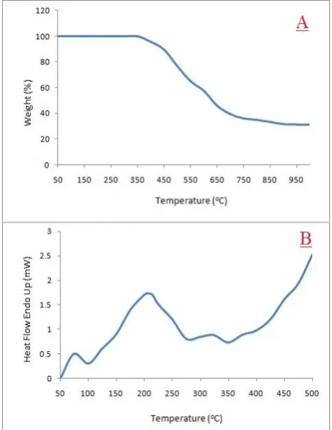
Thermal analysis
The TGA curve recorded the weight loss during heating and the DSC curve describes changes in the reaction enthalpy during the degradation process. The TG thermogram of Ag-PEG-Ch ternary nanocomposite analysis is depicted in Figure 4 A. This nanocomposite exhibits three-stage decomposition process.
Figure 3. TEM images of Ag-PEG-Ch nanocomposite film.
The first stage (100-450 oC; weight loss 10.57%)
decomposition may be due to expulsion of bound water molecules present in the PEG-Chitosan-Silver ternary nanocomposite. In the second stage, decomposition (451-650 oC; weight loss 43.34 %) corresponds to the loss of side
group elimination attached to the aromatic nucleus, and final stage of decomposition (651-950 oC; weight loss 15.08 %) is
probably due to main chain scission, which ultimately results in depolymerization into aromatic moieties. The ceramic yield of synthesized nanocomposite was found about 31.34 %. The result clearly reveals that PEG-Chitosan-Silver ternary nanocomposite exhibit synergistic effect in thermal stability.55,56
By contrast, in the DSC curve, only one thermal event, endothermic, is clearly distinguished up to 510 oC (Figure 4
B).In broad strokes, these thermal events are related to dehydration and thermal degradation processes, including the depolymerization degree at different stages as well as the secondary decomposition stage of pectin present in nanocomposite. According to the DSC curve, this broad endothermic event at 205.13 oC (with area swept in
endotherm 189.436 mJ and enthalpy change 64.6732 J g-1)
Figure 4 A and B. TGA and DSC curve of PEG-Ch_Ag nanocomposite film, respectively.
Antimicrobial Activities
The antimicrobial activity of Ag-PEG-Ch nanocompositeusing C. sativa leaves extract was studied against four pathogenic bacterial strains, two Gram positive (S. aureusMTCC 96 andS. pyogenus MTCC 442) and two Gram negative (E. coli MTCC 443 andP. aeruginosa MTCC 1688) and three fungal strains (C. albicans MTCC 227,
A.niger MTCC 282 and A.clavatus MTCC 1323). Antimicrobial activity of the synthesizedAg-PEG-Ch nanocomposite was assessed in terms of minimum inhibitory concentration (MIC).57 MIC is the lowest
concentration of antibiotic in a culture media that will prevent growth. To find MIC, bacteria/fungi were grown on different concentration of antibiotic and obtained a concentration of antibiotic on which bacteria/fungi can grow:The lowest concentration that inhibited the growth of bacteria/fungi was considered as the MIC.The results of antibacterial and antifungal activity of nanocomposite are shown in Fig. 5 and Table 1.
Ag-PEG-Ch nanocomposite using C. sativa leaves extract shows most effective antibacterial and antifungal activity as compared to C. sativa leaves extract and Ag nanoparticles alone.
CONCLUSION
This study focuses on the green synthesis of silver nanoparticles without any reducing agent by the use of cannabis sativa leaves extract and stabilize them in a polyethylene glycol-chitosan matrix, which are biodegradable polymers. Ultrasound as an energy source has been used in whole process, so this entire process covers the principles of green chemistry. The structure, morphology and thermal stability of the Ag-PEG–Ch were studied by
FTIR, XRD, TEM, TGA and DSC. This supports the strong interaction of PEG with Chitosan molecules, which interferes with its crystalline structure and use of ultrasound in the synthesis of fine Ag nanoparticles (6 nm) with FCC sturcture. C. sativa aqueous leaves extract act as a bioreducing agent to produce silver nanoparticles. Nanocomposite based on chitosan, polyethylene glycol and silver nanoparticles exhibit better thermal stability as compare to Chitosan and PEG alone. This nanocomposite was synergistically active against different bacterial and fungal strains. Therefore, we believe that Ag-PEG-Ch nanocomposite can be used for various biomedical applications.
Table 1. Antibacterial and antifungal activity of Ag-PEG-Ch nanocomposite.
Antibacterial activity Antifungal activity
Compound E.coli P.aeruginosa S.aureus S.pyogenus Compound C.albicans A.niger A.clavatus
Plant extract 125 100 250 200 Plant extract 500 250 250
Ag nanoparticles 100 75 150 90 Ag nanoparticles 500 90 100
Ag-PEG-Ch* 62 30 60 42 Ag-PEG-Ch* 80 75 80
Ampicillin (g mL-1)
100 250 100 Greseofulvin
(g mL-1)
500 100 100
Ciprofloxacin (g mL-1)
25 25 50 50 Nystatin
(g mL-1)
100 100 100
REFERENCES
1Nowack, B., Krug, H. F., Height, M., 120 Years of Nanosilver History: Implications for Policy Makers, Environ. Sci.
Technol., 2011, 45, 1177-1183. DOI: 10.1021/es103316q 2Agnihotri, S., Mukherji, S., Mukherji, S., Size-controlled silver
nanoparticles synthesized over the range 5–100 nm using the same protocol and their antibacterial efficacy, RSC
Adv., 2014, 4, 3974-3983. DOI: 10.1039/C3RA44507K 3Ayala-Núñez, N. V., Villegas, H. H. L., Turrent, Ld. C. I., Padilla,
C. R., Silver Nanoparticles Toxicity and Bactericidal Effect Against Methicillin-Resistant Staphylococcus aureus: Nanoscale Does Matter, Nanobiotech., 2009, 5, 2-9. DOI https://doi.org/10.1007/s12030-009-9029-1
4Abbaszadegan, A., Ghahramani, Y., Gholami, A., Hemmateenejad, B., Dorostkar, S., Nabavizadeh, M., Sharghi, H., The Effect of Charge at the Surface of Silver Nanoparticles on Antimicrobial Activity against Positive and Gram-Negative Bacteria: A Preliminary Study, J. Nanomater., 2015, 8 pages. ID 720654 http://dx.doi.org/10.1155/2015/720654
5Guo, Q., Zhao, Y., Dai, X., Zhang, T., Yu, Y., Zhang, X., Li, C., Functional Silver Nanocomposites as Broad-Spectrum Antimicrobial and Biofilm-Disrupting Agents, ACS Appl.
Mater. Interfaces, 2017, 9, 16834-16847. DOI: 10.1021/acsami.7b02775
6Bandla, M., Abbavaram, B. R., Kokkarachedu, V., Sadiku, R. E., Silver nanoparticles incorporated within intercalated clay/polymer nanocomposite hydrogels for antibacterial studies, Polym. Compos., 2017, 38, E16-E23.
https://doi.org/10.1002/pc.23963
7Castro-Mayorgaa J. L., Randazzo, W., Fabra, M. J., Lagaron, J. M., Aznar, R., Sánchez, G., Antiviral properties of silver nanoparticles against norovirus surrogates and their efficacy in coated polyhydroxyalkanoates systems, LWT - Food Sci.
Tech., 2017, 79, 503-510.
https://doi.org/10.1016/j.lwt.2017.01.065
8Salahuddin, N., Elbarbary, A. A., Alkabes, H. A., Antibacterial and anticancer activity of loaded quinazolinone polypyrrole/chitosan silver chloride nanocomposite, Int. J.
Polym. Mater. Polym. Biomater, 2017, 66, 307-316.
https://doi.org/10.1080/00914037.2016.1201831
9Benyettou, F., Rezgui, R., Ravaux, F., Jaber, T., Blumer, K., Jouiad, M., Motte, L., Olsen, J.-C., Platas-Iglesias, C., Magzoub, M., Trabolsi, A., Synthesis of silver nanoparticles for the dual delivery of doxorubicin and alendronate to cancer cells, J. Mater.
Chem. B, 2015, 3, 7237-7245. DOI: 10.1039/C5TB00994D 10Prabhu, S., Poulose, E. K., Silver nanoparticles: mechanism of
antimicrobial action, synthesis, medical applications, and toxicity effects, Int. Nano Lett., 2012, 2, 1-10. doi:
10.1186/2228-5326-2-32
11Burd, A., Kwok, C. H., Hung, S.C., Chan, H. S., Gu, H., Lam, W. K., Huang, L., A comparative study of the cytotoxicity of silver-based dressings in monolayer cell, tissue explant, and animal models, Wound Repair Regen., 2007, 15, 94-104. DOI:10.1111/j.1524-475X.2006.00190.x
12Liang, W., Wang, L., Zhu, Z., Qian, C., Sun, H., Yang, B., Li, A., In Situ Preparation of Polyethylene Glycol/ Silver Nanoparticles Composite Phase Change Materials with Enhanced Thermal Conductivity, Chem. Select, 2017, 2, 3428-3436. https://doi.org/10.1002/slct.201700381
13Fahmy, A., Zomrawy, A., Saeed, A. M., Sayed, A. Z., El-Arab, M. A. E., Shehata, H. A., One-step synthesis of silver nanoparticles embedded with polyethylene glycol as thin films, J. Adhesion Sci. Tech., 2017, 31, 1422-1440.
https://doi.org/10.1080/01694243.2016.1259728
14Marslin, G., Revina, A. M., Khandelwal, V. K. M., Balakumar, K., Sheeba, C. J., Franklin, G., PEGylated ofloxacin nanoparticles render strong antibacterial activity against many clinically important human pathogens, Coll. Surf. B: Biointerface, 2015, 132, 62-70. DOI:10.1016/j.colsurfb.2015.04.050
15Guibal, E., Cambe, S., Bayle, S., Taulemesse, J.-M., Vincent, T., Silver/chitosan/cellulose fibers foam composites: from synthesis to antibacterial properties, J. Coll. Interface
Sci., 2013, 393, 411-420. DOI: 10.1016/j.jcis.2012.10.057
16Carpio-Perochena, A.-d., Bramante, C. M., Duarte, M. A. H., de Moura, M. R., Aouada, F. A., Kishen, A., Chelating and antibacterial properties of chitosan nanoparticles on dentin,
Restor. Dent. Endod., 2015, 40, 195-201. DOI:10.5395/rde.2015.40.3.195
17Budnyak, T. M., Pylypchuk, I. V., Tertykh, V. A., Yanovska, E. S., Kolodynska, D., Synthesis and adsorption properties of chitosan-silica nanocomposite prepared by sol-gel method,
Nanoscale Res. Lett., 2015, 10, 87. DOI https://doi.org/10.1186/s11671-014-0722-1
18Dragan, E. S., Cocarta, A. I., Composite Beads Based on Chitosan and a Synthetic Polycation with Sorption Properties Modulated by the Cross‐Linking Strategy, Macromol. Symp., 2015, 352, 33-37. https://doi.org/10.1002/masy.201400146
19Souza, V. G. L., Fernando, A. L., Afonso, Pires, J. R., Rodrigues, P. F., Lopes, A. A. S., Fernandes, F. M. B., Physical properties of chitosan films incorporated with natural antioxidants, Ind. Crops Prod., 2017, 107, 565-572.
https://doi.org/10.1016/j.indcrop.2017.04.056
20Kalaycıoğlu, Z., Torlak, E., Akın-Evingür, G., Özen, İ., Erim, F. B., Antimicrobial and physical properties of chitosan films incorporated with turmeric extract, Int. J. Biol. Macromol., 2017, 101, 882-888. DOI: 10.1016/j.ijbiomac.2017.03.174
21Kiskó, G., Sharp, R., Roller, S., Chitosan inactivates spoilage yeasts but enhances survival of Escherichia coliO157:H7 in apple juice, J. Appl. Microbiol., 2005, 98, 872-880. doi:10.1111/j.1365-2672.2004.02527.
22Nehra, P., Chauhan, R. P., Garg, N., Verma, K., Antibacterial and antifungal activity of chitosan coated iron oxide nanoparticles, Brit. J. Biomed. Sci., 2018, 75, 13-18. DOI:10.1080/09674845.2017.1347362
23Sudarshan, N. R., Hoover, D. G., Knorr, D., Antibacterial action of chitosan, Food Biotechnol., 1992, 6, 257-272.
https://doi.org/10.1080/08905439209549838
24Manukumar, H. M., Umesha, S., Kumar, H. N. N., Promising biocidal activity of thymol loaded chitosan silver nanoparticles (T-C@AgNPs) as anti-infective agents against perilous pathogens, Int. J. Biol. Macromol., 2017, 102, 1257-1265. DOI:10.1016/j.ijbiomac.2017.05.030
25Liu, X. F., Guan, Y. L., Yang, D. Z., Li, Z., Yao, K. D., Antibacterial action of chitosan and carboxymethylated chitosan, J. Appl. Polym. Sci., 2001, 79, 1324-1335.
https://doi.org/10.1002/1097-4628(20010214)79:7<1324::AID-APP210>3.0.CO;2-L
26Rabea, E. I., Badawy, M. E. T., Stevens, C. V., Smagghe, G., Steurbaut, W., Chitosan as Antimicrobial Agent: Applications and Mode of Action, Biomacromol., 2003, 4, 1457-1465. DOI: 10.1021/bm034130m
27Li, X., Kong, X., Shi, S., Gu, Y., Yang, L., Guo, G., Luo, F., Zhao, X., Wei, Y., Qian, Z., Biodegradable MPEG-g-Chitosan and methoxy poly(ethylene glycol)-b-poly(ε-caprolactone) composite films: Part 1. Preparation and characterization, Carbohyd. Polym., 2010, 79, 429-436.
https://doi.org/10.1016/j.carbpol.2009.08.032
28Shameli, K., Ahmad, M. B., Zargar, M, Yunus, W. M. Z. W., Ibrahim, N. A., Shabanzadeh, P., Moghaddam, M. G., Int. J. Nanomed., 2011, 6, 271-284.
29Freire, T. M., Dutra, L. M. U., Queiroz, D. C., Ricardo, N. M. P. S., Barreto, K., Denardin, J. C., Wurm, F. R., Sousa, C. P., Correia, A. N., Lima-Neto, P., Fechine, P. B. A., Fast ultrasound assisted synthesis of chitosan-based magnetite nanocomposites as a modified electrode sensor, Carbohyd. Polym., 2016, 151, 760-769. DOI:
30Vinoth, V., Wu, J. J., Asiri, A. M., Anandan, S., Sonochemical synthesis of silver nanoparticles anchored reduced graphene oxide nanosheets for selective and sensitive detection of glutathione, Ultrason. Sonochem., 2017, 39, 363-373. DOI:10.1016/j.ultsonch.2017.04.035
31Bagherzade, G., Tavakoli, M. M., Namaei, M. H., Green synthesis of silver nanoparticles using aqueous extract of saffron (Crocus sativus L.) wastages and its antibacterial activity against six bacteria, Asian Pac. J. Trop. Biomed., 2017, 7, 227-233. https://doi.org/10.1016/j.apjtb.2016.12.014
32He, Y., Wei, F., Ma, Z., Zhang, H., Yang, Q., Yao, B., Huang, Z., Li, J., Zeng, C., Zhang, Q., Green synthesis of silver nanoparticles using seed extract of Alpinia katsumadai, and their antioxidant, cytotoxicity, and antibacterial activities, RSC Adv., 2017, 7, 39842-39851. DOI: 10.1039/C7RA05286C
33Salunke, B. K., Sawant, S. S., Kim, B. S., Potential of Kalopanax septemlobus leaf extract in synthesis of silver nanoparticles for selective inhibition of specific bacterial strain in mixed culture, Appl. Biochem. Biotechnol., 2014, 174, 587-601. DOI:10.1007/s12010-014-1077-x
34Krishnaraj, C., Jagan, E. G., Rajasekar, S., Selvakumar, P., Kalaichelvan, P. T., Mohan, N., Synthesis of silver nanoparticles using Acalypha indica leaf extracts and its antibacterial activity against water borne pathogens, Colloid
Surf. B, 2010, 76, 50-56.
DOI:10.1016/j.colsurfb.2009.10.008
35Prathna, T. C., Chandrasekaran, N., Raichur, A. M., Mukherjee, A., Biomimetic synthesis of silver nanoparticles by Citrus limon (lemon) aqueous extract and theoretical prediction of particle size, Colloids Surf. B, 2011, 82, 152-159. DOI:10.1016/j.colsurfb.2010.08.036
36Niraimathi, K. L., Sudha, V., Lavanya, R., Brindha, P., Biosynthesis of silver nanoparticles using Alternanthera sessilis (Linn.) extract and their antimicrobial, antioxidant activities, Coll. Surf. B, 2013, 102, 288-291. DOI:10.1016/j.colsurfb.2012.08.041
37Medda, S., Hajra, A., Dey, U., Bose, P., Mondal, N. K., Biosynthesis of silver nanoparticles from Aloe vera leaf extract and antifungal activity against Rhizopus sp. and
Aspergillus sp., Appl. Nanosci., 2015, 5, 875-880. https://doi.org/10.1007/s13204-014-0387-1
38Johnson, I., Prabu, H. J., Green synthesis and characterization of silver nanoparticles by leaf extracts of Cycas circinalis, Ficus amplissima, Commelina benghalensis and Lippia nodiflora, Int. Nano. Lett., 2015, 5, 43-51. https://doi.org/10.1007/s40089-014-0136-1
39Anandalakshmi, K., Venugobal, J., Ramasamy, V., Characterization of silver nanoparticles by green synthesis method using Pedalium murex leaf extract and their antibacterial activity, Appl. Nanosci., 2016, 6, 399-408. DOI
https://doi.org/10.1007/s13204-015-0449-z
40Izzo, A. A., Borrelli, F., Capasso, R., Di Marzo, V., Mechoulam, R., Non-psychotropic plant cannabinoids: new therapeutic opportunities from an ancient herb., Trends Pharmacol. Sci., 2009, 30, 515-527. DOI: 10.1016/j.tips.2009.07.006
41Ranganathan, M., D’souza, D. C., The acute effects of cannabinoids on memory in humans: a review,
Psychopharmacology, 2006, 188, 425-444. DOI:10.1007/s00213-006-0508-y
42Ożarowski, M., Mikolajczak, P. Ł., Bogacz, A., Bartkowiak-Wieczorek, J., Kujawski, R., Majchrzycki, M., Wielgus, K., Seremak-Mrozikiewicz, A., Czerny, B., Progress in study of Cannabis sativa leaves extracts without psychotropic cannabinoids in animal model of neuropathic pain, J. Med.
Sci., 2014, 83, 328-335.
43Hazekamp, A.,Grotenhermen, F., Review on clinical studies with cannabis and cannabinoids, Cannabinoids, 2010, 5, 1-21. 44Fischedick, J. T., Hazekamp, A., Erkelens, T., Choi, Y. H.,
Verpoorte, R., Metabolic fingerprinting of Cannabis sativa L., cannabinoids and terpenoids for chemotaxonomic and drug standardization purposes, Phytochem., 2010, 71, 2058-2073. DOI: 10.1016/j.phytochem.2010.10.001
45Costa, B., Trovato, A. E., Comelli, F., Giagnoni, G., Colleoni, M., The non-psychoactive cannabis constituent cannabidiol is an orally effective therapeutic agent in rat chronic inflammatory and neuropathic pain, Eur. J. Pharmacol., 2007, 556, 75-83. DOI:10.1016/j.ejphar.2006.11.006
46Weiss, L., Zeira, M., Reich, S., Har-Noy, M., Mechoulam, R., Slavin, S., Gallily, R., Cannabidiol lowers incidence of diabetes in non-obese diabetic mice, Autoimmunity, 2006, 39, 143-151. DOI:10.1080/08916930500356674
47Zajicek, J. P., Apostu, V., Role of cannabinoids in multiple sclerosis, CNS Drugs, 2011, 25, 187-201. DOI:10.2165/11539000-000000000-00000
48Zuardi, A. W., Crippa, J. A. S., Hallak, J. E. C., Moreira, F. A., Guimarães, F. S., Cannabidiol, a Cannabis sativa constituent, as an antipsychotic drug, Braz. J. Med. Biol. Res., 2006, 39, 421-429. DOI:/S0100-879X2006000400001
49Klauke, A. L., Racz, I., Pradier, B., Markert, A., Zimmera, A. M., Gertsch, J., Zimmer, A., E The cannabinoid CB₂
receptor-selective phytocannabinoid beta-caryophyllene exerts analgesic effects in mouse models of inflammatory and neuropathic pain, Eur. Neuropsychopharmacol., 2014, 24, 608-620. DOI: 10.1016/j.euroneuro.2013.10.008
50Wayne, P. A., Clinical and Laboratory Standard Institute (CLSI)
Performance Standards for Antimicrobial Susceptibility Testing; Twenty-Third Informational Supplement CLSI Document, 2013, M100-S23.
51Ahmad, M. B., Tay, M. Y., Shameli, K., Hussein, M. Z., Lim, J. J., Green Synthesis and Characterization of Silver/Chitosan/Polyethylene Glycol Nanocomposites without any Reducing Agent, Int. J. Mol. Sci., 2011, 12, 4872-4884. https://doi.org/10.3390/ijms12084872