AJPRHC Volume 5 Issue 1 16-23
AJPRHC
Research Article
ACTINOMYCIN “D” FROM MARINE SEDIMENT ASSOCIATED STREPTOMYCES CAPILLISPIRALIS MTCC10471
M.SRINU1*, M. MURALI KRISHNA KUMAR 2, G. GIRIJA SHANKAR2
For Author affiliations see end of the text
This paper is available online at www.jprhc.in
ABSTRACT
In our screening program for new bio-active metabolites from marine actinomycetes, a cyclic depsipeptide was found in the fermentation medium of marine Strepromyces (SS23/4) isolated from sediments collected from Bay off Bengal, vellampattai,Tamilnadu. It showed strong biological activity against gram-positive / gram negative bacteria by agar overlay technique. It was taxonomically characterized by the basis of morphological and phenotypic characteristics, genotypic data and phylogenetic showing Streptomyces sps. Bio active compound was obtained by solvent extraction and purification using column chromatography followed by reverse phase HPLC. The pure compound had potent activity against Mycobacterium tuberculosis and Multi Drug Resistant Mycobacterium tuberculosis strains (437RU) at a concentration of 10 µg/mL, and The minimum inhibitory concentration (MIC) against standard test organisms was found to be 1µg/mL against B.subtilis, E.coli and
Methicillin resistant Staphylococcus aureus. The compound exhibited potent cytotoxic activity against breast carcinoma (MCF-7), melanoma cells (A375), prostate carcinoma (DU145) and lung carcinoma (A549) cells with IC values 20µg/ml. The symbiotic Streptomyces capillispiralis MTCC 10471 produces crude antibiotic 30mg/Lt by using nonoptimized fermentation conditions. The structure of the antibiotic was explained by 1D, 2D NMR and LC-ESI-MS/MS, MALDI-TOF/MS experiments, revealed that it belongs to cyclic ploy peptide Actinomycin D.
KEY WORDS: Antimicrobials, marine sediments, Actinomycetes, polypeptides, HPLC, UV, LC-ESI/MS, Agar overlay, MIC, Fermentation.
INTRODUCTION
The oceans are highly complex and house a diverse assemblage of microbes that occur in environments of extreme variations in pressure, salinity, and temperature. Marine Streptomyces have developed unique metabolic and physiological capabilities that not only ensure survival in extreme habitats, but also offer the potential to produce like antiviral, antibacterial, antitumour, anti-helminthic, insecticidal, modulator, immuno-suppressant compounds. A growing number of bioactive metabolites have been explored from marine
AJPRHC Volume 5 Issue 1 16-23 MATERILAS & METHODS
Collection of marine samples
A set of 63 marine sediment samples were collected in a sterile container by scuba diving at depths of 5 mts, 10 mts, 20 mts, 35 meters at a distance of about 40kms off the sea shore of Bay of Bengal, Coastal areas of Visakhapatnam and Vellampatti for the selective isolation of actinomycetes.
Isolation of actinomycetes:
Sediment sample (1gm) was suspended in 10 mL of sterile 5mM phosphate buffer (pH 7.0) and stirred for 1 min in a super mixer. After a 10-fold serial dilution 1mL of the suspension was spread on Starch Casein Agar plates (g/L: Starch 19, Casein 0.3, KNO3 2, NaCl 2, K2HPO4 2, MgSO4.7H2O 0.05, CaCO3 0.02, FeSO4.7H2O 0.01,
Agar 20) supplemented with cycloheximide 50 µg/mL and 5µg/mL rifampicin to inhibit fungal and bacterial growth respectively and incubated at 280 C for 21 days. After 21 days, actinomycetes colonies were confirmed by microscopic /macroscopic examination, and inoculated on selective media (Starch casein agar / Bennett’s agar or YEME) agar slants. The slants were incubated at 280 C for 2 weeks.
Bioactivity screening:
Agar overlay technique 10
The zone of inhibition of all the test organisms were detected using agar overlay technique. Streptomyces culture was stabbed on nutrient agar plates and incubated. The plates were exposed to chloroform for 40 min, followed by plates were overlaid with 5 ml of nutrient agar (0.7% W/V) inoculated with a test organism. Zones of inhibition around the colonies were recorded after 24 hrs at 370 C (Fig.1.)
Taxonomy
The morphological, cultural, physical characteristics were determined after growth at 300 C for 14 days .The media recommended by ISP (International Streptomyces Project) 11. The utilization of carbon sources was tested by the growth on Pridham and Gottlieb’s medium containing 1 – 0 % carbon source 12. Morphological properties were observed with a scanning electron microscope (SEM) 13.
Molecular characterization 14
Single isolated colonies of the SS23 strain culture was taken from agar plates and suspended in 50 µL of colony lysis solution (10mMol, Tris-HCl, pH 7.5, 10 mM EDTA and 50 µg/mL of proteinase K). The reaction mixture was incubated at 550 C for 15 min followed by proteinase K inactivation at 800 C for 10 min. The reaction mixture was centrifuged at 10,000 rpm at 40C for 15 min. The supernatant containing genomic DNA was directly used as a template in PCR reaction. PCR amplification of full length 16S rRNA gene was carried out with eubacterial specific five different primers i.e., 16seq3F_S
(TACGGCTACCCTTGTTACGACTTCGTCCCAATCGCCAGTCCCACCTTCGACAGCTCC
57),16seq4F_S( ---ATCGCCAGTCCCACCTTCGACAGCTCC 27),INS16S1R_( ---0),16seq2R_S( ---0)16seq4R_S ---0).
A 25 µL reaction volume PCR was done using 10ng of genomic DNA, 1X reaction buffer (10mM Tris HCl, pH
8.8 at 250 C, 1.5mM MgCl2, 50 mM KCl and 0.1% Triton X-100), 0.4mM (each) deoxynucleoside
AJPRHC Volume 5 Issue 1 16-23 Fermentation, Isolation and Extraction:
The strain SS23/4 was subjected to batch fermentation by shake flask method for isolation of antibiotic principles. Spore suspension was prepared, added to 20 mL of the seed medium (g/100mL: Soy bean meal 1g, Corn steep liquor 1g, Glucose 1g, CaCO3 0.5g, pH 7.0 ) in a 100 mL conical flask and incubated in shaker at 30 0
C /220 rpm for 48 hours. 5 ml of seed culture was transferred to a 250mL flask containing 50 mL of the production medium (g/100mL: Soya bean meal 1 g, Corn steep liquor 0.5 g, Soluble starch 1 g, Dextrose 0.5 g, CaCO3 0.7 g, pH 7.2) in a 250ml conical flask and incubated in shaker at 30 0 C /220 rpm for 6 days. After
fermentation, the culture was centrifuged, the supernatant and mycelia was washed twice with hexane and followed by extraction with 20%V/V of ethyl acetate three times, and then samples were evaporated to dryness. The crude antibiotic fraction was loaded on the top of a silica gel column chromatography. The column was developed with increasing solvents polarity (Hexane to Ethyl acetate to Methanol) to give antimicrobial fractions.
HPLC-DAD Analyses:
The chromatography system consisted of an analytical reverse phase C18 column (Agilent 4.6x150mm), attached to a LKB-BROMA 2152 HPLC system. 20 l of the sample was injected into the HPLC column (150×4.6mm) fitted with a guard-column (20×4.6) packed with 5- m Nucleosil-100 C-18 (Varian) and analyzed by gradient elution using 100% water as solvent A and 100% methanol as solvent B in 40 min at a flow rate of 1ml/minute. Constituents eluting from the column were detected at 226nm, 254nm using a Shimadzu SPD -20A UV-VIS detector.
Mass Determination of the Major Fraction:
The mass spectrum of HPLC purified active fraction was acquired on an Ultra flex Bruker LC-ESI/MS/MS mass spectrometer and MALDI-TOF, equipped with a nitrogen laser of wavelength 337nm.
Biological activities:
Purified compound was evaluated for MIC values against various Gram-positive and Gram-negative bacteria test cultures using nutrient broth described by Andrews 15. Bioactive fraction was also tested against
Mycobacterium tuberculosis, Multi Drug Resistant Mycobacterium tuberculosis strains (437RU) and Methicillin resistant Staphylococcus aureus. (Table 5)
CYTOTOXIC ACTIVITY
The antitumor activity of the pure compound was tested according to MTT based cell assay 16 against human cell lines from breast carcinoma (MCF-7), melanoma cells (A375), prostate carcinoma (DU145), lung carcinoma (A549) cells. The cancer cell lines obtained from National Centre for Cell science (NCCS), Pune, India and cultivated in Dulbecco’s Modified Eagle’s red Medium (DMEM).
RESULTS AND DISCUSSION
Taxonomy:
AJPRHC Volume 5 Issue 1 16-23
spore chain held about 10 to 20 spores. The size of each elliptical spore was about 880 nm. The surface of the spore was smooth, no sclerotic granules, sporangia or zoöspores were observed (Fig.2).
Sequence analysis:
The phylogenetic studies revealed that SS23 belongs to the Streptomyces sp. ME03-5709A and Streptomyces sp. VTT E-99-1329 (A52) were interchanged when the Fitch- Margoliash method was employed (Fig.3). The isolate has been deposited in the Microbial Type Culture Collection (MTCC), Chandigarh, India (http://www.mtcc.imtech.res.in) as S.capillispiralis MTCC 10471.
Time course of fermentation for act-D production:
During the course of fermentation, it was a decrease in the pH of the broth until the point of maximum antibiotic titer and afterwards there was an increase in the pH of the broth. The biomass increased during the first six days, remained stable, then decreased after the sixth day (144 h) (Fig.4.)
Purification:
Among the 7 fractions (1-23), fraction VI (6-15) showed strong biological activity, a red coloured solid (190mg) was obtained after concentrating in vacuo (Fig.5). Fraction VI was purified by using semi preparative reverse phase HPLC, the active fraction was eluted at 32.6min/84% methanol: water gradient at 254nm (Fig.6).
Chemical characterization:
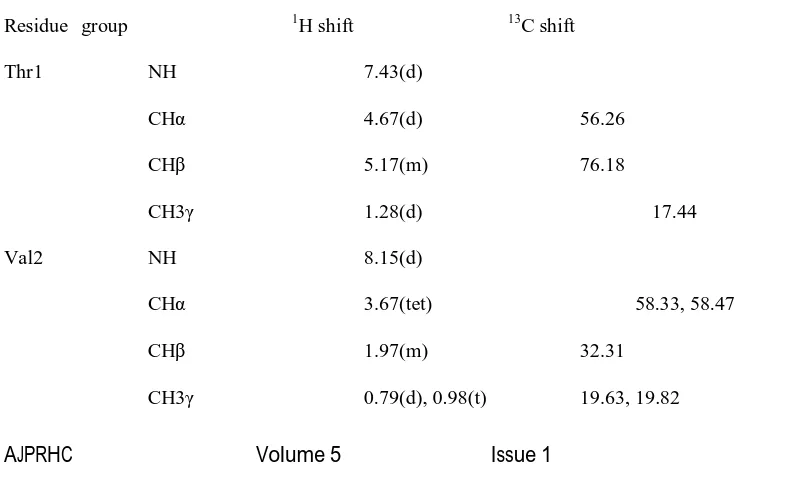
The mass of HPLC active fraction was 1255.602 D determined by high-resolution mass spectrometry LC-ESI-MS/MS and MALDI-TOF. UV absorption bands was observed at 205,210,215,226, 240, 254 and 430 nm indicates the presence of quinoid and benzenoid chromophores, the infrared spectrum showed amide carbonyl bands (1649 1), aromatic absorptions (1463, 1205, 1), and NH and OH (34231), aliphatic (3423 cm-1) functionalities. The proton ( 1H ) and carbon (13C) NMR spectra (Fig.7 &8) data showed the presence of amide protons, 14 methyl groups, 4 methylenes, 25 methine carbons, and 6 quaternary carbons and phenoxazone moiety. The structure was confirmed by comparison of the 13C NMR and 1H NMR data with the literature 17. Analyses by 1H and 13C (Table 3) NMR indicated that bioactive compound is a depsipeptide composed of 10 amino acid residues and chromophore moiety. The structures of all of the amino acids (valine, proline, methyl valine, sracosine, theronine) were determined by 2D-NMR techniques including HMBC, COSY, TOCSY, and NOESY.
Table 3. 1H/13C NMR assignments for actinomycin D
Residue group 1H shift 13C shift
Thr1 NH 7.43(d)
CH 4.67(d) 56.26
CH 5.17(m) 76.18
CH3 1.28(d) 17.44
Val2 NH 8.15(d)
CH 3.67(tet) 58.33, 58.47
CH 1.97(m) 32.31
AJPRHC Volume 5 Issue 1 16-23
Pro3 NH
CH 6.08(d) 58.47
CH2 1.83(m) 32.91
CH2 2.02(m) 23.97
CH2 3.08(m) 52.51
Sar4 N-CH3 2.56(s) 35.52
CH2 3.90(d) 52.51
Metval N-CH3 2.84(d), 2.94(d) 39.34
CH 3.01(d) 71.95
CH 3.29(d) 28.34
CH3 1.12(t) 21.72
Phenoxazone 7H 7.70
8H 7.75
4CH3 2.13 7.58
6CH3 2.70 14.98
AJPRHC Volume 5 Issue 1 16-23
Fig.9. Mass spectrum showing the mass of parent ion (m/z 1255.7) (top), daughter ions obtained from fragmentation of parent ion (Middle) and derived sequence is shown at the bottom
CONCLUSION
AJPRHC Volume 5 Issue 1 16-23
cytotoxic activity profile against cell lines A549, DU-145, A375, MCF-7 in vitro, also exhibited potent activities against Methicillin resistant Staphylococcus aureus,Multi drug resistant Mycobacterium tuberculosis strains (437RU). Previously, S.capliispiralis has never been reported to produce act-D. To our knowledge,
Streptomyces capillispiralis MTCC10471 is the first reported antibiotic producing strain from marine sediments. Our report can be considered as a new source for anti tuberculosis drugs and cytotoxic antibiotics.
Acknowledgments
The authors are thankful to UGC- Kothari post doctoral fellowship for the financial support to carry out this work. We are thankful to SAIF and Proteomics facility, IISc, Bangalore, for providing NMR spectra and LC-ESI/MS, MALDI-TOF spectrum.
REFERENCES
1. Cho, K.W., Seo Y, Youn T, Shin J. Purification and structure determination of antifungal phospholipids from a marine Streptomyces. J. Microbiol. Biotechnol. 1999; 9:705–715.
2. Cho, K.W., Lee HS, Rho JR, Kim TS, Mo S, Shin J. New lactone-containing metabolites from a marine-derived Streptomyces. J. Nat. Prod. 2001; 64:664–667.
3. Mitchell, S.S., Nicholson B, Teisan S, Lam KS, Potts BCM. Aureoverticillactam, a novel 22-atom macro cyclic lactam from the marine actinomycetes Streptomyces aureoverticillatus. J. Nat. Prod. 2004; 67:1400–2.
4. Seo,Y., Cho, K.W., Lee HS, Youn T, Shin J. New polyene macrolide antibiotics from Streptomyces sp. M90025. J. Microbiol. Biotechnology, 2000; 10:176–80.
5. Sujatha, P., Bapi Raju KVVSN, Ramana. T. Studies on a new marine streptomycete BT-408 producing polyketide antibiotic SBR-22 effective against methicillin resistant Staphylococcus aureus. Microbiol. Res. 2005; 25:119–26.
6. Takuya, I., Masahiro K, Hong W, Motomasa, K. Stereo structure of Komodo Quinone A, a Neuritogenic Anthracycline, from marine Streptomyces sp. KS3. Chem. Pharm. Bull. 2003; 51:14024.
7. Piel, J., Hoang K, Moore BS. Natural metabolic diversity encoded by the enterocin biosynthesis gene cluster. J. Am. Chem. Soc. 2000; 122: 5415–6.
8 .Newmans D.J, and Cragg G.M, (2004) Marine natural products and related compounds in clinical and advanced preclinical trials. J.natural products, 67, 1216-1238.
9. Lam, K.S (2006) discovery of novel metabolites from marine actinomycetes. Curr.Opin.Microbiol., 9, 245-251.
10. Janssen A.M, N. L. J. Chin, J. J. C. Scheffer, A. Baerheim Svendsen (1986) Pharmaceutisch Weekblad Screening for antimicrobial activity of some essential oils by the agar overlay technique, 8, 289-292 .
11. Shirling E.B, Gottlieb D. (1966) Methods for characterization of Streptomyces species. Int J Syst Bacteriol 16, 313–340.
12. Pridham T.G, Gottlieb D. (1948) the utilization of carbon compounds by some Actinomycetales as an aid for species determination. J Bacteriol 56, 107–114
AJPRHC Volume 5 Issue 1 16-23
14. Saito H, Miura K.I. (1963) Preparation of transforming deoxyribonucleic acid by phenol treatment. Biochim Biophys Acta 72, 619–629.
15. Andrews.J.M.(2001).Determination of minimum inhibitory concentrations, J. Antimicrobial. Chemotherapy, 48, 5-16.
16. Mossman T (1983).Rapid Colorimetric assay for cellular growth and survival: application to proliferation and cytotoxicity assays.J.immuno methods 65(1-2):55-63.
17. Yu C, Tseng Y. (1992) NMR study of solution confirmation of actinomycin D. Eur J Biochem 209, 181– 187.
18. Kim T.K, Hewavitharana A.K, Shaw N.P. and Fuerst J.A. (2006) Discovery of a new source of rifamycin antibiotics in marine sponge actinobacteria by phylogenetic prediction.Appl Environ Microbiol 72, 2118–2125.
19. Kurosawa K. Bui V.P, Van E.J.L , Willis L.B , Lessard P.A , Ghiviriga I, Sambandan, T.G, Rha C.K. et al. (2006) Characterization of Streptomyces MITKK-103, a newly isolated actinomycin X2-producer. Appl Microbial Biotechnology.72, 145–154.
20. Wagman G.H, Marquez J.A , Watkins P.D, Gentile F, Murawski A, Patel M, and Weinstein M.J. (1976)A new actinomycin complex produced by a Micromonospora Species: fermentation, isolation, and characterization. Antimicrob Agents Chemother 9, 465–469.
21. Caixia Chen , Fuhang Song , Qian Wang (2012) A marine-derived Streptomyces sp. MS449 produces high yield of actinomycin X2 and actinomycin D with potent anti-tuberculosis activity Appl Microbiol Biotechnol,
21. Laatsch H. (2000) AntiBase. A Natural Products Database for Rapid Structure Determination.
22. Lie J, Zhou J.(2002).A marine natural product databse.J.Chem.Inf.Comput.Sci.42 (3), 742-748.
23. Praveen V, C.K.M. Tripathi (2009) Studies on the production of actinomycin-D by Streptomyces griseoruber – a novel source Letters in Applied Microbiology ISSN 0266-8254 450 Journal.
24. Waksman S.A. (1961) Classification, identification and description of genera and species. In The Actinomycetes ed. Waksman, S.A. Vol. 2. pp. 1–363.
AUTHORS AFFILIATION AND ADDRESS FOR CORRESPONDENCE
1. Indian Instuite of Science, Molecular Bio Physics Unit, Bangalore -12, INDIA.
2. College of Pharmaceutical Sciences, Andhra University, Visakhapatnam-03, Andhra Pradesh, INDIA