COVID-19 and Co-Infections: An Overview of
Ace2 Mediated Viral Entry of Sars-Cov-2 and
Multiorgan Failure in Covid-19
Shah Faisal
1, Shahzar Khan
2, Muhammad Ajmal
3, Hasnain Jan
4, Muhammad Taj Akbar
2,
Rashida Ilyas
5, Khadija Bibi
5, Sadeeq ur Rehman
51. Department of Biotechnology, Bacha Khan University, Charsadda, KPK, Pakistan.
2. Department of Microbiology, Abdul Wali Khan University, KPK, Pakistan.
3. Department of Anesthesia, Hayatabad Medical Complex, Peshawar, KPK, Pakistan.
4. Department of Biotechnology, Quaid-i-Azam University, Islamabad 45320, Pakistan.
5. Department of Botany, Govt.Post Graduate College Charsadda, Bacha Khan University Charsadda Khyber Pakhtunkhwa.
Corresponding author:
Shah Faisal
Email: [email protected]
Introduction
In December 2019 a pandemic of acute respiratory distress
syndromes occurred in Wuhan, China, the early evidence suggests
this may be due to exposure to local sea foods in China [1] The
pathogen was isolated from Chinese patients and designated
as severe acute respiratory syndrome corona virus 2
(SARS-COV-2) .WHO announces this as global pandemic on March 11;
2020. COVID-19 mainly affects lungs by producing respiratory
predominant symptoms like fever, cough, flu and dyspnea [2].
The virus primarily attacks the angiotensin converting enzyme-2
receptor (Ace2) of the lung. However recent reports suggested that
COVID-19 infection is not confined only to lungs.As ACE2 receptor
is the binding site of (SARS-COV-2) and there are abundance of
this receptor on different body organs such as in alveolar epithelial
type II cells of lung tissues, as well as extrapulmonary tissues such
as the heart, endothelium, kidneys, and intestines [3, 4, 5, 6]. In
addition to respiratory disease there are a lot of complications
associated with COVID-19. The pandemic
has an adverse effect on the central nervous system. Reported
data have shown neurological manifestations of COVID-19. Much
of the research on corona virus has shown it has capability of
causing brain infections in humans and other animals [7, 8].
The heart infection is highly prevalent in COVID-19 leading
to cardiovascular complications [9,10]. Although lungs are its
primary target but there are many related data supporting the
cutaneous manifestation of COVID-19 [11, 12,]. Patients suffering
from COVID-19 and admitted to intensive care unit have high
incidence of venous and thrombotic complications [13]. Acute
ischemic stroke is another complication of COVID-19 [14].
Enhanced ACE2 receptor expression in kidney supports nephritic
cells damage in kidney due to COVID-19 [15]. This review is
an effort to highlights some of the co-infections associated with
COVID-19 and multi organs failure.
Abstract
COVID-19 is highly fatal disease having a high mortality rate and is declared as pandemic situation by World Health Organization.
It shows a clear indication that every individual is at risk of this pandemic especially older individuals and immunocompromised
persons. It’s casual agent is SARS-Cov-2 and the main target site of this virus is the ACE2 receptor of lungs. But as compared
to lungs ACE2 receptor is highly expressed in other organs i.e. kidney, liver, brain, GI tract, cutaneous, adipose tissues and
cardiovascular system these organs are susceptible to COVID-19 infections because of having ACE2 receptor. Many of the
co-infections associated with COVID-19 are reported i.e. Neurological manifestation of COVID-19,Cutaneous manifestation of
COVID-19, Endothelial cell infection and endotheliitis, adipose tissues infections, Cerebral Hemorrhage, liver injury, cardiovascular
complications, kidney infection, trigger immune system response and subsequent organ failure. In this review we highlights ACE2
mediated viral entry of the SARS-Cov-2 and subsequent multi organs failure in COVID-19.
Keywords:
COVID-19; Co-infections; ACE2 receptor; SARS-Cov-2; Multi organs
Citation:
Faisal S, Khan S, Ajmal M, Jan H, Akbar M, Ilyas R, Bibi K, Rehman S. COVID-19 and Co-Infections: An Overview of
Ace2 Mediated Viral Entry of Sars-Cov-2 and Multiorgans Failure in Covid-19. International Cardiovascular Forum Journal
2020;20:16-21.DOI: 10.17987/icfj.v20i0.693
Background
2.1. Epidemiology
On April 11, 2020 1,610,909 confirmed cases were reported,
with 99,690 (6.19% of confirmed cases) of mortality. All the
ages are susceptible to this pandemic but the risk is higher in
older age and immunocompromised or with co-morbidities. On
the other hand mortality is related with age and more often with
targeted organ failure [16, 17, 18, 19, 20, 21].
2.2. Virology
Corona virus are enveloped, positive stranded RNA virus,
genome size ranges between 26kb to 32 kb, resembles a solar
corona, have four sub families i.e. alpha, beta, gamma and delta
Coronaviruses and was first described by Tyrell and Bynoe in
1996 [22, 23, 24, 25, 26]
2.3. Transmission
The most accepted model at first was animal to human
transmission from Wuhan sea food market. Genome studies have
suggested that animal is the natural reservoir of corona virus like
pigs. But after sometimes person to person transmission model
was the most acceptable mode of transmission i.e. by aerosol,
air droplets, fecal oral transmission has also clarified [27, 28, 29,
30, 31, 32, 33, 34, 35].
3. ACE2 involvement in manifestation of co-infection
in COVID-19 patients
As viral infections need entry into the cell for replications, the
ACE2 receptor act as co receptor for COV-1 and
SAR-COV-2 for entering to lungs and brain cell. ACE2 receptors are
expressed in nearly all tissues and cells of the body i.e. in the
ileum and kidney followed by adipose tissue, heart, brain stem,
lung, vasculature, stomach, liver, and nasal and oral mucosa
[41, 42, 43]. Current studies suggest the presence of viral load of
SAR-COV-2 in brain may be attributed to internalization by ACE2
and transport by cranial nerves. And it may result in neuronal cell
death. Loss of ACE2 receptor from pulmonary tissues may lead
to respiratory distress. As we know that ACE2 receptor is found
in nearly all tissues so the disruption or loss of this receptor may
lead to dysfunction of all the organs [44, 45, 46, 47, 48].
4. Neurological manifestation of COVID-19
Moriguchi et al., 2020 reported a 59 years old male patients
having no familial history of seizures but a complaint of
atrial fibrillation and treated sleep apnea who was admitted
in emergency department on March 26,2020 with fever, dry
cough and headache. His first RT-PCR result was negative for
SAR-C0V-2, but later he was tested positive for SAR-COV-2 by
RT-PCR assay from tracheal secretion and chest CT- SCAN.
From APRIL 10 he started short seizures. When the patients
awoke from seizure a 49 minutes EEG was performed and the
results of the peak confirm the epileptic seizures. He felt first
seizure for six minutes, second seizure for 5 minutes. His brain
MRI was normal, laboratory test was also normal, on APRIL 14
the patient has only one brief episode of seizure but his EEG
was normal then. The result suggests seizures associated with
COVID-19 with no history of meningitis or encephalitis [36]
4.1. Mechanism of seizures induction by COVID-19
The movement of human corona viruses toward brain is supported
by the presence of ACE2 receptor in brain cell. SAR-COV-2 inhibit
the ACE2 receptor which slows the baroreflex which in turn slow
down the blood flow toward brain and thus increases seizures
threshold [37, 38, 39,]
5. Endothelial cell infection and endotheliitis in
COVID-19
As ACE2 receptors are also expressed in endothelial cell.
However vascular damage in COVID-19 by endothelial cell entry
of SAR-COV-2 is unknown. But SARS-COV-2 can directly infect
blood vessels [50, 51, 52, 53]
6. COVID-19 and adipose tissues
According to previous studies it is well known that adipose tissues
are more prone to Influenza virus and HIV. The transmission and
mortality rate of influenza virus is more related to obesity [54,
55, 56, 57]. As ACE2 expression in adipose tissues is much and
higher than lung, kidney and other organs and has higher RNA
level than lungs. There may be some possibilities of SARS-C0V-2
infection in adipose tissues but till now it is not reported [58].
7. Liver and COVID-19
Previous studies have reported liver abnormalities in patients
infected by SARS-coV-2 and COVID-19 medications side effects
on liver have been illustrated previously [59]. In addition after
performing liver biopsy of COVID-19 patients fatty liver and liver
injuries were reported [60].
8. Cerebral Hemorrhage a complication of COVID-19
patients
In a study a COVID-19 patients was reported whose first symptom
was cerebral hemorrhage. When craniotomy was performed the
patient has high fever and the case was challenging to treat. After
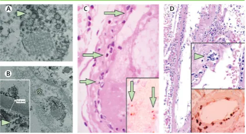
Figure 2.
Pathology of endothelial cell dysfunction in COVID-19 and viral
inclusion in kidney cells and also viral particles in endothelial
cells adapted from [49]
Figure 1.
CSF testing the intracranial infections was reported and it shows
a clear evidence for the involvement of COVID-19 in cerebral
hemorrhage and cerebral blood vessels damage [61]
9. Cardiovascular complications in COVID-19
According to previous studies and results of previous outbreaks
such as SARS-Cov-1 and MERS cardiovascular complications
are associated with COVID-19 [63]. The actual mechanism
through which COVID-19 induces cardiovascular infections is
not understood yet but there are some proposed mechanism i.e.
by inducing direct myocardial injury or through indirect injury by
cytokine storms [63, 64, 65]
10. Cutaneous manifestation of COVID-19
There are many case reports on cutaneous manifestations of
COVID-19 [68, 69]. For the diagnosis of different infectious disease
cutaneous manifestations are very important. As COVID-19
remains asymptomatic for 14 days cutaneous manifestations may
be an indicator of infection cause by SARS-Cov-2 [70, 71, 72]
Table 1
Total COVID-19 infected
patients having
cardiovascular complications
138
Percent patients presented
with shock
8.7%
Percent patients with acute
cardiac injury
7.2%
No of patients received
extracorporeal membrane
oxygenation
4 out of 138
New onset heart failure or
cardiomyopathy in patients
with COVID-19
25%
Postulated mechanism for
cardiovascular system failure
in COVID-19
direct myocardial injury, indirect
injury through cytokine release,
a prothrombotic state causing
microvascular thrombosis, and
exacerbation of underlying
cardiovascular disease e.g.
plaque rupture in susceptible
patients
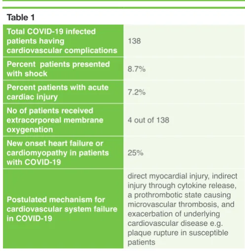
Table 1.
shows percent COVID-19 infected patients with
cardiovascular complication and propose mechanism for
cardiovascular system failure in COVID-19 patients [63, 64, 65,
66, 67].
Table 2.
Total patients under study for
Cutaneous manifestation of
COVID-19
72
Patients showing mobilliform
36.1% (26/72) patients
Patients with papulovesicular rash
(34.7%, 25/72
urticaria
(9.7%, 7/72)
painful acral red purple papules
(15.3%, 11/72
livedoreticularis lesions
(2.8%, 2/72)
petechiae
(1.4%, 1/72)
Table 2 shows different cutaneous lesions in patients infected
with COVID-19 [73]
Table 3.
Common sites of lesion in
COVID-19
Trunk, head, feet, skin
Trunk
(66.7%, 50/72)
Head and feet
19.4% (14/72)
skin
12.5% (9/72)
Table 3 shows common sites of cutaneous manifestation of
COVID-19 [74]
Figure 3.
Shows the CT scan image of lung and brain of COVID-19 patient
who have the complication of cerebral hemorrhage[61]
Table 4.
Total no of COVID-19
patients admitted with kidney
abnormalities
A total of 701 patients were
included in a study
Glomerular filtration rate
recorded in most of COVID-19
patients
< 60 ml/min per 1.73 m2
was reported in 13.1% of
patients
Percentage of patients having
elevated creatinine and blood
urea nitrogen level
14.4% and 13.1% of the
patients
Patients with proteniurea
43.9% of patients
Patients with hematuria
(26.7%)
11. COVID-19 and kidney
In previous epidemics of SARS and MERS kidney infections was
reported as one of the most drastic complications of the epidemic
with high mortality rate up to 90% due to kidney involvement and
acute kidney injury [73, 74]
12. COVID-19 and gastrointestinal complications
A number of patients tested positive for COVID-19 by
performing RT-PCR from their stool sample shows many GI tract
complications. The reason for GI tract complications is clear as
ACE-2 receptor is highly expressed in gastrointestinal tract and
thus GI tract serves as target site for SARS-Cov-2. [77, 78]
13. COVID-19 and immune system
Immune system is greatly influenced by any infection in the body.
In COVID-19 pandemic immune system triggers cytokine storms
and induces inflammatory responses and in multi organs hence
result in multi organ failure. Concentration of serum inflammatory
mediators like interleukin ((IL)-2, IL-7, IL-10) and interferon’s are
highly elevated in sera of patients infected with COVID-19 [79].
Conclusion
From the above study it is concluded that as ACE2 is involved
in viral entry of SARS-Cov-2 and as this receptor is present on
many organs so all these organs are susceptible to COVID-19.
And these co-morbidities help in the progression of the disease
leading to a high mortality rate. Acute kidney failure, liver injury,
nervous system damage, cutaneous lesion, cardiovascular
system failure all these are the outcomes of COVID-19. All those
patients having co-morbidities and infected with COVID-19 need
intensive care because they are at high risk. This paper adds in
highlighting co-morbidities association with COVID-19.
Conflict of interest
The authors declare no conflicts of interest.
Acknowledgements
The authors state that they abide by the “Requirements for Ethical
Publishing in Biomedical Journals” [83].
References
1. C.I. Paules, H.D. Marston, A.S. Fauci, Coronavirus infections – more than just the common cold, JAMA (2020) Epub ahead of print.
2. J. Yang, Y. Zheng, X. Gou, et al., Prevalence of comorbidities in the novel Wuhan coronavirus (COVID-19) infection: a systematic review and meta-analysis, Int. J. Infect. Dis. (2020) Epub ahead of print.
3. D. Wang, B. Hu, C. Hu, et al., Clinical characteristics of 138 hospitalized patients with novel coronavirus-infected pneumonia in Wuhan, China, JAMA (2020) Epub ahead of print.
4. Xu X, Chen P, Wang J, et al. Evolution of the novel coronavirus from the ongoing Wuhan outbreak and modeling of its spike protein for risk of human transmission. Sci China Life Sci 2020;63:457–60. https://doi.org/10.1007/ s11427-020-1637-5.
5. Zhao Y, Zhao Z, Wang Y, Zhou Y, Ma Y, Zuo W. Single-cell RNA expression profiling of ACE2, the putative receptor of Wuhan 2019-nCov. bioRxiv. 2020. https://doi. org/10.1101/2020.01.26.919985.
6. Ding Y, He L, Zhang Q, et al. Organ distribution of severe acute respiratory syndrome (SARS) associated coronavirus (SARS-CoV) in SARS patients: implications for pathogenesis and virus transmission pathways. J Pathol 2004;203:622–30. https://doi.org/ 10.1002/path.1560.
7. Hamming I, Timens W, Bulthuis ML, Lely AT, Navis G, van Goor H. Tissue distribution of ACE2 protein, the functional receptor for SARS coronavirus. A first step in understanding SARS pathogenesis. J Pathol. 2004;203:631– 7. https://doi.org/ 10.1002/path.1570.
8. J. R. Lechien, C. M. ChiesaEstomba, D. R. De Siati, et al., Olfactory and gustatory dysfunctions as a clinical presentation of mild to moderate forms of the coronavirus disease (COVID 19): A multicenter European study, European Archives of Oto-Rhino-Laryngology (2020) Published online April 2, 2020.
9. Zhou F, Yu T, Du R, et al. Clinical course and risk factors for mortality of adult inpatients with COVID-19 in Wuhan, China: a retrospective cohort study. Lancet 2020; 395: 1054–62.
10. Horton R. Offline: COVID-19—bewilderment and candour. Lancet 2020; 395: 1178
11. P. Zhai, Y. Ding, X. Wu, J. Long, Y. Zhong, Y. Li, The epidemiology, diagnosis and treatment of COVID-19, Int. J. Antimicrob. Agents 28 (March) (2020) 105955, doi:http://dx.doi.org/10.1016/j.ijantimicag.2020.105955 [Epub ahead of print].
12. R. Gianotti, P. Zerbi, R. Dodiuk-Gad, Histopathological study of skin dermatoses in patients affected by COVID-19 infection in the Northern part of Italy, J. Cosmet. Dermatol. Sci. Appl. (2020) In press
13. F.A. Klok, M.J.H.A. Kruip, N.J.M. van der Meer, M.S. Arbous, D.A.M.P.J. Gommers, K.M. Kant, F.H.J. Kaptein, J. van Paassen, M.A.M. Stals, M.V. Huisfman, H. Endeman, Incidence of thrombotic complications in critically ill ICU patients with COVID-19, Thromb. Res. (2020), https://doi. org/10.1016/j.thromres.2020. 04.013
Table 5.
Percent patients infected with
SARS and having chances to
develop acute kidney injury
6.7% of patients with SARS
developed acute kidney
injury
Percent mortality of COVID-19
patients having acute kidney
injury
mortality of those with AKI
was 91.7%
Table 5 Comparison of acute kidney injury and mortality rate with
SARS-Cov [76]
Table 6.
SARS-Cov-2 target site on GI
tract
ACE2 receptor of GI tract
Common symptoms associated
with GI tract complications in
COVID-19 patients
Nausea, vomiting and
abdominal pain
Table 6 represents target site of SARS-Cov-2 on GI tract and
common symptoms of GI tract complication in COVID-19 [77, 78]
Table 7.
COVID-19 and immune system
Induces triggers immune
response to produce
inflammation in multi organ
Types of serum inflammatory
mediators associated with
cytokine storms in COVID-19
interleukin (IL)-2, IL-7,
IL-10, granulocyte colony
stimulating factor,
interferon-inducible protein 10,
monocyte chemo attractant
protein 1, macrophage
inflammatory protein 1a,
and tumor necrosis factor a
(TNF-a).
Lymphocyte hyper activation in
COVID-19
CD4þ and CD8þ cell counts
in peripheral blood were
greatly reduced, (CD4
3.47%) and CD38 (CD8
39.4%) are highly increased
which shows lymphocytes
hyper activation
Response of pro-inflammatory
T-cells
14. Abdulkadir, T. U. N. Ç., ÜNLÜBAŞ, Y., ALEMDAR, M., & AKYÜZ, E. (2020). Coexistence of Covid-19 and Acute Ischemic Stroke Report of Four Cases. Journal of Clinical Neuroscience.
15. Naicker S, Yang CW, Hwang SJ, Liu BC, Chen JH, Jha V. The Novel Coronavirus 2019 epidemic and kidneys [published online ahead of print, 2020 Mar 7]. Kidney Int 2020. https://doi.org/10.1016/j.kint.2020.03.001. 16. WHO. Coronavirus-19 (COVID-19) situation report—82. 2020. Available
at: https:// www.who.int/docs/default-source/coronaviruse/situation-reports/20200411-sitrep82-covid-19.pdf?sfvrsn=74a5d15_2. Assessed April 12, 2020.
17. Li Q, Guan X, Wu P, et al. Early transmission dynamics in Wuhan, China, of novel coronavirus-infected pneumonia. N Engl J Med 2020;382:1199–207. https://doi.org/ 10.1056/NEJMoa2001316.
18. Backer JA, Klinkenberg D, Wallinga J. Incubation period of 2019 novel coronavirus (2019-nCoV) infections among travellers from Wuhan, China, 20-28 January 2020. Euro Surveill 2020;25:2000062. https://doi. org/10.2807/1560-7917.ES.2020.25.5.2000062. ARTICLE IN PRESS CurrProblCardiol, 00 2020 15
19. Wu D, Wu T, Liu Q, Yang Z. The SARS-CoV-2 outbreak: what we know [published online ahead of print, 2020 Mar 11]. Int J Infect Dis 2020. https:// doi.org/10.1016/j. ijid.2020.03.004.
20. Wu Z, McGoogan JM. Characteristics of and important lessons from the coronavirus disease 2019 (COVID-19) outbreak in China: summary of a report of 72 314 cases from the Chinese Center for Disease Control and Prevention [published online ahead of print, 2020 Feb 24]. JAMA 2020. https://doi.org/10.1001/jama.2020.2648.
21. Weiss P, Murdoch DR. Clinical course and mortality risk of severe COVID-19. Lancet 2020;395:1014–5. https://doi.org/10.1016/S0140-6736(20)30633-4. 22. Update on the prevalence and control of novel coronavirus-induced
pneumonia as of 24:00 on February 21. http://www.nhc.gov.cn/xcs/ yqtb/202002/543cc50897 8a48d2b9322bdc83daa6fd.shtml (accessed February 23, 2020). (in Chinese).
23. Huang, C. L. et al. The Lancet https://doi.org/10.1016/S0140-6736(20)30183-5 (2020)
24. Tyrrell DA, Bynoe ML. Cultivation of viruses from a high proportion of patients with colds. Lancet 1966: 1: 76–77.
25. GISAID Global Initiative on Sharing All Influenza Data. Phylogeny of SARS-like betacoronaviruses including novel coronavirus (nCoV). (Available from: https://nextstrain. org/groups/blab/sars-like-cov).
26. Zhou P, Yang XL, Wang XG et al. A pneumonia outbreak associated with a new coronavirus of probable bat origin. Nature 2020. https://doi. org/10.1038/s41586-020-2012-7
27. Zhou F, Yu T, Du R, et al. Clinical course and risk factors for mortality of adult inpatients with COVID-19 in Wuhan, China: a retrospective cohort study [published correction appears in Lancet. 2020 Mar 28;395(10229):1038] [published correction appears in Lancet. 2020 Mar 28;395(10229):1038]. Lancet 2020;395:1054–62. https://doi.org/10.1016/S0140-6736(20)30566-3. 28. Zhang T, Wu Q, Zhang Z. Probable Pangolin origin of SARS-CoV-2
associated with the COVID-19 outbreak. CurrBiol 2020;30. https://doi. org/10.1016/j.cub.2020.03.022. 13461351.e2.
29. Rothan HA, Byrareddy SN. The epidemiology and pathogenesis of coronavirus disease (COVID-19) outbreak. J Autoimmun 2020;109:102433. https://doi.org/10.1016/ j.jaut.2020.102433.
30. van Doremalen N, Bushmaker T, Morris DH, et al. Aerosol and surface stability of SARS-CoV-2 as compared with SARS-CoV-1 [published online ahead of print, 2020 Mar 17]. N Engl J Med 2020. https://doi.org/10.1056/ NEJMc2004973. NEJMc2004973. ARTICLE IN PRESS 16 CurrProblCardiol, 00 2020
31. Yeo C, Kaushal S, Yeo D. Enteric involvement of coronaviruses: is faecal-oral transmission of SARS-CoV-2 possible? Lancet GastroenterolHepatol 2020;5:335–7. https://doi.org/10.1016/S2468-1253(20)30048-0.
32. Zhang H, Kang Z, Gong H, et al. The digestive system is a potential route of 2019- nCov infection: a bioinformatics analysis based on single-cell transcriptomes. bioRxiv. 2020. https://doi.org/10.1101/2020.01.30.927806. 33. Chen Y, Peng H, Wang L, et al. Infants born to mothers with a new
coronavirus (COVID-19). Front Pediatr 2020;8:104. https://doi.org/10.3389/ fped.2020.00104. Published 2020 Mar 16.
34. Chen H, Guo J, Wang C, et al. Clinical characteristics and intrauterine vertical transmission potential of COVID-19 infection in nine pregnant women: a retrospective review of medical records [published correction appears in Lancet. 2020 Mar 28;395 (10229):1038] [published correction appears in Lancet. 2020 Mar 28;395(10229):1038]. Lancet 2020;395:809– 15. https://doi.org/10.1016/S0140-6736(20)30360-3.
35. Schwartz DA. An analysis of 38 pregnant women with COVID-19, their newborn infants, and maternal-fetal transmission of SARS-CoV-2: maternal coronavirus infections and pregnancy outcomes [published online ahead of print, 2020 Mar 17]. Arch Pathol Lab Med 2020;10. https://doi.org/10.5858/ arpa.2020-0901-SA. 5858/ arpa.2020-0901-SA.
36. Moriguchi T, Harii N, Goto J, Harada D, Sugawara H, Takamino J, et al. A first case of meningitis/encephalitis associated with SARS-coronavirus-2. Int J Infect Dis 2020;94:55–8. https://doi.org/10.1016/j.ijid.2020.03.062.
37. Helms J, Kremer S, Merdji H, Clere-Jehl R, Schenck M, Kummerlen C, et al. Neurologic features in severe SARS-CoV-2 infection. N Engl J Med 2020. https:// doi.org/10.1056/NEJMc2008597.
38. Mao L, Jin H, Wang M, Hu Y, Chen S, He Q, et al. Neurologic manifestations of hospitalized patients with coronavirus disease 2019 in Wuhan, China. JAMA Neurol 2020. https://doi.org/10.1001/jamaneurol.2020.1127. Xia H, Lazartigues E. Angiotensin-converting enzyme 2 in the brain: properties and future directions. J Neurochem 2008;107:1482–94. https://doi. org/10.1111/ j.1471-4159.2008.05723.x
39. Hamming I, Timens W, Bulthuis MLC, Lely AT, Navis GJ, van Goor H. Tissue distribution of ACE2 protein, the functional receptor for SARS coronavirus. A first step in understanding SARS pathogenesis. J Pathol 2004;203:631–7. https://doi.org/10.1002/path.1570.
40. Chappell MC, Marshall AC, Alzayadneh EM, Shaltout HA, Diz DI. Update on the Angiotensin converting enzyme 2-Angiotensin (1-7)-MAS receptor axis: fetal programing, sex differences, and intracellular pathways. Front Endocrinol (Lausanne) 4: 201–215, 2014. doi:10.3389/fendo. 2013.00201. [41] Chappell MC. Emerging evidence for a functional ACE2-angiotensin- (1– 7)-Mas receptor axis: more than regulation of blood pressure? Hypertension 50:596–603,2007.doi:10.1161/HYPERTENSIONAHA.106.076216. doi:10.1161/HYPERTENSIONAHA.106.076216.
42. Sánchez-Aguilar M, Ibarra-Lara L, Del Valle-Mondragón L, RubioRuiz ME, Aguilar-Navarro AG, Zamorano-Carrillo A, Ramírez-Ortega MD, Pastelín-Hernández G, Sánchez-Mendoza A. Rosiglitazone, a ligand to PPAR, improves blood pressure and vascular function through Renin-Angiotensin System regulation. PPAR Res 2019: 1371758, 2019. doi:10.1155/2019/1371758.
43. Wang W, McKinnie SM, Farhan M, Paul M, McDonald T, McLean B, Llorens-Cortes C, Hazra S, Murray AG, Vederas JC, Oudit GY. Angiotensin-Converting Enzyme 2 Metabolizes and Partially Inactivates Pyr-Apelin-13 and Apelin-17: Physiological Effects in the Cardiovascular System. Hypertension 68: 365–377, 2016. doi:10.1161/ HYPERTENSIONAHA.115.06892. 44. Wang X, Ye Y, Gong H, Wu J, Yuan J, Wang S, Yin P, Ding Z, Kang L, Jiang
Q, Zhang W, Li Y, Ge J, Zou Y. The effects of different angiotensin II type 1 receptor blockers on the regulation of the ACEAngII-AT1 and ACE2-Ang(1-7)-Mas axes in pressure overload-induced cardiac remodeling in male mice. J Mol Cell Cardiol 97: 180 –190, 2016. doi:10.1016/j.yjmcc.2016.05.012. 45. Williams VR, Scholey JW. Angiotensin-converting enzyme 2 and renal
disease. CurrOpinNephrolHypertens 27: 35–41, 2018. doi:10.1097/ MNH.0000000000000378.
46. Wösten-van Asperen RM, Lutter R, Specht PA, Moll GN, van Woensel JB, van der Loos CM, van Goor H, Kamilic J, Florquin S, Bos AP. Acute respiratory distress syndrome leads to reduced ratio of ACE/ACE2 activities and is prevented by angiotensin-(1-7) or an angiotensin II receptor antagonist. J Pathol 225: 618 –627, 2011. doi:10.1002/ path.2987. 47. Xu P, Sriramula S, Lazartigues E. ACE2/ANG-(1-7)/Mas pathway in the
brain: the axis of good. Am J PhysiolRegulIntegr Comp Physiol 300: R804 –R817, 2011. doi:10.1152/ajpregu.00222.2010.
48. Yuan YM, Luo L, Guo Z, Yang M, Ye RS, Luo C. Activation of renin-angiotensin-aldosterone system (RAAS) in the lung of smokinginduced pulmonary arterial hypertension (PAH) rats. J Renin Angiotensin Aldosterone Syst 16: 249 –253, 2015. doi:10.1177/1470320315576256.
49. Varga, Z., Flammer, A. J., Steiger, P., Haberecker, M., Andermatt, R., Zinkernagel, A. S., ...&Moch, H. (2020). Endothelial cell infection and endotheliitis in COVID-19. The Lancet, 395(10234), 1417-1418.
50. Zhou F, Yu T, Du R, et al. Clinical course and risk factors for mortality of adult inpatients with COVID-19 in Wuhan, China: a retrospective cohort study. Lancet 2020; 395: 1054–62.
51. Horton R. Offline: COVID-19—bewilderment and candour. Lancet 2020; 395: 1178.
52. Ferrario CM, Jessup J, Chappell MC, et al. Effect of angiotensin-converting enzyme inhibition and angiotensin II receptor blockers on cardiac angiotensin-converting enzyme 2. Circulation 2005; 111: 2605–10. 53. Monteil V KH, Prado P, Hagelkrüys A, et al. Inhibition of SARS-CoV-2
infections in engineered human tissues using clinical-grade soluble human ACE2. Cell 2020; published online in press. https://www.cell.com/ pbassets/products/coronavirus/CELL_ CELL-D-20-00739.pdf (accessed April 17, 2020).
54. Couturier J, Suliburk JW, Brown JM, et al. Human adipose tissue as a reservoir for memory CD4+ T cells and HIV. AIDS (London, England) 2015; 29(6): 667-74.
55. Nishimura H, Itamura S, Iwasaki T, Kurata T, Tashiro M. Characterization of human influenza A (H5N1) virus infection in mice: neuro-, pneumo- and adipotropic infection. The Journal of general virology 2000; 81(Pt 10): 2503-10. Preprints (www.preprints.org) | NOT PEER-REVIEWED | Posted: 23 February 2020 doi:10.20944/preprints202002.0315.v1
56. Milner JJ, Rebeles J, Dhungana S, et al. Obesity Increases Mortality and Modulates the Lung Metabolome during Pandemic H1N1 Influenza Virus Infection in Mice. J Immunol 2015; 194(10): 4846-59.
57. Maier HE, Lopez R, Sanchez N, et al. Obesity Increases the Duration of Influenza A Virus Shedding in Adults. J Infect Dis 2018; 218(9): 1378-82. 58. Bourgeois C, Gorwood J, Barrail-Tran A, et al. Specific Biological Features
59. Zhang C, Shi L, Wang F-S. Liver injury in COVID-19: management and challenges. The Lancet Gastroenterology &Hepatology.
60. Xu Z, Shi L, Wang Y, Zhang J, Huang L, Zhang C, et al. Pathological findings of COVID-19 associated with acute respiratory distress syndrome. The Lancet Respiratory Medicine. 2020.
61. Bao, Y., Lin, S. Y., Cheng, Z. H., Xia, J., Sun, Y. P., Zhao, Q., & Liu, G. J. (2020). Clinical Features of COVID‐19 in a Young Man with Massive Cerebral Hemorrhage: Case Report.
62. Rabi, F. A., Al Zoubi, M. S., Kasasbeh, G. A., Salameh, D. M., & Al-Nasser, A. D. (2020). SARS-CoV-2 and coronavirus disease 2019: What we know so far. Pathogens, 9(3), 231.
63. Xiong T-Y, Redwood S, Prendergast B, Chen M. Coronaviruses and the cardiovascular system: acute and long-term implications. Eur Heart J 2020 [Epub ahead of print].
64. Wang D, Hu B, Hu C et al. Clinical Characteristics of 138 Hospitalized Patients With 2019 Novel Coronavirus–Infected Pneumonia in Wuhan, China. JAMA 2020;323:1061-69.
65. Zhou F, Yu T, Du R et al. Clinical course and risk factors for mortality of adult inpatients with COVID-19 in Wuhan, China: a retrospective cohort study. Lancet 2020;395:1054-62.
66. Arentz M, Yim E, Klaff L et al. Characteristics and Outcomes of 21 Critically Ill Patients With COVID-19 in Washington State. JAMA 2020 [Epub ahead of print].
67. Madjid M, Safavi-Naeini P, Solomon SD, Vardeny O. Potential Effects of Coronaviruses on the Cardiovascular System. JAMA Cardiol 2020 [Epub ahead of print].
69. R. Gianotti, P. Zerbi, R. Dodiuk-Gad, Histopathological study of skin dermatoses in patients affected by COVID-19 infection in the Northern part of Italy, J. Cosmet. Dermatol. Sci. Appl. (2020) In press.
69. A.V. Marzano, G. Genovese, G. Fabbrocini, P. Pigatto, G. Monfrecola, B.M. Piraccini, et al., Varicella- like exanthem as a specific COVID-19-associated skin manifestation: multicenter case series of 22 patients, J. Am. Acad. Dermatol. (April 16) (2020), doi:http://dx.doi.org/10.1016/j.jaad.2020.04.044 pii: S0190- 9622(20)30657-5. [Epub ahead of print].
70. A.W. Swarbrick, S.P. Kumarasinghe, Toxic shock syndrome: a dermatological emergency, Australas. J. Dermatol. 59 (May (2)) (2018) 154–155, doi:http:// dx. doi.org/10.1111/ajd.12730 Epub 2017 Sep 27
71. S. Basetti, J. Hodgson, T.M. Rawson, A. Majeed, Scarlet fever: a guide for general practitioners, London J. Prim. Care (Abingdon) 9 (August (5)) (2017) 77–79, doi:http://dx.doi.org/10.1080/17571472.2017.1365677 eCollection 2017 Sep..
72. B. Joob, V. Wiwanitkit, COVID-19 can present with a rash and be mistaken for dengue, J. Am. Acad. Dermatol. 82 (May (5)) (2020) e177, doi:http:// dx.doi.org/ 10.1016/j.jaad.2020.03.036 Epub 2020 Mar 22
73. Wang D, Hu B, Hu C, et al. Clinical characteristics of 138 hospitalized patients with 2019 novel coronavirus-infected pneumonia in Wuhan, China. JAMA. https://doi.org/10.1001/jama.2020.1585. Accessed March 2, 2020. 73. Chen N, Zhou M, Dong X, et al. Epidemiological and clinical characteristics
of 99 cases of 2019 novel coronavirus pneumonia in Wuhan, China: a descriptive study. Lancet. 2020;395:507–513.
76. Chu KH, Tsang WK, Tang CS, et al. Acute renal impairment in coronavirus-associated severe acute respiratory syndrome. Kidney Int. 2005;67: 698– 705
77. Wong SH, Lui RN, Sung JJ. Covid-19 and the digestive system [published online ahead of print, 2020 Mar 25]. J GastroenterolHepatol 2020. https:// doi.org/10.1111/jgh.15047.
78. Holshue ML, DeBolt C, Lindquist S, et al. First case of 2019 novel coronavirus in the United States. N Engl J Med 2020;382:929–36. https:// doi.org/10.1056/NEJMoa2001191.
79. Huang C, Wang Y, Li X, et al. Clinical features of patients infected with 2019 novel coronavirus in Wuhan, China. Lancet. 2020;395:497e506. https://doi. org/10.1016/S0140-6736(20) 30183-5.
80. Xu Z, Shi L, Wang Y, et al. Pathological findings of COVID-19 associated with acute respiratory distress syndrome. Lancet Respir Med. 2020;8:420e422. https://doi.org/10.1016/S2213- 2600(20)30076-X.
81. Cheng YC, Luo R, Wang K, et al. Kidney Impairment Is Associated with In-Hospital Death of COVID-19 Patients; 2020. https://www.medrxiv.org/ content/10.1101/2020.02.18. 20023242v1. Accessed February 24, 2020. 82. Chen N, Zhou M, Dong X, et al. Epidemiological and clinical characteristics
of 99 cases of 2019 novel coronavirus pneumonia in Wuhan, China: a descriptive study. Lancet. 2020;395:507e513. https://doi.org/10.1016/ S0140-6736(20)30211-7.
![Table 7 shows activation different types of inflammatory mediators and lymphocyte hyper activation in COVID-19 which results in inflammatory response against multi organs and susceptible organs failure in COVID-19 [79, 80, 81, and 82].](https://thumb-us.123doks.com/thumbv2/123dok_us/7831438.2089253/4.637.63.308.317.618/activation-different-inflammatory-mediators-lymphocyte-activation-inflammatory-susceptible.webp)
