61
© 2017 by the Serbian Biological Society How to cite this article: Li X, Yang M, Ke Y, Liu M, Wang Y, Liu S, Liu B, Chen Z. Hfq mutation confers increased cephalosporin resistance in Klebsiella pneumoniae. Arch Biol Sci. 2017;69(1):61-9.
Hfq mutation confers increased cephalosporin resistance in
Klebsiella pneumoniae
Xinran Li1,3, Mingjuan Yang2, Yuehua Ke2, Mingyuan Liu4, Yufei Wang5, Shiwei Liu6,*, Bo Liu1,4,# and Zeliang Chen2,3,§
1College of Animal Sciences, Jilin University, Changchun, China
2Department of Infectious Disease Control, Institute of Disease Control and Prevention, Academy of Military Medical
Sciences, Beijing, China
3Key Laboratory of Zoonosis of Liaoning Province, College of Animal Science and Veterinary Medicine, Shenyang
Agriculture University, Shenyang, China
4Key Laboratory for Zoonosis Research, Ministry of Education, Institute of Zoonosis, Jilin University, Changchun, China
5Department of Laboratory medicine, the General Hospital of Chinese People’s Armed Police Forces, Beijing, China
6Wangjing Hospital, Academy of Chinese Traditional Medicine, Beijing, China
Corresponding authors: *[email protected]; #[email protected]; §[email protected]
Received: January 26, 2016; Revised: March 20, 2016; Accepted: April 30, 2016; Published online: September 14, 2016
Abstract: Klebsiella pneumoniae (K. pneumoniae), is an opportunistic pathogen raising significant public health concerns owing to its multi-drug resistance. Hfq, one of the main RNA-binding proteins, is a key post-transcriptional regulator. This protein is closely related to virulence and resistance in various pathogenic bacteria. Although the role of hfq in K.
pneumoniae virulence has been explored, its influence on resistance remains largely unknown. The aim of this study was
to investigate the role of hfq in the resistance of K. pneumoniae to cephalosporins. An hfq mutant was constructed, and its resistance to cephalosporins was investigated. The hfq mutant exhibited over 16-fold higher cephalosporin resistance than that exhibited by the wild type. Time-kill curve analysis showed that the hfq mutant could survive under higher concentra-tions of cephalosporins than the wild-type strain could. Quantitative RT-PCR showed that expression levels for 8 out of the 9 penicillin-binding proteins, which are the targets of cephalosporins, were downregulated in the hfq mutant. Taken together, contrary to its role in many other bacteria, hfq is involved in a negative regulation of K. pneumoniae resistance to cephalosporins by downregulating the expression of penicillin-binding proteins
Key words:Cephalosporins; hfq; Klebsiella pneumoniae; penicillin-binding proteins; resistance
INTRODUCTION
Klebsiella pneumoniae, a gram-negative bacterium, is a major opportunistic pathogen, causing a variety of human diseases such as pneumonia [1], urinary tract infection [2], bacterial meningitis [3], septicemia [4], and even liver abscess [5]. Infections range from local to life-threatening disseminated diseases, especially at the hospital and community levels. Even with ap-propriate antibiotic treatment, in vulnerable patients the mortality rate of hospital-acquired pneumonia due to K. pneumoniae exceeds 50% [6]. K. pneumoniae is one of the main causes of carbapenem-resistant bac-terial infections, which have no effective treatment. This pathogen can resist third-generation
cephalo-sporins and carbapenems, which are the last line of defense against severe infections [6]. K. pneumoniae poses grave public health risks owing to its multidrug-resistance and global reach. In light of the high patho-genicity and dangerous resistance of K. pneumoniae, it is necessary to understand more about the resistance mechanisms of this pathogen.
coli [19,20]. There have been many reports of the ef-fects of Hfq on bacterial growth, virulence and bio-film formation; this protein is a key virulence- and stress-response factor [21]. Hfq facilitates the interac-tion between small non-coding RNAs (sRNAs) and mRNA, which can impact the stability and translation of mRNA. Through its influence on gene expression, Hfq affects multiple cellular processes and physiologi-cal reactions, particularly virulence and resistance [21-24]. This vital role in regulating virulence has been described in a variety of bacteria. For K. pneumoniae, Chiang et al. [25] reported that the Hfq protein is a key factor in the regulation of gene expression, being criti-cal for virulence, and is involved in many stress condi-tions. However, the relationship between hfq and drug resistance in K. pneumoniae remains largely unknown. Cephalosporins are one of the most commonly used antibiotics, and K. pneumoniae resistance to this type of antibiotic is becoming a public health prob-lem. Several explanations for cephalosporin resistance have been developed over the years. The expression of chromosome- or plasmid-mediated β-lactamases, which would bind and hydrolyze β-lactam antibiotics, including cephalosporins, could provide protection to penicillin-binding proteins (PBPs), the targets of cepha-losporin, thereby conferring cephalosporin resistance [26]. Alternatively, modifications to cephalosporin tar-gets may influence resistance. The modification of PBPs was mediated by the expression of low-affinity PBPs or acquisition of supplementary cephalosporin-insensitive PBPs [27]. Moreover, a number of recent studies have demonstrated that changes in PBP expression levels can impact cephalosporin resistance [28-30].
In the present study, we investigated the role of hfq in K. pneumoniae resistance to cephalosporins. We also examined PBP expression levels for indications of regulatory processes, since these proteins can directly influence cephalosporin resistance.
MATERIALS AND METHODS
Strains, plasmids and growth conditions
The K. pneumoniae strains and plasmids used in this study are listed in Table 1. We used K. pneumoni-ae from the wild-type (WT) isolate K. pneumoniae
WJ101, which was a kind gift from Y. F. Wang. All strains were maintained in Luria-Bertani agar (LB agar: 1% tryptone, 0.5% yeast extract, 1% NaCl and 3% agar) plates and stored at 4°C during the experi-mental period. Mueller-Hinton broth (MHB) was used as the medium for susceptibility testing.
Antibacterial agents
Antibiotics for plasmid maintenance were added into the medium at the following concentrations: kanamy-cin 60 µg/mL and tetracycline 40 µg/mL. All antimi-crobial agent stock solutions were maintained at -20°C.
Construction of K. pneumoniae WJ101 hfq -mutant and complemented strain
The genome sequence of K. pneumoniae NTUH-K2044 (GenBank accession number NC_012731.1) was re-trieved from the NCBI GenBank database and used for primer design. All primers used in this study were synthesized commercially (Sangon Biotech Co., Ltd., Shanghai, China). These primers are listed in Table 2.
The deletion of the hfq gene from K. pneumoniae WJ101 chromosomal DNA was performed using the Red recombinase system [31]. Since the ampicillin re-sistance marker was invalid for K. pneumoniae, a new recombinant plasmid, pKD46T, was constructed. The tetracycline resistance gene fragment was cut from pBR322 and inserted at the XmnI site of pKD46 [32]. Thus, ampicillin resistance was replaced with
tetra-Table1. Strains and plasmids used in this study.
Strains or
plasmids Descriptiona Reference or source
Strains
WJ101 K. pneumoniaeWJ101 wild-type This study WJ101ΔH WJ101 derivative; hfq::KmR This study
WJ101ΔHCH WJ101 derivative; harboring
pHSGCH This study
Plasmids
pKD46 pKD46T pBRR1mcs2
λ recombination plasmids; ApR
modified pKD46 plasmids; TR
cloning vector;
[32] This study This study pHSG298 cloning vector; This study pHSGCH pHSG298 derivative; containing hfq fragments This study pBR332 cloning vector; This study
ApR − resistance to ampicillin; KmR − resistance to kanamycin;
cycline resistance. The preparation of K. pneumoniae electrocompetent cells was conducted following the procedure described by Wei et al. [31], with slight modification. Briefly, K. pneumoniae WJ101 was in-oculated into a flask containing 100 mL of LB, and incubated on ice for 30 min after the cell density value (OD600) was approximately 0.7. The K. pneumoniae WJ101 cells were collected by centrifugation at 5000×g for 10 min at 4°C, and washed twice using ice-cold sterile ddH2O. Next, the cells were resuspended in ice-cold sterile ddH2O to a final volume of 0.5 mL. To construct the Δhfq mutant, a disruption cassette consisting of a kanamycin-resistance marker flanked by hup and hdown (the upstream and downstream fragments of hfq) was constructed by fusion PCR us-ing the primer set KPhfq-N-F/R and KPhfq-C-F/R. Purified pKD46T was electroporated into competent K. pneumoniae cells using a Bio-Rad MicroPulser, fol-lowed by transformation of the disruption cassette. The recombinant K. pneumoniae were screened on LB plates supplemented with kanamycin. The pKD46T plasmid was eliminated from K. pneumoniae by cul-turing this recombinant strain at 42°C. The hfq -com-plemented strain was constructed according to the method reported by Jayol et al. [33], to demonstrate the specificity of deletion. Amplification of the hfq fragment from the genomic DNA of K. pneumoniae
WJ101 was performed using primers hfqF and hfqR, which contained the EcoRI and XbaI cleavage sites, respectively. Next, this hfq fragment was digested with EcoRI and XbaI, and cloned into the pHSG298 plas-mid, which contains a kanamycin-resistance gene. The recombinant plasmid pHSG298CH was transferred into K. pneumoniae WJ101 hfq mutant strains, yield-ing the complemented strains. The recombinant K. pneumoniae WJ101ΔHCHwere screened on LB plates supplemented with kanamycin in higher concentra-tions (125 µg/mL) than was once used. All constructed strains were identified by PCR and further verified by DNA sequencing (Supplementary Figs. 1S and 2S).
Antimicrobial susceptibility
Cefazolin sodium (CFZN), cefotaxime (CTX), ce-furoxime sodium (CXMN) and ceftazidime (CAZ) were used in this study. These antimicrobial agents (all ≥98% pure) were purchased from the National Insti-tute for the Control of Pharmaceutical and Biological Products, Beijing, China. Sterile water was used to prepare stock solutions of these drugs (1024 µg/mL), which were maintained at -20°C.
The minimum inhibitory concentrations (MICs) of cephalosporins against the K. pneumoniae strains
Table 2. Oligonucleotides used for PCR in this study.
Primers name Forward primer (5'-3') Reverse primer (5'-3') Reference PTF/R a AACTGCAGGTTTGACAGCTTATCATCG AACTGCAGGAGTGGTGAATCCGTTAGCG This study
KPhfq-N-F/R ACTGACGCAAACGTCAGGAG
GCTTCTCAGTGCGTTACATAGCGTTCAG-GAACGGATCTTG This study
KPhfq-C-F/R
GCCTACACAATCGCTCAAGTCTTCTGC-GCCGCAACAAGAC AGTCGCCATATGGCGCAGC This study
KANA-F/R GCCACCTGGGATGAATGTC CGGTCATTTCGAACCCCAGA This study KPhfq-I-F/R TCAGCAATTACAGGAGATCG ATCTGCATGATACGGTTACG This study Kana-I-F/R ATCAGGACATAGCGTTGGC GGCAAGAAAGCCATCCAGT This study KPhfqF/R GCTCTAGAGCAAACGTCAGGAG GGAATTCCAGTCGCCATATGGCGCAGC This study rpoB-F/R GCGGTTGGTCGTATGAAGTT TGGCGTTGATCATATCCTGA [34] PBP1a-F/R CCCTTGTCGGCGGGTTTGA TCGGCACATCGTTGAGCAT This study PBP1b- F/R GTTTCATGGCCCGGTGAGC CGCCGAGGATTAGCGACAGA This study PBP2- F/R ACGGCCATCTGAACGTCAC TAGCCAAACTTGCTCATCCAC This study PBP3- F/R AAACAACGGTGGTCTGACATAG CGACGGTACGAACGAGTGA This study PBP4- F/R ATCGACTATCACAGCCAGCAAA TTTCAGCGTCGCCACCATA This study PBP5- F/R CTCTTTCACCGCTCGTTTACT TCCAGGATTCGGCGTCTAT This study PBP6a- F/R TCGGTGATGTTCCTCAAGCC GGACGGTCATAAAGGTGGTGT This study PBP6b-F/R GCTGTATCTTTGCTCGCTTCG GGTCAGACTGGCAGGATTGC This study PBP7- F/R GTTCCGCAACACCAACCATC AAGGCATCCATCACCACCAG This study
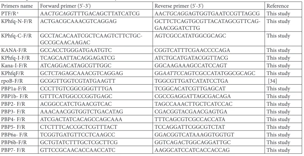
were determined using the standard broth microdilu-tion method, as described by the Clinical and Labora-tory Standards Institute (CLSI, formerly the National Committee for Clinical Laboratory Standards). All test isolates were diluted with NaCl 0.85% medium (BIOMERIEUX) to a turbidity of 0.5 McFarland. These dilute samples were further diluted (1:1000) into fresh Mueller-Hinton (MH) medium in the plates, and incubated for 18h at 37°C. The MIC was defined as the lowest concentration of antibacterial agent that showed no visible growth compared to that of the drug-free control. Meanwhile, the minimum bactericidal concentrations (MBCs) were determined using the colony-counting method. The complete con-tent of every dilution was spread on an LB agar plates and incubated at 37°C for 24 h. We counted the num-ber of colonies on each plate. The MBC was defined as the lowest concentration of bactericide with which no colonies appear on the LB agar plate. Each assay was performed in triplicate.
Time-kill curves
Time-kill curves for CFZN and CXMN against WT K. pneumoniae WJ101 and the hfq mutant, respec-tively, were analyzed. The test isolates were diluted to 0.5 McFarland turbidity using NaCl 0.85% medium (BIOMERIEUX), and then inoculated (1:500) into fresh MH medium in 5-mL test tubes. The tubes contained 0.5×drug concentrations obtained from the previous experiment. We incubated these tubes at 37°C. At the 0, 4, 8, 12, and 24 h time points, a 100-µL aliquot was removed from each test tube and seri-ally diluted 10-fold in sterile water. One hundred mL of each dilution was spread on the LB agar plates and incubated at 37°C for 18 h. Subsequently, we counted the colonies on each plate. Each assay was performed in triplicate.
PCR
We used PCR to confirm the presence of PBP genes in K. pneumoniae WJ101. Genomic DNA from WJ101 was extracted using the TIANamp bacteria DNA kit (TianGen), and this DNA was the PCR template. The reaction system consisted of 12.5 µL 2×Taq Master-Mix, 1 µL of each primer (PBPs-F/R primers, Table 2), 1 µL genomic DNA as template, and ddH2O, for a
final volume of 25 µL. The PCR was performed using the GeneAmpPCR system 9700 (Applied Biosystem) according to following program: 5 min of predenatur-ation at 94°C, followed by 35 cycles of denaturpredenatur-ation for 30 s at 94°C, primer annealing for 30 s at 55°C and extension for 30s at 72°C. The final step was ex-tension for 5 min at 72°C. Five mL of each reaction mixture was electrophoresed in 1% agarose gel con-taining ethidium bromide (EB) at 125 V for 35 min. The DNA bands were imaged using Gel DocTM XR
System (Bio-Rad Laboratories).
Quantitative reverse transcription PCR
RNA was isolated from overnight bacterial cultures using TRIzol reagent (Invitrogen). The RNA yield and purity were determined using a NanoDrop ND-1000 (Thermo Scientific). The purified RNA was reverse transcribed to cDNA using an ImProm-IITM Reverse
Transcription System (Promega). Quantitative reverse transcription PCR (qRT-PCR) was carried out to de-tect PBP gene expression levels in WJ101 and the hfq mutant. The primers and their target genes are listed in Table 2. RT-PCR was performed on an IQ5 real-time PCR detection system (Bio-Rad) using the SYBR Premix Ex TaqTM (TliRNaseH Plus) (Takara).
Ampli-fication was performed in a 25-μL reaction system consisting of 12.5 μL Ex Taq mixture, 0.6 μL of each primer, 1μL cDNA and ddH2O. We used the following program: predenaturation for 30 s at 95°C, followed by 40 cycles of denaturation for 30 s at 95°C and primer annealing for 30 s at 60°C. To acquire melting curves, we performed 71 denaturation-annealing cycles to confirm that this amplification was specific. The anal-yses of the expression levels were conducted on the following targets: PBP1a, PBP1b, PBP2, PBP3, PBP4, PBP5, PBP6a and PBP7. We normalized measure-ments to those for the housekeeping gene rpoB [34] and used the 2-ΔΔt method. Each assay was performed
in triplicate.
Statistical analysis
RESULTS
Hfq mutant of K. pneumoniae exhibits increased MIC for cephalosporin
To test the possible roles of the Hfq protein in K. pneumoniae resistance to cephalosporins, a deletion mutant of hfq was constructed. The hfq open reading frame (ORF) was deleted using a lambda red-based replacement system. PCR and DNA sequencing con-firmed that the hfq deletion was successful (Supple-mentary Figs. 1S and 2S). Next, the antibacterial activ-ities of cephalosporins against K. pneumoniae and the hfq mutant were assessed in a microtiter plate model. The MIC values of the two strains against four cepha-losporin antibiotics are shown in Table 3. MIC values for the WT strain ranged from 0.0625 of CAZ to 16 of CTX. For the hfq mutant, the MIC ranged from 1 of CAZ to 1024 of CFZN. The cephalosporin MIC against the hfq mutant was significantly higher than against WT K. pneumoniae. With respect to the WT MICs, the CTX, CFZN, CXMN and CAZ MICs for the hfq mutant increased by 32-, 1024-, 128- and 16-fold, respectively (Table 3). This effect was impaired by an hfq complementary strain (Table 3).
Increased MBC of the hfq-mutant of K.
pneumoniae to cephalosporin
To further confirm the increased resistance of the hfq mutant to cephalosporin antibiotics, the MBCs for the hfq mutant and WT strains were tested. MBCs for CTX, CFZN, CXMN and CAZ against the WT strain were 32, 16, 4 and 0.25 µg/mL, respectively. MBCs for the hfq mutant were 512, 1024, 64 and 1 µg/mL, respectively (Table 4). Compared with those of the WT strain, the MBCs for these antibiotics in the hfq mutant increased between 4-fold (CAZ) and 64-fold (CFZN). These results demonstrated that the deletion of hfq increased the MBCs for K. pneumoniae.
Time-kill curves
To assess the role of hfq in K. pneumoniae cephalospo-rin insensitivity, the antimicrobial activities of CFZN and CXMN against WJ101 and its hfq mutant, respec-tively, were determined using the time-kill approach.
The results are shown in Fig. 1. As presented in the figure, although the curves of WJ101and the hfq mu-tant show a similar trend, the hfq mutant exhibited a slow-growing state when compared with WT WJ101. While CFZN exhibited excellent bactericidal activity against WJ101 at 1/2×hfq-mutant MIC during 12 h, the mutant exhibited only a minor growth inhibition during the same period. There was no apparent im-pact on the mutant when we treated it with CFZN at 1/2×WT MIC concentration, whereas WT WJ101 exhibited significant growth inhibition. As Fig. 1 il-lustrates, there was a parallel trend for CXMN against WJ101 and the hfq-mutant at 1/2×hfq-mutant MIC and 1/2×WT MIC. These time-kill curves confirmed that hfq plays an important role in resistance of K. pneumoniae to cephalosporins.
Decreased expression level of PBP genes in the
hfq mutant
Bacterial resistance to cephalosporins is usually medi-ated by PBPs. Decreased expression of PBPs results in increased resistance to the antibiotics. To test whether the hfq deletion influenced the expression of PBPs, the distribution of PBPs and their expression were deter-mined. Firstly, K. pneumoniae WJ101 was detected by PCR to confirm the presence of genes that encode PBPs. As shown in Fig. 2(a), all 9 genes encoding PBPs
Table 3. MIC fold increases for hfq mutant over wild-type K. pneumoniae
Antibioticsa WJ101 WJ101ΔH WJ101ΔHCHMIC (µg/ml) Increased folds
CTX 16 512 — 32
CFZN 1 1024 1 1024
CXMN 0.5 64 0.5 128
CAZ 0.0625 1 0.625 16
CTX − cefotaxime; CFZN − cefazolin sodium; CXMN − cefuroxime sodium; CAZ − ceftazidime.
Table 4. MBC fold increases for hfq mutant over wild-type K. pneumoniae.
Antibioticsa WJ101MBC(µg/mL)WJ101ΔH Fold increase
CTX 32 512 16
CFZN 16 1024 64
CXMN 4 64 16
CAZ 0.25 1 4
were successfully detected in the WJ101 strain, indi-cating that these genes do exist in the strain and it was feasible to analyze their expression. To test the influence of hfq on PBPs, gene expression of was ana-lyzed by qRT-PCR in WT and the hfq-mutant strain as described in the Materials and Methods. As shown in Fig. 2(b), compared with that in the WT strain, the expressions of most of these genes, including 1a, 1b, 2, 3, 5, 6a, 6b and 7, were decreased. The expression levels of these genes were inhibited from 0.3- to 5-fold.
Among these, PBP1b, PBP3, PBP6b and PBP7 showed the greatest decrease. The only exception was PBP4, which showed increased expression.
DISCUSSION
Cephalosporins, which have a broad spectrum of anti-microbial activity against a large variety of pathogens, are widely used as first-line drugs for both the
preven-Fig. 1. Survival curves of WJ101 and hfq mutant treated with CFZN and CXMN, respectively. a – 512 µg/mL (1/2×hfq mutant MIC of CFZN); b – 0.5 µg/mL (1/2×WT MIC of CFZN); c – 32 µg/mL (1/2×hfq
mutant MIC of CXMN); d – 0.25 µg/mL (1/2×WT MIC of CXMN).
Fig. 2. Expression levels of PBPs were changed in the hfq mutant of K. pneumoniae. a – all major PBPs are present in K. pneumoniae WJ101. M − marker; 1 − PBP1a; 2 − PBP1b; 3 − PBP2; 4 − PBP3; 5 − PBP4; 6 − PBP5; 7 − PBP6a; 8 − PBP6b; 9 − PBP7; N − negative control; P − positive control (rpoB);
tion and the treatment of bacterial infection. However, recent studies have shown that they have a reduced ef-fect against many drug-resistant pathogens, such as K. pneumoniae. In the present work, we have investigated the role of hfq in the regulation of K. pneumoniae re-sistance to cephalosporins. One interesting finding here was that an hfq mutant exhibited increased resis-tance to cephalosporins in K. pneumoniae.
There have been many reports describing the spe-cific mechanism of the antimicrobial effect of cepha-losporins. In brief, this kind of antibiotic can inhibit the activity of the penicillin-binding proteins, which is necessary for the biosynthesis of peptidoglycan, the main component in the negative-bacteria cell wall. Hence, cephalosporins can influence cell-wall integ-rity, resulting in damage to the pathogen cell. As an important factor contributing to the multifactorial mechanism of cephalosporin resistance, the changes in PBP level have been found to be involved in drug susceptibility in some bacteria. Another interesting finding here is that the inactivation of hfq leads to altered expression levels of PBPs, the targets of ceph-alosporins in K. pneumoniae. This finding implies that the increased resistance might be mediated by an altered expression of PBPs, the main mediators of resistance. The reduction or loss of one or more PBPs has been identified as a reason for the increased resis-tance to β-lactam in many reports. Examples include PBP3 for clinical ceftazidime-resistance in Burkhold-eria pseudomallei [30] and cefotaxime-resistance in Streptococcus pneumoniae [29], and PBP4 for trig-gering overproduction of AmpC in Enterococcus faecium [35]. In addition, the similar phenomenon where increased resistance accompanies a decreased expression level of most PBPs was also observed by Vashist et al. [36] in Acinetobacter baumannii. Con-trary to the above results, Mottl et al. [37] and Sanders et al. [38] reported respectively that the loss of PBP4 would result in diminishing the induction of AmpC in Escherichia coli, which is in line with the increased expression of PBP4 in the cephalosporin-resistant hfq mutant.
In conclusion, one key finding in this investiga-tion is that hfq negatively regulates the sensitivity of K. pneumoniae to cephalosporins. Inactivation of hfq leads to reduced expression levels of most of the
PBPs, which is the main mechanism of resistance to cephalosporin. Therefore, hfq mediated the sensitiv-ity of K. pneumoniae to cephalosporins probably by decreasing the expression of PBPs. Because the roles of the Hfq protein are mainly mediated by sRNA, our future work is to identify sRNA involved in expression regulation of PBPs.
Acknowledgments: This work was supported by the National
Natural Science Foundation of China (81401646, 81171530).
Conflict of interest disclosure: The authors declare no conflict of interest related to this work.
REFERENCES
1. Banapurmath CR, Kallinath S, Banapurmath S, Kalliath A, Kesaree N. Congenital pneumonia caused by Klebsiella pneu-moniae. Indian Pediatr. 1994;31(10):1264-6.
2. Kil KS, Darouiche RO, Hull RA, Mansouri MD, Musher DM. Identification of a Klebsiella pneumoniae strain associated with nosocomial urinary tract infection. J Clin Microbiol. 1997;35(9):2370-4.
3. Soscia JL, Dibenedetto R, Crocco J. Klebsiella pneumoniae
meningitis. Report of a Case and Review of the Literature. Arch Intern Med. 1964;113:569-72.
4. Huck RF. Cholecystitis, septicemia, and cystitis due to Klebsi-ella pneumoniae. U S Armed Forces Med J. 1956;7(9):1368-72. 5. Casanova C, Lorente JA, Carrillo F, Perez-Rodriguez E, Nunez
N. Klebsiella pneumoniae liver abscess associated with septic endophthalmitis. Arch Intern Med. 1989;149(6):1467. 6. World Health Organization. Antimicrobial Resistance: Global
Report on Surveillance. 2014. Geneva, Switzerland: World Health Organization; 2014. [cited 6 December 2015]. 232 p. Available from: http://www.who.int/drugresistance/docu-ments/surveillancereport/en/.
7. Sonnleitner E, Hagens S, Rosenau F, Wilhelm S, Habel A, Jager KE, Blasi, U. Reduced virulence of a hfq mutant of Pseu-domonas aeruginosa O1. Microb Pathog. 2003;35(5):217-28. 8. Christiansen JK, Larsen MH, Ingmer H, Sogaard-Andersen
L, Kallipolitis BH. The RNA-binding protein Hfq of Listeria monocytogenes: role in stress tolerance and virulence. J Bac-teriol. 2004;186(11):3355-62.
9. Ding Y, Davis BM, Waldor MK. Hfq is essential for Vibrio cholerae virulence and downregulates sigma expression. Mol Microbiol. 2004;53(1):345-54.
10. Sharma AK, Payne SM. Induction of expression of hfq by DksA is essential for Shigella flexneri virulence. Mol Micro-biol. 2006;62(2):469-79.
11. Sittka A, Pfeiffer V, Tedin K, Vogel J. The RNA chaperone Hfq is essential for the virulence of Salmonella typhimurium. Mol Microbiol. 2007;63(1):193-217.
mutants in Salmonella: implications for virulence and global protein translation. PLoS One. 2009;4(3):e4809.
13. Dietrich M, Munke R, Gottschald M, Ziska E, Boettcher JP, Mollenkopf H, Friedrich A. The effect of hfq on global gene expression and virulence in Neisseria gonorrhoeae. FEBS J. 2009;276(19):5507-20.
14. Fantappie L, Metruccio MM, Seib KL, Oriente F, Cartocci E, Ferlicca F, Giuliani MM, Scarlato V, Delany I. The RNA chaperone Hfq is involved in stress response and virulence in
Neisseria meningitidis and is a pleiotropic regulator of protein expression. Infect Immun. 2009;77(5):1842-53.
15. Geng J, Song Y, Yang L, Feng Y, Qiu Y, Li G, Guo J, Bi Y, Qu Y, Wang W, Wang X, Guo Z, Yang R, Han Y. Involvement of the post-transcriptional regulator Hfq in Yersinia pestis virulence. PLoS One. 2009;4(7):e6213.
16. Schiano CA, Bellows LE, Lathem WW. The small RNA chap-erone Hfq is required for the virulence of Yersinia pseudotu-berculosis. Infect Immun. 2010;78(5):2034-44.
17. Meibom KL, Forslund AL, Kuoppa K, Alkhuder K, Dubail I, Dupuis M, Forsberg A, Charbit A. Hfq, a novel pleiotropic regulator of virulence-associated genes in Francisella tularen-sis. Infect Immun. 2009;77(5):1866-80.
18. Zeng Q, McNally RR, Sundin GW. Global small RNA chaperone Hfq and regulatory small RNAs are important virulence regulators in Erwinia amylovora. J Bacteriol. 2013;195(8):1706-17.
19. Kendall MM, Gruber CC, Rasko DA, Hughes DT, Sperandio V. Hfq virulence regulation in enterohemorrhagic Escherichia coli O157:H7 strain 86-24. J Bacteriol. 2011;193(24):6843-51. 20. Simonsen KT, Nielsen G, Bjerrum JV, Kruse T, Kallipolitis
BH, Moller-Jensen J. A role for the RNA chaperone Hfq in controlling adherent-invasive Escherichia coli colonization and virulence. PLoS One. 2011;6(1):e16387.
21. Chao Y, Vogel J. The role of Hfq in bacterial pathogens. Curr Opin Microbiol. 2010;13(1):24-33.
22. Van Assche E, Van Puyvelde S, Vanderleyden J, Steenackers HP. RNA-binding proteins involved in post-transcriptional regulation in bacteria. Front Microbiol. 2015;6:141.
23. Faner MA, Feig AL. Identifying and characterizing Hfq-RNA interactions. Methods. 2013;63(2):144-59.
24. Vogel J, Luisi BF. Hfq and its constellation of RNA. Nat Rev Microbiol. 2011;9(8):578-89.
25. Chiang MK, Lu MC, Liu LC, Lin CT, Lai YC. Impact of Hfq on global gene expression and virulence in Klebsiella pneu-moniae. PLoS One. 2011;6(7):e22248.
26. Kalant H. The pharmacology of semisynthetic antibiotics. Can Med Assoc J. 1965;93(16):839-43.
27. Livermore DM. Mechanisms of resistance to cephalosporin antibiotics. Drugs. 1987;34(Suppl.2):64-88.
28. Torok ME, Chantratita N, Peacock SJ. Bacterial gene loss as a mechanism for gain of antimicrobial resistance. Curr Opin Microbiol. 2012;15(5):583-7.
29. Krauss J, Hakenbeck R. A mutation in the D,D-carboxypepti-dase penicillin-binding protein 3 of Streptococcus pneumoniae
contributes to cefotaxime resistance of the laboratory mutant C604. Antimicrob Agents Chemother. 1997;41(5):936-42. 30. Chantratita N, Rholl DA, Sim B, Wuthiekanun V,
Limmathu-rotsakul D, Amornchai P, Thanwisai A, Chua HH, Ooi WF, Holden MT, Day NP, Tan P, Schweizer HP, Peacock SJ. Anti-microbial resistance to ceftazidime involving loss of penicil-lin-binding protein 3 in Burkholderia pseudomallei. Proc Natl Acad Sci U S A. 2011;108(41):17165-70.
31. Wei D, Wang M, Shi J, Hao J. Red recombinase assisted gene replacement in Klebsiella pneumoniae. J Ind Microbiol Bio-technol. 2012;39(8):1219-26.
32. Doublet B, Douard G, Targant H, Meunier D, Madec JY, Cloeckaert A. Antibiotic marker modifications of lambda Red and FLP helper plasmids, pKD46 and pCP20, for inactivation of chromosomal genes using PCR products in multidrug-resistant strains. J Microbiol Methods. 2008;75(2):359-61. 33. Jayol A, Poirel L, Brink A, Villegas MV, Yilmaz M,
Nord-mann P. Resistance to colistin associated with a single amino acid change in protein PmrB among Klebsiella pneumoniae
isolates of worldwide origin. Antimicrob Agents Chemother. 2014;58(8):4762-6.
34. Srinivasan VB, Rajamohan G. KpnEF, a new member of the
Klebsiella pneumoniae cell envelope stress response regulon, is an SMR-type efflux pump involved in broad-spectrum antimicrobial resistance. Antimicrob Agents Chemother. 2013;57(9):4449-62.
35. Moya B, Dotsch A, Juan C, Blazquez J, Zamorano L, Haussler S, Oliver A. Beta-lactam resistance response triggered by inactivation of a nonessential penicillin-binding protein. PLoS Pathog. 2009;5(3):e1000353.
36. Vashist J, Tiwari V, Das R, Kapil A, Rajeswari MR. Analysis of penicillin-binding proteins (PBPs) in carbapenem resistant
Acinetobacter baumannii. Indian J Med Res. 2011;133:332-8. 37. Mottl H, Nieland P, de Kort G, Wierenga JJ, Keck W. Dele-tion of an addiDele-tional domain located between SXXK and SXN active-site fingerprints in penicillin-binding protein 4 from
Escherichia coli. J Bacteriol. 1992;174(10):3261-9.
Supplementary Figures
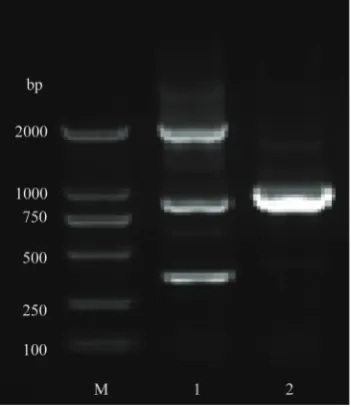
Fig. 1S.hfq mutant strain was identified by PCR. M − marker; 1 − upstream of the identification fragment; 2 − downstream of the identification fragment.