13
© 2018 by the Serbian Biological Society How to cite this article: Jovanović NŽ, Trbojević-Stanković JB, Nešić DM, Obrenović RŽ, Boričić NI, Stojimirović BB. Cancer antigen 125 concentrations in patients on chronic peritoneal dialysis: Relationship with dialysis quality and membrane transport properties. Arch Biol Sci. 2018;70(1):13-20.
Cancer antigen 125 concentrations in patients on chronic peritoneal dialysis:
relationship with dialysis quality and membrane transport properties
Nataša Ž. Jovanović1,*, Jasna B. Trbojević-Stanković2, Dejan M. Nešić3, Radmila Ž. Obrenović4, Novica I. Boričić5 and Biljana B. Stojimirović1
1Clinic of Nephrology, Clinical Center of Serbia, Faculty of Medicine, University of Belgrade, Serbia 2Department of Dialysis, University Clinical Center “Dr Dragiša Mišović, Dedinje”, Faculty of Medicine,
University of Belgrade, Serbia
3Institute of Medical Physiology, Faculty of Medicine, University of Belgrade, Serbia 4Center for Medical Biochemistry, Clinical Center of Serbia, Belgrade, Serbia 5Institute for Pathology, School of Medicine, University of Belgrade, Serbia
*Corresponding author: [email protected]
Received: March 24, 2017; Revised: May 7, 2017; Accepted: May 9, 2017; Published online: June 12, 2017
Abstract: The aim of this study was to evaluate longitudinal changes in drained dialysate cancer antigen 125 (dCA-125) levels and to assess relationships between dCA-125 and dialysis quality, peritoneal membrane transport rates, dialysate glucose load, peritonitis and use of erythropoiesis stimulating agents (ESA), angiotensin-converting-enzyme inhibitors (ACEi) and statins in patients with end-stage renal failure during the first 6 months of peritoneal dialysis (PD) treatment. This prospective study included 20 patients (11 males and 9 females; mean age 62.90±12.69 years) who were followed-up during the first 6 months of PD using conventional low pH glucose-based dialysis fluids. The concentration of dCA-125 was measured in all patients, and the peritoneal equilibration test (PET), peritoneal dialysis treatment adequacy (Kt/V), normalized protein catabolic rate (nPCR), and total, peritoneal and residual clearances of urea and creatinine were calcu-lated. Information on peritonitis occurrence, the use of ESA, ACEi and statins were collected. Data were analyzed by the Mann-Whitney test, Wilcoxon matched pairs test and Spearman’s rank correlation. The concentration of dCA-125 signifi-cantly decreased during the follow-up (p=0.016). After 6 months of PD treatment, the concentration of dCA-125 decreased significantly (p=0.016) in all patients. The decrease was present in all patients, but was statistically significant in patients on ACEi therapy (p=0.006) and in patients not using statins (p=0.005) or ESA (p=0.012). No correlation was found between dCA-125 and glucose load, but a statistically significant negative correlation between dCA-125 and the PET for creatinine was observed (p=0.013). These findings challenge the role of dCA-125 in predicting mesothelial cell integrity in PD patients.
Key words: peritoneal dialysis; cancer antigen 125; ACE inhibitors; erythropoietin; statins
INTRODUCTION
Cancer antigen125 (CA-125) or carcinoma antigen 125 or carbohydrate antigen 125 is a transmembrane mucin-type glycoprotein. In humans, it is encoded by the MUC16 gene, also known as mucin 16 (MUC16) [1-3]. CA-125 was first detected in 1981 [4]; however, its physiological function is still unknown. Some au-thors have suggested that this massive glycoprotein forms a lubricating barrier against particles and infec-tious agents on mucosal surfaces [5]. Its function may be to protect the apical surface from pathogen
adhe-sion due to its negatively charged structure and ligand properties for specific receptors on certain cells [6,7].
cells also express CA-125 and are even more potent producers than ovarian cancer cells [9]. The presence of fluid in the serosal cavities may stimulate CA-125 production. During chronic PD, mesothelial cells come into direct contact with the dialysate and par-ticipate in the overall response. The concentration of CA-125 in the drained dialysate (dCA125) was long considered a mesothelial cell mass marker in stable PD patients, since it tends to decline in parallel with the duration of peritoneal dialysis, suggesting a loss in mesothelial cell mass due to the depletory influence of conventional PD fluid [10,11]. However, authors have reported a lack of correlation between the concentra-tion of CA-125 and the number of mesothelial cells in peritoneal drained dialysate [12]. Even more so, the concentrations of dCA-125 were observed to be elevated during peritonitis episodes. This has put into question whether this marker might actually reflect mesothelial cell damage or even death, rather than mesothelial cell health [13]. All things considered, there is still much uncertainty regarding the physi-ologic and diagnostic significance of dCA-125 in peri-toneal dialysis patients.
The aim of this investigation was to evaluate the changes in the concentrations of drained dialysate dCA-125 in end-stage renal failure patients during the first six months of PD, and to study the relation-ships between dCA-125 and PD quality, exposure of the peritoneum to glucose from the dialysate in peri-tonitis, as well as the relationships between dCA-125 and statin, erythropoietin and ACE-inhibitor use.
MATERIALS AND METHODS
Patients
The study was approved by the Ethical Committee of the Faculty of Medicine, University of Belgrade, and all patients provided informed consent for participa-tion. This prospective study included 20 end-stage re-nal failure patients (11 males and 9 females; mean age 62.90±12.69 years, body surface area 1.81±0.15 m2, body weight 69.99±8.97 kg, body height 170.6±7.96 cm), assessed at the beginning and after six months of peritoneal dialysis treatment. The patients were free of clinical and laboratory signs of infection within 4 weeks before enrollment. They used at least 8 L of
conventional low-pH, glucose-based dialysis fluids (glucose concentration ranged from 1.25-2.5%) per day and drained a larger amount of dialysate than in-stilled liquid (Table 1). The underlying kidney disease was diabetes mellitus-linked in 8 patients, nephroan-giosclerosis was due to long-lasting hypertension in 3, glomerular disease in 1, obstructive nephropathy in 1, and chronic undetermined nephropathy in 7 patients. During the follow-up period, 3 patients de-veloped peritonitis; however, none of them had scle-rosing peritonitis.
Blood sample analyses
At the beginning and after six months of PD, fast-ing mornfast-ing blood, urine and dialysate samples were taken to determine urea and creatinine. CA-125 con-centrations were measured in the drained dialysate af-ter the overnight dwell time. Data concerning statins, ACEi and ESA usage, glucose load and occurrence of peritonitis were collected. PD quality and peritoneal membrane transport properties were assessedat base-line and after 6 months follow-up.
and drained dialysate samples, and concentrations of urea and creatinine in serum assessed during the same time interval. Kt/V is normalized to the volume of total body water, which approximately reflects the vol-ume of urea distribution and is calculated using the Watson formula [15]. ClCr is normalized by the body surface area, calculated using the DuBois-DuBois formula [16]. Fasting venous blood samples to assess serum levels of glucose, urea, creatinine and albumin were taken in biochemistry vacutainer vials and cen-trifuged at 3000 rpm for 10 min. Concentrations of glucose, urea, creatinine and albumin were assessed in samples of 24-h urine and drained dialysate col-lections. All parameters were determined using the Biochemical analyzer ARCHITECT ci8200 (Abbott Diagnostics, Wiesbaden, Germany). Residual renal function was calculated as a mean value of residual clearance of urea and residual clearance of creatinine. The protein catabolic rate (PCR) and normalized protein catabolic rate (nPCR) were calculated using the Bergstrom or Randerson formula [17]. Perito-neal membrane transport characteristics were exam-ined using the standard peritoneal equilibration test (PET) according to Twardowski [18]. After an 8-h night dwell, 2 L of conventional dialysate with glucose concentration between 2.2 and 2.5% were instilled for 4 h. Glucose and creatinine concentrations in blood and in drained dialysate samples were assessed at the start of the test, and again after 2 and 4 h. Peritoneal membrane transport characteristics for glucose and creatinine were calculated based on D/D0 and D/P ratio at the beginning of the test, after 2 and 4 h of instillation of dialysis solution.
Statistical analysis
Data are presented as means±standard deviation (SD) or median where appropriate. Differences between groups were assessed using the non-parametric Mann Whitney test and Wilcoxon matched pairs test. Correla-tional analysis was performed using the Spearman test.
RESULTS
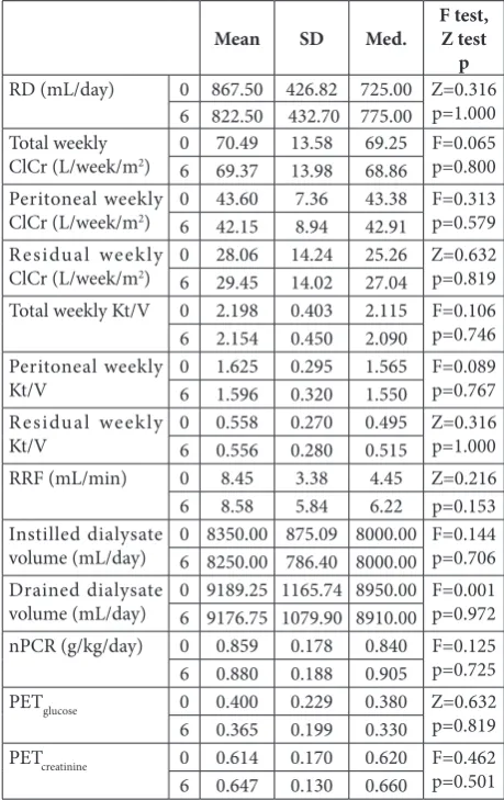
The concentration of dCA-125 decreased significantly (p=0.016) from 28.83±25.35 at baseline to 16.56±14.65 U/mL after 6 months of PD (Fig. 1A). Residual diuresis
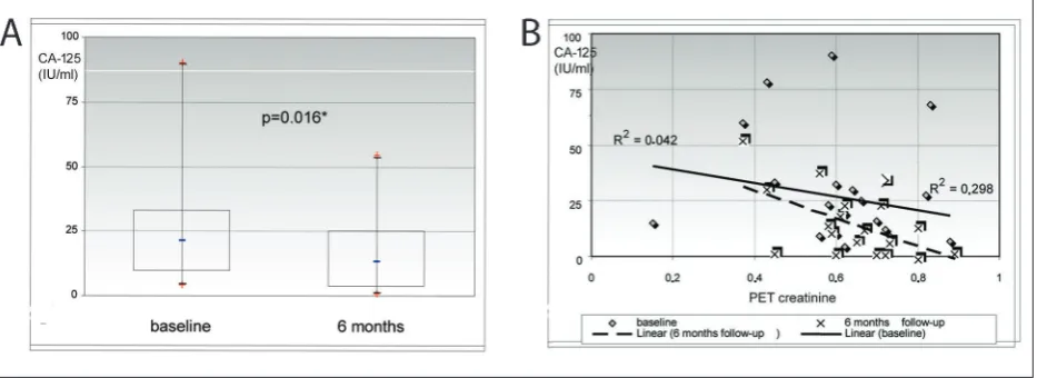
and mean values of total weekly clearances of urea and creatinine were in agreement with the recommenda-tions for dialysis quality (Table 1). Mean values of nor-malized protein catabolic rate (nPCR) were lower than 1 g/kg/day at baseline and after 6 months, and they did not differ significantly (Table 1). The PET for glucose and creatinine did not differ significantly at the begin-ning and after six months of peritoneal dialysis (Table 1). We observed that the concentrations of dCA-125 did not correlate with the nPCR, total, peritoneal and residual clearances of urea and creatinine, neither at the beginning nor after 6 months of chronic PD (Table 2). At the beginning of chronic PD, the concentrations of dCA-125 did not correlate significantly with PETglucose, PETcreatinine, residual diuresis (RD) and residual renal
Table 1. Parameters of dialysis quality at the beginning (0) and after six months (6) of PD.
Mean SD Med. F test,Z test
p
RD (mL/day) 0 867.50 426.82 725.00 Z=0.316
p=1.000 6 822.50 432.70 775.00
Total weekly
ClCr (L/week/m2) 06 70.4969.37 13.5813.98 69.25 F=0.06568.86 p=0.800 Peritoneal weekly
ClCr (L/week/m2) 06 43.6042.15 7.368.94 43.38 F=0.31342.91 p=0.579 Residual weekly
ClCr (L/week/m2) 06 28.0629.45 14.2414.02 25.26 Z=0.63227.04 p=0.819
Total weekly Kt/V 0 2.198 0.403 2.115 F=0.106
p=0.746
6 2.154 0.450 2.090
Peritoneal weekly
Kt/V 06 1.6251.596 0.2950.320 1.565 F=0.0891.550 p=0.767 Residual weekly
Kt/V 06 0.5580.556 0.2700.280 0.495 Z=0.3160.515 p=1.000
RRF (mL/min) 0 8.45 3.38 4.45 Z=0.216
6 8.58 5.84 6.22 p=0.153
Instilled dialysate
volume (mL/day) 0 8350.00 875.09 8000.00 F=0.1446 8250.00 786.40 8000.00 p=0.706 Drained dialysate
volume (mL/day) 0 9189.25 1165.74 8950.00 F=0.0016 9176.75 1079.90 8910.00 p=0.972
nPCR (g/kg/day) 0 0.859 0.178 0.840 F=0.125
p=0.725
6 0.880 0.188 0.905
PETglucose 0 0.400 0.229 0.380 Z=0.632
p=0.819
6 0.365 0.199 0.330
PETcreatinine 0 0.614 0.170 0.620 F=0.462
p=0.501
6 0.647 0.130 0.660
function (RRF) (Table 3). After 6 months of chronic PD, no significant correlation was found between dCA-125 concentration and PETglucose, residual diuresis (RD) and RRF (Table 3), while a statistically significant in-verse correlation (R=-0.546; p=0.013) was found be-tween dCA-125 concentration and PETcreatinine (Fig. 1B). In our study cohort, there was no significant correlation between dCA-125 and peritoneal glucose exposure (at baseline: R=-0.195, p=0.409, and after 6 months it was R=-0.262, p=0.265). The concentrations of dCA-125 did not differ significantly between patients with and without peritonitis during the follow-up (Table 4). The concentrations of dCA-125 in patients on ESA, ACEi and statin therapy and in those not receiving these
medications were not significantly different, neither at baseline nor after 6 months of PD (Table 5). During the 6-month follow-up, the concentrations of dCA-125 de-creased in all patients, the difference being statistically significant in patients not treated with ESA or statins and in patients receiving ACE inhibitors (Table 5).
DISCUSSION
Highly glycosylated CA-125 creates a hydrophilic envi-ronment on the epithelial cell apical membrane, acting as a lubricating barrier against foreign particles and infectious agents in different epithelia, such as the
ocu-Table 2. Correlation between drained dialysate CA-125 (dCA-125) and Kt/V, ClCr and nPCR at the beginning (0) and after six months (6) of PD.
nPCR
(g/kg/day) total weekly Kt/V per. w.Kt/V res w.Kt/V
total weekly ClCr
(l/w./m2)
per. w. ClCr (l/w./m2)
res. w. ClCr (l/w./m2)
dCA-125 (U/mL)
R 0 0.152 -0.066 -0.366 0.275 0.257 0.000 -0.378
R 6 0.401 -0.015 0.025 -0.057 -0.116 -0.046 -0.319
p 0 0.522 0.782 0.112 0.241 0.274 1.000 0.101
p 6 0.080 0.951 0.916 0.810 0.628 0.848 0.171
nPCR – normalized protein catabolic rate; w. – weekly; per. – peritoneal; res. – residual; ClCr – creatinine clearance
Table 3. Correlation of peritoneal membrane transport properties. Residual diuresis and residual renal function with drained dialysate CA-125 (dCA-125) at the beginning (0) and after six months (6) of PD.
PETglucose PETcreatinine RD (ml/day) RRF (ml/min)
0 6 0 6 0 6 0 6
dCA-125
(U/mL) Rp 0.0360.880 0.3500.131 -0.4430.050 0.013*-0.546 0.1850.434 0.9320.020 0.3940.202 0.0630.791
PET – peritoneal equilibration test; RD – residual diuresis; RRF – residual renal function
lar, the respiratory and the female reproductive system [19]. Elevated CA-125 has served as a tumor marker in gynecological malignancies, lung cancer, mediasti-nal teratoma and even in non-Hodgkin’s lymphoma [20], and serum CA-125 levels are helpful in monitor-ing, risk stratification and prognosis after treatment of ovarian cancer [21]. Elevated CA-125 levels can be seen in endometriosis, acute pancreatitis, peritonitis, inflammatory pelvic disease, ascites of nonmalignant origin, cirrhosis, as well as in physiological conditions, including menstruation and early pregnancy [6]. Dur-ing the last decade, CA-125 has been accepted as a marker of the integrity and/or regeneration of me-sothelial cells in in vitro conditions [12,19,20], and dCA-125 can be considered a marker of mesothelial cell mass in stable PD patients [10]. During chronic PD, the peritoneal membrane is continuously exposed to bioincompatible dialysate solutions and sporadi-cally to peritonitis episodes. These conditions cause adverse structural and functional changes that limit the membrane’s efficacy as a dialyzing organ [21,22].
In vitro studies have shown that conventional dialysis solutions are toxic to cultured mesothelial cells: the combination of low-pH lactate with high glucose and
glucose-degradation product (GDP) concentrations and hyperosmolarity reduce cell viability, inhibit the synthesis of interleukins and prostaglandins and in-duce apoptosis [22,23]. Chronic peritoneal dialysis treatment is accompanied by a decrease in dCA-125, which points to a loss of mesothelial cell mass [10]. We observed that dCA-125 concentrations significantly decreased from 28.83±25.35 to 16.56±14.65 IU/mL after 6 months of PD, which is in agreement with lit-erature data. Several cross-sectional studies reported a significant relationship between dCA-125 and the duration of PD; however, some researchers did not confirm this finding, probably because of the large interindividual variability. In a cross-sectional study including 31 patients, the mean dCA-125 concentra-tion was 13.8 U/mL, range 1.1-96.5 U/mL. In a stable subgroup of 26 patients during the second year of PD, the mean dCA-125 concentration was 22.1±2.1 U/mL, with a calculated decreasing annual trend of 2.2% [24]. The application of theoretically more-biocompatible PD solutions with a low content of GDPs and a higher pH is associated with an increase in dCA-125 as com-pared to treatment with conventional fluid [22-25]. In a 3-month longitudinal follow-up study of 35 patients treated with neutral pH and a low-GDP PD fluid, a significant increase in dCA-125 from 12 (ranging from 3.93-85) U/mL to 48 (ranging from 14-373) U/mL was shown. Another study that followed 36 peritoneal di-alysis patients treated with conventional didi-alysis fluid showed a decrease in dCA-125 from 38 (ranging from 3.42-122) U/mL to 13 (ranging from 4-44) U/mL [21]. However, after the follow-up period of 12 months, no significant differences were observed between dCA-125 levels at the beginning and at the end of the study (8.26±5.22 U/mL vs. 9.08±6.8 U/mL) [26].
In our study, no significant correlation was ob-served between dCA-125 concentrations and total, residual and peritoneal clearances of urea and creati-nine, RD and RRF during the first six months of PD. These findings could be explained by the compara-tively short duration of dialysis and may suggest that structural and functional changes of the peritoneal membrane had not developed during the first months of PD, in spite of the significant changes in dCA-125 concentrations. This could suggest that changes in dCA-125 do not have a prognostic value, at least not in the early period of PD. In our study cohort, a
sig-Table 4. Influence of peritonitis after six months of PD on drained dialysate CA-125 (dCA-125) concentrations.
peritonitis N Mean±SD p
dCA-125 (U/mL) No 17 17.56±15.54 p=0.78
Yes 3 10.90±7.00
N – number of patients
Table 5. Statin influence on drained dialysate CA-125 (dCA-125) concentrations. ESA and ACEi treatment at the beginning (0) and after six (6) months of PD.
Therapy N dCA-125 (mean±SD) pW
0 6
ESA Yes 12 27.67±23.35 16.26±12.08 0.071
No 8 30.56±29.69 17.03±18.80 0.012*
pMW 1.000 0.734
ACEi Yes 18 28.15±25.32 14.90±12.22 0.006*
No 2 34.90±35.07 31.55±32.31 0.180
pMW 0.801 0.313
Statins Yes 6 40.82±34.92 23.63±15.46 0.173
No 14 23.69±19.37 13.54±13.57 0.005*
pMW 0.284 0.161
ESA – erythropoiesis stimulating agents; ACEi – angiotensin converting enzyme inhibitors; pW – probability Wilcoxon matched pairs test; pMW –
nificant inverse correlation was found between the concentration of dCA-125 and PETcreatinine after the 6-months follow-up. Lower dCA-125 concentrations are related to higher creatinine transport rates, which is an unfavorable characteristic of the mesothelial lay-er, thus implying that a low dCA-125 concentration is associated with a poor prognosis. The majority of studies found no relationship between dCA-125 and peritoneal transport rates [10,27]. One case-control study showed that an appearance rate of dCA-125 less than 33 U/min, combined with the appearance rate of interleukin 6 (IL-6) above 350 pg/min, was predictive for the development of encapsulating peritoneal scle-rosis – a deleterious complication of chronic PD oc-curring in 2 to 3 years, with a sensitivity of 70% and a specificity of 89%. The temporal trend of dCA-125 is a useful predictor of encapsulating peritoneal sclerosis, which has a very poor prognosis when diagnosed [28].
Only 3 patients from our study group developed peritonitis during the follow-up. Concentrations of dCA-125 were assessed in patients free of peritonitis and/or signs of other infection within 4 weeks prior to the examination. The concentration of dCA-125 was slightly lower in patients who developed peri-tonitis during the follow-up vs. the periperi-tonitis-free group. Some investigators reported increased levels of dCA-125 during acute peritonitis, probably due to necrosis of mesothelial cells [10]. Other studies did not find any additional diagnostic or prognostic benefit of measuring dCA-125 in patients on PD pre-senting acute peritonitis when compared to standard investigations, including peritoneal white cell count and serum C-reactive protein [29]. No differences in peritoneal dCA-125 concentrations were found in patients with Gram-positive, Gram-negative, fungal and polymicrobial peritonitis, nor in patients who responded successfully to intraperitoneal antibiotics and those with treatment failure [30]. No correlation was observed between dCA-125 levels and the number of peritonitis episodes, accumulated days of perito-neal inflammation and ultrafiltration capacity [31]. However, in a patient with five episodes of peritonitis, ultrafiltration failure was diagnosed; this was preceded by a gradual and marked decline in CA-125 levels dur-ing a 3-year follow-up [32,33].
In our study cohort, dCA-125 concentrations were not significantly different between the patients on
ESA, ACEi and statin therapy and patients that were not receiving these medications, both at baseline and after 6 months of PD. The concentration of dCA-125 decreased in all patients during the 6-month PD treat-ment, and the difference was significant in patients receiving ACEi therapy and in those not receiving statins or ESA therapy. No literature data are available concerning the influence of ACEi on dCA-125 levels during PD, but it was found that ACEi play a protec-tive role in preserving residual renal function and the transport rates of the peritoneal membrane during long-term PD, indirectly suggesting better preserva-tion of the mesothelial membrane [34,35]. Our find-ings of significantly lower dCA-125 concentrations in patients on ACEi therapy after the 6-month follow-up are not in agreement with the suggested protective role of ACEi, so possibly a longer follow-up would be required to better define the correlation between ACEi therapy and dCA-125 levels.
Erythropoietin is a highly specialized growth fac-tor. In our group of patients, ESA therapy was not significantly associated with changes in dCA-125 con-centrations.However, patients not receiving ESA had significantly reduced dCA-125 levels after 6 months of PD, suggesting that ESA has a protective roll in preserving mesothelial cell mass.
In our patients who were not using statins dur-ing the entire follow-up period, the concentrations of dCA-125 were insignificantly lower than in those treated with statins. After six months of PD, in both groups the concentrations of dCA-125 were lower than at baseline, and the difference was significant in the group that was not treated with statins. While the literature data concerning this relationship are lim-ited, it was published that the use of atorvastatin is associated with increased dCA-125 levels, which may be linked with the anti-inflammatory and antihyper-lipidemic effects of the drug [36].
Concerning the protective effect of some medi-cations on mesothelial cell mass during chronic PD treatment, it has also recently been found that spironolactone therapy is significantly correlated with higher dCA-125 concentrations [37]. This effect, how-ever, was not investigated in our study.
con-centrations in our study. Previous studies showed a significant decline in dCA-125 levels, reflecting the loss of mesothelial cell mass in children (>5 years) under-going long-term PD, but this was not related to peri-toneal glucose exposure [38]. On the other hand, the concentration of dCA125 increased significantly from baseline values in patients with a low-GDP level [39].
CONCLUSION
In our study in which we examined the effects of the use of conventional peritoneal dialysis solutions, the drained dialysate CA-125 levels decreased significant-ly during the first 6 months of PD treatment, they did not correlate with membrane transport rates and the quality of dialysis and they were not influenced by peritoneal glucose exposure. These results challenge the role of dCA_125 as an indicator of mesothelium integrity in chronic PD patients. The use of statins and ESA appeared to be protective for mesothelial cell mass while ACEi did not exhibit such an effect.
Acknowledgments: The study was supported by the research grant from the Ministry of Education, Science and Technologi-cal Development of the Republic of Serbia No 145070.
Author contributions: Nataša Jovanović and Biljana Stojimirović were responsible for conceiving and designing the study and the drafting of the manuscript. Jasna Trbojević-Stanković and Dejan Nešić collected the literature. Nataša Jovanović and Bil-jana Stojimitović made critical revisions to the paper. Radmila Obrenović and Novica Boričić provided statistical expertise. Conflict of interest disclosure: None to declare
REFERENCES
1. Yin BW, Lloyd KO. Molecular cloning of the CA125 ovarian cancer antigen: identification as a new mucin, MUC16. J Biol Chem. 2001;276(29):27371-5.
2. Yin BW, Dnistrian A, Lloyd KO. Ovarian cancer antigen CA125 is encoded by the MUC16 mucin gene. Int J Cancer. 2002;98(5):737-40.
3. Duraisamy S, Ramasamy S, Kharbanda S, Kufe D. Dis-tinct evolution of the human carcinoma-associated trans-membrane mucins, MUC1, MUC4 AND MUC16. Gene. 2006;373:28-34.
4. Bast RC, Feeney M, Lazarus H, Nadler LM, Colvin RB, Knapp RC. Reactivity of a monoclonal antibody with human ovarian carcinoma. J Clin Invest. 1981;68(5):1331-7. 5. Miralles C, Orea M, Espana P, Provencio M, Sancjez A,
Cantos B, Cubedo R, Carcereny E, Bonilla F, Gea T. ancer
antigen 125 associated with multiple benign and malignant pathologies. Ann Surg Oncol. 2003;10(2):150-4.
6. Ercan S, Kaymaz O, Yucel N, Orcun A. Serum concentra-tions of CA 125, CA 15-3, CA 19-9 and CEA in normal pregnancy: a longitudinal study. Arch Gynecol Obstet. 2012;285(3):579-84.
7. Mitić N, Janković M. Activity profile of the CA 125 antigen towards human red blood cells. Arch Biol Sci. 2010;62(2):271-80.
8. Hung C-L, Hung T-C, Lai Y-H, Lu C-S, Wu Y-J, Yeh H-I. Beyond malignancy: the role of carbohydrate antigen 125 in heart failure. Biomark Res.2013:1:25.
9. Zeimet AG, Marth C, Offner FA, Obrist P, Uhl_Steidl M, Feichtinger H. Human peritoneal mesothelial cells are more potent than ovarian cancer cells in producing tumor marker CA-125. Gynecol Oncol. 1996;62:384-89.
10. Krediet RT. Dialysate cancer antigen 125 concentra-tion as marker of peritoneal membrane status in patients treated with chronic peritoneal dialysis. Perit Dial Int. 2001;21(6):560-7.
11. Fusshöller A, Grabensee B, Plum J. Effluent CA 125 concen-tration in chronic peritoneal dialysis patients: influence of PD duration, peritoneal transport and PD regimen. Kidney Blood Press Res. 2003;26(2):118-22.
12. Breborowicz A, Breborowicz M, Pyda M, Połubinska A, Oreopoulos D. Limitations of CA125 as an index of peritoneal mesothelial cell mass. Nephron Clin Pract. 2005;100:c46-c51.
13. Cheema H, Bargman JM. Cancer antigen 125 as a biomerker in peritoneal dialysis mesothelial cell health or death? Perit Dial Int. 2013;33(4):349-52.
14. Gotch FA, Sargent JA. A mechanistic analysis of the National Cooperative Dialysis Study (NCDS). Kidney Int. 1985;28:526-34.
15. Watson PE, Watson ID, Batt RD. Total body water volumes for adult males and females estimated from simple anthro-pometric measurements. Am J Clin Nutr. 1980;33:27-39. 16. Dubois D, Dubois EF. A formula to estimate the
approxima-tive surface area if height and weight are known. Arch Intern Med. 1960;17:863-71.
17. Mandolfo S, Zucchi A, Cavalieri D, Oro L, Corradi B, Imbas-ciati E. Protein nitrogen appearance in CAPD patients: what is the best formula. Nephrol Dial Transplant. 1996;11:1592-6.
18. Twardowski ZJ, Nolph KD, Khanna R, Prowant BF, Ryan LP, Moore HL, Nielsen MP. Peritoneal equilibration test. Perit Dial Bull. 1987;7:138-47.
19. Perez BH, Gipson IK. Focus on Molecules: human mucin MUC16. Exp Eye Res. 2008; 87 (5): 400-1.
20. Bast RC, Xu FJ, Yu YH, Barnhill S, Zhang Z, Mills GB. CA 125: the past and the future. Int J Biol Markers. 1998;13(4):179-87.
21. Topley N, Michael D, Chalice T. Ca125: Holy grail or poi-soned chalice. Nephron 2005; 100: c52-4.
23. Trbojević-Stanković J, Obradović M, Cemerikić-Martinović V, Trpinac D, Lausević Z, Stojimirović B. Immunohisto-chemical study of pathological alterations of peritoneum in patients with terminal renal insufficiency and on peritoneal dialysis. Vojnosanit Pregl. 2011;68(7):556-60.
24. Pannekeet, Marja M. Ho-Dac; Hiralall JK, Struijk DG. Krediet RT. Longitudinal follow-up of CA125 in peritoneal effluent. Kidney Int. 1997;51(3):888-93.
25. Williams JD, Topley N, Craig KJ, Mackenzie RK, Pischetsrie-der M, Lage C, Passlick-Deetjen J and the Euro Balance Trial Group. The euro-balance trial: the effect of a new biocom-patible peritoneal dialysis fluid (balance) on the peritoneal membrane. Kidney Int. 2004;66:408-18.
26. Fusshoeller A, Plail M, Grabensee B, Plum J. Biocompatibil-ity pattern of a bicarbonate /lactate-buffered peritoneal dial-ysis fluid in APD: a prospective randomized study. Nephrol Dial Transplant. 2004;19:2101-6.
27. Rodrigues A, Martins S, Santos MJ: Evaluation of effluent markers cancer antigen 125, vascular endothelial growth fac-tor and interleukin-6. Relationship with peritoneal trans-port. Adv Perit Dial. 2004;20:8-12.
28. le Poole CY, Welten AGA, Piet M. ter Wee, Paauw NIJ., Djorai AN, Valentijn RM, Beelen RHJ, van den Born J, van Ittersum FJ. A peritoneal dialysis regimen low in glucose and glucose degradation products results in increased can-cer antigen 125 and peritoneal activation. Perit Dial Int. 2012;32(3):305-15.
29. Passadakis P, Panagoutsos S, Thodis E, Tsivara I, Sopassi F, Kartali S, Vargemezis V. Evaluation of changes in serum and dialysate levels of cancer antigen 125 in stable continu-ous ambulatory peritoneal dialysis patients. Adv Perit Dial. 1999;15:40-4.
30. Barreto DL, Coester AM, Noordzij M, Smit W, Struijk DG, Rogers S, de Waart DR, Krediet RT. Variability of efflu-ent cancer antigen 125 and interleukin-6 determination in peritoneal dialysis patients. Nephrol Dial Transplant. 2011;26:3739-44.
31. Panorchan K, Davenport A. Diagnostic and prognostic role of peritoneal Ca 125 in peritoneal dialysis patients present-ing with acute peritonitis. BMC Nephrol. 2014;15:149. 32. Jiménez C, Díaz C, Selgas R, Auxiliadora Bajo M, del Peso
G, Sánchez-Tomero JA, Gonzalez-Gancedo P. Peritoneal kinetics of cancer antigen 125 in peritoneal dialysis patients: the relationship with peritoneal outcome. Adv Perit Dial. 1999;15:36-9.
33. Barreto DL, Krediet RT. Current status and practical use of effluent biomarkers in peritoneal dialysis patients. Am J Kidney Dis. 2013;62(4):823-33.
34. Duman S, Günal AI, Sen S, Asçi G, Ozkahya M, Terzioglu E, Akçiçek F, Atabay G. Does enalapril prevent peritoneal fibrosis induced by hypertonic (3.86%) peritoneal dialysis solution? Perit Dial Int. 2001;21:219-24.
35. Duman S, Sen S, Duman C, Oreopoulos DG. Effect of val-sartan versus lisinopril on peritoneal sclerosis in rats. Int J Artif Organs. 2005;28:156-63.
36. Özlem Tir, YAki Celalettin Usalan. The effect of atorvas-tatin therapy in CAPD Patients on inflammatory markers (CRP) and dialysate IL-6 and Ca-125 Level. Turk Neph Dial Transpl. 2011;20(3):284-9.
37. Yelken B, Gorgulu N, Gursu M, Yazici H, Caliskan Y, Telci A, Ozturk S, Kazancioglu R, Ecder T, Bozfakioglu S. Effects of spironolactone on residual renal function and perito-neal function in peritoperito-neal dialysis patients. Adv Perit Dial. 2014;30:305-10.
38. Candan C1, Turhan P, Sever L, Civilibal M, Canpolat N, Caliskan S, Kasapcopur O, Arisoy N. Dialysate CA125 lev-els after 5 years on continuous peritoneal dialysis. Pediatr Nephrol. 2011;26(5):783-8.