Address for correspondence Col. (Dr.) Asher Ahmed Mashhood Classified Skin Specialist
CMH, Multan
E-mail: [email protected]
Original Article
Is slit-skin smear an alternate to histopathology
in the diagnosis of cutaneous leishmaniasis?
Asher Ahmed Mashhood, Salman Ahmad, Muhammad Mumtaz Malik
Department of Dermatology, CMH, Multan
Abstract Objective To determine how effective is slit-skin smear as a diagnostic test in comparison with the histopathology.
Methods This was a validation study, carried out in Combined Military Hospital (CMH) Kohat from 1st September 2011 to 30th June 2012. The study population was the patients who were clinically diagnosed as cutaneous leishmaniasis. Slit-skin smears were performed in all and a biopsy was sent for histopathology. The results were analyzed by cross tabs and chi square test.
Results A total of 50 patients were included. They were all adult males. Leishman-Donovan (LD) bodies were seen in histopathology slides in 31 patients (62%) and were not detected in 19 patients (38%). The slit-smear showed LD bodies in 18 smears (36%) and negative in 32 smears (64%) [p=0.048].
Conclusion Histopathology is statistically superior investigation than slit-skin smear in the diagnosis of cutaneous leishmaniasis.
Key words
Cutaneous leishmaniasis, slit-skin smear, histopathology, Leishman-Donovan bodies.
Introduction
The first descriptions of cutaneous leishmaniasis can be traced back to the 9th century (Balkh sore), but now it is a major world health problem especially in Pakistan. It has now become the most common chronic granulomatous infection in our country. The clinical history, appearance, duration of illness and acquisition of the disease in an endemic area all helps in the diagnosis. However, there are many patients in whom the diagnosis is not straight forward, histopathology and/or slit-skin smears are used for the confirmation. The latter has an advantage that it requires only Leishman staining and a good
quality microscope equipped with oil immersion lens. The yield of the test, however, depends upon the quality of prepared smear, microscope and expertise of the pathologist. There is a lot of false positivity due to wrong technique of smear preparation, poor fixing and staining, less sensitive microscope, and misinterpretation of Leishman-Donovan (LD) bodies in the smear.
The objective of the study was to determine if the slit-skin smears could be an alternative to histopathology in the diagnosis of cutaneous leishmaniasis, and to demonstrate by photographs the true appearance of LD bodies in a smear.
Methods
from 1st September 2011 to 30th June 2012. The study population was the patients who reported to the skin outpatient department of CMH, Kohat, a tertiary care hospital of Pakistan Army. The inclusion criteria were mainly clinical. This was presence of one or more ulcerated or scaly plaques on exposed body areas for more than a month and history of acquisition of disease from a known endemic area. The exclusion criteria were those patients who had lesions which were clinically doubtful, or those who had received some definitive treatment.
For the calculation of the sample size, Altman’s nomogram was used. The sample size was calculated as 50. The study group comprised of 50 male patients. All the patients were admitted, slit-skin smear done, biopsied and specimen submitted for histopathology. The slit-smears were done at CMH, Kohat and histopathology was done at CMH, Peshawar.
As already proven, histopathology was taken as the gold standard. Only those cases were declared positive in which clearly demonstrable LD bodies were seen in hematoxylin-eosin (H&E) sections. Sometimes Giemsa stain was used for confirmation. Those cases in which LD bodies were not seen were declared as negative. Slit-skin smear was stained by Leishman stain and seen by one dermatopathologist on Olympus microscope (CX-21).
The results were analyzed by cross tab. The sensitivity and specificity of slit-smear was calculated by keeping the histopathology as gold
standard. The results were further evaluated by applying chi-square test.
Results
The study group comprised of 50 male patients having ages between 12-46 years (mean 26.62 years, SD 7.70). All the patients acquired the infection from North or South Waziristan. The duration of illness was from 3-24 weeks (mean 10.12 weeks, SD 5.32). Number of lesions per patient ranged between 1-20 (mean 2.98, SD 4.32), [Table 1].
There were in total 111 lesions in 50 patients. As far as the site of lesions is concerned; there were 6 lesions on face (5.4%), 17 lesions on left hand (15.3%), 15 on right hand (13.5%), 9 on left forearm (8.1%), 9 on right forearm (8.1%), 6 on right leg (5.4%), 3 on left leg (2.7%), 29 on right foot (26.1%) and 17on left foot (15.3%).
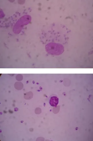
LD bodies were seen in histopathology slides in 31 patients (62%) and were not detected in 19 patients (38%). The slit-smear showed LD bodies in 18 smears (36%) and negative in 32 smears (64%). The LD bodies in a smear were seen under X100 or oil immersion lens. At this magnification, they were seen as an oval body with clear nucleus and sometimes a small kinetoplast. They were not seen in isolation, but were always be in a group, in the cytoplasm of a macrophage (Figure 1)
Statistical analysis
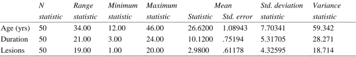
The sensitivity and specificity of slit-skin smear Table 1 Descriptive statistics.
N Range Minimum Maximum Mean Std. deviation Variance
statistic statistic statistic statistic Statistic Std. error statistic statistic Age (yrs) 50 34.00 12.00 46.00 26.6200 1.08943 7.70341 59.342
Duration 50 21.00 3.00 24.00 10.1200 .75194 5.31705 28.271
Table 2 Sensitivity and specificity of skin-split smear as compared to histopathology, the gold standard (n=50). Histopathology proven cases Histopathology negative cases Total
Slit-skin smear positive cases 13 (a) 5 (b) 18
Slit-skin smear negative cases 18 (c) 14 (d) 32
Total 31 19 50
Sensitivity = a / a+c x 100 = 41.94 %, specificity = d / d+b x100 = 73.68%, positive predictive value = a / a+b x 100 = 72.22%, negative predictive value = d / c+d x 100 = 43.75 %
Figure 1 Different images of Leishman-Donovan bodies as seen with oil immersion lens (100X).
were calculated in comparison with the histopathology, which was taken as gold standard; and plotting the values in 2x2 chart as given in Table 2.
On the basis of statistical tests, slit-skin smear had a low sensitivity and high specificity, showing the true nature of this test. Chi square test was applied and the Pearson chi square value came out as 3.92 and the p value was 0.048, which showed that there was statistically significant difference between the two tests.
Discussion
Cutaneous leishmaniasis (CL) is an emerging global hazard. There are about 1.5 million new cases of CL leishmaniasis each year of which over 90% occur in Afghanistan, Algeria, Iran, Iraq, Saudi Arabia, Syria, Brazil and Peru.1 It is now a serious health issue in Pakistan also, but due to the lack of any epidemiological study the true incidence of the disease is not known.
The geographical distribution of CL is mainly determined by the sandfly vectors (Phlebotomus
or Lutzomyia spp.). They live in dark, damp places, are relatively weak flyers, and due to their small size (2-3mm long) can penetrate mosquito nets. They are most active in the evening and at night. Sandfly numbers are related to natural factors such as rainfall1,2 and during summer. Decreased insecticide spraying for malaria, poor waste disposal and heaps of construction waste encourage breeding, and has led to an increase in the prevalence of the disease in urban areas.3
In Pakistan, CL is seen mainly in travelers returning from endemic areas, such as those conducting rural field studies, tourists and military personnel. Unfortunately many of those infected are ignorant of the risks, take no personal protective measures and experience delays in diagnosis followed by inappropriate treatment upon their return.4
hallmark of the disease, although at times they are difficult to detect. Infected macrophages measure 20-30 µm across whereas amastigotes (known as Leishman-Donovan bodies) are round to oval structures measuring only 2-5µm. Although H & E will demonstrate amastigotes, a Giemsa stain is used in difficult cases.5,6
The diagnosis is often made on the basis of clinical appearance, bite over an exposed area and a history of acquiring infection from a known endemic area. Although the appearance mimics lupus vulgaris and deep mycosis but on the basis of the clinical experience of the last 10 years it can be confidently stated that CL remains the first diagnosis of any granulomatous skin infection fulfilling the above criteria. Since the treatment is expensive and potentially toxic pathological confirmation should be made preferably by demonstrating the organism in tissue and /or smear.
There are several methods to prepare a smear. One of these is “impression smear”, made by gently pressing the inner side of the skin biopsy against a glass microscope slide 4-5 times after which the slide is dried in air then fixed in 95% ethanol for 3 minutes. Alternative methods are needle aspirates and slit-skin smears. A needle aspirate is obtained by using a 2ml syringe with a 20 gauge needle, containing 0.3ml 0.9% saline. The needle is inserted through intact skin and the saline is injected into the edge of the lesion. The needle is then moved back and forth rotating it and applying suction at the same time to cut small pieces of tissue from the edge of the needle track, which are then aspirated. The aspirate is used to prepare smears. Slit-skin smears are made by squeezing the edge of the lesion between thumb and forefinger, making a shallow slit 2-3 mm deep in the pinched skin with a scalpel and then scrapping the cut edge at
the right angle and spreading the smear over a glass slide.7
Most Old World lesions heal appropriately without treatment, but the spontaneous healing is often associated with unsightly scarring. Lesions on cosmetically or functionally important sites, such as the face or hands, those associated with lymphangitis or sporotrichoid spread and multiple or persistent lesions must be treated promptly.
the toxicity.12 Rifampicin has been reported to be efficacious but there are no good controlled trials to support its use and furthermore its use in areas where tuberculosis and leprosy are endemic should be avoided.13 Alternative systemic agents include aminosidine (paromomycin), pentamidine, ketoconazole and itraconazole, but they have yet to find a place in routine clinical practice.8 The most promising oral drug today is miltefosine. In an open label study in Colombia, four weeks treatment with 133 and 150mg of miltefosine daily cured 100% and 89% respectively.14 An experience of its use in 20 patients as a part of multi-departmental trial, has similar results, but in patients with one or a few lesions.
Local and topical therapy is an important option for those with a single non-inflamed lesion. Local injection of sodium stibogluconate, or meglumine antimoniate, is useful for this purpose. Technique is important – infiltrate 1-3ml around the lesion to produce complete blanching at the base. It may be repeated on alternate days or given weekly until they heal.9,15 Aminosidine (paromomycin) ointment (15% paromomycin, 12% methyl benzethonium chloride in white soft paraffin) applied twice daily for 10 days are effective for L. major16 (not available in Pakistan). Cryotherapy is also useful for small lesions (<1cm diameter). Apply two 20s freeze-thaw cycles to the whole lesion, assess response at intervals of 3 weeks and repeat once or twice if necessary.16 Lesions can also be treated by local excision, curettage or by electrodessication but are probably associated with a higher risk of relapse.8 The most recent topical agent to be investigated is the topical immunomodulator, imiquimod, which was developed to treat genital warts. It produced a 90% cure rate in patients who had failed to respond to antimonials alone when it was used
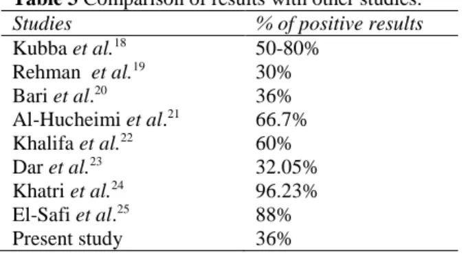
Table 3 Comparison of results with other studies. Studies % of positive results
Kubba et al.18 50-80%
Rehman et al.19 30% Bari et al.20 36% Al-Hucheimi et al.21 66.7% Khalifa et al.22 60% Dar et al.23 32.05% Khatri et al.24 96.23% El-Safi et al.25 88%
Present study 36%
in conjunction with antimonials.17
Patients should be monitored until the lesions have fully healed and the infiltration has resolved. Thereafter patient should be warned that relapse is possible and observe their scar for thickening, ulceration or crusting. Those who relapse should be given a full course of systemic antimony as in the first instance.
Table 3 shows a comparison of the positive
yield in picking LD bodies in slit-skin smears, between the present and previous study. It is quite obvious that the results are not uniform. In two studies the yield is unusually high i.e. around 90%, while in 3 studies it was around 60%. There were 3 studies in which the yield was around 30% just like the present study.
Conclusion
On the basis of the results and the statistical tests, it may be concluded that histopathology is a superior investigation than slit-skin smear, but the later was found to be very specific for the diagnosis of cutaneous leishmaniasis. The results of this study were in comparison with majority of the previous studies.
References
and number of cases of leishmaniasis. Parasitol Today. 1992;8:104-5.
2. Gonzalez R, De Sousa L, Devera RJA, Ledezema E. Seasonal and nocturnal domiciliary human landing/biting behaviour of Lutzomyia (lutzomyia) evansi and Lutzomyia (Psychodopygus) panamensis (diptera; Psychodidae) in a periurban area of a city on the Caribbean coast of eastern Venezuela (Barcelona; Anzoategui State). Trans R Soc Trop Med Hyg. 1999;93:361-4. 3. Brandao-Filho SP, Campbell-Lendrum D,
Brito ME et al. Epidemiological surveys confirm an increasing burden of cutaneous leishmaniasis in north-eastern Brazil. Trans R Soc Trop Med Hyg. 1999;93:488-94. 4. Herwaldt BL, Stokes SL, Juranek DD.
American cutaneous leishmaniasis in U.S. travellers. Ann Intern Med. 1993;118:779-84.
5. Kalter DC. Cutaneous and mucocutaneous leishmaniasis. Progr Derm. 1989;23:1-11. 6. Lever WF, Schamberg-Lever G.
Histopathology of the Skin. 7th edn. Philadelphia: Lippincott; 1990.
7. Evans D. Handbook on Isolation, Characterisation and Cryopreservation of Leishmania. Geneva: UNDP/World Bank/WHO(TDR); 1989.
8. Gilles HM, editor. Protozoal Diseases. London: Arnold; 1999.
9. Bryceson A. Therapy in man. In: Peters W, Killick-Kendrick R, editors. The Leishmaniases in Biology and Medicine. London: Academic Press; 1987. pp.847-907. 10. Berman JD. Chemotherapy for leishmaniasis: Biochemical mechanisms, clinical efficacy and future strategies. Rev Infect Dis. 1988;10:560-86.
11. Hepburn NC, Tidman MJ, Hunter JAA. Cutaneous leishmaniasis in British troops from Belize. Br J Dermatol. 1993;128:63-8.
12. Herwaldt BL, Berman JD.
Recommendations for treating leishmaniasis with sodium stibogluconate (Pentostam) and review of pertinent clinical studies. Am J Trop Med Hyg. 1992;46:296-306.
13. Herwaldt BL, Arana BA, Navin TR. The natural history of cutaneous leishmaniasis in Guatemala. J Infect Dis. 1992;165:518-27. 14. Soto J, Toledo J, Gutierrez P et al.
Treatment of American cutaneous leishmaniasis with miltefosine, an oral agent. Clin Infect Dis. 2001;33:e57-61.
15. WHO. Control of the leishmaniases. Technical report series 793. Geneva: WHO; 1990.
16. Uzun S, Uslular C, Yucel A et al. Cutaneous leishmaniasis: evaluation of 3074 cases in the Cukurova region of Turkey. Br J Dermatol. 1999;140:347-50.
17. Arevalo I, Ward B, Miller R et al. Successful treatment of drug resistant cutaneous leishmaniasis in humans by use of Imiquimod, an immunomodulator. Clin Infect Dis. 2001;33:1847-51.
18. Kubba R, Al-Gindan Y, El-Hassan AM, Omer AHS. Clinical diagnosis of cutaneous leishmaniasis (oriental sore). J Am Acad Dermatol. 1987;16:1183-9.
19. Rahman S, Bari A. Laboratory profile in patients of cutaneous leishmaniasis from various regions of Pakistan. J Coll Physicians Surg Pak. 2003;13:313-6. 20. Bari AU, Azam S, Ejaz A, Mahmood T.
Comparison of various cytodiagnostic tests in the rapid diagnosis of cutaneous leishmaniasis. J Pak Assoc Dermatol. 2010;20:63-9.
21. Al-Hucheimi SN, Sultan BA, Al-Dhalimi MA. A comparative study of the diagnosis of Old World cutaneous leishmaniasis in Iraq by polymerase chain reaction and microbiologic and histopathologic methods. Int J Dermatol. 2009;48:404-8.
22. Sharquie KE, Hassen AS, Hassan SA, Al-Hamami IA. Evaluation of diagnosis of cutaneous leishmaniasis by direct smear, culture and histopathology. Saudi Med J. 2002;23:925-8.
23. Dar NR, Khurshid T. Comparison of skin smears and biopsy specimens for demonstration of Leishmania tropica bodies in cutaneous leishmaniasis. J Coll Physicians Surg Pak. 2005;15:765-7. 24. Khatri ML, Di Muccio T, Gramiccia M.
Cutaneous leishmaniasis in North-Western Yemen: a clinicoepidemiologic study and Leishmania species identification by polymerase chain reaction-restriction fragment length polymorphism analysis. J Am Acad Dermatol. 2009;61:e15-21. Epub 2009 Aug 19.