Address for correspondence Dr. Ali H. Al-Ghamdi
Dean of the Faculty of Medicine, Al-Baha University
Al-Baha, Saudi Arabia E-mail:[email protected]
Case Report
Urticaria
pigmentosa
in a child with
sporadic
bilateral diffuse conjunctival redness:
Case
report and
review of the literature
Ali H. Ghamdi*, Ahmed Hassan Ghamdi**, Hasan S. Ghamdi***, Aziz A. A. Al-Sohaimi¶, Mesfer Al-Ghamdi¶¶
* Dean of the Faculty of Medicine, Al-Baha University, Al Baha, Saudi Arabia ** Head of Pediatric Department, Al-Baha University, Al Baha, Saudi Arabia *** Head of Dermatology Department, Baljorashi Hospital, Saudi Arabia ¶ King Fahad Hospital, Saudi Arabia
¶¶ Baljorashi Hospital, Saudi Arabia
Abstract
We present a case of a 2-year-old girl with u rticaria pigmentosa. The clinical features indicated disseminated, discrete, non-clustered, reddish-brown macules, papules, and plaques on her body (0.5-1 cm in diameter). S k i n lesions occurred in the second month of life. There was associated sporadic bilateral lid swelling, diffuse conjunctival redness, and watery discharge. Darier's sign was positive. Diagnosis of mastocytosis was confirmed histopathologically. To the best of our knowledge, this information has not been previously reported.Key words
Mast cells, mastocytosis, urticaria pigmentosa.
Introduction
Mastocytosis is a hematopoietic disorder, which is usually sporadic and characterized by an increasing number and accumulation of mast cells in one or more organ.1 It can be divided
into cutaneous and systemic mastocytoses. Urticaria pigmentosa is the most common pattern of cutaneous mastocytosis in both children and adults.2 It develops in the first year
of life in 84% of 67 children.3 The most common
age of onset for adult urticaria pigmentosa is 20-40 years.4 Numerous reddish-brown or pale,
monomorphic maculopapules, plaques or nodules appear in a symmetrical distribution in various locations on the body (except the face,
head, palms, and soles) with the highest concentration usually being on the trunk and thighs. The edges of the lesions are not completely sharp. Mucous membranes may be involved.5
Urticaria pigmentosa was first described by Nettleship and Tay in 1869 as a rare form of urticaria.6 Unna in 1887 demonstrated the
relationship between urticaria pigmentosa and mast cells.7 In cutaneous mastocytosis, histamine
release from their granules led to urticarial edema of the lesions, which results in secondary hyperpigmentation due to melanocytic activity at the dermoepidermal junction.8 Mast cells were
first described by Erhlich.9,10 They contain
Urticaria pigmentosa most often affects infants. In approximately 55% of patients, the onset occurs before the age of two years, and for 10% of the patients, the onset is between two and 15 years of age.5 The condition affects all races, and does
not exhibit any gender predilection. There is no familial predisposition.
Clinical features appear within the first two years of life, in the form of multiple oval or round hyperpigmented macules, papules, or patches that are brown-red-yellow and 2-4 mm in diameter.11 They urticate within a few minutes of
gentle rubbing (Darier’s sign) causing localized itch, redness, and wealing, which subsides within an hour. Stroking may also produce wealing in clinically unaffected skin. Darier’s sign is not always demonstrable, especially in those with a long history of the disorder and is not 100% specific for mastocytosis, since it has also been described rarely in juvenile xanthogranuloma and acute lymphoblastic leukemia of neonates.12,13 Lesions may blister in
infancy or childhood and may be the presenting feature but heal without scarring. The clinical diagnosis of urticaria pigmentosa should be confirmed histopathologically.11,14
Although in 90% of patients there is no evidence of systemic involvement, sometimes urticaria pigmentosa may be associated with ulcers, malabsorption, bone abnormalities hepatomegaly, splenomegaly, lymphadenopathy, peripheral blood abnormalities, elevated levels of plasma or urinary histamine or histamine metabolites, prostaglandin D2, and other metabolites in urine or plasma.2,15
Case Report
A 2-year-old female child presented with generalized eruptions all over her body that had started at two months. She was born following a normal, full-term pregnancy. Her skin was
months when the first skin lesions appeared on the left leg. The lesions then spread to other body parts.
There was associated itching and the lesions became occasionally raised, especially after pressure or exposure to heat (Figure 1). The parents noticed that, there was watery discharge from both eyes associated with redness and burning sensation, which lasted for three days. There was history of recurrence reported by the parents, especially with the flare up of skin lesions. There was no fever, weight loss, or any other systemic complaints, and there was no other history of skin problems. There was no history of contact with animals. A review of other systems showed no specific fever, flushing, diarrhea, emesis, pain, respiratory problems or syncope. Past medical history was unremarkable, and there was no family history of similar disease.
The physical examination showed a healthy appearing female who was scratching her right arm. Her vital signs were normal. Her skin examination showed discrete, non-clustered, reddish-brown macules and plaques on her body ranging in size from 0.5 to 1.0 cm, which were scattered over the face, chest, abdomen, trunk, upper limbs, and legs.
On stroking the individual lesion, there was formation of a weals and erythema in addition to severe itching (Darier's sign positive), (Figure 2). There was no hepatosplenomegaly or lymphadenopathy. Hair and nails were normal.
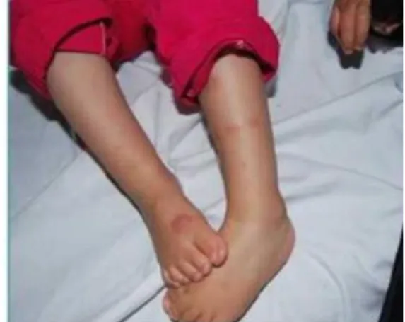
Figure 1 Multiple reddish-brown macules and patches on the back of the body.
Figure 3 Histopathology revealed mast cell infiltrate in papillary dermis (hematoxylin-eosin, low power).
Figure 5 Conjunctival redness and watery discharge.
metachromatic granules. An immunohistochemical study of CD117 was also positive.
An ophthalmology examination showed bilateral lid swelling, diffuse conjunctival redness, and
Figure 2 Darier's sign positive.
Figure 4 Histopathology revealed mast cell infiltrate in papillary dermis (hematoxylin-eosin. high power).
watery discharge (Figure 5), which was treated with topical antihistamines and cromolyn sodium. Other causes of conjunctivitis were excluded by the ophthalmologist.
A computed tomography (CT) scan was done for both eyes. Axial and sagittal reconstructed images showed normal eyes and anoptic nerve with no intra-conal lesion or enlargement of the lacrimal gland.
Discussion
Mastocytosis represents a spectrum of clinical disorders with a common phenotype of tissue
mast cell hyperplasia. Patients with this disorder were initially thought to have only cutaneous disease, but it was then discovered that mastocytosis patients could have involvement of multiple organs. The two common clinical presentations of childhood mastocytosis are solitary tan, yellow-tan, or brown plaques or nodules (mastocytoma) and variable numbers of tan to brown macules or papules as our case presented here. The lesions of urticaria pigmentosa usually develop during early childhood and most commonly appear on the trunk, classically sparing the central face, scalp, palms and soles. Some infants and children with urticaria pigmentosa develop non-scarring vesicles or bullae. Bullous lesions of mastocytosis usually resolve by 3-5 years and are believed to result from the release of mast cell serine proteases. The presence of mast cell hyperplasia in the skin of mastocytosis patients can be confirmed clinically by firmly rubbing a characteristic lesion. The formation of an urticarial weal (Darier’s sign) at the lesion site is indicative of mast cell mediator release. Systemic involvement is rare in urticaria pigmentosa.
In our patient, this disease occurred in the second month of life. Clinical features typical for urticaria pigmentosa include reddish-brown macules and slightly raised plaques on the skin of the entire body. In addition to bilateral lid swelling, diffuse conjunctival redness and watery discharge from both eyes, which is not that typical, had occurred.16 Darier's sign was positive.
The ocular involvement in mastocytosis has been described as solitary mastocytoma of the
lesions, and nyctalopia caused by the malabsorption of vitamin A.17-19
In our case, the eye symptoms increased especially with the flare up of skin lesions possibly due to histamine release into the mucous membrane.5 Complete clearance after
treatment with topical antihistamines and mast cell stabilizer (cromolyn sodium) point to a role for histamine and elevated total serum tryptase level.
Because of the eye symptoms, she was examined by hematology-oncology for systemic involvement, which was negative, but her serum tryptase was elevated (12.5 ng/ml). Total serum tryptase can be used indirectly to assess mast cell burden, and its level has been correlated with the extent of mast cell disease.20
Blood cell count was within normal values, which is in accordance with cutaneous mastocytosis; in systemic mastocytosis, blood cell counts may reveal anemia, thrombocytopenia, thrombocytosis, leukocytosis, and eosinophilia.
If a mastocytosis diagnosis is uncertain, the detection of circulating mast cell mediators and/or their metabolites can offer indirect evidence of mastocytosis. Serum tryptase levels are elevated in patients with mastocytosis. Our patient also had elevated serum tryptase levels.
Total (α and β combined) serum tryptase levels have been correlated with the extent of mast cell disease. Fifty percent of patients with total serum tryptase levels between 20 and 75 ng/ml had evidence of systemic mastocytosis, whereas all patients with levels >75 ng/ml had proven systemic involvement. A total serum tryptase level >20 ng/ml indicates one of the minor criteria for systemic mastocytosis.21 Urinary
methylimidazole acetic acid (MeImAA) levels correlated with the extent of mast cell disease; patients with widespread systemic involvement had the highest concentrations, whereas patients with only cutaneous disease had normal or only slightly elevated MeImAA.22
In conclusion, according to the clinical presentation, positive Darier's sign, elevated serum tryptase levels, and the presence of mast cells in the papillary dermis histopathologically, the diagnosis of urticaria pigmentosa was confirmed. The prognosis is good for this form of non-systemic cutaneous mastocytosis with mucous membrane involvement only. It can be expected that lesions will heal within the course of a few years.
References
1. Akaglu G, Erkin G, Cakir B, Boztepe G, Sahin S, Karaduman A et al. Cutaneous mastocytosis: demographic aspects and clinical features of 55 patients. J Eur Acad Dermatol Venereol. 2006;20:969-73. 2. Metcalfe DD. The mastocytosis syndrome.
In: Fitzpatrick TB, Eisen AZ, Wolff K, Freedberg IM, Austen K, editors.
Dermatology in General Medicine. New York: McGraw-Hill; 1993. P. 2017-23. 3. Azaña JM, Torrelo A, Mediero IG,
Zambrano A. Urticaria pigmentosa: a review of 67 pediatric cases. Pediatr Dermatol.
1994;11:102-6.
4. Soter NA. The skin in mastocytosis. J Invest Dermatol. 1991;96:32S-39S.
5. Paller AS, Mancini AJ. Hurwitz Clinical
Pediatric Dermatology, 4th ed. New York: Elsevier; 2011.
6. Nettleship E, Tay W. Rare form of urticaria.
Br Med J. 1869;2:323-30.
7. Unna PG. Anatomie und pathogenese der urticaria simplex und pigmentosa.
Monatschr Prakt Dermatol. 1887;6:EH1 8. Braun-Falco O, Plewig G, Wolff HH,
Winkelmann RK. Mastocytosis, editors.
Dermatology. Berlin: Springer-Verlag; 1991. P. 1107-12.
9. Ehrlich P. Beitrege zur Kenntnis der Anilinfaerbungen und ihrer Verwendung in
der mikroskopischen Technik. Arch Mikr Anat. 1877;13:263-77.
10. Erlich P. Uber die specifichen granulationen das blutes. Archiv Physiologie. 1879, 571-9. 11. Seitz CS, Rose C, Brocker EB, Trautmann A. Intertriginous urticaria pigmentosa.
Dermatology. 2005;210:77-9. http:// dx.doi.org/10.1159/000081493
12. Nagayo K, Sakai M, Mizuno N. Juvenile xanthogranuloma with Darier’s sign. J Dermatol. 1983;10:283-5.
13. Yen A, Sanchez R, Oblender M, Raimer S. Leukemia cutis: Darier’s sign in a neonate with acute lymphoblastic leukemia. J Am Acad Dermatol 1996; 34:375-8.
14. Garriga MM, Friedman MM, Metcalfe DD. A survey of the number and distribution of mast cells in the skin of patients with mast cell disorders. J Allergy Clin Immunol.
1988;82:425.
15. Greaves MW. Mastocytoses. In: Champion RH, Burton JL, Ebling FJG, editors.
Textbook of Dermatology. Oxford: Blackwell Scientific Publications; 1992. P. 2065-72.
16. Shaikh A, Benjamin L. Allergic eye disease associated with mastocytosis. Eye. 2003;17:788-90.
17. Scheck O, Horny HP, Ruck P, Schmelzle R, Kaiserling E. Solitary mastocytoma of the eyelid. A case report with special reference to the immunocytology of human mast cells and a review of the literature. Virchows Arch A Pathol Anat Histopathol.1987;412:31-6. 18. Jacoby BG, Wesley RE. Painful orbital
inflammatory lesions and mastocytosis. Ann Ophthalmol 1987;149(4):146-7.
19. Lesser RL, Brodie SE, Sugin SL. Mastocytosis-induced nyctalopia. J Neuroophthalmol. 1996;16:115-9.
20. Tharp MD. Mastocytosis. Bolognia JL, Jorizzo JL, Schaffer JV, editors.
Dermatology, Volume two, 3rd edn. Philadelphia: Elsevier; 2012. P. 1993-2000. 21. Horny H-P, Akin C, Metcalfe DD, Bennett
JM, Valent P, Bain BJ. Mastocytosis. In: Swerdlow SH, Campo E, Harris NL, Jaffe ES, Pileri SA, Stein Het al., editors. WHO
Classification of Tumours of Haematopoetic and Lymphoid Tissues. Lyon, France: IARC Press; 2008. P. 54-63.