Address for correspondence
Dr. Sabrina Yesmin, Professor and Head, Department of Medicine,
Dr. Sirajul Islam Medical College & Hospital, 12/3, New Circular Road, Malibagh, Dhaka-1217. Ph: +8801755557790
Case Report
WILD syndrome: A rare presentation of primary
lymphedema, generalized warts and immune
deficiency diagnosed in Bangladesh
Sabrina Yesmin1, AKM Ruhul Amin2, Towhida Noor3, SM Khodeza Nahar Begum4, Tareq M Bhuiyan5, Mehtab Al-Wadud Khan6, Rozina Akhter7, Md. Selim Mahmod8, Jahid Hasan1, MA Baqui2
1 Department of Medicine, Dr. Sirajul Islam Medical College & Hospital, Dhaka 2 Department of Surgery, Dr. Sirajul Islam Medical College & Hospital, Dhaka
3 Department of Dermatology & Venereology, Dr. Sirajul Islam Medical College & Hospital, Dhaka 4
Department of Pathology, Dr. Sirajul Islam Medical College & Hospital, Dhaka
5 Department of Gastroenterology, Bangladesh Institute of Research and Rehabilitation in Diabetes,
Endocrine and Metabolic Disorders, Dhaka
6 Department of Anesthesiology, Dr. Sirajul Islam Medical College & Hospital, Dhaka 7
Department of Paediatrics, Dr. Sirajul Islam Medical College & Hospital, Dhaka
8 Department of Cardiology, Dr. Sirajul Islam Medical College & Hospital, Dhaka
Abstract WILD syndrome is a rare disease characterized by warts, impaired cell mediated immunity, primary lymphedema and anogenital dysplasia. Our patient who is a 13 year old boy presented with swelling of right lower limb since birth. The swelling gradually involved both upper limbs and right side of his face. The swelling over the right lower limb grew to such an extent that he has been unable to do his daily activities for the last 3 years and warty skin lesions over his whole body for the last 5 years. At the same time he has swelling of his external genitalia and warty lesion around the anus. He has recurrent painless swelling of his abdomen with scanty micturition. On examination, he was mildly anemic, non-icteric, non-pitting odema of right leg, normal vital parameters, right sided pleural effusion, pericardial effusion and ascites, no organomegaly. Swelling and distortion of right lower limb with nodular lesions over the right leg, generalized hyper and hypopigmented verrucous papules and paques over whole body, swelling of the glans and flat warts around the anus. His investigations revealed leucopenia, chylous pleural effusion, lymphedema of right lower limb with venous incompetency and normal arterial supply, reduced CD4 and CD8 T cell count, normal immunoglobulin levels, negative ICT for filarial, histology of skin lesion revealed elephanthiasis verrucosa nostra, and anogenital dysplasia with a large number of koilocytes.
Key words
Generalized warts, primary lymphedema, impaired cell mediated immunity, anogenital dysplasia, koilocytes.
Introduction
WILD syndrome is a rare disease characterized by generalized warts, impaired cell mediated immunity, primary lymphedema and anogenital
dysplasia. A very few cases has been reported worldwide and its genetic inheritance has not been determined yet. It has features similar to GATA2 gene deficiency. The first patient with WILD syndrome was reported by Ostrow et al. thirty years back.1
Case report
presented for evaluation of persistent generalized warts that appeared at the age of eight, initially affecting the right leg. He was the
5th issue of consanguineously married parents,
and none of his relatives had similar findings. But his two siblings and an aunt had suspected neuromuscular disorder. Swelling of right lower limb was noticed at birth, and later progress to involve both the upper limbs and right side of his face. At the age of eight, the patient developed disseminated skin colored flat warts over the swollen right leg, gradually involving the whole body including palms, soles and perianal region along with disfigurement of his external genital organ. The swelling of the right lower limb was extensive and deformed with foul smelling discharge that had impaired his daily activities for the last several years. He had history of recurrent painless abdominal swelling with scanty micturition for the last three years which used to subside after taking oral steroid and diuretics. There was frequent passage of whitish loose stools for the same duration.
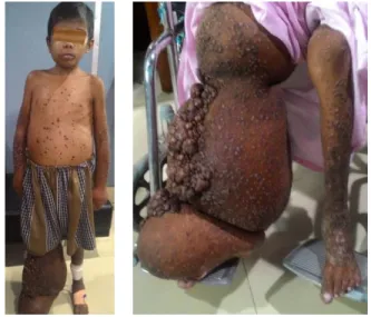
His physical examination revealed mild anemia and swelling of his right cheek, arms and forearms on both sides; hugely distended and deformed right lower limb (Figure 1). All the swellings were painless and non-pitting. There were multiple grouped skin colored painless nodules over the right leg extending from knee to dorsum of foot with foul smelling whitish
discharge from the clefts of the foot (Figure 2).
Both hypopigmented and hyperpigmented
verrucous papules and plaques were distributed
over whole body (Figure 3), with flat warts
around the anus (Figure 4) and swelling of his glans.
He had pericardial and right sided pleural effusion, and ascites with no organomegally. Results of laboratory investigations revealed hemoglobin 11.5 g/dl, WBC 12,000/cmm, differential count: neutrophil 89%, lymphocyte
Figure 1 Swelling of the right lower limb, right cheek and both hands
Figure 2 Multiple grouped skin colored painless nodules over the enlarged deformed right leg extending from knee to dorsum of foot and verrucous plaques over the left leg
Figure 3 Hypopigmented papules and plaques over both hands
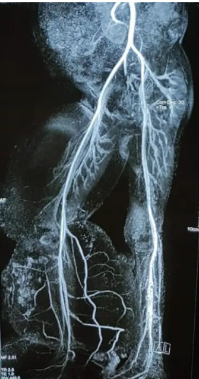
Figure 5 MRA showing normal arterial supply Figure 6 HPV cytopathic changes
05%, monocyte 03%, eosinophil 03%, low serum albumin level of 1.6 gm/dl, trace of protein in urine, and fat globules in stool. Serum creatinine and SGPT were within normal reference range. ICT for filaria was negative, pleural fluid study was chylous in nature. HIV 1&2, HBsAg, anti HCV were negative. Notably decreased CD4 T cells of 138/μl (ref range 410-1590/μl) and CD8 T cells of 91/μl (ref range 150-1000/μl), but immunoglobulin levels were within reference range.
Chest X-ray revealed right-sided moderate pleural effusion and pericardial effusion. X-ray of right lower limb showed no bone deformity. Magnetic resonance arteriography (MRA) of
lower limbs was normal (Figure 5). Color
duplex study of right lower limb revealed incompetent long saphenous and common femoral venous valves. Lymphoscintigraphy of lower extremities showed lymphatic obstruction. Histological evaluation of nodules on this limb revealed Elephanthiasis verrucosa nostra, warty lesions revealed features of HPV associated verruca vulgaris as evidenced by presence of
koilocytes (Figure 6), and perianal skin lesion revealed low grade dysplasia. Due to lack of facility HPV typing could not be done in this patient.
With the features of primary lymphedema,
generalized warty lesion, depressed cell
mediated immunity and anogenital dysplasia, we have finally diagnosed the boy as WILD syndrome.
For improving the patient’s wellbeing, multi stage bulk reduction from the enlarged and deformed right lower limb and anastomosis of the lymphatic channel with venous system has been planned. Cryotherapy and electrosurgery will be used for removing the warts.
Discussion
which overlap with WILD syndrome. The age of clinical presentation ranges from early childhood to late adulthood, with most occurring in
adolescence to early adulthood.2
As WILD syndrome is a rare disease, we have considered a number of differentials before considering our final diagnosis.
Epidermodysplasia verruciformis is a
genodermatosis where abnormal susceptibility to infection by various HPV types occurs, which normally does not occur in immunocompetent individuals. It occurs due to mutation of EVER1 and EVER2 genes. Patients usually presents with disseminated flat warts at an early age and about 30% develops skin cancer. There is no association of primary lymphedema in this syndrome.3,4
WHIM syndrome is an acronym derived from the major features of the disorder that include
warts, hypogammaglobulinemia, recurrent
bacterial infection and myelokathexis (apoptosis of mature myeloid cells in the bone marrow). Here mutation occurs in chemokine receptor
CXCR4.5
Milroy’s disease is an autosomal dominant disorder presents with unilateral or bilateral lymphedema at or soon after birth. Skin changes due to lymphatic obstruction are present, also known as elephantiasis verrucosa nostra and chylous discharge may be present. Generalized skin lesions are usually absent.6
Klippel-Trenauny syndrome is characterized by nevus flammeus, venous malformation and soft tissue hypertrophy of the affected extremity, in 95% cases affecting the lower extremity. The most common and earliest sign is nevus flammeus, usually confined to one extremity, and the involved limb is larger and longer than
the other. Generalized skin lesions are usually absent.7
WILD syndrome is characterized by generalized warts, depressed cell mediated immunity, primary lymphedema and anogenital dysplasia.
Patients usually present with primary
lymphedema at or soon after birth which may affect single limb or both limbs or all four limbs
or with systemic lymphatic channel
involvement. Generalized warty lesions appear during adolescent, involving palms, soles and anogenital region. Histological evaluation of anal region reveals dysplasia.
Thirty years ago Ostrow et al. reported the case of a patient with similar features of WILD syndrome. A 37-year-old white man without family history of lymphedema presented with
congenital lymphatic disease on all 4
extremities, and disseminated flat warts and pityriasis versicolor–like papules developed during adolescence. Further features included anergy to routine skin testing, depressed mitogen-stimulated lymphocyte transformation, severe CD4T-cell and B-cell depletion, low albumin and low total serum protein levels, and condylomatous (partially dysplastic) lesions in
the anogenital region.1
of the red and brown wart like lesions showed characteristic of cutaneous warts. Specimens of several verrucous lesions of the anogenital region including the vulva, perianal skin, and anal canal revealed intraepithelial neoplasia grades I to II.8
In comparison to the above two cases, our patient had similar features with systemic involvement of primary lymphedema.
Conclusion
We can finally conclude that this patient is suffering from WILD syndrome although further molecular investigations like genetic mutation studies are needed to confirm which are not available in our country.
References
1. Ostrow RS, Manias D, Mitchell AJ et al. Epidermodysplasiaverruciformis: a case associated with primary lymphatic dysplasia, depressed cellmediated immunity, and Bowen’s disease containing human
papillomavirus 16 DNA. Arch Dermatol 1987; 123(11): 1511-16.
2. Hasu AP, McReynolds LJ, Holland SM. GATA 2 Deficiency. CurrOpin Allergy Clin Immunol 2015; 15(1): 104-109.
3. Sá NB, Guerini MB, Barbato MT et al. Epidermodysplasia verruciformis: clinical presentation with varied forms of lesions. An Bras Dermatol 2011; 86(4Supl1): S57-60.
4. Shruti S, Siraj F, Singh A et al. Epidermodysplasia verruciformis: three case reports and a brief review. Acta Dermatovenerol Alp Pannonica Adriat 2017; 26(3): 59-61.
5. Kawai T, Malech HL. WHIM Syndrome: Congenital Immune Deficiency Disease. Curr Opin Hematol 2009; 16(1): 20-26. doi:10.1097/MOH.0b013e32831ac557 6. James WD, Berger TG, Elston DM et al.,
Andrews’ Diseases of the Skin: Clinical Dermatology. 12th edn. Philadelphia: ELSEVIER; 2016. p.853.
7. Zea MI, Hanif M, Habib M et al. Klippel-Trenaunay Syndrome: a case report with brief review of literature. J Dermatol Case Rep 2009; 3(4): 56–59.