R E S E A R C H
Open Access
The role of lipid droplet formation in the
protection of unsaturated fatty acids
against palmitic acid induced lipotoxicity to
rat insulin-producing cells
Thomas Plötz, Magnus Hartmann, Sigurd Lenzen and Matthias Elsner
*Abstract
Background:Type 2 diabetes is associated with increased plasma concentrations of non-esterified fatty acids (NEFAs), which trigger pancreaticβ-cell dysfunction and apoptosis. Only long-chain saturated NEFAs induced lipotoxicity in rat insulin-producing cells in in vitro experiments, whereas unsaturated NEFAs were not toxic. Some unsaturated NEFAs even protected against lipotoxicity. In former studies it was suggested that long-chain unsaturated NEFAs, which induce the formation of lipid droplets, can cause sequestration of palmitic acid into lipid droplets. In the present structure-activity-relationship study the correlation between lipid droplet formation and the protection against palmitic acid induced lipotoxicity by unsaturated NEFAs in rat insulin-producing cells was examined.
Methods:Rat insulin-producing RINm5F and INS-1E tissue culture cells were incubated in the presence of
palmitic acid and unsaturated NEFAs with different chain lengths and different numbers of double bonds. The expression of the lipid droplet associated proteins perilipin 1 and 2 was repressed by the shRNA technique and the expression analyzed by qRT-PCR and Western blotting. Viability was measured by MTT assay and the
accumulation of lipid droplets was quantified by fluorescence microscopy after Oil Red O staining.
Results:Long-chain unsaturated NEFAs strongly induce the formation of lipid droplets in rat insulin-producing RINm5F and INS-1E cells. In RINm5F cells incubated with 11-eicosenoic acid (C20:1) 27 % of the cell area was covered by lipid droplets corresponding to a 25-fold increase in comparison with control cells. On the other hand the saturated NEFA palmitic acid only induced minor lipid droplet formation. Viability analyses revealed only a minor toxicity of unsaturated NEFAs, whereas the cells were markedly sensitive to palmitic acid. Long-chain unsaturated NEFAs antagonized palmitic acid induced lipotoxicity during co-incubation, whereby no
correlation existed between protection and the ability of lipid droplet formation. Perilipin 1 and 2 expression was decreased after incubation with C20:1 to about 80 % by shRNA. For the protective effect of long-chain unsaturated NEFAs against lipotoxicity of saturated NEFAs repression of perilipin was not of crucial importance. Conclusions:Long-chain unsaturated fatty acids protected rat insulin-producing cells against lipotoxicity of saturated fatty acids. This protective effect was not dependent on lipid droplet formation. Thus lipid droplet formation is apparently not essential for the protective effect of unsaturated NEFAs against palmitic acid toxicity.
Keywords:Type 2 diabetes, Non-esterified fatty acids, Lipid droplets, Perilipin, Pancreaticβ-cell, Peroxisomalβ-oxidation
* Correspondence:elsner.matthias@mh-hannover.de
Institute of Clinical Biochemistry, Hannover Medical School, 30623 Hannover, Germany
Background
Type 2 diabetes mellitus (T2DM) is one of the most prevalent metabolic diseases worldwide caused by in-sulin resistance of the peripheral tissues and impaired insulin secretion of pancreatic β-cells [1]. Physical in-activity and hypercaloric diets, rich in carbohydrates and saturated fats, are thought to be responsible for an increase in body weight and can cause the metabolic syndrome [2–4], which is characterized by obesity, hypertension and dyslipidemia, and often precedes the manifestation of T2DM. Chronically elevated plasma levels of non-esterified fatty acids (NEFAs), which are associated with the metabolic syndrome [5], can sup-press insulin secretion and cause β-cell dysfunction and loss by apoptosis. This so-called lipotoxicity is sub-ject to intensive research and scientific debate. Only long-chain saturated NEFAs induce lipotoxicity in ro-dent insulin-producing cells in in-vitro experiments, whereas unsaturated NEFAs are not toxic [6]. Short-and medium-chain fatty acids are metabolized in the mitochondrial β-oxidation, whereas long-chain fatty acids can be metabolized in the mitochondria but also in the peroxisomes when cells were exposed to high NEFA concentrations [7, 8]. Very long-chain fatty acids are exclusive substrates for the peroxisomal β-oxidation [8–10]. The electron transfer in the first step of the per-oxisomal β-oxidation catalyzed by the enzyme acyl-CoA-oxidase results in high concentrations of hydrogen peroxide, which can cause cellular damage [8]. In con-trast to other cell types pancreatic β-cells do not ex-press the H2O2 inactivating enzyme catalase [11, 12].
This can explain why long-chain saturated fatty acids are harmful to rat insulin-producing β-cells and can cause apoptosis [7].
On the other hand unsaturated NEFAs are not toxic to rat insulin-producing cells and can even protect against lipotoxicity. The molecular mechanism of pro-tection has not yet been fully elucidated, but experi-mental evidence has been provided that the formation of lipid droplets (LDs) by unsaturated NEFAs may be involved in the protection against lipotoxicity [13]. Spe-cifically it has been suggested that long-chain unsatur-ated NEFAs can induce the formation of LDs, which may allow the sequestration of the toxic NEFA palmitic acid (PA) into these droplets. NEFAs converted into tri-glycerides and stored in LDs have been considered to be less toxic than NEFAs, which are readily metabolized [13]. For the formation of LDs perilipins are crucially important [14–17]. These proteins are associated with the LD surface and protect the stored triglycerides against hydrolysis by the hormone-sensitive lipases. Perilipins are thus important regulators of intracellular lipid storage [18, 19]. In the present study we therefore suppressed perilipin 1 and 2 expression in rat
insulin-producing cell lines using the shRNA technique and analyzed the effects on LD formation and PA-induced toxicity. In a comprehensive approach we correlated the structural requirements of saturated and unsatur-ated NEFAs with different chain lengths between C12 and C24 with respect to LD formation and toxicity in rat insulin-producing cells.
Our results show that unsaturated NEFAs were able to antagonize palmitic acid induced lipotoxicity. However, we observed no correlation between protection and the ability of lipid droplet formation in rat insulin-producing cells.
Methods
Tissue culture of insulin-producing cells
The stable insulin-producing RINm5F cell line was estab-lished from a radiation-induced rat islet cell tumor by Gazdar et al. [20] and purchased from ATCC (American Type Culture Collection, Manassas, VA, USA; order# ATCC CRL-11605). Cells were cultured in RPMI 1640 medium supplemented with 10 mM glucose, 10 % (v/v) fetal calf serum (Biowest, Nuaillé, France), penicillin and streptomycin in a humidified atmosphere at 37 °C and 5 % CO2 [12]. The insulin-producing INS-1E cell line was
clonally selected from a radiation-induced rat insulinoma and showed a stable phenotype for more than 100 pas-sages [21, 22]. INS-1E cells were kindly provided by Prof. Claes Wollheim (University of Geneva, Switzerland). These cells were cultured in RPMI 1640 medium sup-plemented with 10 mM glucose, 10 % fetal calf serum (FCS), penicillin and streptomycin, 10 mM Hepes (Serva, Heidelberg, Germany), 2 mM glutamine, 1 mM sodium-pyruvate (Sigma-Aldrich, Munich, Germany), and 50 μM of 2-mercaptoethanol in a humidified at-mosphere at 37 °C and 5 % CO2.
NEFA incubation
acid/arachidic acid), C20:1 (cis-11-eicosenoic acid/gon-doic acid), C20:2 (cis-11, 14-eicosadienoic acid), C20:3 (cis-8, 11, 14-eicosatrienoic acid), C20:4 (cis-5, 8, 11, 14-eicosatetraenoic acid/arachidonic acid), C20:5 (cis-5, 8, 11, 14, 17-eicosapentaenoic acid), C22:0 (docosanoic acid/behenic acid), C22:1 (cis-13-docosenoic acid/eru-cic acid), C22:2 (cis-13, 16-docosadienoic acid), C22:4 (cis-7, 10, 13, 16-docosatetraenoic acid), C22:6 (cis-4, 7, 10, 13, 16, 19-docosahexaenoic acid), C24:0 (tetracosa-noic acid/lignoceric acid), C24:1 (cis-15-tetracose(tetracosa-noic acid/nervonic acid) were purchased from Sigma-Aldrich (Munich, Germany) or Larodan (Malmö, Sweden).
Lipid droplet staining with Oil Red O
RINm5F cells were cultured for 24 h and afterwards ex-posed to 100 μM of different unsaturated NEFAs with different chain lengths and different numbers of double bonds (C18:1 to 18:4, C20:1 to C20:5, C22:1 to C22:6) for another 24 h. Cells were trypsinized and fixed in paraformaldehyde for 15 min at room temperature. Thereafter cells were stained with Oil Red O solution (Sigma-Aldrich, Munich, Germany) and washed twice with PBS (phosphate buffered saline). Lipid droplet for-mation was analyzed by fluorescence microscopy and the area within the cells was quantified by the use of the Olympus xcellence software (Olympus, Hamburg, Germany) at 546 nm excitation and 580 nm emission. For each incubation condition five to seven coinciden-tally selected images, each containing 30 to 100 cells, were used to quantify the proportion of the lipid drop-let area to the total cell area with the phase analysis module of the xcellence software.
Assessment of cell viability
RINm5F insulin-producing cells were seeded at 2.5 × 10⁴ cells/well in 100 μl culture medium onto 96-well plates and allowed to attach for 24 h before they were incubated at 37 °C with combinations of different unsat-urated NEFAs and PA (palmitic acid/hexadecanoic acid/ C16:0) for further 24 h. Cell viability and the ability to protect against PA-induced toxicity was determined by a microplate-based MTT assay (3-(4,5-dimethylthiazol-2-yl)-2,5-diphenyl tetrazolium bromide, Serva, Heidelberg, Germany) [23].
Lentivirus preparation and transduction of insulin-producing cells
Lentiviral vector particles were produced as described [24]. In brief, 5x10⁶ 293FT cells were transfected with the packaging plasmid pPAX2 (37.5 μg), the envelope plasmid pcDNA-MDG (7.5 μg), and the transfer plas-mids sh-rPlin1, sh-rPlin2 or pLV-U6-sh-NTC (22.5 μg) by calcium phosphate precipitation. The transfer plasmids were produced by Sirion Biotech
(Munich, Germany) and validated for suppression effi-ciencies > 85 %. The virus particles were harvested from the culture medium 48 h after transfection and purified by ultrafiltration columns at 3000 g for 25 min (Amicon Ultra Ultracel-100 K, Millipore, Schwalbach, Germany). Virus titers (typically 5x10⁷ infectious particles) were quantified by a TaqMan qPCR assay as described else-where [24]. RINm5F-Tet3G and INS-1E-Tet3G cells were transduced with either the sh-rPlin1, sh-rPlin2 or sh-NTC virus with a multiplicity of infection (MOI) followed by a selection with puromycin (2.5μg/ml).
Gene expression analyses
RNA from RINm5F and INS-1E cells was isolated with the RNeasy Kit (Qiagen, Hilden, Germany) according to the manufacturer’s instructions. The quality of the total RNA was verified by comparing the intensities of the 28S and 18S rRNA bands in agarose gel electrophoresis. Two microgram total RNA was reverse transcribed into cDNA using the RevertAid H Minus M-MuLV Reverse Tran-scriptase (Thermo Fisher Scientific, Schwerte, Germany) and random hexamer primers (Life Technologies, Karlsruhe, Germany). The Plin1 or Plin2 expression in 10 ng cDNA was quantified by a SYBR Green based assay (GoTaq Green Master Mix; Promega, Mannheim, Germany) and performed on an Opticon fluorescence de-tection system (Biorad, Munich, Germany) with the follow-ing protocol: Samples were initially denaturated at 95 °C for 2 min followed by up to 40 PCR cycles. Each PCR cycle comprised a denaturation at 94 °C for 30 s, an annealing at 60 °C for 30 s, and an extension at 72 °C for 30 s. The spe-cificity of the amplification was verified by melting point analysis. For each sample amplification was performed in triplicate. The Plin1 and Plin2 expression data were nor-malized against the geometric mean of the three reference genesactin beta(Actb), peptidylprolyl isomerase A (PPIA),
and TATA box binding protein (TBP) with the software
qBasePlus (Biogazelle, Zwijnaarde, Belgium). Primers for qPCR analyses were synthesized by Life Technologies (Karlsruhe, Germany): Plin1-fw (5′GATTCTGCTTTGCA GCGTGAA 3′), Plin1-rv (5′CAGGACTCTCTGGAGCA CATTC 3′), Plin2-fw (5′TCGTCTCTCAGCTCTCCTGT 3′), Plin2-rv (5′ TAGGTGGAGCTCACCAAGGG 3′), Actb-fw (5′GCGTCCACCCGCGAGTACAA 3′), Actb-fw (5′TTGCACATGCCGGAGCCGTT 3′), PPIA-fw (5′TT GCAGACGCCGCTGTCTCTT 3′), PPIA-rv (5′ TGGAA CTTTGTCTGCAAACAGCTCG 3′) TBP-fw (5′CAGCT CGGCGCACCGTACAT 3′), TBP-rv (5′ TCTGGGTTAT CGTCACGCACCA 3′).
Western blot analyses
recommendation (Sigma) supplemented with complete protease inhibitor mixture (Roche Diagnostics, Mannheim, Germany). Protein content was determined by the BCA assay (Thermo Fisher Scientific, Rockford, IL, USA). 30 μg of total protein were separated by a 10 % SDS-PAGE and electroblotted to nitrocellulose membranes (GE Healthcare, Buckinghamshire, UK). Nonspecific binding sites of the membranes were blocked with 5 % nonfat dry milk for 1 h at room temperature. The membranes were incubated with specific primary anti-bodies overnight at 4 °C. The following antianti-bodies from Santa Cruz Biotechnology (Santa Cruz, CA, USA) were used: Perilipin 1 (H-300, diluted 1:2000), anti-Perilipin 2 (H-80, diluted 1:200), and anti-β-actin (sc-1615, diluted 1:250). The excess of primary antibody was removed by three washing steps with washing buf-fer (PBS, 0.1 % Tween 20, 0.1 % BSA). Subsequently, the membranes were incubated with peroxidase-labeled secondary anti-rabbit antibodies at a dilution of 1:20,000 at room temperature for 1 h. Protein bands were visual-ized by chemiluminescence using the ECL detection sys-tem (GE Healthcare). As a loading control the expression ofβ-actin was analyzed after stripping the blots with Re-blot Pus solution (Merck-Millipore, Darmstadt, Germany) according to the manufacturer’s manual. Protein band in-tensity was quantified and normalized to β-actin though densitometry with the Gel-Pro Analyzer program (version 6.0, Media Cybernetics, Silver Spring, MD, USA).
Statistical analysis
Data are expressed as means ± SEM. Statistical ana-lyses were performed using ANOVA plus Dunnett’s or Bonferroni’s test for multiple comparisons, unless stated otherwise (GraphPad Prism5, San Diego, CA, USA).
Results
Induction of lipid droplet formation by NEFAs in insulin-producing cells
Long-chain monounsaturated NEFAs (C18:1, C20:1, C22:1) strongly induced the formation of LDs in rat insulin-producing RINm5F cells. More than 25 % of the cell area of RINm5F cells incubated with the long-chain NEFA gondoic acid (C20:1, 100 μM) was covered by LDs, which corresponds to a 16-fold increase in comparison to untreated cells. On the other hand incu-bation with medium-chain and long-chain monoun-saturated NEFAs (C12:1, C14:1, C16:1) and with a very long-chain NEFA (C24:1) showed no significant in-crease in LD formation in comparison to control cells (Fig. 1). In contrast to unsaturated NEFAs none of the analyzed saturated NEFAs (C12:0 to C24:0) caused sig-nificant increases in LD content in RINm5F cells.
Effect of saturation level of unsaturated NEFAs on lipid droplet formation in insulin-producing cells
In the group of unsaturated NEFAs with the chain length of C18 with different numbers of double bonds (C18:x) only C18:1 and C18:2 revealed a significant up to 15 % increase in the cell area covered with LDs (Fig. 2a). However, the unsaturated NEFAs C18:3 and C18:4 showed no increase in LD formation compared to the control condition. With 27 % LDs of the cell area C20:1 induced maximal lipid droplet formation. With an increasing number of double bonds the LD area declined to < 4 % after incubation with C20:4 or C20:5 (Fig. 2b), which was not significantly higher than in the control condition. The unsaturated C22:x NEFA group revealed the same tendency with the greatest LD formation after incubation with the monounsaturated C22:1 and a steady decrease with increasing numbers of double bonds up to C22:6 (Fig. 2c).
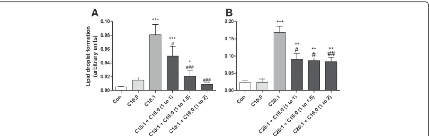
Antagonistic effect of saturated fatty acids against lipid droplet formation
Long-chain unsaturated NEFAs induced LD formation, whereas long-chain saturated NEFAs were not able to form LDs. A co-incubation experiment of both saturated and unsaturated NEFAs revealed a decline of LD forma-tion typically induced by long-chain unsaturated NEFAs. Palmitic acid (C16:0), which is the physiologically and nutritionally most important saturated fatty acid, antago-nized the LD formation by C18:1 and to a lesser extent
Con C12:
0 C12:
1 C14:
0 C14:
1 C16:
0 C16:
1 C18:
0 C18:
1 C20:
0 C20:
1 C24:
0 C24:
1 0
2 4 6 8 20 25 30 35
***
***
## # ###
###
#
R
eal
ti
ve l
ip
id
dr
opl
et
ar
ea
(%
)
by C20:1 (Fig. 3a-b). The antagonistic effect increased with the increase of the proportion of C16:0 in the mix-tures with C18:1 and C20:1.
Toxicity of long-chain NEFAs in insulin-producing cells
Long-chain saturated NEFAs at a 100 μM concentration were highly toxic to insulin-producing RINm5F cells. On the other hand the corresponding unsaturated NEFAs with the same chain lengths showed only a minimal or no significant toxicity at a concentration of 100 μM com-pared to the toxic effect of palmitic acid with a viability of 31 % (Additional file 1: Figure S1). Generally no correl-ation between the toxicity and the number of double bonds could be identified.
Protective capacity of long-chain unsaturated NEFAs against the toxicity of palmitic acid in insulin-producing cells in dependence on chain length and saturation level
The protective capacity of long-chain unsaturated NEFAs was analyzed in co-incubation experiments of 200μM PA with increasing concentrations of unsaturated NEFAs to determine the unsaturated NEFA concentration necessary to achieve a 50 % viability reduction (Table 1). The best protection against PA-induced toxicity in RINm5F insulin-producing cells was provided by C20:5 with a half-maximally effective protective concentration of 5.4μM. Generally NEFAs of the C20:x group revealed the highest protective potency. In the NEFA groups of C20:x and C22:x the monounsaturated NEFAs were less protect-ive than the polyunsaturated NEFAs. A concentration of
Con C18:1 C18:2 -C18:3
α γ-C18:3 C1 8:4
0 10 20 30 40
** ***
A
R
e
a
lt
iv
e
lip
id
d
ro
p
le
t a
re
a
(%
)
Con C20:1 C20:2 C20:3 C20:4 C20:5
0 10 20 30 40
***
***
***
B
Con C22:1 C22:2 C22: 4
C22:6
0 10 20 30 40
C
***
*** ***
***
Fig. 2Lipid droplet formation in rat insulin-producing RINm5F cells after incubation with unsaturated NEFAs with a different degree of unsaturation. RINm5F cells were incubated with C18:x (a), C20:x (b) and C22:x (c) NEFAs (100μM) with different numbers of double bonds for 24 h. Thereafter cells were fixed and stained with Oil Red O. Lipid droplet formation was analyzed by fluorescence microscopy and quantified using the software xcellence rt. Data are given as means ± SEM from 5 to 8 individual experiments. **p< 0.01, ***p< 0.001 compared with control cells (Dunnett’s Multiple Comparison Test)
A
B
17.2μM for C20:1 and of 88.6μM for C22:1 respectively was required to antagonize PA toxicity by 50 %. With a rise in the number of double bonds in these two groups the protective potential increased. The group of C18:x un-saturated NEFAs revealed the weakest protection of all tested groups. There were no significant differences in the protective potency against PA-induced toxicity between the omega-9 fatty acid oleic acid (C18:1), the omega-6 fatty acidγ-linolenic acid (γ-C18:3) and the omega-3 fatty acidα-linolenic acid (α-C18:3). The lowest protective po-tency exhibited the omega-3 fatty acid C18:4.
Lack of correlation between protective potency against PA-induced toxicity and lipid droplet formation in insulin-producing cells
The most efficient unsaturated NEFA with respect to the ability to form lipid droplets was C20:1, which mediated only a moderate protection against PA-induced toxicity. The highest protective potency against PA-induced tox-icity revealed C20:5, but it showed no significant in-crease in lipid droplet formation compared to control conditions without NEFAs. To examine, whether there was a correlation between the protective potency and lipid droplet formation in the different unsaturated NEFA groups (C18:x, C20:x, C22:x) the corresponding correlation coefficients and thepvalues were calculated. In none of the groups the correlation coefficient was significant (Table 1). A calculation of an overall coeffi-cient between lipid droplet formation and the protective
potency of the different NEFA groups revealed a correl-ation coefficient of 0.04.
Gene expression analyses of perilipin 1 or 2 in
insulin-producing cells after suppression of perilipin 1 or 2
To verify the efficiency of the shRNA mediated knock-down of perilipin 1 or 2 (shRNA-Plin1 or shRNA-Plin2) gene expression in insulin-producing RINm5F and INS-1E cells was analyzed after incubation with PA, OA (oleic acid/cis-9-octadecenoic acid/C18:1), a combin-ation of PA and OA, or GA (gondoic acid/cis-11-eicose-noic acid/C20:1). Control INS-1E and RINm5F cells as well as non-target shRNA control cells showed a signifi-cant 3- to 4-fold increase in perilipin 1 gene expression after incubation with OA, PA + OA, or GA in compari-son to control conditions without NEFAs. In shRNA-Plin1 cells no significant increase was detectable (Fig. 4a-c). Similar results were obtained in shRNA-Plin2 cells. Only in control cells and non-target shRNA con-trol cells a significant increase in perilipin 2 expression was detectable, whereas the gene expression in shRNA-Plin2 was not significantly increased after incubation with OA, PA + OA, or GA in comparison to control conditions (Fig. 4b-d).
In addition to the gene expression quantification the mRNA level, perilipin 1 and 2 protein expression in insulin-producing RINm5F and INS-1E cells was lyzed by immunoblotting (Fig. 5). In Western blot ana-lyses two bands in the range between approx. 60 kDa
Table 1Protective effect of unsaturated NEFAs against PA-induced toxicity and relative lipid droplet area linked with the correlation coefficient for each group
NEFA Half-maximal protective
concentration (μM)
Lipid droplet area after
incubation with 100μM
NEFA (% of cell area)
Correlation coefficient for each group
Pvalue for
each group
C18:1 32.8 ± 2.2 (6) 7.3 ± 1.3 (5) C18-FFA: +0.52 C18-FFA: 0.37
C18:2 11.4 ± 1.2 (5) 15.1 ± 2.8 (5)
α-C18:3 24.0 ± 1.8 (4) 2.4 ± 0.6 (5)
γ-C18:3 27.2 ± 1.6 (4) 2.6 ± 0.5 (5)
C18:4 > 100.0 (5) 1.9 ± 0.3 (5)
C20:1 17.2 ± 1.6 (6) 26.5 ± 4.9 (5) C20-FFA:−0.63 C20-FFA: 0.25
C20:2 6.0 ± 0.6 (4) 19.2 ± 2.9 (5)
C20:3 10.4 ± 1.2 (4) 8.1 ± 1.3 (5)
C20:4 8.6 ± 0.6 (4) 2.7 ± 0.6 (5)
C20:5 5.4 ± 0.6 (4) 3.9 ± 1.0 (5)
C22:1 88.6 ± 22.8 (6) 19.7 ± 4.1 (5) C22-FFA:−0.89 C22-FFA: 0.11
C22:2 42.0 ± 6.8 (4) 18.9 ± 2.7 (5)
C22:4 8.6 ± 1.4 (5) 12.1 ± 2.5 (5)
C22:6 10.8 ± 3.2 (5) 11.8 ± 2.4 (5)
Total value 0.04 0.89
and 65 kDa were detectable for perilipin 1. This finding confirms earlier immunoblotting analyses of INS-1E cells [13]. For perilipin 2 the immunoblotting revealed a specific band at a molecular weight of 48 kDa. Through shRNA mediated suppression of perilipin 1 or perilipin 2 a 60 to 70 % reduction both on the mRNA and protein level was achieved after incubation with OA, PA + OA, or GA, when compared to non-targeted control cells (Fig. 5). PA alone did not induce expression of perilipin 1 or 2. This low expression level was also not affected by shRNA mediated perilipin suppression.
Quantification of lipid droplet formation in insulin-producing cells after exposure to oleic acid
In order to verify the effect of perilipins on LD forma-tion Plin1 or Plin2 RINm5F and shRNA-Plin1 or shRNA-Plin2 INS-1E cells were incubated with the physiologically most important NEFAs, the saturated palmitic acid, (PA, C16:0) and the monounsaturated oleic acid (OA, C18:1) as well as a combination of both. Maximal LD formation was induced in RINm5F and INS-1E control cells or shRNA non-target controls (shNTC) after OA incubation in comparison to cells incubated without this NEFA (Fig. 6a and b). In shRNA-Plin1 or
shRNA-Plin2 cells C18:1 exposure resulted in a non-significant increase in LD formation in comparison to cells not incubated with OA. Furthermore shRNA-Plin1 or shRNA-Plin2 cells generated significantly fewer LDs in re-sponse to OA when compared to non-suppressed perilipin cells. The reduction in LD formation was in INS-1E-shPlin cells approx. 80 % (Fig. 6a) and in RINm5F-INS-1E-shPlin cells approx. 50 % (Fig. 6b). Incubation of RINm5F and INS-1E cells with knockdown of perilipin 1 or 2 in the presence of PA (200μM) plus OA (100 or 200μM) also caused a significant reduction of lipid droplet formation in the range of 50–70 % in comparison with the respective shNTC control cells (Fig. 6c and d). PA (200 μm) alone did not cause lipid droplet formation (data not shown).
Palmitic acid-induced lipotoxicity in perilipin suppressed insulin-producing cells
Perilipin suppressed RINm5F and INS-1E cells were used to analyze differences in the sensitivity against PA-induced toxicity. Neither perilipin 1 suppression nor perilipin 2 suppression showed a significantly higher sensitivity when compared to non-target shRNA control cells after incuba-tion with the toxic saturated NEFA PA (C16:0) (Fig. 7a and b). Furthermore the cell lines were co-incubated with
Contro l PA OA PA+OA GA 0 2 4 6 * * * * * * # # # RINm5F Con RINm5F-shNTC RINm5F-shPlin1 R e la ti ve gene expr essi o n o f p e rilip in 1 Contro l PA OA PA+OA GA 0 2 4 6 RINm5F Con RINm5F-shNTC RINm5F-shPlin2 * * * * * * # # # Rel a ti ve gene expr essi on o f p e rilip in 2 Control PA OA PA+OA GA 0 2 4 6 * * * * * * # # # INS-1E Con INS-1E-shNTC INS-1E-shPlin1 R e la ti ve gene expr essi o n o f p e rilip in 1 Contro l PA OA PA+OA GA 0 2 4
6 INS-1E Con
INS-1E-shNTC INS-1E-shPlin2 * * * * * * # # # Rel a ti ve gene expr essi on o f p e rilip in 2
B
A
C
D
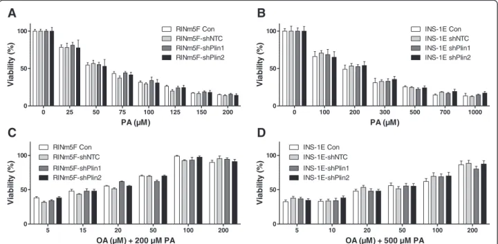
200 μM PA and increasing concentrations of OA (up to 200μM) demonstrating the protective effect of the mono-unsaturated NEFA OA on PA-induced toxicity. In these experiments there were also no significant differences in toxicity detectable between perilipin 1 or 2 suppressed cell lines detectable (Fig. 7c and d). Incubation with OA (up to 500 μM) alone did not induce significant toxicity in the cell lines (data not shown).
Discussion
Obesity and the metabolic syndrome are associated with increased plasma concentrations of non-esterified fatty acids (NEFAs) concentrations in the circulation,
which can cause T2DM by inducing insulin resistance in peripheral tissues as well as damage to pancreatic β -cells [1, 2]. In several in vitro studies on lipotoxicity in rodent β-cells long-chain saturated NEFAs were found to be responsible for the induction of apoptosis by ER-stress [25–27] or the formation of toxic reactive oxygen species (ROS) [7, 28–30]. In contrast, long-chain unsat-urated NEFAs are less toxic or non-toxic to ratβ-cells and can even provide protection against the toxicity in-duced by long-chain saturated NEFAs. We recently re-ported that the PA-induced formation of cytotoxic H2O2 in the peroxisomal β-oxidation is inhibited by
long-chain unsaturated NEFAs [27]. Pancreatic β-cells
Contro l
PA OA
PA+O
A GA
0 2 4 6
* *
*
# # #
INS-1E-shNTC INS-1E-shPlin1
Contro l
PA OA
PA+O
A GA
0 2 4 6
* *
*
#
# # RINm5F-shNTC RINm5F-shPlin1
Relative protein expression
of perilipin 1
60 kDa
42 kDa
48 kDa
42 kDa
B
A
C
D
Contro l
PA OA
PA+O
A GA
0 2 4
6 INS-1E-shNTC
INS-1E-shPlin2
* *
*
# # #
Control
PA OA
PA+O
A GA
0 2 4
6 RINm5F-shNTC
RINm5F-shPlin2
* *
*
# # #
Relative gene expression
of perilipin 2
do not express the H2O2 detoxifying enzyme catalase
which contributes to the exceptional sensitivity towards ROS mediated toxicity of this cell type [11, 31].
Another concept offered to explain the protection of long-chain unsaturated NEFAs against lipotoxicity is the formation of cytosolic lipid droplets (LDs). NEFAs in the cytosol can be sequestrated to LDs after esterification in triglycerides and could therefore not express their cyto-toxic action [13, 32, 33]. PA is known to be incorporated into LDs to a significantly lesser extent and evokes a greater lipotoxic effect than OA [32–34]. Furthermore, when cells are incubated with PA and OA simultan-eously, OA induces LD formation and facilitates the in-corporation of PA into the LDs, and thereby is thought to decrease the lipotoxic action of PA [33]. It was there-fore the aim of the present study to verify if this hypoth-esis is applicable not only to OA but also to other unsaturated NEFAs with different chain lengths and sat-uration levels in rat insulin-producing cells.
In a comprehensive experimental approach we ana-lyzed for the first time a group of 16 different saturated as well as mono- and polyunsaturated NEFAs with chain lengths ranging from C12 to C24 by fluorescence microscopy for their ability to form LDs in
insulin-producing cells. Only long-chain mono- and polyunsat-urated NEFAs were found to cause LD formation whereas the saturated NEFAs were ineffective. Such a difference had been reported in previous studies only between the unsaturated NEFA OA and the unsatur-ated NEFA PA [13, 32, 33, 35]. Our detailed compari-son of different mono- and polyunsaturated NEFAs with chain lengths of C18, C20 and C22 revealed no correlation (correlation coefficientr= 0.04) between ex-tent of NEFAs saturation and LD formation.
These results are in disagreement with the previ-ously proposed hypothesis that LD formation is re-sponsible for the protective effect of unsaturated NEFAs against toxicity of long-chain saturated NEFAs [13, 32, 33, 35]. To obtain further evidence for a lack of association between LD formation and a possible protective effect of unsaturated NEFAs against the toxicity of the saturated NEFA PA we determined in RINm5F insulin-producing cells in co-incubation ex-periments with 200 μM PA the half maximal protect-ive concentration for each of the 14 unsaturated NEFAs with different chain lengths and saturation levels and correlated these data with the LD forma-tion. Interestingly, there was no correlation detectable
between toxicity and LD formation. This conclusion is in contradiction to the hypothesis of a crucial role of LDs for the protection against PA toxicity [13, 31, 32], which was based only on analyses of the effects of PA, OA and linoleic acid [13, 35]. Thus a valid conclusion must be based upon the analyses of more than these three NEFAs.
Additional evidence that LD formation is not a cru-cial protective mechanism against lipotoxicity was pro-vided by the perilipin 1 and 2 repression experiments in insulin-producing cells. Perilipin repression was used as an additional experimental tool to study the effect of LD formation on lipotoxicity. Perilipins are proteins which are involved in the biogenesis of LDs and located in the phospholipid mono layer of the LDs [16, 17]. Like other investigators [13] we were able to detect perili-pin 1 and 2 gene expression in rat insulin-producing RINm5F and INS-1E cells on the mRNA as well as on the protein level. Incubation with OA, PA + OA, or GA led to an approx. 3 to 4-fold increase in perilipin 1 and 2 mRNA and protein expression. This induced perilipin 1 and 2 ex-pression was diminished by the shRNA technique by 60 % to 80 %. This gene suppression caused also a 50-80 % re-duction in LD formation in insulin-producing cells indi-cating that perilipins are involved in LD formation. If LD formation would be of crucial importance for lipotoxicity of long-chain saturated NEFAs it should be expected, that
the protection of OA against PA-induced toxicity was re-duced in perilipin 1 and 2 repressed cells which was not the case in our experiments. On the other hand we ob-tained also no experimental evidence for a short-term detri-mental effect on cell viability of LD storing unsaturated fatty acids. This does not exclude that long-term intracellu-lar storage of fatty acids in the form of triglycerides may have unfavorable effects on the cellular function; in particu-lar in diseases affecting other organs such as non-alcoholic fatty liver [36] and cardiovascular diseases [37], which go along with pathological intracellular fat accumulation.
Conclusions
The results document convincingly that LD formation plays no essential role in the protective effect of unsatur-ated NEFAs against the toxicity of palmitic acid in rat insulin-producing cells.
Additional file
Additional file 1: Figure S1.Viability of long-chain unsaturated NEFAs in comparison to the long-chain saturated NEFA PA (C16:0). RINm5F cells were incubated with 100μM of the long-chain saturated NEFA or 100μM of the unsaturated NEFAs as depicted for 24 h. Thereafter viability was measured by MTT assay. Data are means ± SEM ofn= 5 to 8. *p< 0.05, **p< 0.01, ***p< 0.001 compared to cells incabated under control condition without NEFAs (Dunnett’s Multiple Comparison Test). (DOCX 34 kb)
5 10 20 50 100 200
0 50 100
INS-1E Con
INS-1E-shPlin1 INS-1E-shPlin2 INS-1E-shNTC
OA (µM) + 500 µM PA
V
ia
b
ilit
y
(%
)
0 25 50 75 100 125 150 200
0 50
100 RINm5F Con
RINm5F-shPlin1 RINm5F-shPlin2 RINm5F-shNTC
PA (µM)
V
ia
b
ility
(%
)
0 100 200 300 500 700 1000
0 50
100 INS-1E Con
INS-1E shPlin1 INS-1E shPlin2 INS-1E shNTC
PA (µM)
V
ia
b
ility
(%
)
B
A
D
C
5 15 20 50 100 200
0 50 100
RINm5F Con
RINm5F-shPlin1 RINm5F-shPlin2 RINm5F-shNTC
OA (µM) + 200 µM PA
V
ia
b
ilit
y
(%
)
Abbreviations
GA:gondoic acid; LDs: lipid droplets; MOI: multiplicity of infection; NEFA: non-esterified fatty acid; NTC: non-target control; OA: oleic acid; PA: palmitic acid; ROS: reactive oxygen species; T2DM: type 2 diabetes mellitus.
Competing interests
The authors declare that they have no competing interests
Authors’contributions
TP performed lipid droplet analyses and cell viability assays, participated in data analyses and interpretation. MH carried out the shRNA and qPCR experiments and analyzed the data. TP and ME prepared the manuscript. ME was responsible for the experimental design and the interpretation of the data. SL was responsible for the design of the research and reviewed the manuscript. All authors read and approved the final manuscript.
Acknowledgements
We are grateful to Maren Böger and Martin Wirth for their skillful technical assistance. This work was supported by the European Union (Collaborative Project BetaBAT in the Framework Programme 7, grant agreement 277713), the German Diabetes Association (Hans-Christian-Hagedorn project grant), and the German Research Foundation (DFG) within the Research Training Group (GRK) 1947, project A4.
Received: 10 June 2015 Accepted: 18 February 2016
References
1. Stumvoll M, Goldstein BJ, van Haeften TW. Type 2 diabetes: principles of pathogenesis and therapy. Lancet. 2005;365:1333–46.
2. Vinciguerra F, Baratta R, Farina MG, Tita P, Padova G, Vigneri R, et al. Very severely obese patients have a high prevalence of type 2 diabetes mellitus and cardiovascular disease. Acta Diabetol. 2013;50:443–9.
3. Hamilton MT, Hamilton DG, Zderic TW. Role of low energy expenditure and sitting in obesity, metabolic syndrome, type 2 diabetes, and cardiovascular disease. Diabetes. 2007;56:2655–67.
4. Lottenberg AM, Afonso Mda S, Lavrador MS, Machado RM, Nakandakare ER. The role of dietary fatty acids in the pathology of metabolic syndrome. J Nutr Biochem. 2012;23:1027–40.
5. Sabin MA, De Hora M, Holly JM, Hunt LP, Ford AL, Williams SR, et al. Fasting nonesterified fatty acid profiles in childhood and their relationship with adiposity, insulin sensitivity, and lipid levels. Pediatrics. 2007;120:1426–33. 6. Dhayal S, Welters HJ, Morgan NG. Structural requirements for the
cytoprotective actions of mono-unsaturated fatty acids in the pancreatic beta-cell line, BRIN-BD11. Br J Pharmacol. 2008;153:1718–27.
7. Elsner M, Gehrmann W, Lenzen S. Peroxisome-generated hydrogen peroxide as important mediator of lipotoxicity in insulin-producing cells. Diabetes. 2011;60:200–8.
8. Kunau WH, Dommes V, Schulz H. Beta-oxidation of fatty acids in mitochondria, peroxisomes, and bacteria: a century of continued progress. Prog Lipid Res. 1995;34:267–342.
9. Van Veldhoven PP. Biochemistry and genetics of inherited disorders of peroxisomal fatty acid metabolism. J Lipid Res. 2010;51:2863–95.
10. Fransen M, Nordgren M, Wang B, Apanasets O. Role of peroxisomes in ROS/ RNS-metabolism: implications for human disease. Biochim Biophys Acta. 1822;2012:1363–73.
11. Lenzen S, Drinkgern J, Tiedge M. Low antioxidant enzyme gene expression in pancreatic islets compared with various other mouse tissues. Free Radic Biol Med. 1996;20:463–6.
12. Tiedge M, Lortz S, Drinkgern J, Lenzen S. Relation between antioxidant enzyme gene expression and antioxidative defense status of insulin-producing cells. Diabetes. 1997;46:1733–42.
13. Borg J, Klint C, Wierup N, Strom K, Larsson S, Sundler F, et al. Perilipin is present in islets of Langerhans and protects against lipotoxicity when overexpressed in the beta-cell line INS-1. Endocrinology. 2009;150:3049–57. 14. Grahn TH, Zhang Y, Lee MJ, Sommer AG, Mostoslavsky G, Fried SK, et al.
FSP27 and PLIN1 interaction promotes the formation of large lipid droplets in human adipocytes. Biochem Biophys Res Commun. 2013;432:296–301.
15. McIntosh AL, Senthivinayagam S, Moon KC, Gupta S, Lwande JS, Murphy CC, et al. Direct interaction of Plin2 with lipids on the surface of lipid droplets: a live cell FRET analysis. Am J Physiol Cell Physiol. 2012;303:C728–42. 16. Brasaemle DL. Thematic review series: adipocyte biology. The perilipin
family of structural lipid droplet proteins: stabilization of lipid droplets and control of lipolysis. J Lipid Res. 2007;48:2547–59.
17. Hsieh K, Lee YK, Londos C, Raaka BM, Dalen KT, Kimmel AR. Perilipin family members preferentially sequester to either triacylglycerol-specific or cholesteryl-ester-specific intracellular lipid storage droplets. J Cell Sci. 2012; 125:4067–76.
18. Brasaemle DL, Rubin B, Harten IA, Gruia-Gray J, Kimmel AR, Londos C. Perilipin A increases triacylglycerol storage by decreasing the rate of triacylglycerol hydrolysis. J Biol Chem. 2000;275:38486–93.
19. Martinez-Botas J, Anderson JB, Tessier D, Lapillonne A, Chang BH, Quast MJ, et al. Absence of perilipin results in leanness and reverses obesity in Lepr(db/db) mice. Nat Genet. 2000;26:474–9.
20. Gazdar AF, Chick WL, Oie HK, Sims HL, King DL, Weir GC, et al. Continuous, clonal, insulin- and somatostatin-secreting cell lines established from a transplantable rat islet cell tumor. Proc Natl Acad Sci U S A. 1980;77:3519–23. 21. Asfari M, Janjic D, Meda P, Li G, Halban PA, Wollheim CB. Establishment of 2-mercaptoethanol-dependent differentiated insulin-secreting cell lines. Endocrinology. 1992;130:167–78.
22. Merglen A, Theander S, Rubi B, Chaffard G, Wollheim CB, Maechler P. Glucose sensitivity and metabolism-secretion coupling studied during two-year continuous culture in INS-1E insulinoma cells. Endocrinology. 2004;145:667–78. 23. Mosmann T. Rapid colorimetric assay for cellular growth and survival:
application to proliferation and cytotoxicity assays. J Immunol Methods. 1983;65:55–63.
24. Elsner M, Terbish T, Jörns A, Naujok O, Wedekind D, Hedrich HJ, et al. Reversal of diabetes through gene therapy of diabetic rats by hepatic insulin expression via lentiviral transduction. Mol Ther. 2012;20:918–26. 25. Cunha DA, Hekerman P, Ladriere L, Bazarra-Castro A, Ortis F, Wakeham MC,
et al. Initiation and execution of lipotoxic ER stress in pancreatic beta-cells. J Cell Sci. 2008;121:2308–18.
26. Karaskov E, Scott C, Zhang L, Teodoro T, Ravazzola M, Volchuk A. Chronic palmitate but not oleate exposure induces endoplasmic reticulum stress, which may contribute to INS-1 pancreatic beta-cell apoptosis. Endocrinology. 2006;147:3398–407.
27. Gehrmann W, Würdemann W, Plötz T, Jörns A, Lenzen S, Elsner M. Antagonism between saturated and unsaturated fatty acids in ROS mediated lipotoxicity in rat insulin-producing cells. Cell Physiol Biochem. 2015;36:852–65.
28. Gehrmann W, Elsner M, Lenzen S. Role of metabolically generated reactive oxygen species for lipotoxicity in pancreatic beta-cells. Diabetes Obes Metab. 2010;12 Suppl 2:149–58.
29. Schönfeld P, Wojtczak L. Fatty acids as modulators of the cellular production of reactive oxygen species. Free Radic Biol Med. 2008;45:231–41. 30. Newsholme P, Haber EP, Hirabara SM, Rebelato EL, Procopio J, Morgan D, et al. Diabetes associated cell stress and dysfunction: role of mitochondrial and non-mitochondrial ROS production and activity. J Physiol. 2007;583:9–24. 31. Lenzen S. Oxidative stress: the vulnerable beta-cell. Biochem Soc Trans.
2008;36:343–7.
32. Cnop M, Hannaert JC, Hoorens A, Eizirik DL, Pipeleers DG. Inverse relationship between cytotoxicity of free fatty acids in pancreatic islet cells and cellular triglyceride accumulation. Diabetes. 2001;50:1771–7. 33. Listenberger LL, Han X, Lewis SE, Cases S, Farese Jr RV, Ory DS, et al.
Triglyceride accumulation protects against fatty acid-induced lipotoxicity. PNAS. 2003;100:3077–82.
34. Listenberger LL, Ory DS, Schaffer JE. Palmitate-induced apoptosis can occur through a ceramide-independent pathway. The Journal of biological chemistry. 2001;276:14890–5.
35. Moffitt JH, Fielding BA, Evershed R, Berstan R, Currie JM, Clark A. Adverse physicochemical properties of tripalmitin in beta cells lead to morphological changes and lipotoxicity in vitro. Diabetologia. 2005;48:1819–29.
36. Parekh S, Anania FA. Abnormal lipid and glucose metabolism in obesity: implications for nonalcoholic fatty liver disease. Gastroenterology. 2007;132: 2191–207.