MJCCA9 – 727 ISSN 1857-5552 e-ISSN 1857-5625
Received: July 23, 2016 DOI:10.20450/mjcce.2017.1001
Accepted: February 20, 2017 Original scientific paper
MOLECULAR STRUCTURE, VIBRATIONAL SPECTRA, NBO, FUKUI
FUNCTION, HOMO-LUMO ANALYSIS AND MOLECULAR DOCKING STUDY OF
6-[(2-METHYLPHENYL)SULFANYL]-5-PROPYLPYRIMIDINE-2,4(1
H
,3
H
)-DIONE
Haitham Al-Rabiah1, S. Muthu2, Fatmah A. M. Al-Omary1, Abdul-Malek S. Al-Tamimi3, M. Raja4, R. Raj Muhamed4, Ali A. El-Emam1*
1
Department of Pharmaceutical Chemistry, College of Pharmacy, King Saud University, Riyadh 11451, Saudi Arabia
2
Department of Physics, Arignar Anna Govt. Arts College, Cheyyar, 604407, Tamilnadu, India
3
Department of Pharmaceutical Chemistry, College of Pharmacy, Prince Sattam bin Abdulaziz University, Alkharj 11942, Saudi Arabia
4
Department of Physics, Jamal Mohamed College, Tiruchirappalli 620020, Tamilnadu, India *aelemam@ksu.edu.sa; elemam5@hotmail.com
Theoretical and experimental FT-IR and FT-Raman vibrational spectral analysis of 6-[(2-methylphenyl) sulfanyl]-5-propylpyrimidine-2,4(1H,3H)-dione have been recorded in the region 4000– 400 cm–1 and 4000–100 cm–1 insolid phase. The molecular geometric parameters bond length, bond angle and vibrational wave numbers, as well as harmonic vibrational frequency were investigated using the density functional theory B3LYP method with the 6-311++G(d,p) basis set. The stability of the molecule has been investigated using the natural bond orbital (NBO) analysis. The electronic properties such as HOMO-LUMO energies were determined by the time-dependent DFT approach. The thermodynamic properties and the first order hyperpolarizability and molecular electrostatic potential (MEP) of the title compound were also studied. The electron density-based local reactivity descriptors such as the Fukui functions were calculated to explain the chemical selectivity or reactivity site in the molecule. The mole-cule orbital contributions were investigated using the total density of states (TDOS), the sum of 𝛼 and 𝛽 electron density of states (𝛼𝛽DOS). The molecular docking (ligand-protein) simulations have been per-formed using the AutoDock 4.2.6. Binding energy, bonded residues and donor-acceptor bond length val-ues revealed that title compound can act as potential inhibitor against HIV-1 protease.
Keywords: FT-IR; FT-Raman; NBO; MEP; Fukui function; pyrimidine-2,4(1H,3H)-dione;
molecular docking
МОЛЕКУЛСКА СТРУКТУРА, ВИБРАЦИОНИ СПЕКТРИ, NBO, ФУНКЦИЈА НА ФУКУИ, HOMO-LUMO АНАЛИЗА И ИСПИТУВАЊЕ НА МОЛЕКУЛСКО ПРИПОЈУВАЊЕ
НА 6-[(2-МЕТИЛФЕНИЛ)СУЛФАНИЛ]-5-ПРОПИЛПИРИМИДИН-2,4-(1Н,3Н)-ДИОН
засновани на електронската густина како што се функциите на Фукуи беа пресметани со цел да се објасни хемиската селективност или реактивните места во молекулата. Придонесите на молекулските орбитали беа изучувани со употреба на состојбите на вкупната густина (TDOS), како збир на 𝛼 и 𝛽 состојбите на електронската густина (𝛼𝛽DOS). Симулациите на молекулско припојување (лиганд-протеин) беа извршени со помош на AutoDock 4.2.6. Сврзувачката енергија, сврзаните остатоци, како и вредностите на донорно-акцепторните должини на врски укажуваат дека ова соединение може да биде потенцијален инхибитор на протеазата на HIV-1.
Клучни зборови: FT-IR; FT на Раман; NBO; MEP; функција на Фукуи; пиримидин-2,4(1H,3H)-дион;
молекулско припојување
1. INTRODUCTION
Pyrimidine and its related derivatives like uracil and thymine occupy a distinct position in the field of chemotherapy. Several pyrimidine and py-rimidine-related drugs are currently employed for the treatment of various diseases. Numerous py-rimidine-based derivatives have been developed as antiviral agents against human immunodeficiency viruses (HIV) [1–5], hepatitis B viruses (HBV) [6, 7], hepatitis C viruses (HCV) [8] and herpes sim-plex viruses (HSV) [9, 10]. In addition, several pyrimidine derivatives have long been utilized as potent anticancer drugs [11–14]. Moreover, potent antibacterial [15–19], fungicidal [20, 21] and anti-protozoal activities [22–25] were recognized by several pyrimidine derivatives. 1-[(2-Hydroxy-ethoxy)methyl]-6-(phenylthio)thymine (HEPT) and its related analogues [26–29] were discovered as potent and selectively active agents against HIV-1 infections. In continuation of our ongoing interest in the pharmacological and structural properties of pyrimidine and uracil derivatives [3, 4, 17, 18, 30– 35], we synthesized the title compound as the more lipophilic 5-propyl HEPT analog for evaluation as a potential chemotherapeutic agent [36].
The FT-IR-Raman spectroscopy combined with quantum chemical computations have been recently used as an effective tool in the vibrational analysis of drugs. The search for new materials with nonlinear optical (NLO) properties has been the subject of intense research due to their applica-tion in a wide range of technologies such as optical computing and communications [37, 38]; much attention has been paid to organic NLO materials due to their promising applications in optoelectron-ic technology [39, 40]. Vibrational spectral studies of molecules can provide more in depth knowledge about the relationships between molecular archi-tecture, nonlinear response, and hyperpolarizabili-ty, supporting the efforts towards the discovery of new efficient materials for technological applica-tions.
In the present study, the IR and FT-Raman spectral investigation of the title compound has been performed using the density functional theory (DFT). A complete vibrational analysis of the molecule was performed by combining the ex-perimental and theoretical information using Pu-lay’s DFT based scaled quantum mechanical (SQM) approach. The change in electron density (ED) in the * and * anti-bonding orbitals and the stabilization energies E(2) have been calculated by NBO analysis to acquire clear evidence of stabi-lization originating in the hyperconjugation of the hydrogen-bonded interaction. In addition, the elec-tron density-based local reactivity descriptor such as the Fukui functions are calculated employing the Mulliken charges to explain the chemical selec-tivity or reacselec-tivity sites in the title molecule. The geometric structure, highest occupied molecular orbital (HOMO) energy, lowest unoccupied molecu-lar orbital (LUMO) energy, and molecumolecu-lar electro-static potential (MEP) analyses of the title com-pound were also performed to predict information regarding charge transfer within the molecule.
2. EXPERIMENTAL DETAILS
The title compound (C) was prepared start-ing with 5-propylbarbituric (A) acid with phospho-rus oxychloride and trace amounts of water to yield 5-propyl-6-chlorouracil (B), which was further reacted with o-thiocresol in ethanolic potassium hydroxide to yield the target compound (Scheme 1). The reaction was carried out via heating a mix-ture of 6-chloro-5-propyluracil (943 mg, 0.005 mol), o-thiocresol (621 mg, 0.005 mol) and potas-sium hydroxide (281 mg, 0.005 mol), in ethanol (10 ml), under reflux for three hours. The solvent was then distilled off in vacuo and the residue was washed with cold water, dried and recrystallized from ethanol to yield 940 mg (68%) of the title compound (C14H16N2O2S) as colorless needle
and single crystal X-ray diffraction [36]. 1H NMR (DMSO-d6, 500.13 MHz): 0.84 (t, 3H, CH2CH3,
J = 7.0 Hz), 1.37–1.40 (m, 2H, CH2CH3), 2.33 (s,
3H, Ar-CH3), 2.43 (t, 2H, CH2CH2CH3, J = 7.0
Hz), 6.92–7.02 (m, 3H, Ar-H), 7.26–7.28 (m, 1H, Ar-H), 10.91 (s, 1H, NH), 11.24 (s, 1H, NH). 13C NMR (DMSO-d6, 125.76 MHz): 13.72
(CH2CH3), 22.06 (CH2CH3), 20.12 (Ar-CH3),
28.22 (CH2CH2CH3), 117.44 (Pyrimidine C-5),
125.90, 126.50, 129.88, 130.20, 133.18, 140.56 (Ar-C), 143.02 (Pyrimidine C-6), 150.53 (C=O), 163.23 (C=O).
HN
N H O
O
O
HN
N H O
O
Cl POCl3
SH
KOH
HN
N H O
O
S
A B
C H2O
Scheme 1. Synthesis of the title compound
The FT-IR spectrum has been recorded in the region 4000–400cm–1 in evacuation mode us-ing KBr pellet technique with 1.0 cm–1 resolution on a Perkin Elmer FT-IR spectrophotometer. The FT-Raman spectrum was recorded in the region 4000–100 cm–1 in pure mode using Nd:YAG Laser of 100 mW with 2 cm–1 resolution on a Brucker RFS 27 Raman spectrophotometer.
3. COMPUTATIONAL DETAILS
The entire calculations were performed at the DFT (B3LYP) method 6-311++G(d,p) basis set on a personal computer using the Gaussian 03W [41, 42] program package invoking gradient geom-etry optimization [43]. The optimized molecular structural parameters were used in the harmonic vibrational frequency. The IR Intensity and Raman
activity were calculated using the B3LYP/6-311++G(d,p) basis set by combining the results of the Gauss-view program with the harmonic vibra-tional frequency assignments with a high degree of accuracy. The PEDs are computed from the quan-tum chemically-calculated vibrational frequencies using the VEDA program [44]. The natural bond-ing orbital (NBO) calculations were performed using the NBO 3.1 program [45] implemented in the Gaussian 03 package at the B3LYP /6-311++G(d,p) level in order to understand various second-order molecular interactions between the filled orbitals of one subsystem and the vacant or-bitals of another subsystem, which is a measure of the intermolecular delocalization or hyperconjuga-tion. Molecular docking (ligand-protein) simulations were performed using AutoDock 4.2.6. It should be noted that the Gaussian 03 package does not calcu-late the Raman intensity. The calcucalcu-lated Raman ac-tivities (Si) with the Gaussian 03W program have
been converted to the relative Raman Intensities (Li)
using the following relationship derived from the basis theory of Raman scattering [46, 47]:
Ii = 10
-12
(n0 – ni) 4
(1/ni). S
where Ii is the Raman intensity, S is the Raman scattering activities, ni is the wave number of
nor-mal modes, and no denotes the wave number of the
excitation laser.
4. RESULTS AND DISCUSSION
4.1. Molecular geometry
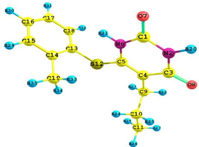
The optimized molecular geometry of the ti-tle compound was obtained from the Gaussian 03W program with the atom numbering scheme shown in Figure 1. The comparative optimized structural parameters such as bond length, bond angle along with its experimental data [36] are pre-sented in Table 1. The most optimized geometry is performed at the B3LYP /6-311++G(d, P) basis set of the title molecule. The molecule has 35 atoms and 99 normal modes of fundamental vibrations. All 99 vibrations are active in both IR and Raman.
The theoretical values for the title molecule were found to be closely related to the experi-mental XRD values. The maximum bond lengths were calculated for the C5-C12, S12-C13 and found to
be 1.792 Å, 1.792 Å (experimental) and 1.787 Å, 1.801 Å (theoretical), respectively. The calculated bond lengths C3-C4 (1.386 Å, 1.462 Å), C4-C9
devi-ations when compared with the experimental data, and these differences are probably due to the in-termolecular or intramolecular interactions in the solid state. The computed bond lengths and bond angles are in reasonable agreement with the corre-sponding experimental values. The experimental values of the bond angles correlate well to the the-oretical values (DFT) in the solid phase. The minor deviation can be attributed to the fact that the theo-retical calculations were aimed at the isolated mol-ecules in the gaseous phase and the experimental results were aimed at the molecule in the solid state. Despite these differences, the calculated ge-ometric parameters represent a good approxima-tion, and are the basis for the calculation of other parameters such as polarizability, vibrational fre-quencies and thermodynamic properties.
Fig. 1. Optimized ground state structure of the title compound (B3LYP/6-311++G(d,p)
T a b l e 1
The optimized parameters (bond lengths and bond angle of the title compound
Parameters B3LYP/6-311++G(d,p) Experimental*
1 2 3
Bond length (Å)
C1-N2 1.378 1.362
C1-N6 1.389 1.381
C1-O7 1.214 1.223
N2-C3 1.408 1.380
N2-H20 1.012 0.830
C3-C4 1.462 1.386
C3-O8 1.220 1.223
C4-C5 1.361 1.398
C4-C9 1.507 1.499
C5-N6 1.380 1.381
C5-S12 1.787 1.792
N6-H21 1.011 0.830
C9-C10 1.541 1.534
C9-H22 1.094 0.970
C9-H23 1.095 0.097
C10-C11 1.531 1.459
C10-H24 1.096 0.970
C10-H25 1.094 0.970
C11-H26 1.093 0.970
C11-H27 1.094 0.970
C11-H28 1.095 0.970
S12-C13 1.801 1.792
C13-C14 1.411 1.521
C13-C18 1.400 1.386
C14-C15 1.399 1.386
C14-C19 1.507 1.521
C15-C16 1.393 1.386
C15-H29 1.085 0.970
C16-C17 1.392 1.386
C16-H30 1.084 0.970
C17-C18 1.392 1.386
C17-H31 1.084 0.970
C18-H32 1.083 0.970
C19-H33 1.094 0.970
C19-H34 1.093 0.970
1 2 3 Bond angle (◦)
N2-C1-N6 112.8 114.51
N2-C1-O7 124.5 122.14
C1-N2-C3 127.5 126.26
C1-N2-H20 116.2 115.4
N6-C1-O7 122.7 123.35
C1-N6-C5 124.4 123.55
C1-N6-H21 115.4 115.4
C3-N2-H20 116.3 115.4
N2-C3-C4 115.4 116.35
N2-C3-O8 119.5 120.1
C4-C3-O8 125.1 123.4
C3-C4-C5 118.1 121.60
C3-C4-C9 117.4 117.4
C5-C4-C9 124.5 124.1
C4-C5-N6 121.8 121.6
C4-C5-S12 120.8 122.6
C4-C9-C10 113.6 117.4
C4-C9-H22 107.8 108.9
C4-C9-H23 110.3 109.3
N6-C5-S12 117.4 115.5
C5-N6-H21 120.2 120.4
C5-S12-C13 103.4 100.7
C10-C9-H22 108.8 108.9
C10-C9-H23 109.4 109.3
C9-C10-C11 112.4 117.4
C9-C10-H24 109.1 109.3
C9-C10-H25 108.7 108.9
H22-C9-H23 106.7 107.7
C11-C10-H24 109.8 109.3
C11-C10-H25 109.8 109.3
C10-C11-H26 111.2 109.3
C10-C11-H27 111.1 109.3
C10-C11-H28 111.3 109.3
H24-C10-H25 106.9 107.7
H26-C11-H27 107.7 107.7
H26-C11-H28 107.7 107.7
H27-C11H28 107.7 107.7
S12-C13-C14 121.4 120.2
S12-C13-C18 117.5 118.7
C14-C13-C18 120.9 120.5
C13-C14-C15 117.3 117.3
C13-C14-C19 122.5 122.0
C13-C18-C17 120.4 120.6
C13-C18-H32 119.2 119.2
C15-C14-C19 120.2 120.0
C14-C15-C16 121.8 122.0
C14-C15-H29 118.7 119.7
C14-C19-H33 111.6 109.5
C14-C19-H34 111.2 109.5
C14-C19-H35 110.6 109.5
C16-C15-H29 119.5 119.7
C15-C16-H17 120.1 119.7
C15-H16-H30 119.7 119.7
C17-C16-H30 120.2 119.7
C16-C17-H18 119.4 119.7
C16-C17-H31 120.7 119.7
C18-C17-H31 120 119.7
C17-C18-H32 120.4 119.7
H33-C19-H34 106.9 109.5
H33-C19-H35 107.8 109.3
H34-C19-H35 108.6 109.3
4.2. Vibrational assignments
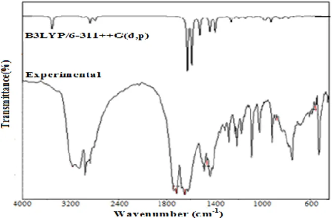
The title molecule consists of 35 atoms, which has 99 normal modes of vibration. The ex-perimental FT-IR and FT-Raman spectra with their corresponding theoretically-simulated IR and Ra-man spectra are shown in Figures 2 and 3, respec-tively. The calculated vibrational frequencies (un-scaled and (un-scaled), IR intensity, and Raman activi-ty are shown in Table 2.
Fig. 2. Experimental and theoretical (B3LYP/6-311++G(d,p) FT- IR spectra of the title compound
Fig. 3. Experimental and theoretical (B3LYP/6-311++G(d,p) FT- Raman spectra of the title compound
4.2.1. N-H vibrations
The N-H stretching vibrations of the hetero-cyclic compounds always occur in the region of 3500–3000 cm–1 [48–50]. The bands observed at 3440 and 3159 cm–1 in the FT-IR and at 3475, 3445 and 3076 cm–1 in the FT Raman spectra with 100, 100, 100, 100 and 97% of PED contribution are attributed to the N-H groups of the pyrimidine
ring. The theoretically-calculated values of the N-H vibrations by the B3LYP/6-311++G(d,P) in high frequency region are reliable with the experimental results and these assignments are in good agree-ment with the literature data [35].
4.2.2. C-C vibrations
The ring C-C aromatic vibrations give rise to characteristic bands in both the observed FT-IR and FT-Raman spectra covering the spectral range from 1600 to 1400 cm–1 [50]. In the present study, the C-C stretching vibration bands are calculated at 1591, 1522, 1497 and 1395 cm–1 in the FT-Raman spectra with major PED contributions of 11%. The calculated bands at the B3LYP/6-311++G(d,P) levels are in excellent agreement with the experi-mental observations of both the IR and FT-Raman spectra of title compound. The ring C-C vibrations have given rise to weak bands across the low frequency region below 1320 cm–1. The bands at 1263, 1178, 1155, 1090, 1047, 1010 and 535 cm–1 in the FT-IR and 1180, 1094, 1033 and 1003 cm–1 in the FT-Raman spectra have been assigned to the C-C stretching vibrations with 74, 56, 10, 20, 18, 31,17 and 56, 32, 17 and 92% contributions of PED, respectively.
4.2.3. Aromatic C-H vibrations
The aromatic structure shows the presence of C-H stretching vibrations in the region 3100– 3000 cm–1, which is the characteristic region for identification of the aromatic C-H stretching vibra-tions [51]. In this region, the bands are not affected appreciably by the nature of the constituents. In the present investigation, the aromatic C-H stretching vibrations were observed at 3073, 3063, 3054 and 3043 cm–1 by the B3LYP/6-311++G(d,P) method which showed excellent agreements with the bands observed in the recorded FT-Raman spectrum at 3076, 3067, 3054 cm–1 and with the FT-IR bands at 3159, 3050 cm–1. The PED corresponding to this pure mode of contributing to 94%, as shown in Table 2.
4.2.4. Propyl vibrations
T a b l e 2
Observed and calculated vibrational frequency of the title compound at the B3LYP method with 6-311++G(d,P) basis set
Mode
Experimental wave number (cm–1)
Theoretical wave
number (cm–1) IR intensityc
Raman intensityb
Vibrational assignments (PED)a
FT-IR FT- Raman Unscaled Scaledd
1 2 3 4 5 6 7 8
1 – 3475 3603 3462 9 43 γ NH(100)
2 3440 3445 3592 3452 21 23 γ NH(100)
3 3159 3076 3198 3073 1 100 γ CH(97)
4 – 3067 3187 3063 1 40 γ CH(87)
5 – 3054 3178 3054 1 42 γ CH(100)
6 3050 – 3166 3043 0 19 γ CH(93)
7 – – 3116 2994 2 21 γ CH3(97)
8 3440 3445 3592 3452 21 23 γ NH(100)
9 – 2980 3088 2968 7 17 γ CH3(84)
10 – – 3086 2966 5 36 γ CH3(80)
11 – 2962 3086 2965 1 15 γ CH3(92)
12 2961 – 3075 2955 0 3 γ CH3(99)
13 – 2931 3054 2935 1 38 γ CH3(72)
14 2929 – 3034 2916 4 17 γ CH2(95)
15 2912 – 3033 2915 1 72 γ CH3(96)
16 – 2871 3021 2903 2 42 γ CH3(87)
17 1719 1719 1790 1721 100 10 γ OC(74)
18 1662 1649 1734 1666 89 41 γ OC(81)
19 – 1591 1629 1565 5 20 γ CC(57)
20 – 1552 1622 1558 31 23 γ CC(62)
21 – 1497 1604 1541 0 10 γ CC(47)
22 1463 – 1507 1449 1 1 β HCH(62)
23 – 1454 1503 1445 2 2 β HCC(24)+ β HCH(38)
24 – – 1500 1441 1 3 β HCH(75)+τ HCCC(14)
25 1439 – 1494 1436 8 5 β HNC(17)+ β HCH(26)
26 – – 1493 1435 7 5 β HNC(14)+ β HCH(39)
27 – 1431 1492 1434 8 4 β HNC(12)
28 – 1425 1482 1424 4 2 β HCH(63)+ τ HCCC(13)
29 1417 – 1482 1424 1 8 β HCH(62)
30 – 1395 1459 1402 0 0 γ CC(11)+ β HCC(55)
31 – – 1422 1366 2 3 β HCH(85)
32 1363 – 1415 1360 28 1 γ NC(31)+ β HNC(11)
33 – 1352 1412 1357 1 0 β HCH(89)
34 – – 1403 1348 0 4 β HNC(69)
35 1314 1317 1383 1329 0 3 τ HCCC(66)
36 1284 1287 1338 1285 0 5 β HCC(57)+τ HCCC(11)
37 – – 1317 1266 0 0 β HCC(10)+τ HCCC(37)
38 1263 – 1312 1261 1 2 γ CC(74)
39 – – 1301 1250 0 1 γ CC(14)+ β HCC(55)
40 1230 1232 1295 1244 1 2 β HCC(26)
41 1214 1211 1237 1189 1 7 γ NC(10)+β HCC(12)
42 1178 1180 1223 1176 0 11 γ CC(56)+β HCC(16)
43 – 1169 1208 1161 10 1 γ NC(45)+ γ CC(29)
44 1155 – 1188 1142 0 2 γ CC(10)+ β HCC(73)
45 1117 1123 1160 1115 3 2 γ NC(46)
46 – 1094 1152 1107 0 2 γ CC(33)+ β HCC(35)
47 1090 – 1108 1065 1 4 γ CC(20)+ τ HCCC(35)
48 1047 – 1098 1056 3 2 γ CC(18)+ β HNC(11)
49 – 1033 1071 1029 0 3 γ CC(17)+βHCC(16)+ β CCC(16)
50 1027 – 1063 1021 1 0 βHCH(14)+τHCCC(59)+
ωCCCC(11)
51 1010 – 1054 1013 2 16 γ CC(31)+ γ SC(11)+ β CCC(29)
52 – 1003 1038 997 0 2 γ CC(92)
53 984 – 1016 976 3 4 γ NC(28)+ β HNC(11)+ βCNC(16)
1 2 3 4 5 6 7 8
55 958 – 1007 968 0 0 β HCH(10)+ τ HCCC(45)
56 921 924 972 934 0 0 τ HCCC(86)
57 894 895 908 873 2 5 γ NC(15)+ γ SC(16)
58 880 – 897 862 0 4 γ CC(38)+ τ HCCC(13)
59 – 855 886 852 0 0 τ HCCC(86)
60 839 835 861 828 1 1 τ HCCC(14)
61 783 798 813 781 0 4 γ CC(25)+ β CCC(47)
62 754 – 799 768 1 0 γ CC(13)+ β NCN(22)+ω
ONCC(11)
63 735 – 772 742 5 0 τ HCCC(74)
64 – 728 763 733 1 0 ω ONCC(49)
65 725 – 750 721 5 0 ω ONCC(93)
66 – 708 742 713 1 0 β HCC(18)+ τ HCCC(28)
67 698 – 730 702 1 0 τ CCCC(58)
68 668 674 687 661 0 3 γ SC(16)+ β CCC(54)
69 645 640 669 643 10 0 τ HNCN(70)+ ω CNSC(12)
70 610 608 645 619 1 0 β OCN(46)
71 590 589 588 565 1 1 β CNC(11)+ τ HNCN(14)+ ω
ONCC(10)
72 568 553 586 563 0 3 τ HNCN(28)+ ω CNSC(14)
73 555 531 559 537 4 0 τ HNCN(53)
74 535 – 559 537 0 4 γ CC(17)+ β CCC(27)
75 505 – 531 510 1 1 τCCCC(14)+ωCCCC(10)+ωSCCC
(19)
76 470 485 522 501 3 1 β NCN(10)+ β CNC(10)+ τ
HNCN(14)+ ω CNSC(15)
77 459 449 470 452 1 0 τ CCCC(10)+ ω CCCC(24)
78 – – 449 432 0 1 γ SC(21)+ β CCC(19)
79 – 396 434 418 4 1 γ SC(21)+ β OCN(15)
80 – – 388 373 0 0 β CCC(28)+ β SCC(15)
81 – – 376 361 1 0 β CCC(12)+ ω CCCC(21)+ ω
CNSC(10)
82 – 331 370 355 1 1 γ SC(12)+ β OCN(29)
83 – 302 324 311 0 1 β CCC(38)
84 – – 291 280 0 1 γ CC(11)+ β CCC(41)
85 – 254 258 248 1 0 τ CCCC(10)+ ω CCCC(17)
86 – – 250 240 0 0 β CCC(12)+ β SCC(46)
87 – – 241 232 0 0 τ HCCC(69)
88 – 185 189 182 0 1 β NCS(22)
89 – – 166 160 0 0 β CCC(12)+ τ CCCC(13)+ τ
NCNC(13)+ τ CNCS(10)+ ω CCCC(10)
90 – – 152 146 0 0 τ CCCC(20)+ τ CNCN(32)
91 – 131 149 143 0 0 β NCS(10)+ β CCC(16)+ τ
CCCC(12)+ τ CNCN(14)+ ω CCCC(10)
92 – – 125 120 0 0 τ NCNC(15)+ τ CNCS(32)+ τ
CNCN(21)
93 – – 119 115 0 0 τ HCCC(52)+ τ CNCS(12)+ ω
CCCC(10)
94 – – 88 85 0 0 τ HCCC(10)+ τ CCCC(73)
95 – – 60 58 0 1 τ NCNC(30)+ τ
CNCS(20)+τCNCN(14)+ωCCCC(1 3)
96 – – 56 54 0 1 β CSC(28)+ω SCCC(26)
97 – – 42 41 0 1 β CCC(10) +τ CCCC(47)+
τCSCC(17)
98 – – 33 31 0 2 τ CSCC(64)
99 - – 17 16 0 0 τ NCSC(82)
a γ: Stretching, γa: symmetrical stretching, γas: symmetrical stretching, β: in-plane bending , ω: out-plane bending, τ: torsion b Scaling factor : 0.961 for B3LYP/6-311+G(d,p)
c
Relative absorption intensities normalized with the highest peak absorption equal to 100. d
In aromatic compounds, the methyl asym-metric vibrations are expected near in the range 3000–2925 cm–1 and the symmetric CH3 vibrations
in the range 2940–2905 [53, 54]. In the title com-pound, the asymmetric stretching vibration was identified at 2968, 2966, 2965 cm–1 in the B3LYP/6-311++G(d,p) method which showed good agreement with recorded FT-Raman spec-trum at 2980, 2962 cm–1, and were not detected in the in the FT-IR spectrum. The CH3 symmetric
stretching was found at 2915, 2901 cm–1 in the B3LYP/6-311++G(d,p) method and at 2912, 2870 cm–1 in FT-IR spectrum. The PED corresponding to asymmetric and symmetric stretching vibration is almost 85% and 97%, as shown in Table 2.
For the assignments of CH2 group
frequen-cies, basically, six fundamentals can be associated to each CH2 group which is expected to be
depo-larized [55]. The CH2 asymmetrical stretching
vi-bration is generally observed above 3000 cm–1, while the symmetrical stretch would appear be-tween 3000 and 2900 cm–1 [56]. In this study, the asymmetric stretching vibrations were observed at 3166, 3075, 3074 cm–1 in the B3LYP/6-311++G(d,p) method, 2961 cm–1 in the FT-IR spectrum and 2931 cm–1 in the FT-Raman spec-trum. The symmetric CH2 stretching vibrations are
identified at 2916, 2903 cm–1 in the B3LYP/6-311++G(d,p) method, 2929 cm–1 in the FR-IR spectrum and 2871 cm–1 in the FT-Raman spec-trum.
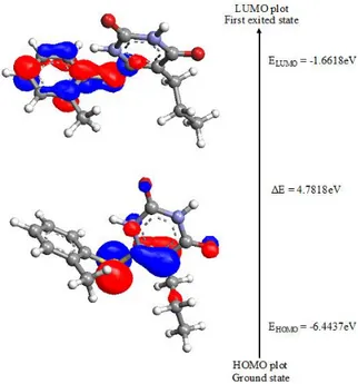
4.3. HOMO-LUMO energy
The fundamental importance of the HOMO (highest occupied molecular orbital) and LUMO (lowest unoccupied molecular orbital) is under-standing the chemical stability and reactivity of many organic molecules [57]. HOMO, the outer-most orbital containing electrons, tends to give these electrons; on the other hand, LUMO, the in-nermost orbital, contains free places to accept elec-trons [58]. HOMO and LUMO are directly related to the ionization potential and electron affinity.
The energy difference between HOMO and LUMO orbits is called energy gap which is consid-ered as important for indicator for compound sta-bility [59]. The total energy, HOMO and LUMO energies, the energy gap (ΔE), the ionization po-tential (I), the electron affinity (A), the absolute electronegativity (χ), the absolute hardness (η) and softness (S) of the title molecule have been calcu-lated at the B3LYP/6-311++G(d, P) basis set (Fig. 4); the results are shown in Table 3.
Fig. 4. Atomic orbital HOMO-LUMO composition of the frontier molecular orbital of the title compound
T a b l e 3
Comparison of the HOMO, LUMO, energy gaps and ionization potentials of the title compound
Basis set B3LYP/6-311++G(d, p)
EHomo -6.4437
ELomo -1.6618
Ionization potential 6.4437
Electron affinity 1.6618
Energy gap 4.7818
Electronegativity 4.0527
Chemical potential 4.0527
Chemical hardness 2.3909
Chemical softness 0.2091
Electrophilicity index 3.4347
4.4. Local reactivity descriptors
The local quantities such as Fukui function and local softness define the reactivity/selectivity of a specific site in a molecule. The Fukui function is defined as follows:
ƒ( ⃗)=( 𝜕ρ/𝜕N ) 𝜐 ⃗ =( ⃗ )N .
T a b l e 4
Condensed Fukui function ƒr and new descriptor (sƒ)r of the title compound
Atoms ƒr+ ƒr- ƒr0 sr+ƒr+ sr–ƒr– sr0 ƒr0
C1 0.0016 0.0015 0.0015 0.0003 0.0003 0.0003
N2 –0.0010 –0.0089 –0.0049 0.0002 0.0018 0.0010
C3 –0.0231 –0.0087 –0.0159 0.0048 0.0018 0.0033
C4 –0.0314 –0.0461 –0.0388 0.0065 0.0096 0.0081
C4 0.0310 –0.0091 0.0109 0.0064 0.0019 0.0022
N6 –0.0371 –0.0509 –0.0440 0.0077 0.0106 0.0092
O7 –0.0559 –0.1110 –0.0835 0.0117 0.0232 0.0174
O8 –0.1010 –0.0971 –0.0991 0.0211 0.0203 0.0207
C9 0.0097 0.0086 0.0092 0.0020 0.0018 0.0019
C10 –0.0081 0.0179 0.0049 0.0017 0.0037 0.0010
C11 –0.0071 –0.0390 –0.0230 0.0014 0.0081 0.0048
S12 –0.1761 –0.1814 –0.1787 0.0368 0.0379 0.0373
C13 0.5241 0.1789 0.3515 0.1096 0.0374 0.0735
C14 –0.0787 –0.0297 –0.0542 0.0164 0.0062 0.0113
C15 –0.1221 –0.0333 –0.0777 0.0255 0.0069 0.0162
C16 –0.0191 –0.0163 –0.0177 0.0040 0.0034 0.0037
C17 –0.0643 –0.0473 –0.0558 0.0134 0.0099 0.0116
C18 –0.1715 –0.0768 –0.1242 0.0358 0.0160 0.0259
C19 –0.0288 –0.0197 –0.0242 0.0060 0.0041 0.0050
H20 –0.0417 –0.0372 –0.0394 0.0087 0.0077 0.0082
H21 –0.0028 –0.0243 –0.0136 0.0006 0.0050 0.0028
H22 –0.0626 –0.0530 –0.0578 0.0131 0.0111 0.0121
H23 –0.0249 –0.0173 –0.0211 0.0052 0.0036 0.0044
H24 –0.0120 –0.0123 –0.0122 0.0025 0.0025 0.0025
H25 0.0028 –0.0153 –0.0062 0.0006 0.0032 0.0013
H26 –0.0264 –0.0327 –0.0296 0.0055 0.0068 0.0061
H27 –0.0189 –0.0208 –0.0198 0.0039 0.0043 0.0041
H28 –0.0070 –0.0124 –0.0097 0.0014 0.0026 0.0020
H29 –0.0549 –0.0406 –0.0478 0.0114 0.0085 0.0100
H30 –0.0593 –0.0462 –0.0527 0.0124 0.0096 0.0110
H31 –0.0501 –0.0365 –0.0433 0.0104 0.0076 0.0090
H32 –0.1687 –0.0215 –0.0951 0.0352 0.0045 0.0198
H33 –0.0384 –0.0300 –0.0342 0.0080 0.0062 0.0071
H34 –0.0444 0.0071 –0.0186 0.0093 0.0014 0.0039
H35 –0.0306 –0.0376 –0.0341 0.0064 0.0078 0.0071
The Fukui function (ƒ+( ⃗), ƒ-( ⃗), ƒ0( ⃗)), ( ) [61] for selected atomic sites in the title molecule have been listed in Table 4. Yang and Mortier [62] have given a simple procedure for the calculation of the atomic condensed Fukui function indices based on the MPA and the three possible forward, backward, and central finite dif-ference approximations to the derivatives [63]. The Fukui functions are calculated using the following equation:
ƒ+
( ⃗) = for nucleophilic attack
ƒ
-( ⃗) = for electrophilic attack
ƒ0
( ⃗)= for radical attack
In these equations, the qr is the atomic
charge (evaluated from the Mulliken population analysis, electrostatic derived charge, etc.) at the rth atomic site is the neutral (N), anionic (N+1), cati-onic (N–1) chemical species.
The local softness is related to Fukui func-tion as follows:
s( ⃗)=( ⃗ ) 𝜐 ⃗ = ( ⃗ ) 𝜐 ⃗ ( ) 𝜐 ⃗ = ƒ( ⃗)S
s=( ) ∫ ⃗ ⃗
The local softness can be represented as:
for nucleophilic attack
for electrophilic attack
for radical attack
where +, –, 0 signs show nucleophilic, electro-philic and radical attack, respectively.
Table 4 shows the fr and (srfr) values for the
title compound, using which one can find the com-plexities associated with fr values due to the
nega-tive values being removed in the (srfr) values. It has
been found that the MAP schemes predicting C13
to have a higher value indicate possible sites for nucleophilic attack. From the values reported in Table 4, the MAP schemes predict the reactivity order for the nucleophilic case to be C13 > C5 > C9
> H28 > C1. The observation of the reactive sites by
is found almost identical to . Even though the (srfr) values are numerically lower, it is worth
nothing that the values are positive and the order-ing of the reactivity has not changed in any case. The calculated – values predicts that the possible sites for electrophilic attack are C13 > C10 > C9 >
H34 > C1 and the radical attack was predicted at C13
> C5 > C9 > C10 > C1 site. It could therefore be
con-cluded that the possibility of nucleophilic attack is higher than that of electrophilic and radical attack.
4.5. Natural bond orbital (NBO) analysis
In order to investigate the intra- and inter-molecular interactions, the stabilization energies of the title compound were performed by using the second-order perturbation theory. The change in electron density (ED) in the (𝜎*
,*) anti-bonding orbitals and E(2) energies have been calculated by the natural bond orbital analysis [64]. The second order Fock matrix was carried out to evaluate the donor-acceptor interactions in the NBO analysis [65]. The NBO analysis provides the most accurate possible ‘natural Lewis structure’ picture of ɸ be-cause all of the orbital details are mathematically chosen to include the highest possible percentage of electron density.
The natural bond orbital analysis provides an efficient method for studying intra- and
inter-molecular bonding and interaction among bonds and also provides a convenient basis for investigat-ing charge transfer or conjugative interaction in molecular systems. Some electron donor orbital, acceptor orbital and interacting stabilization energy resulting from the second-order micro-disturbance theory have been reported [66]. For each donor (i) and acceptor (j), the stabilization energy E [2] as-sociated with the delocalization i, j estimated as:
where qi is the donor orbital occupancy, Ei and Ej
are diagonal elements and F(i,j) is the off-diagonal NBO Fock matrix elements.
The larger the E(2) value, the more intensive the interaction between electron donors and elec-tron acceptors is; i.e. the high donating tendency from electron donors to electron acceptors and the greater the extent of conjugation of the whole sys-tem. The delocalization of electron density be-tween the occupied Lewis-type (bond or lone pair) NBO orbitals and formally unoccupied (anti-bond or Rydgberg) non-Lewis NBO orbitals correspond to a stabilizing donor-acceptor interaction. The NBO analysis has been performed on the title mol-ecule at the B3LYP/6-311++G(d,p) level in order to determine the delocalization of electron density within the molecule.
The strong intramolecular hyperconjugative interaction of the s and p electrons of C-C to the anti C-C bond of the ring leads to stabilization of some part of the ring, as shown in Table 5. The intramolecular hyperconjugative interaction of σ (C4-C5) distributes to σ
*
(C3–C4), (C1-N2), (C1-N6)
leading to stabilization of 8.32 KJ/mol. This en-hances further conjugation with anti-bonding or-bital of π*
(C3-O8), (C17-C18) which lead to strong
delocalization of 20.67 KJ/mol, respectively. The σ system shows some contribution to the delocaliza-tion corresponds to the donor-acceptor interacdelocaliza-tions are (N2-H20) (C5-S12), (C5-S12) (C1-C2), (C1
-O7) (C1-N6), (C5-N6) (C4-C5), (C4-C9) (C1
-N6), (C3-C4) (N2-H20) bondings are shown in
Table 5. The LP (1) N6 anti-bonding acceptor π *
(C1-O7) of the title compound energy of 108.49
KJ/mol. The LP (1) N2 of the NBO orbital further
conjugate with π* (C1-O7) resulting in an enormous
T a b l e 5
Second order perturbation theory analysis of Fock matrix in NBO basis of the title compound
Donor (i) Type ED/e Acceptor (i) Type ED/e E(2)a(kjmol–1) E(j)-E(i)b(a.u.) F(i,j)c(a.u.)
C1-N2 σ 1.9818 C1-O7 σ
*
0.0172 4.23 1.66 0.075
σ N2-C3 σ* 0.0872 4.05 1.47 0.070
C1-N6 σ 1.9759 N2-H20 σ* 0.0244 3.33 1.19 0.056
σ C5-S12 σ* 0.0488 3.99 0.99 0.056
C1-O7 σ 1.9843 C1-N2 σ* 0.0498 5.20 1.78 0.087
C1-O7 π 1.9868 C1-O7 π* 0.4167 3.77 0.38 0.038
N2-C3 σ 1.9833 C1-N2 σ* 0.0498 4.34 1.59 0.075
σ C1-O7 σ* 0.0172 3.61 1.49 0.066
N2-H20 σ 1.9726 C1-N6 σ* 0.0876 8.32 1.11 0.087
C3-C4 σ 1.9726 N2-H20 σ
*
0.0244 3.14 1.00 0.050
C3-O8 σ 1.9934 C3-C4 σ
*
0.0770 2.05 1.48 0.050
C3-O8 π 1.9827 C4-C5 π
*
0.2423 4.13 0.41 0.039
C4-C5 σ 1.9754 C4-C9 σ
*
0.0187 4.07 1.22 0.063
σ N6-H21 σ* 0.0325 3.24 1.17 0.055
C4-C5 σ 1.8445 C3-O8 π
*
0.2952 17.63 0.31 0.068
C4-C9 σ 1.9714 C4-C5 σ
*
0.0260 4.79 1.27 0.070
σ C5-N6 σ* 0.0375 4.37 1.10 0.062
C5-N6 σ 1.9852 C4-C9 σ
*
0.0187 3.01 1.31 0.056
C5-S12 σ 1.9655 C1-N6 σ
*
0.0876 3.22 1.07 0.053
σ C3-C4 σ* 0.0770 4.58 1.03 0.062
N6-H21 σ 1.9806 C4-C5 σ
*
0.0260 3.39 1.33 0.060
C9-H22 σ 1.9679 C4-C5 π
*
0.0242 4.29 0.50 0.440
C9-H23 σ 1.9746 C3-C4 σ
*
0.0770 4.76 0.88 0.059
S12-C13 σ 1.9687 C14-C15 σ
*
0.0236 3.64 1.17 0.058
C13-C14 σ 1.9736 C13-C18 σ
*
0.0260 3.33 1.24 0.057
C13-C14 π 1.6682 C15-C16 π
*
0.3149 16.17 0.28 0.610
π C17-C18 π
*
0.0327 20.67 0.28 0.680
C14-C15 σ 1.9705 S12-C13 σ
*
0.0364 4.45 0.08 0.055
σ C13-C14 σ
*
0.0359 3.90 1.23 0.062
C15-C16 π 1.6650 C13-C14 π
*
0.3877 23.20 0.27 0.071
π C17-C18 π
*
0.3277 19.39 0.27 0.064
C15-H29 σ 1.9804 C13-C14 σ
*
0.0359 4.39 1.06 0.061
C17-C18 σ 1.9755 S12-C13 σ* 0.0364 4.40 0.86 0.550
C17-C18 π 1.6770 C13-C14 π* 0.3877 17.47 0.27 0.063
π C15-C16 π* 0.3149 19.30 0.28 0.065
C18-H32 σ 1.9682 N6-H21 σ* 0.0325 6.18 0.94 0.068
C19-H34 σ 1.9855 C14-C15 σ* 0.0260 4.75 1.04 0.063
C19-H35 σ 1.9761 C13-C14 π* 0.3877 4.01 0.51 0.045
N2 LP (1) 1.5884 C1-O7 π* 0.4167 108.49 0.31 0.163
C3-O8 π* 0.2952 51.76 0.30 0.115
N6 LP (1) 1.6576 C1-O7 π* 0.4167 54.27 0.30 0.115
LP (1) C4-C5 π* 0.2423 42.62 0.31 0.105
O7 LP (2) 1.8731 C1-N2 σ* 0.0498 18.50 0.98 0.122
LP (2) C1-N6 σ* 0.0876 25.56 0.68 0.199
O8 LP (2) 1.8609 N2-C3 σ* 0.0872 28.08 0.07 0.013
LP (2) C3-C4 σ* 0.0770 19.99 0.66 0.104
S12 LP (2) 1.8723 C4-C5 π
*
0.2423 6.33 0.28 0.039
C3-O8 π
*
0.2952 C4-C5 π* 0.2423 92.15 0.02 0.071
C13-C14 π* 0.3877 C4-C5 π* 0.2423 5.73 0.01 0.015
a
E(2): Energy of hyperconjugative interaction (stabilization energy). b
Energy difference between donor and acceptor I and j NBO orbitals. c
4.6. Hyperpolarizability calculations
The first order hyperpolarizability (βtotal) of
the title compound along with the related proper-ties (µ, α and ∆α) are calculated by using the DFT- B3LYP method with 6-311++G(d,P) basis set based on the finite-field approach. The NLO ef-fects arise from the interactions of electromagnetic fields in various media to produce new fields al-tered in phase, frequency, amplitude or other prop-agation characteristics from the incident fields. NLO is at the forefront of current research because of its importance in providing the key functions of frequency shifting, optical modulation, optical switching, optical logic, and optical memory for emerging technologies in areas such as telecom-munications, signal processing, and optical inter-connections [67].
The non-linear optical response of an isolat-ed molecule in an electric field Ei (ω) can be
repre-sented as a Taylor series expansion of the total di-pole moment, µtot, induced by the field:
tot = 0 + ijEj + BijkEjEk + …,
where α is the linear polarizability, µ0 is the
per-manent dipole moment and βijk are the first
hy-perpolarizability tensor components. The isotropic (or average) linear polarizability is defined as:
𝛼 = .
The first hyperpolarizability is a third rank tensor that can be described by 3×3×3 matrix. The 27 components of 3D matrix can be reduced to 10 components due to the Kleinman symmetry [68].
Components of the first hyperpolarizability can be calculated using the following equation:
𝛽 𝛽 ∑ 𝛽 𝛽 𝛽
Using the x, y and z components of β, the magnitude of the first hyperpolarizability tensor can be calculated by:
𝛽 √ 𝛽 𝛽 𝛽
The complete equation for calculating the magnitude of β from Gaussian 03W output is given as follows:
𝛽
√(𝛽 𝛽 𝛽 ) (𝛽 𝛽 𝛽 )
𝛽 𝛽 𝛽
The calculations of the total molecular di-pole moment (µ), linear polarizability (α) and first-order hyperpolarizability (β) from the Gaussian output have been previously explained in detail [69], and the DFT has been extensively used as an effective method to investigate the organic NLO materials [70,71]. In addition, the polar properties of the title compound were calculated at the DFT (B3LYP)/6-311++G(d,p) level using Gaussian 03W program package.
Urea is one of the prototypical molecules used in the study of the NLO properties of the molecules. Therefore, it was frequently used as a threshold value for comparative purposes.
T a b l e 6
The values of calculated dipole moment (D), polarizability (0),
the first order hyperpolarizability (βtot) components of the title compound
Parameters B3LYP/6-311++G(d,p) Parameters B3LYP/6-311++G(d,p)
x 0.1418 xxx 87.4914
y 1.4189 βxxy 209.2697
z 0.8954 βxyy 38.0065
(D) 1.6837 βyyy –169.2489
xx 246.9805 βzxx 38.1262
xy 1.2806 βxyz –1.042
yy 191.87 βzyy –41.707
xz 19.83073 βxzz 20.3421
yz 31.6945 βyzz 78.9891
zz 186.4042 βzzz 21.3964
e.s.u 3.0888×10⁻²³ βtot (e.s.u) 1.6335×10⁻³⁰
The calculated dipole moment and hyperpo-larizability values obtained from B3LYP/6-31++G(d,p) methods are shown in Table 6. The total molecular dipole moment of the title molecule from the B3LYP with 6-311++G(d,p) basis set is 1.2 times greater than that of urea (µ = 1.3732 D). Similarly, the first order hyperpolarizability of the title molecule with B3LYP/6-311++G(d,p) basis set (1.6335 × 10⁻³⁰) is four times greater than the value of urea (βo = 0.372 × 10⁻³⁰ esu). From the
computation, the high values of the hyperpolariza-bilities of the title molecule are probably attributed to the charge transfer existing between the phenyl rings within the molecular skeleton. This is evi-dence for the nonlinear optical (NLO) property of the molecule.
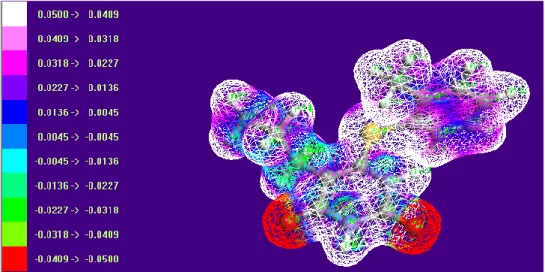
4.7. Molecular electrostatic potentials (MEP)
The MEP and electrostatic potential are use-ful quantities to illustrate the charge distributions of molecules and used to visualize variably charged regions of a molecule. Therefore, the charge distributions can give information about how the molecules interact with another molecule. MEP is widely used as a reactivity map displaying the most probable regions for the electrophilic at-tack of charged point-like reagents on organic molecule [72, 73]. The molecular electrostatic po-tential V(r) at a given point r(x,y,z) in the vicinity of a molecule, is defined in terms of the interaction energy between the electrical charge generated from the molecule electrons and nuclei and posi-tive test charge (a proton) located at r [74]. Unlike many of the other quantities used at present and earlier as indices of reactivity, V(r) is a real physi-cal property that can be determined experimentally by diffraction or computational methods. For the
systems studied, the MEP values were calculated as previously described, using the following equa-tion [75]:
V(r) = ∑ZA (RA r) ρ(rˈ) (r r)dr
where the summation runs over all the nuclei A in the molecule and polarization and reorganization effects are neglected. ZA is the charge of the nucle-us A, located at RA and ρ(r ) is the electron density function of the molecule.
It provides a visual method to understand the relative polarity of the molecule, as shown in Figure 5. The different values of the electrostatic potential are represented by different colors; red represented the regions of the most negative electrostatic poten-tial, white represents the regions of the most posi-tive electrostatic potential and blue represents the region of zero potential. The potential increases in the order red < green < blue < pink < white. It can be seen that the negative regions are mainly over the O7 and O8 atoms. The negative (red color) and
posi-tive (white) regions of the MEP are related to the electrophilic and nucleophilic reactivity. The pre-dominance of the light green region MEP surface corresponds to a potential half way between two extremes red and white color. The negative electro-static potential corresponds to an attraction of the proton by the aggregate electron density in the mol-ecule (shades of red), while the positive electrostatic potential corresponds to the repulsion of the protons by the atomic nuclei (shades of white). According to the calculated results, the MEP map shows that the negative potential sites are on oxygen atoms and the positive potential sites are also around the hydrogen atoms. These sites give information about the region from where the compound can have non-covalent interactions.
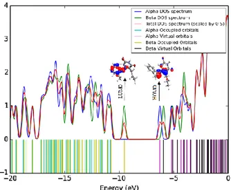
4.8. Total, sum of alpha plus beta electrons (DOS)
The consideration of only HOMO and LU-MO may not yield a realistic description of the frontier orbitals because, in the boundary region, neighboring orbitals may show quasi-degenerate energy levels. For this reason, the total (TDOS), sum of 𝛼 and 𝛽 electron density of states, in terms of Mulliken population analysis, are calculated and created by convoluting the molecular orbital in-formation with the Gussian curves of unit height and full width at half maximum (FWHM) of 0.3 eV by using the Gauss Sum 2.2 program [76,77]. The TDOS and 𝛼𝛽DOS of the title molecule are plotted in Figures 6 and 7.
Fig. 6. The sum of alpha plus beta electrons DOS diagram of the title compound
Fig. 7. Calculated total electronic density of states (TDOS) for the title compound
They provide a pictorial representation of molecule orbital (MO) compositions and their con-tributions to the chemical bonding. The most im-portant application of the DOS plots is to demon-strate the MO compositions and their contributions to the chemical bonding through the positive and negative charges provided by 𝛼𝛽DOS and TDOS diagrams. The 𝛼𝛽DOS shows the bonding and sum of positive and negative electrons with the nature of the interaction of the two orbitals, atoms or groups. In this case, the title molecule consists of 73 𝛼-electrons and 73 𝛽-electrons; in total 146 electrons are occupied in density of states. The way we designate a pictorial representation for cat-ions and ancat-ions is essentially similar to that for neutral atoms in their ground state. A positive val-ue of the 𝛼𝛽DOS indicates a bonding interaction, negative values mean that there is an anti-bonding interaction and zero values indicate non-bonding interactions [78].
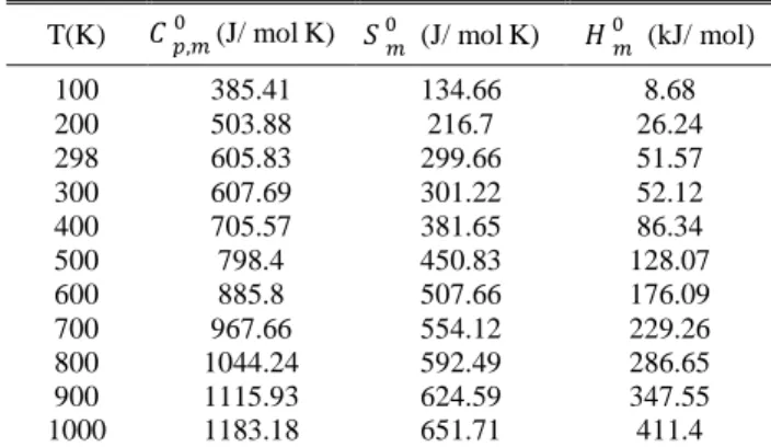
4.9. Thermodynamic properties
On the basis of vibrational analysis, the stat-ically thermodynamic functions: heat capacity ( ), entropy ( ) and enthalpy changes( ) were computed using the B3LYP/6-311++G(d,p) basis set by using Perl script THERMO.PL and are listed in Table 7. From the results in Table 7, it can be observed that the values of all the thermody-namic functions are increasing with the tempera-ture ranging from 100 to 1000 K due to the fact that the molecular vibration intensities increase with temperature. The correlation equation be-tween heat capacities, entropies, enthalpy changes and temperatures were fitted by quadratic formulas and the corresponding fitting factors (R2); these thermodynamic properties are 0.9999, 0.9995 and 0.9994, respectively. The correlations plot of those shown in Figure 8. The thermodynamic correlation fitting equations are as follows:
( ) = 273.02 + 1.1957T - 2.8756 x 10⁻⁴
T2 (R2 = 0.9999)
( ) = 28.374 + 1.05038T – 4.2828 x 10⁻⁴
T2 (R2 = 0.9995)
( ) = - 12.228 + 0.1365T - 2.9108 x 10⁻⁴
T a b l e 7
Thermodynamic properties for the title compound (B3LYP/6-311++G(d,p)
T(K) (J/ molK) (J/ molK) (kJ/ mol)
100 385.41 134.66 8.68
200 503.88 216.7 26.24
298 605.83 299.66 51.57
300 607.69 301.22 52.12
400 705.57 381.65 86.34
500 798.4 450.83 128.07
600 885.8 507.66 176.09
700 967.66 554.12 229.26
800 1044.24 592.49 286.65
900 1115.93 624.59 347.55
1000 1183.18 651.71 411.4
Fig. 8. Correlation plot of the thermodynamic properties at different temperature of the title compound
The thermodynamic data provide very help-ful information for further investigation of the title molecule. They can be used to compute the ther-modynamic energy according to the relationship between thermodynamic functions and to evaluate the directions of chemical behavior according to the second law of thermodynamics in the thermo-chemical field [79].
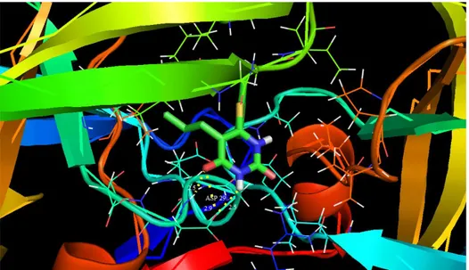
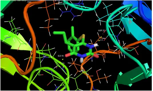
4.10. Molecular docking studies
Molecular docking has recently been used as a convenient tool to obtain insights into the molec-ular mechanism of protein-ligand interactions. Mo-lecular docking studies were carried out to evaluate the binding affinity of the ligand with the active site of c-south African HIV-1 protease (C-SA HIV PR) [80]. The title molecule was chosen to be docked into the active site of different receptors 1HXW, 1D4H, 1D4J and 1EBW anti-Human im-mune deficiency virus activity against HIV-1 Prote-ase (HIV-PR) obtained from Protein Data Bank (PDB). The docking calculations were carried out using the AutoDock 4.2 package software [81]. The Lamarckian genetic algorithm [82] was applied to search for protein-ligand interaction with and for visualization performed using PyMOL [83]. The ligand was docked into the functional sites of the respective proteins individually and the docking energy was examined to achieve a minimum value. AutoDock results indicated the binding position and bound conformation of the peptide, together with a rough estimate of its interaction. The docked con-formation which had the lowest binding energy was chosen to investigate the mode of binding.
T a b l e 8
Binding energy, bonded residues and donor-acceptor bond lengths of the title compound against various protein targets
Ligand PDB code
for targets Bonded residues
Estimated inhibition Constant
(μm)
Binding energy (Kcal/mol)
Reference RMSD
(Å)
Donor- acceptor bond lengths (Å)
Title
compound 1HXW
ASP 29 (NH…O) ASP 29 (O…O)
ASP 29 (O…HN) 908.70 –4.15 24.75
2.5 2.9 2.2 Title
compound 1D4H
ILE 50 (O…HN) ILE 50 (O…HN) ASP 125 (NH…O)
24.35 –6.29 24.40
2.2 1.9 1.9
Title
compound 1D4J
ASP 130 (O…O) ASP 130 (O…N) ASP 129 (NH…O) ASP 129 (O…O) ARG 8 (O…NH) GLY 148 (NH…O)
9.64 –6.84 23.97
2.7 2.9 2.5 2.8 2.6 2.2 Title
compound 1EBW
ASP 25 (NH…O)
Fig. 9. Docking and hydrogen bond interactions of the title compound with chain A,B of 1HXW protein structure
Fig. 10. Docking and hydrogen bond interactions of the title compound with chain A,B of 1D4H protein structure
Fig. 12. Docking and hydrogen bond interactions of the title compound with chain 1EBW protein structure
The molecular docking binding energies (kcal/mol) and inhibition constants (μm) were also obtained and listed in Table 8. Among them, 1D4J exhibited the lowest free energy at –6.84 kcal/mol and most docked inhibitors interacted with the lig-and within the 1D4J binding site. They exhibited up to six donor- acceptor interactions involving two ASP 130, two ASP 129, one ARG 8 and one GLY 148 with an RMSD of 23.97Å. The docking simulation shows the binding mode of the title compound into 1D4J. The ligand interactions with different receptors are shown in Figures 9–12.
5. CONCLUSIONS
In the present study, the title compound, namely 6-[(2-methylphenyl)sulfanyl]-5-propylpy-rimidine-2,4(1H,3H)-dione, is theoretically opti-mized using the B3LYP method with the 6-311++G(d,p) basic set and the optimized geometry is compared with the experimental XRD data and well discussed. The vibrational IR and FT-Raman spectra of compounds were recorded; a detailed description of vibrational modes was as-signed with the aid of normal coordinate analysis. The contributions to each of the observed frequen-cies showed the reliability and accuracy of the spectral analysis. The NBO analysis revealed that there is an efficient intramolecular charge transfer (ICT) within the molecule. The MEP map showed that the negative potential sites are on oxygen and nitrogen atoms, and the positive potential sites are also the NH hydrogen atoms. The TDOS and 𝛼𝛽 DOS, were also calculated. The calculated
differ-ence in the HOMO and LUMO energies supported the charge transfer occurring within the molecule. The Fukui function and the first order hyperpolar-izability were determined. The thermodynamic functions of the molecule at different temperatures have been calculated. The heat capacities, entro-pies and enthalentro-pies increased with increasing tem-perature owing to the intensities of the molecular vibrations. Moreover, the molecular docking simu-lations have been performed to predict the biologi-cal inhibition activity against HIV-1 protease. The results showed that the title molecule has potential activity against HIV-1 protease.
Acknowledgment. The authors extend their appreci-ation to the deanship of scientific research and the research center, college of pharmacy, King Saud University for funding this research.
REFERENCES
[1] R. Ragno, A. Mai, S. Sbardella, M. Artico, S. Massa, C. Musiu, M. Mura, T. Marceddu, A. Cadeddu, P. La Colla, Computer-aided design, synthesis, and anti-HIV-1 activity
in vitro of 2-alkylamino-6-[1-(2,6-difluorophenyl)alkyl]-3,4-dihydro-5-alkylpyrimidin-4(3H)-ones as novel potent non-nucleoside reverse transcriptase inhibitors also ac-tive against the Y181C variant, J. Med. Chem. 47, 928– 934 (2004). DOI: 10.1021/jm0309856
[2] X. Lu, Y. Chen, Y. Guo, Z. Liu, Y. Shi, Y. Xu, X. Wang, Z. Zhang, J. Liu, The design and synthesis of N-1-alkylated-5-aminoaryalkylsubstituted-6-methyluracils as potential non-nucleoside HIV-1 RT inhibitors,
Bioorg. Med. Chem. 15, 7399–7407 (2007). DOI: 10.1016/j.bmc.2007.07.058
and related derivatives as potential antiviral agents, Bull. Kor. Chem. Soc. 25, 991–996 (2004).
DOI: 10.5012/bkcs.2004.25.7.991
[4] N. R. El-Brollosy, O. A. Al-Deeb, A. A. El-Emam, E. B. Pedersen, P. La Colla, G. Collu, G. Sanna, L. Roberta, Synthesis of novel uracil non-nucleoside derivatives as potential reverse transcriptase inhibitors of HIV-1, Arch. Pharm. 342, 663–670 (2009).
DOI: 10.1002/ardp.200900139
[5] M. Artico, S. Massa, A. Mai, M.E. Marongiu, G. Piras, E. Tramontino, P. La Colla, 3,4-Dihydro-2-alkyloxy-6-benzyl-4-oxoypyrimidines (DABOs): a new class of specific inhibitors of human immunodeficiency virus type 1, Antiviral Chem. Chemother. 4, 361–368 (1993). DOI: 10.1177/095632029300400608
[6] R. Kumar, W. Semaine, M. Johar, D. L. J. Tyrrell, B. Agrawal, Effect of various pyrimidines possessing the 1-[(2-hydroxy-1-(hydroxymethyl)ethoxy)methyl] moiety, able to mimic natural 2‘-deoxyribose, on wild-type and mutant hepatitis B virus replication, J. Med. Chem.49, 3693–3700 (2006). DOI: 10.1021/jm010410d
[7] M. N. Brunelle, J. Lucifora, J. Neyts, S. Villet, A. Holy, C. Trepo, F. Zoulim, In vitro activity of 2,4-diamino-6-[2-(phosphonomethoxy)ethoxy]-pyrimidine against mul-tidrug-resistant hepatitis B virus mutants, Antimicrob. Agents Chemother. 51, 2240–2243 (2007).
DOI: 10.1128/AAC.00138-06
[8] Y. Ding, J. L. Girardet, K. L. Smith, G. Larson, B. Prigaro, J. Z. Wu, N. Yao, Parallel synthesis of 5-cyano-6-aryl-2-thiouracil derivatives as inhibitors for hepatitis C viral NS5B RNA-dependent RNA polymerase,
Bioorg. Chem. 34, 26–38 (2006). DOI: 10.1016/j.bioorg.2005.10.001
[9] K. K. Gauni, H. Kohlhage, Reassessment of the ra-tionale for the combinations of sulphonamides with di-aminopyrimidines, Chemotherapy 14, 158–169 (1969). DOI: 10.1159/000220625
[10] P. Russ, P. Schelling, L. Scapozza, G. Folkers, E. De Clercq, V. E. Marquez, Synthesis and biological evalua-tion of 5-substituted derivatives of the potent antiherpes agent (north)-methanocarbathymine, J. Med. Chem.46, 5045–5054 (2003). DOI: 10.1021/jm00128a029 [11] R. S. Klein, M. Lenzi, T. H. Lim, K. A. Hotchkiss, P.
Wilson, E. L. Schwartz, Novel 6-substituted uracil ana-logs as inhibitors of the angiogenic actions of thymidine phosphorylase, Biochem. Pharmacol. 62, 1257–1263 (2001). DOI: 10.1016/S0006-2952(01)00783-3
[12] O. N. Al-Safarjalani, X. Zhou, R. H. Rais, J. Shi, R. F. Schinazi, F. N. M. Naguib, M. H. El Kouni, 5-(Phenylthio)acyclouridine: a powerful enhancer of oral uridine bioavailability: relevance to chemotherapy with 5-fluorouracil and other uridine rescue regimens, Cancer Chemother. Pharmacol. 55, 541–551 (2005).
DOI: 10.1007/s00280-004-0967-y
[13] K. Ghoshal, S. T. Jacob, An alternative molecular mech-anism of action of 5-fluorouracil, a potent anticancer drug, Biochem. Pharmacol. 53, 1569–1575 (1997). DOI: 10.1016/S0006-2952(97)00040-3
[14] N. Sirisoma, S. Kasibhatla, B. Nguyen, A. Pervin, Y. Wang, G. Claassen, B. Tseng, J. Drewe, S. X. Cai, Dis-covery of substituted 4-anilino-2-(2-pyridyl)pyrimidines
as a new series of apoptosis inducers using a cell- and caspase-based high throughput screening assay. Part 1: Structure-activity relationships of the 4-anilino group,
Bioorg. Med. Chem. 14, 7761–7773 (2006). DOI: 10.1016/j.bmc.2006.08.002
[15] H. H. Locher, H. Schlunegger, P. G. Hartman, P. Anghern, R. L. Then, Antibacterial activities of epiro-prim, a new dihydrofolate reductase inhibitor, alone and in combination with dapsone, Antimicrob. Agents Chemother. 40, 1376–1381 (1996).
[16] C. A. Sincak, A novel diaminopyrimidine for the treat-ment of resistant Gram-positive infections, Ann. Pharma-cother. 43, 1107–1114 (2009). DOI: 10.1345/aph.1L167 [17] E. S. Al-Abdullah, A. A. Al-Turkistani, O. A. Al-Deeb,
N. R. El-Brollosy, E. E. Habib, A. A. El-Emam, Pyrimi-dine-5-carbonitriles, II: Synthesis and antimicrobial ac-tivity of novel 6-alkyl-2,4-disubstituted pyrimidine-5-carbonitriles, Drug Res. 64, 31–39 (2014).
DOI: 10.1055/s-0033-1351315
[18] E. S. Al-Abdullah, A. R. Al-Obaid, O. A. Al-Deeb, E. E. Habib, A. A. El-Emam, Synthesis of novel 6-phenyl-2,4-disubstituted pyrimidine-5-carbonitriles as potential an-timicrobial agents, Eur. J. Med. Chem. 46, 4642–4647 (2011). DOI: 10.1016/j.ejmech.2011.08.003
[19] W. Brumfitt, J. M. Hamilton-Miller, Reassessment of the rationale for the combinations of sulphonamides with diaminopyrimidines, J. Chemother. 5, 465–469 (1993). DOI: 10.1080/1120009X.1993.11741097 [20] D. Tassel, M. A. Madoff, Treatment of candida sepsis
and cryptococcus meningitis with 5-fluorocytosine. A new antifungal agent, J. Am. Med. Assoc.206, 830–832 (1968). DOI: 10.1001/archinte.1975.00330020035003 [21] A. Mai, D. Rotili, S. Massa, G. Brosch, G. Simonetti, C.
Passariello, A. Palamara, Discovery of uracil-based his-tone deacetylase inhibitors able to reduce acquired anti-fungal resistance and trailing growth in candida albicans,
Bioorg. Med. Chem. Lett. 17, 1221–1225 (2007). DOI: 10.1016/j.bmcl.2006.12.028
[22] A. F. Cowman, M. J. Morry, B. A. Biggs, G. A. Cross, S. J. Foote, Amino acid changes linked to pyrimetham-ine resistance in the dihydrofolate reductase-thymidylate synthase gene of Plasmodium falciparum, Proc. Natl. Acad. Sci. USA 85, 9109–9113 (1988).
[23] A. Sardarian, K. T. Douglas, M. Read, P. F. G. Sims, J. E. Hyde, P. Chitnumsub, R. Sirawaraporn, W. Sirawara-porn, Pyrimethamine analogs as strong inhibitors of double and quadruple mutants of dihydrofolate reductase in human malaria parasites, Org. Biomol. Chem. 1, 960– 964 (2003). DOI: 10.1039/B211636G
[24] C. Sirichaiwat, C. Intaraudom, S. Kamchonwongpaisan, J. Vanichtanankul, Y. Thebtaranonth, Y. Yuthavong, Target guided synthesis of 5-benzyl-2,4-diamono-pyrimidines: Their antimalarial activities and binding af-finities to wild type and mutant dihydrofolate reductases from Plasmodium falciparum, J. Med. Chem. 47, 345– 354 (2004). DOI: 10.1021/jm0303352
[25] B. K. Singh, M. Mishra, N. Saxena, G. P. Yadav, P. R. Maulik, M. K. Sahoo, R. L. Gaur, P. K. Murthy, R. P. Tripathi, Synthesis of 2-sulfanyl-6-methyl-1,4-dihydro-pyrimidines as a new class of antifilarial agents, Eur. J. Med. Chem. 43, 2717–2723 (2008).