The Manifestation of Middle Ear Pathology in an Elderly Group
Ν J Cilliers, Β Log (Pretoria)
A van der Merwe, D Phil (Pretoria)
Μ Hurter, Β Log (Pretoria)
Department of Speech Pathology and Audiology,
University of Pretoria
Ο Nel, MB ChB (Pretoria)
Department of Otorhinolaryngology,
University of Pretoria
ABSTRACT
There is a lack of clarity in the literature regarding the manifestation of the structural changes due to aging in the middle ear and the pathology which occurs in the elderly. In order to determine the incidence and manifestation of middle ear problems in an elderly group, acoustic immittance measurements and otoscopy were carried out on 94 subjects over the age of 65 years. Thirty eight percent of all the subjects tested had abnormal tympanometry results in one or both ears. These abnormal results were due to either pathologies which were medically diagnosed, or other unidentified factors such as possible structural changes in the middle ear as a result of increased age. Otoscopy and acoustic immittance measures should always be carried out as part of the test battery for the elderly.
OPSOMMING
In die literatuur is daar geen duidelikheid oor die manifestasie van strukturele veranderinge in die middeloor weens veroudering en mid-deloorpatologie by die geriatriese bevolking nie. Ten einde die insidensie en manifestasie van middeloorprobleme by 'n geriatriese groep na te gaan, is akoestiese immittansiemetings en otoskopiese ondersoeke uitgevoer op 94 proefpersone oor die ouderdom van 65 jaar. Agt en dertig persent van al die proefpersone het abnormale timpanometriese resultate in een of beide ore vertoon. Die abnormale resultate was die gevolg van verskillende middeloorpatologiee wat gediagnoseer is asook onge'identifiseerde faktore soos moontlike strukturele veranderinge in die middeloor weens veroudering. Otoskopiese ondersoeke en akoestiese immittansiemetings behoort deel uit te maak van die toetsbattery vir geriatriese persone.
Many physical and behavioural changes differentiate the elderly population from younger persons (Wofford, 1981). These physical changes include structural changes, specifi-cally in the middle ear, such as degeneration of muscles and the stiffening of ossicular joints.
There is no consensus in the literature as to the significance of these structural changes. Such changes could have an ef-fect on the functioning jof the middle ear and should be detectable through acoustic immittance measurements. There is also a lack of clarity in the literature as to whether these structural changes] lead to an increase in middle ear pathology (Chermack, 1981).
Some studies have examined acoustic immittance measure-ments in the elderly. Blood and Greenburg (1977) examined the static acoustic immittance measurements in persons be-tween the ages of 50 and 70. They found a significant de-crease in values and concluded that there was a need for a different set of norms to be used when testing people over 70 years of age. Nerbonne, Bliss and Schow (1978) investi-gated the static acoustic immittance measurements in sub-jects between the ages of 20 and 79 years. They found a slight but non-significant tendency for values to decrease with age. They recommended, however, that further study is neccessary in subjects over 79 years of age.
Degeneration of middle ear muscles results in an increase in acoustic reflex thresholds (Chermack, 1981). However, stu-dies by Gelfand and Piper (1981) indicated that there was no difference between the acoustic reflex thresholds of an el-derly population with normal hearing and that of a young
population with normal hearing.
These studies have only investigated single aspects such as static immittance measurements or acoustic reflexes. The optimal use of acoustic immittance measurement is achiev-ed when all three tympanometric parameters are assessachiev-ed, viz. tympanometric peak pressure, tympanometric shape, as well as static immittance (Margolis and Shanks, 1985). It is also still uncertain whether there is an increase in mid-dle ear pathology due to the structural changes. Turner (1982), states that middle ear pathology is common among older patients. He found perforations of the tympanic mem-brane, otitis media with effusion, usually resulting from up-per respiratory infection of influenza and otosclerosis. Otitis media often occurs because of Eustachian tube dysfunction (Meyerhoff and Paparella, 1978). Eustachian tube dysfunc-tion in the elderly is frequently a result of the degeneradysfunc-tion of the veli palatine muscles (Chermack, 1981).
The literature indicates therefore that middle ear pathology does in fact occur in the elderly. There is no indication of the incidence of the pathology or whether the incidence is significant.
These shortcomings in the literature have important impli-cations for the audiologist. Firstly, if these structural changes in the middle ear have a significant effect on immit-tance measurement, a different set of acoustic impedance (immittance) norms would be necessary for the elderly. Se-condly, if these structural changes lead to an increase in pathology, the audiologist should identify potential dis-Die Suid-Afrikaanse Tydskrif vir Kommunikasieafwykings, Vol. 35, 1988 © SASH 1988
ce
d
by
S
ab
in
et
G
at
ew
ay
u
nd
er
li
ce
nc
e
gr
an
te
d
by
th
e
Pu
bl
is
he
r (
da
te
d
20
12
orders for referral, as many of these problems can be remedied medically or surgically (Turner, 1982). Thirdly, a significant incidence of middle ear disorders can motivate the inclusion of acoustic immittance measurement in the test battery for the elderly.
The need for further study in this field is evident. The goals of this study are, firstly, to determine how structural changes in the middle ear affect acoustic immittance measurement. This evaluation will include tympanometric peak pressure, tympanometric shape, static immittance and acoustic reflexes. Secondly, this study will examine the in-cidence of middle ear pathology within the test group (Cilliers, 1987).
METHODOLOGY Goals
The goals of the study are:
— To determine the types of tympanograms which occur-red most frequently in a specific group of elderly sub-jects. This can supply information regarding the in-fluence of age on the tympanogram.
— To relate the acoustic immittance results to the diagnosis of pathology in order to determine whether the results are influenced by pathology or other factors such as structural changes due to aging.
— To determine what effect aging has on the static immit-tance values and, if this varies significantly from the prescribed norms, a different set of norms for the elderly should be considered.
— To record the presence or absence of acoustic reflex measures in the group of elderly subjects.
Experimental design
A one-group design was used for this study. The same group of subjects was subjected to the test battery and each subject underwent the same procedure.
Subjects
Criteria for Selection
— Age: The subjects had to consist of persons over the age of 65 years as sixty-five was accepted for medical-legal pur-poses as the start of "old age". Many physiological [changes occur in this age group. These include structural changes of the auditory system (Wofford, 1981). — Health: All the subjects had to be capable of undergoing
acoustic immittance testing and an otoscopic examina-tion.
I /' I
— Sex: The subjects could be either male or female. This is essential as some research claims that there is a dif-ference between static immittance values of males and females (Jerger, Jerger and Mauldin, 1976). Therefore • both male and female subjects were selected for this
study.
Selection of Subjects
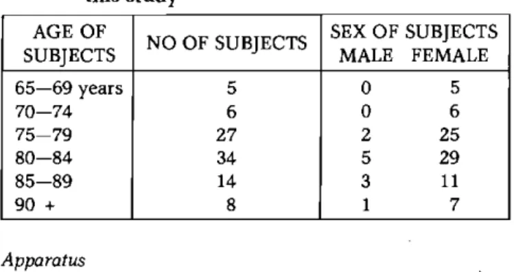
Ninety-four people from three old age homes in Pretoria were randomly selected for this study. The ages of persons in the experimental group varied between 66 and 95 years of age. There were 11 male subjects and 83 female subjects.
The age spread and sex of the subjects are presented in table 1.
Table 1: The age-spread and sex of subjects used for this study
AGE OF
SUBJECTS NO OF SUBJECTS SEX OF SUBJECTS MALE FEMALE
65—69 years 5 0 5
7 0 - 7 4 6 0 6
7 5 - 7 9 27 2 25
8 0 - 8 4 34 5 29
8 5 - 8 9 14 3 11
90 + 8 1 7
Apparatus
Acoustic immittance data were obtained on each subject with a calibrated (to standard -ISO 389, 1979) Grason Stadler GSI 28A Auto Tymp, utilizing a 226 Hz probe tone. Acoustic reflex measures were obtained at 500, 1000, 2000 and 4000 Hz.
Experimental procedure
Each subject tested underwent the following procedure: A short interview was carried out to obtain personal infor-mation, viz. name and date of birth.
A preliminary visual examination of the external auditory canal had to be carried out to determine whether immit-tance measurement could be obtained. Immitimmit-tance measurements cannot be carried out if fluid is running from the ear canal or if there is an obstruction (Meyer, Hurter and Van Rensburg, 1987).
The acoustic immittance protocol was then carried out. The probe was placed in the ear canal and an airtight seal was obtained. The test sequence proceeded automatically. The pressure sweep began at + 200 daPa and proceeded in a ne-gative direction until it reached -400 daPa. The ear canal volume in ml, immittance peak in ml, and the pressure peak in daPa were recorded (Margolis and Shanks, 1985). Acoustic reflex measures were then obtained ipsilaterally at 500, 1000, 2000 and 4000 Hz in dB HL (Hearing level), i
I I
An otoscopic examination was then carried out by a clinical assistant in otorhinolaryngology in order to determine the presence of any middle ear pathology.
/
Data analysis
Since all the acoustic immittance measures were carried out on the GSI 28A, the norms were used as set out in the in-struction manual.
— Static immittance: The normal range is 0,3 ml to approxi-mately 1,8 ml. A static immittance peak which falls be-tween these two ranges indicates normal mobility of the middle ear system.
— Pressure peak: For most applications a normal pressure range of -150 daPa to +100 daPa is used (Margolis and Shanks, 1985). However, strict rules for a normal middle The South African Journal of Communication Disorders, Vol. 35, 1988
ce
d
by
S
ab
in
et
G
at
ew
ay
u
nd
er
li
ce
nc
e
gr
an
te
d
by
th
e
Pu
bl
is
he
r (
da
te
d
20
12
ear pressure indicate a pressure range of -50 daPa to + 50 daPa (Brooks, 1981).
— Tympanometric shape:
— Type A tympanogram has static immittance values of be-tween 0,3 ml and 1,8 ml and has a pressure peak at or near 0 daPa although a range between -150 daPa and + 100 daPa is considered normal (Hodgson, 1980; Margo-lis and Shanks, 1985).
— Type Ad signifies a tympanogram with an unusually high static immittance peak of 1,8 ml or more. Peak pressure is usually at or near 0 daPa (Hodgson, 1980; Margolis and Shanks, 1985).
— Type As denotes a tympanogram with reduced amplitude of 0,2 ml or less. The pressure peak is usually at or near 0 daPa (Margolis and Shanks, 1985).
— Type Β tympanogram is flat, typified by the absence of a pressure peak (Hodgson, 1980; Margolis and Shanks, 1985).
— Type C tympanogram has a negative tympanometric peak pressure, usually smaller than -150 daPa (Hodgson, 1980, Margolis and Shanks, 19^5).
— Acoustic reflex measures: The reflex usually occurs be-tween 70 and 90 dB HL above the hearing threshold in normal;hearing people (Wiley and Block, 1985).
Data Processing
For the analysis of the data obtained in this study descrip-tive statistical techniques were used. The aim of descripdescrip-tive statistics is to sum up and condense the measurable charac-teristics of results obtained. The following techniques were used: percentages and frequency tables.
I
RESIJLTS j
/
/ · I
The results are presented in the sequence outlined accor-ding to the goals of this study.
The types.of -tympanograms which occurred
Table 2 presents a summary of the salient data obtained iq this study. It shows the number of subjects with normal acoustic immittance results bilaterally, with abnormal acoustic immittance results unilaterally and with abnormal immittance results bilaterally. The types of tympanograms which occurred' are indicated as well as the number of ears which presented each type of tympanogram. Table 2 also shows the various types of pathology which were diagnos-ed.
Of the 94 subjects tested 58 (62%) had Type A tympanograms bilaterally. Type A tympanograms are associated with normal middle ear function. This pattern reflects normal mobility and peak pressure (Hodgson, 1980). Twenty three of the sub-jects (24%) had abnormal results unilaterally and 13 subsub-jects
(14%) had abnormal results bilaterally. Therefore 38% of all the subjects tested had abnormal acoustic immittaijce results in one or both ears. These results are extremely high, compared with a study by Jerger (1976). Jerger found that the highest
in-cidence of abnormal tympanometric results (31%) occurred in the age group 2 to 5 years. It was suggested th^t tnis age group is the highest risk group for otitis media. Jerger found that there was a gradual decrease in abnormal results with an increase in age.
) With reference to the number of ears tested in this study 188 ears were examined in total. Of these 139 (74%) yielded Type A tympanograms. Of the remaining ears 38 (26%) presented Type As tympanograms. Type As denotes a tympanogram with a reduced amplitude characteristic of ossicular fixation, tympa-nosclerosis and some forms of otitis media (Margolis and Shanks, 1985). The Type As tympanogram therefore predomi-nates over the other abnormal tympanograms in this elderly group. This differs from findings in children where tympano-grams associated with otitis media, eg. Type Β and Type C tym-panograms, occur most frequently (Hodgson, 1980). The reason that the Type As tympanograms occurred more often in the elderly group could be due to the calcification and ossifica-tion of the joints between the ossicles in the middle ear (Kahane, 1981).
There were 3 Type Β tympanograms. Type Β tympanograms occur in the presence of middle ear effusion and other space-occupying lesions of the middle ear. They can also occur in cases of tympanic membrane perforation and impacted cerumen (Hodgson, 1980; Margolis and Shanks, 1985). Most of these pathologies associated with Type Β tympanograms can be remedied medically or surgically and therefore can be identi-fied and referred for further treatment (Turner, 1982). There were 5 ears with Type C tympanograms. This type of tympanogram is often an indication of Eustachian tube dys-function or otitis media (Hodgson, 1980; Margolis and Shanks, 1985).
Of all the ears tested tympanograms with no peak were record-ed in 3 ears. This could indicate a perforation or impactrecord-ed cerumen (Margolis and Shanks, 1985).
Correlation between the acoustic immittance results and the types of pathology diagnosed
The question as to why this high percentage of abnormal acoustic immittance results occurred was posed and thus the acoustic immittance results are discussed in terms of the different types of pathology which were identified. This is also presented in table 2.
Type A tympanograms
Of the 139 ears which yielded Type A tympanograms 96 were diagnosed as having normal middle ear function. Forty of the remaining ears with Type A tympanograms were diagnosed as having cerumen, ear canal collapse or both. Although cerumen and ear canal collapse can affect pure tone audiometric results (Wofford, 1981), they do not necessarily affect acoustic immit-tance results (Randolph and Schow, 1983; Wofford, 1981). This could explain why Type A tympanograms occurred.
A further 2 ears that presented a Type A tympanogram were diagnosed as having healed perforations in the tympanic mem-brane. A mildly scarred tympanic membrane can result in in-creased mobility in the tympanic membrane (Jerger, Anthony, Jerger and Mauldin, 1976). In these 2 cases it appears as though the healed tympanic membrane had not necessarily affected the tympanometric results.
The South African Journal of Communication Disorders, Vol. 35, 1988 .
ce
d
by
S
ab
in
et
G
at
ew
ay
u
nd
er
li
ce
nc
e
gr
an
te
d
by
th
e
Pu
bl
is
he
r (
da
te
d
20
12
Ό <u
a.
Si bO jS "o Λ <3 Ο, <υα
Si <υ Λ "Όα
<3 4) u 4) υα
<3 S <Λ 3 Ο υ < Ν Ο Ξ <3 Η Type s o f patholog y diagnose d Surger y o n Tym . Mem . 7—1 Type s o f patholog y diagnose d Otiti s Medi a7—1 Ν
Type s o f patholog y diagnose d Repaire d membran e
7—1 7—1
Type s o f patholog y diagnose d Cerume n + retracte d membran e 7—1 Type s o f patholog y diagnose d Perforatio n
- 7—1 τ—I
Type s o f patholog y diagnose d Cerume n + tympano -sclerosi s 7—1 Type s o f patholog y diagnose d Tympano -sclerosi s \
- 7—1
Type s o f patholog y diagnose d Cerume n + ea r cana l collaps e
Ν Ν CO τ—I
Type s o f patholog y diagnose d Ea r cana l collaps e <N τ—1
7—1 τ—I CO Ν
Cerume
n
impactio
n
CO - 7—1
Cerume
n
l£>
7—1 7—1 7—1 τ—1
Norma
l
Ο
00 l£> 7—1 CO
7—1 oo No . o f ear
s l£>
τ—1
τ—1
CO Ν oo
7—1
τ—1 CO -1 ο
Ν
Ν Ν CM
Typ e o f tympano -gra m Typ e A Typ e A Typ e A s Typ e Β Typ e C N o pea k Typ e A s Typ e Β Type C N o pea k No . o f subject s oo
in CO Ν
o-Norma l acousti c immittanc e result s bilaterall y Abnorma l acousti c immittanc e result s unilaterall y Abnorma l acousti c immittanc e result s bilaterall y
Die Suid-Afrikaanse Tydskrif vir Kommunikasieafwykings, Vol. 35, 1988
ce
d
by
S
ab
in
et
G
at
ew
ay
u
nd
er
li
ce
nc
e
gr
an
te
d
by
th
e
Pu
bl
is
he
r (
da
te
d
20
12
)
The Manifestation of Middle Ear Pathology in an Elderly Group One ear with a Type A tympanogram was diagnosed as having
otitis media. It should be noted that a peak pressure of -119 daPa was recorded. This could imply that the peak pres-sure norms that were used, viz. a prespres-sure range of -150 to + 100 daPa, are not sensitive enough to identify all cases of otitis media.
Type As tympanograms
Thirty eight of the ears tested yielded a Type As tympanogram. Of these 21 were diagnosed as having normal middle ear func-tion, 5 were diagnosed as having cerumen, 1 had impacted cerumen, 3 had ear canal collapses, 5 had both cerumen and ear canal collapses, 2 had tympanosclerosis and 1 ear had had surgery to the tympanic membrane.
The percentage of ears with Type As tympanograms that were diagnosed as normal is 55. This implies that factors other than pathology influenced the tympanometric results. Structural changes due to aging such as stiffening of the tympanic mem-brane and ossicular joints could have resulted in the Type As tympanogram (Wofford, 1981). The possibility that structural changes due to aging may have affected the tympanometric results, has important implications for the audiologist, as there will be a high percentage of abnormal tympanograms with no evidence of middle ear disorder. This implies that the occur-rence of a Type As tympanogram does not always indicate the presence of middle ear pathology among the elderly. Impacted cerumen can cause a Type As tympanogram (Margo-lis and Shanks, 1985). This accounts for the single case of cerumen impaction which yielded as Type As tympanogram. Ear canal collapse and cerumen do not necessarily influence the acoustic immittance results (Randolph and Schow, 1983; Wofford, 1981).
There were 13 ears with Type As tympanograms that were diagnosed as having cerumen, ear canal collapse or both. These tympanometric results could therefore be caused by factors other than pathology. Structural changes due to aging can /cause stiffening of the middle ear system, which could result in / Type As tympanograms.
There were also 2 ears which had tympanosclerosis and 1 case of surgery to the tympanic membrane that yielded Type As tympanograms. According to Margolis and Shanks (1985), both of these diagnoses could] result in a Type As tympanogram. Type Β tympanograms
There were 3 ears with Type Β tympanograms. One was diag-nosed as having a perforated tympanic membrane and the third had a retracted tympanic membrane and cerumen. All three of these types of pathology correlate with the acoustic im-mittance results (Margolis and Shanks, 1985). Referral for fur-ther medical attention is essential in these cases (Turner, 1982). Type C tympanograms
Of the 5 ears which yielded C tympanograms 2 had otitis media. The remaining 3 ears all had an ear canal collapse. An ear canal collapse cannot influence tympanometric results (Wofford, 1981). The type C tympanogram could therefore be caused by Eustachian tube dysfunction.
Tympanograms with no peak
Three ears with tympanograms which had no peak were diag-nosed. Perforated tympanic membranes were found in 2 of the ears. The third ear had cerumen and an ear canal collapse. No peak is registered in tympanometric measurement when the pressure that is required for the measurement of the middle ear function cannot be built up (Hodgson, 1980). Both the perfora-tions and the ear canal collapse could result in no pressure be-ing built up. There is therefore a correlation between the tym-panometric results and the presence of pathology.
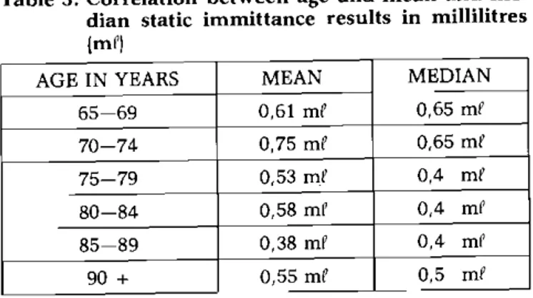
The effect of aging on static immittance values
In order to determine the effect of aging on the static immit-tance values a correlation was drawn between the two. Table 3 gives the mean and median static immittance values as a func-tion of age.
Table 3: Correlation between age and mean and me-dian static immittance results in millilitres
(mP)
AGE IN YEARS MEAN MEDIAN
65-69 0,61 mP 0,65 mP
7 0 - 7 4 0,75 mP 0,65 mP
75-79 0,53 mP 0,4 mP
8 0 - 8 4 0,58 mP 0,4 mP
85-89 0,38 mP 0,4 mP
90 + 0,55 mP 0,5 mP
The results show that there is a significant decrease in both the mean and median static immittance values in the sub-jects of 75 years and older. This decrease does not, however, continue with an increase in age. There is in fact, a slight in-crease in the static immittance values of subjects over 90 years of age. This tendency contrasts with the findings by Blood and Greenburg (1977). They found a significant de-crease in static immittance values. Their study examined subjects between the ages of 50 and 70 years. The fact that their population was considerably younger than the popula-tion used in this study may explain the resulting difference in findings.
The tendency for static immittance values to decrease slight-ly and then level off as was observed in this study, correlates with findings by Nerbonne, et al. (1978). They found a slight but non-significant tendency for values to decrease with age. Their subjects ranged in age from 20 to 79 years. They did recommend that further study of individuals over 79 years of age was necessary. This study examined subjects between the ages of 65 and 95 years, but the results did not differ from those found by Nerbonne, et al. (1978). A separate set of static immittance norms for the elderly is not neces-sary, as the mean and median values for all the age groups fall within the normal range of 0,3 mP — 1,8 mP.
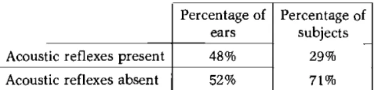
Acoustic reflex measures
Table 4 presents the percentage of subjects as well as the percentage of ears with absent acoustic reflex measures. Die Suid-Afrikaanse Tydskrif vir Kommunikasieafwykings, Vol. 35, 1988
ce
d
by
S
ab
in
et
G
at
ew
ay
u
nd
er
li
ce
nc
e
gr
an
te
d
by
th
e
Pu
bl
is
he
r (
da
te
d
20
12
Table 4: Percentage of subjects and ears with absent and present acoustic reflexes
Percentage of
ears Percentage of subjects
Acoustic reflexes present 48% 29%
Acoustic reflexes absent 52% 71%
The acoustic reflex is usually elicited at 70—90 dB HL above pure tone threshold. The most likely cause of the absent re-flexes is an increased pure tone threshold. Most elderly peo-ple have a sensory neural hearing loss due to presbycusis. Since pure tone audiometric testing was not carried out, the exact cause of the absent acoustic reflexes is unknown. The possibility exists that the absent reflexes could also be the result of the degeneration of the middle ear muscles. All the muscles in the body degenerate with increasing age. The muscles in the middle ear should not be an exception.
DISCUSSION OF RESULTS
The manifestations of outer and middle ear pathology Outer and middle ear pathology occurred in 38% of all the ears tested. This high occurrence can partially be explained as a function of increased age.
The type of pathology which occurred most frequently was excessive cerumen which may obstruct the external auditory canal partially or completely (Cohn, 1981). Copious secretion of wax occurs in individuals of all ages; Marshall (1985), however, indicates that excessive cerumen is more common among older people. An overaccumulation of cerumen can result in a conductive hearing loss and ab-normal acoustic immittance results, depending on the de-gree of obstruction (Wofford, 1981). Excessive cerumen can also influence ear mould impressions for hearing aids. Twenty two percent of all the ears tested were diagnosed as having cerumen, and of these, 64% had Type A tympano-grams. The acoustic immittance results indicate that even a total occlusion of the ear canal due to cerumen impaction does not always result in abnormal tympanograms. The paction may, however, influence the results, and this im-plies that an otoscopic examination should always be car-ried out prior to acoustic immittance testing in an elderly group. Excessive cerumen can be treated quickly and suc-cessfully.
Ear canal collapse or stenosis occurred in 17% of all the ears which were examined. Although ear canal collapse can oc-cur in any age group, it is usually associated with old age which causes a loss of elasticity in the dermis. The cartilagi-nous portion also becomes more flexible (Wofford, 1981). The external auditory, canal closes when an earphone is placed over it; this can result in a mild to moderate conduc-tive hearing loss (Randolph and Schow, 1983). A Type A tympanogram can occur with reduced ear canal volume measurements. This correlates with the findings of this study where 64% of the ears diagnosed as manifesting an ear canal collapse had Type A tympanograms. The remaining 36% had additional factors which affected the results. In order to prevent incorrect interpretation of audiometric test-ing, otoscopy and tympanometry should always be per-formed prior to audiometric testing. A collapse of an ear canal during audiometric testing can be prevented by using circuaural or postaural cushions (Marshall and Grossman, 1982).
There were 3 cases of otitis media diagnosed and 1 case of retracted tympanic membrane, indicative of an early stage of otitis media (Cohn, 1981). The development of otitis media is most frequently related to Eustachian tube dysfunction. The Eustachian tube maintains middle ear ventilation and facilitates the clearing of foreign material as well as pro-viding immunological defence (Cohn, 1981). Eustachian tube dysfunction is often associated with old age because the veli palatini muscles which open the Eustachian tube may degenerate or atrophy. Kahane (1981), says that degeneration of all the muscles occurs with old age. Otitis media may therefore be caused indirectly by increased age. However, it is also possible that these subjects have a history of otitis media, and thus the condition is not as a result of old age. Otitis media cannot always be detected through pure tone audiometry. It can yield a conductive hearing loss. Otoscopy and acoustic immittance testing can detect the presence of otitis media. As mentioned previous-ly, a Type Β or C tympanogram usually occurs (Hodgson,
1980) and this correlates with the tympanometric results ob-tained in this study. It is therefore essential that these ex-aminations be carried out as part of the test battery for the elderly. Otitis media is therefore not exclusively associated with the very young, but can also occur among the elderly. There were 3 ears with perforated tympanic membranes. Tympanic membrane perforation can result from excessive effusion, the erosive effect of middle ear lesions such as cholesteatomas or an external trauma (Wofford, 1981). In the 3 cases diagnosed in this study, the most likely cause was excessive effusion. A perforation may affect the audio-metric results, depending on the size of the perforation (Wofford, 1981).
A perforation can be identified through otoscopy and acoustic immittance measurement. If the 3 cases identified in this study had perforations due to excessive effusion, the possibility exists that the otitis media was caused, by a Eustachian tube dysfunction. The Eustachian tube dysfunc-tion, as mentioned earlier, could be the result of increased age.
Tympanosclerosis and healed perforations occurred in 4 ears. Tympanosclerosis and healed perforations may result after tympanic membrane rupture and healing (Wofford, 1981). Tympanosclerosis can include stiffening of the tym-panic membrane, tymtym-panic mucosa, ossicular ligaments1,
tendons of the stapedius and tensor tympani muscles and fixing the malleus andincus in the epitympanic area. A scar·! red tympanic membrane is indicative of a healed perfora-; tion. The most likfely cause of a healed perforation is
ef-fusive otitis media. - j It is essential that the persdn with evidence of a healed per-j
foration undergoes regular ,'otoscopic examinations and1
acoustic immittance testing to detect the recurrence of otitis 1
media. / /
It is evident; therefore, that much of the pathology which occurs in' elderly individuals could be as a result of in-creased age. Otoscopy and acoustic immittance testing are
essential for the diagnosis of most of these disorders and must therefore be carried out prior to pure tone audiometric testing. Repeated testing is necessary to detect the recur-rence of otitis media. The nursing staff who work in old age homes could also be trained to identify symptoms related to outer and middle ear disorders for early identification and referral.
The effect of structural changes due to aging on, the acoustic immittance measurements
Eleven percent of abnormal acoustic immittance results The South African Journal of Communication Disorders, Vol. 35, 1988
ce
d
by
S
ab
in
et
G
at
ew
ay
u
nd
er
li
ce
nc
e
gr
an
te
d
by
th
e
Pu
bl
is
he
r (
da
te
d
20
12
were diagnosed as having no middle ear disorders. This im-plies that there are factors other than a middle ear pathology which resulted in the abnormal acoustic immittance results. One of the possible causes of these abnormal results could be attributed to structural changes to the middle ear system. Kahane (1981), noted that the structure of the adult laryn-geal cartilages changes with increasing age. Calcification or ossification caused stiffening of the laryngeal joints. These changes do not only occur in the larynx, but occur in all the joints of the body including those in the middle ear. It seems possible, therefore, that a stiffening of the middle ear system could cause abnormal acoustic immittance results. Fifty five percent of the Type As tympanograms which occurred had a diagnosis of normal middle ear condition.
Abnormal results could also have been caused by a history of pathological conditions which are no longer evident, but which have caused a permanent effect on the middle ear system.
Since biological changes are known to occur with increasing age, it is unlikely that the middle ear should remain unaf-fected. These non-pathological changes which influence acoustic immittance measures have important implications for the audiologist. There will thus be a percentage of ab-normal tympanograms with no middle ear disorders in the testing of the elderly. Although this percentage is not high enough to warrant separate static immittance or tympano-metric norms for the elderly, it nevertheless should be taken into consideration. >
It is also of interest that the static immittance values decreased in subjects over 75 years of age. This decrease did not continue with an increase in age. In addition, these values increased slightly in the age group over 90 years. The mean and median static immittance values of each age group fell within the normal range, i.e. 0,3 ml — 1,8 ml. CONCLUSION \
In this group of elderly subjects, acoustic immittance results indicating a normal middle ear system (Type A tympano-gram) occurred most frequently. Of these normal acoustic immittance results, 43 earsjwere, however, medically diag-nosed as having excessive cerumen, ear canal collapse, heal-ed perforations or otitis mheal-edia. These diagnoses do not ne-cessarily influence acoustic immittance measurements. Thirty-eight percent of the subjects had abnormal tympano-grams in one or both ears. Of these tympanotympano-grams indicat-ing middle ear dysfunction; eight ears presented with Type Β or C tympanograms or had no peak, confirming the medi-cal diagnosis of conditions such as otitis media. Two of the ears had Type C tympanograms but there was otoscopically no indication of middle ear pathology. These results could indicate a Eustachian tube dysfunction possibly caused by degeneration of the veli-palatini muscles.
Of the abnormal tympanograms, Type As, indicating reduc-ed amplitude, occurrreduc-ed most often. This was otoscopically verified in 45% of the cases where a diagnosis of pathology such as tympanosclerosis or impacted cerumen occurred. However, 55% of these Type As tympanograms had no evidence of middle ear disorder. These results could there-fore be due to unidentified causes such as non-pathological changes, associated with old age, which lead to a stiffening of the middle ear system.
The fact that there was a correlation between the decrease in static immittance values and an increase in age up to 90 years of age, seems to indicate that aging had an effect on the results.
Otoscopy and acoustic immittance measures should be car-ried out as part of the test battery for the elderly. Identifica-tion of pathology, which may influence further audiometric testing, eg, ear canal collapse, is essential as the effect of these disorders on audiometric testing can be prevented. Middle ear pathology increases the total hearing loss in the elderly. There is a high incidence of sensory neural hearing loss in the aged. The conductive component of the hearing loss can be remedied medically or surgically. The audiolo-gist therefore has an important role to play in the identi-fication ot these conductive components.
The last few decades have seen a rapid increase in the number of people over 65 years. The elderly can no longer be ignored, but must be recognised for the contribution they make to society. There is a shift in the orientation of the field of audiology towards the evaluation and rehabilitation of the elderly. The audiologist plays an important role in the identification and referral of individuals who have patholo-gical conditions in the outer and middle ear.
REFERENCES
Blood, I. & Greenburg, H. Acoustic admittance of the ear in the geriatric person. Journal of the American Audiological Society, 2, 185—187, 1977.
Brooks, D. Impedance measurement in the elderly. In R. Penha & P. de Ν Pizarro (Eds.) Proceedings of the fourth international
symposium in acoustic impedance measurement. Lisbon:
Lissa-bon Universidade, Nova de Lisboa, 1981.
Chermack, G. Handbook of clinical audiological rehabilitation. Illi-nois: Charles C Thomas, 1981.
Cilliers, N.J. The manifestation of middle ear pathology in an elderly
group. Unpublished B.(Log.) dissertation, University of
Pretoria, 1987.
Cohn, A. Etiology and pathology of disorders affecting hearing. In F.N. Martin (Ed.) Medical Audiology Disorders of Hearing. New Jersey: Prentice-Hall, Inc., 1981.
Gelfand, S. & Piper, N. Acoustic reflex thresholds in young and elderly subjects with normal hearing. Journal of the
Acousti-cal Society of America, 69, 295-297, 1981.
Hodgson, W.R. Basic Audiological Evaluation. Baltimore: Williams and Wilken Co., 1980.
Jerger, J.F. Clinical experience with impedance audiometry. In J.L. Northern (Ed.) Selected Readings in Impedance Audiometry. New York: American Electromedics Corporation, 1976. Jerger, J.F., Anthony, L., Jerger, S. & Mauldin, L. Studies in
impe-dance audiometry III. Middle ear disorders. In J.L. Northern (Ed.) Selected Readings in Impedance Audiometry. New York: American Electromedics Corporation, 1976.
Jerger, J.F., Jerger, S. & Mauldin, L. Studies in impedance audio-metry I. Normal and sensori-neural ears. In J.L. Northern (Ed.) Selected Readings in Impedance Audiometry. New York: American Electromedics Corporation, 1976.
Kahane, J.C. Anatomic and physiological changes in the aging peri-pheral speech mechanism. In D.S. Beasley and G.A. Davis
(eds.). Aging Communication Processes and Disorders. New York: Grune & Stratton, 1981. ' / Margolis, R.H. & Shanks, J.E. Tympanometry. In J. Katz (Ed.)
Hand-book of Clinical Audiology. Baltimore: Williaps and Wilken
Co., 1985.
Marshall, L. Audiological assessment of older adults. Seminars in
Hearing, 6, 2, 161-175, 1985.
Marshall, L. & Grossman, M.A. Management of ear canal collapse.
Archives of Otolaryngology, 108, 357-361, 1982.
Meyer, S., Hurter, M. & Van Rensburg, F. Gehoorsiftingsresultate van 'n groep kleurlingleerlinge. The South African Journal of
Communication Disorders, 34, 43—47, 1987.
Meyerhoff, W.L. & Paparella, W.M. Diagnosing the cause of hear-ing loss. Geriatrics, 33, 95—98, 1978.
Nerbonne, M., Bliss, A. & Schow, R.L. Acoustic impedance values in the elderly. Journal of the American Audiotory Society, 4, 57-59, 1978.
Randolph, L.J. & Schow, R.L. Threshold inaccuracies in an elderly clinical population: ear canal collapse as a possible cause.
Journal of Speech and Hearing Research. 26, 54—58, 1983.
Die Suid-Afrikaanse Tydskrif vir Kommunikasieafwykings, Vol 35, 1988
ce
d
by
S
ab
in
et
G
at
ew
ay
u
nd
er
li
ce
nc
e
gr
an
te
d
by
th
e
Pu
bl
is
he
r (
da
te
d
20
12
Turner, J.S. Treatment of hearing loss, ear pain and tinnitus in older patients. Geriatrics, 37, 107-118, 1982.
Wiley, T.L. & Block, M.G. Overview and basic principles of acous-tic immittance measurements. In J. Katz (Ed.) Handbook of
Clinical Audiology. Baltimore: Williams & Wilkens Co., 1985.
Wofford, M. Audiological evaluation and management of hearing disorders. In F.N. Martin (Ed.) Medical Audiology Disorders of
Hearing. New Jersey: Prentice-Hall Inc., 1981.
Talking to Professionals
I'hc Needier Westdene Organisation
P.O. Box 28975 Sandrinaham 2131
Telephone (Oil) 485-1302/3/4/5
TECHNIMARK 560
The South A frican Journal of Communication Disorders, Vol. 35, 1988