Serum Albumin as a Carrier of Immunostimulatory Oligodeoxynucleotides for Cancer Therapy
Patrick C. Guley
A dissertation submitted to the faculty of the University of North Carolina at Chapel Hill in partial fulfillment of the requirements for the degree of Doctor of Philosophy in the School of Pharmacy.
Chapel Hill 2013
ABSTRACT
PATRICK C. GULEY: Serum Albumin as a Carrier of Immunostimulatory Oligonucleotides for Cancer Therapy
(Under the direction of Moo J. Cho, Ph.D.)
Toll-like receptor 9 (TLR9) is an endosomal receptor expressed on immune cells. The receptor recognizes microbial DNA, which contains a higher frequency of unmethylated CpG sequences than host DNA. Oligodeoxynucleotides containing such CpG motifs (CpG) are potent activators of TLR9, causing release of inflammatory cytokines and initiating both the innate and adaptive immune response. CpG has been successfully used to treat solid tumors, but its use is limited by its unfavorable pharmacokinetics. In preclinical studies, CpG was effective only when injected peritumorally; i.e., ineffective when administered systemically. In an attempt to overcome this limitation, CpG was derivatized with a maleimide moiety to allow a chemical reaction with free thiols. The derivative is referred to as CpG-mal throughout this dissertation.
iii
between the CpG-mal and albumin was fast enough to outcompete the high renal clearance of CpG, with the reaction complete within minutes. The new CpG-albumin conjugate displayed a similar distribution pattern and plasma half-life as albumin; half-life was increased 70-fold and tumor accumulation was increased 30-fold over control CpG.
In vitro plasma stability studies showed that albumin conjugation lead to a 1.5-fold reduction in the rate of enzymatic degradation of phosphodiester CpG. An in vitro macrophage activation assay indicated that phosphodiester CpG-albumin conjugates were weak agonists of TLR9. However, phosphorothioate CpG-albumin adducts were able to interact with TLR9 to initiate cytokine release from J774 macrophage cells, although this activity was reduced compared to control phosphorothioate CpG. The activation was independent of crosslinker length, and introducing a reducible disulfide crosslinkage did not enhance the activity. Pharmacokinetics and biodistribution of phosphorothioate CpG were measured using [3H]-labeled CpG. The [3H]-CpG-mal had a longer plasma half-life than control CpG, however, the liver accumulation was significantly increased. This liver uptake led to a less striking increase in tumor accumulation of CpG-mal than control CpG.
v
ACKNOWLEDGEMENTS
No man is an island; and none of this would have been possible without a considerable amount of direct and indirect assistance by many people. Obviously, my advisor Dr. Cho has had a huge impact on my training and development as a scientist and an intellectual (I use the word liberally). He gave me the freedom to experiment which often times lead to failure but he was always offering encouragement and challenging me to do better. My only hope is that the time spent was not a one-way street.
I would like to thank the members of my committee for providing insight and assistance with designing experiments. Additionally, I want to thank all the professors in the School of Pharmacy for teaching me diverse knowledge in the field of pharmaceutics. The staff and administrative assistants (Kathryn, Kim, Jubina, Ning, Rod, Amber, and Ain) deserve praise for making the bureaucratic red tape manageable and enabling quick ordering of supplies.
I would like to thank the other graduate students and research scientists in the School of Pharmacy for the many discussions and friendships we’ve had. I would like to thank Feng Liu for letting me use his cell hood and Yang Liu for helping me on short notice with some final experiments. I especially want to thank former Cho lab members: Kevin Han, Kayla Knilans, Roland Cheung, John An, Brad Gustafson, and June Lee. Tip of the hat to Shyam Joolakankti for teaching me tips and tricks of chemical synthesis. I particularly want to acknowledge my friendship with Michael Hackett, we’ve had plenty of deep scientific and philosophical conversations in addition to the many memories outside of lab.
I would like to thank my parents for their support during the many trials and tribulations I encountered during the past few years and most importantly for providing the many educational opportunities that led me here.
I am indebted to my wife, Natalie, whom I met during my first year of graduate school. She is the constant balancing force in my life and has provided unconditional positive support. I can’t wait for the birth of our first daughter in the coming weeks.
vii
TABLE OF CONTENTS
ABSTRACT ... ii
ACKNOWLEDGEMENTS ...v
LIST OF TABLES ...x
LIST OF FIGURES ... xi
LIST OF ABBREVIATIONS AND SYMBOLS ... xiii
CHAPTER I: INTRODUCTION ...1
1.1 Cancer and Immunotherapy ... 1
1.2 Biological Response Modifiers ... 4
1.3 CpG Oligodeoxynucleotides ... 6
1.4 Serum Albumin ... 8
1.5 Albumin and Cancer ... 11
1.6 Proposed Studies ... 12
REFERENCES ...15
CHAPTER II: SYNTHESIS AND APPLICATION OF A HETEROTRIFUNCTIONAL CROSSLINKER FOR 124 I-BASED PET IMAGING...20
2.1 Overview ... 20
2.2 Introduction ... 20
2.3 Experimental Procedures ... 22
2.3.1 Crosslinker Synthesis ... 22
2.3.2 ODN Chemistry ... 25
2.3.3 Pharmacokinetic Experiments ... 27
2.4 Results ... 29
2.4.1 Crosslinker Synthesis ... 29
2.4.2 ODN Preparation and Iodination ... 30
2.5 Discussion ... 31
REFERENCES ...38
CHAPTER III: PHARMACOKINETICS/BIODISTRIBUTION AND PHARMACODYNAMICS OF MALEMIDE DERIVATIZED OLIGODEOXYNUCLEOTIDES WITH PHOSPHODIESTER BACKBONE IN TUMOR BEARING MICE ...41
3.1 Overview ... 41
3.2 Introduction ... 41
3.3 Experimental Procedures ... 43
3.3.1 CpG ODN Chemistry ... 43
3.3.2 Pharmacokinetics and Biodistribution ... 45
3.3.3 In Vitro Plasma Stability ... 48
3.3.4 In Vitro Macrophage Activation ... 50
3.4 Results ... 51
3.4.1 In Vitro Plasma Stability ... 51
3.4.2 Plasma Pharmacokinetics... 52
3.4.3 Biodistribution and Tumor Accumulation ... 52
3.4.4 4T1 Tumor Growth Study ... 53
3.4.5 In Vitro Cytokine Release ... 53
3.5 Discussion ... 54
REFERENCES ...63
CHAPTER IV: PHARMACOKINETICS/BIODISTRIBUTION AND PHARMACODYNAMICS OF MALEMIDE-DERIVATIZED OLIGODEOXYNUCLEOTIDES WITH PHOSPHOROTHIOATE BACKBONE IN TUMOR-BEARING MICE...65
4.1 Overview ... 65
4.2 Introduction ... 65
4.3 Experimental Procedures ... 67
4.3.1 In Vitro Cytokine Release ... 69
4.3.2 Pharmacokinetic and Biodistribution Study ... 71
4.3.3 CT26 Tumor Growth Study ... 72
ix
4.4 Results ... 73
4.4.1 In Vitro Macrophage Activation ... 73
4.4.2 Pharmacokinetics and Biodistribution ... 74
4.4.3 Tumor Growth Studies ... 74
4.5 Discussion ... 75
REFERENCES ...88
CHAPTER V: CONCLUSIONS & FUTURE DIRECTION ...91
REFERENCES ...98
APPENDIX A ...100
LIST OF TABLES
xi
LIST OF FIGURES
Figure 1.1. The cellular mechanisms of immune activation by CpG ODN. ...14
Scheme 2.1. Synthetic scheme for the synthesis of the heterotrifunctional crosslinker used throughout this study. ...34
Figure 2.2. Chemical structures of the modified ODN synthesized for the study. ...35
Figure 2.3. Normalized whole body PET/CT images of biodistribution of the 124I radiolabeled treatments after 20 min post injection. ...36
Figure 2.4. Time activity curves for the blood and urine during 1 h post injection. ...37
Figure 3.1. In vitro plasma stability of PO [3H]-CpG. ...56
Figure 3.2. Blood concentration of 124I labeled CpG. ...57
Figure 3.3. Tumor time activity curve showing tumor uptake of 124I labeled CpG. ...59
Figure 3.4. Terminal biodistribution of 124I-labeled CpG measured by ex vivo gamma counting. ...60
Figure 3.5. Survival after surgical resection of 4T1 primary tumor. ...61
Figure 3.6. In vitro IL-12 and IL-6 release from J444 cells. ...62
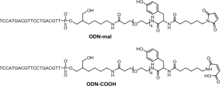
Figure 4.1. Chemical structures and synthetic scheme for the synthesis of CpG-mal and CpG-COOH...79
Figure 4.2. In vitro IL-12 and IL-6 release from CpG-albumin conjugates. ...80
Figure 4.3. Blood concentration of [3H]-CpG. ...81
Figure 4.4. Tumor accumulation of PS [3H]-CpG...83
Figure 4.5. Biodistribution of [3H]-labeled CpG. ...84
Figure 4.6. Tumor growth inhibition of 4T1 tumors. ...85
Figure 4.7. CT26 tumor growth curves. ...86
Figure A.1. 1H NMR spectrum of Mal-Tyr(tBu)-OtBu (1)...101
Figure A.2. 13C NMR of Mal-Tyr(tBu)-OtBu (1) ...102
Figure A.3. 1H NMR of Mal-Tyr (2) ...103
Figure A.4. 13C NMR of Mal-Tyr (2)...104
Figure A.5. Mass Spectrum of Mal-Tyr (2) ...105
Figure A.6. 1H NMR spectrum of Mal-Tyr-TEG-COOH (3) ...106
Figure A.7. 13C NMR spectrum of Mal-Tyr-TEG-COOH (3) ...107
Figure A.8. Mass spectrum of Mal-Tyr-TEG-COOH (3) ...108
Figure A.9. 1H NMR of Mal-Tyr-TEG-NHS (4) ...109
Figure A.10. 13C NMR of Mal-Tyr-TEG-NHS (4) ...110
Figure A.11. Mass spectrum of Mal-Tyr-TEG-NHS (4) ...111
xiii
LIST OF ABBREVIATIONS AND SYMBOLS
ACN Acetonitrile
AcOH Acetic Acid
APC Antigen presenting cell
BD Biodistribution
BSA Bovine Serum albumin
CpG Cytidine-phosphate-Guanosine CT Computer assisted tomography DCC N,N’-Dicyclohexylcarbodiimide
DCM Dichloromethane
DIEA N,N-Diisopropylethylamine
DNP 2,4 Dinitrophenol
EDC N-(3-Dimethylaminopropyl)-N′-ethylcarbodiimide EDTA Ethylenediaminetetraacetic acid
ELISA Enzyme Linked Immunosorbent Assay EMCS N-(ε-Maleimidocaproyloxy)succinimide EtOAc Ethyl acetate
EtOH Ethanol
g Gravitational constant
HPLC High Performance Liquid Chromatography
HSA Human Serum Albumin
IL-12 Interleukin 12
iv intravenous
LPS Lipopolysaccharide
mal maleimide
MeOH methanol
MSA Mouse Serum albumin
NEM N-ethylmaleimide
NHS N-hydroxysuccinimide
NK Natural Killer cell ODN Oligodeoxynucleotide
PAMP Pathogen-associated molecular pattern PBS Phosphate Buffered Saline
PD Pharmacodynamics
PET Positron Emission Tomography
PK Pharmacokinetics
PO Phosphodiester
PS Phosphorothioate
sc subcutaneous
TEA Triethylamine
TEAA Triethylammonium acetate TFA Trifluroacetic acid
THF Tetrahydrofuran
xv TLR9 Toll-like receptor 9
TMS Tetramethyl silane
CHAPTER I: INTRODUCTION
For every minute of 2013, it is predicted that one United States citizen will succumb to cancer.(Siegel, Naishadham et al., 2013) The total direct and indirect economic impact of cancer-related healthcare in the United States for 2008 totaled $200 billion and will undoubtedly rise in the future.(Society, 2013) Society needs better treatments for cancer, especially for younger patients. Ideally, these treatments should be as cost effective as possible. One potential way to reduce the cost of therapy and increase the effectiveness is to utilize existing natural defense mechanisms to our advantage.
1.1 Cancer and Immunotherapy
Current medical understanding of the pathophysiology of cancer suggests we are constantly under assault from cancer causing agents, be they ultraviolent rays from the sun, chemical carcinogens, radiation exposure, reactive oxygen species, or viral and bacterial infections. Any of these agents can cause the genetic mutations that transform normal cells into cancer cells. However, as these mutations occur, the human body is not constantly developing clinical tumors. This implicates the existence of a controlling mechanism. The cancer-controlling mechanism largely consists of two components: an intracellular and an extracellular mechanism.
2
Hickson, 2001) The majority of transformed cells are thought to be repaired or eliminated by these intracellular mechanisms. Some cells can accumulate mutations in these controlling genes and are rendered neoplastic.
The extracellular phase is known as immunosurveillance and, more recently, immunoediting(Dunn, Old et al., 2004); the immune system is able to differentiate and destroy these abnormal cells before they develop into a tumor. A number of experimental studies using genetically-engineered knockout mice with incomplete immune systems have an increase in tumor incidence. (Shankaran, Ikeda et al., 2001; Enzler, Gillessen et al., 2003; Street, Hayakawa et al., 2004) Additionally, it is known that humans with immunodeficiencies have a greater chance of developing tumors.(Rabkin, Biggar et al., 1991; Grulich, van Leeuwen et al., 2007) In some instances, abnormal cells are able to escape this control mechanism and their unchecked growth leads to cancer pathology. The exact nature of how some cells escape the immunoediting mechanism is not currently well understood. One school of thought involves a selective proliferation of non-immunogenic cells lacking specific tumor antigens. The consensus view of tumor immunobiology is that the lack of immune response is not merely due to an absence of immune cells in the vicinity of the tumor. In fact, most tumors contain large amounts of immune cells.(Lin and Pollard, 2004) Rather, the issue appears to be that the cells have been anergized and no longer function as active immune cells. They have been regulated by a sub population of T cells commonly known as Treg and other immunosurpessive cells that tell them that the tumor is not a threat.(Wolf, Wolf et al., 2003)
in the late 1890s by Dr. William Coley, who recognized that patients who suffered from bacterial infections showed a regression in tumor size. He hypothesized that bacteria was the cause of tumors, and could be treated by injection of bacterial extracts. His extracts contained two strains of bacteria and were injected in or around the tumor. As a result, the patients would become febrile and develop flu-like symptoms. In cases that responded positively, the tumors would quickly undergo liquefactive necrosis and begin to dissipate. Repeated injections over the span of multiple months provided complete remission for his patients who initially had a positive response. In patients that did not respond to the initial treatment, subsequent treatments were also ineffective, and these patients were given different therapies.
Cancer vaccines are another type of heavily investigated- and thus heavily invested- immunotherapy. This approach appears flawed because, by their very nature, cancers are heterogeneous and thus do not express the same antigens across the population. The second issue with cancer vaccines is that clinicians have no way of knowing which antigens are present without a biopsy. Additionally, tumors contain a heterogeneous population of cells. A single cancer vaccine can target a single antigen, but will not affect the tumors cells which do not express the specific antigen. This would require a multi-vaccine approach, which further increases cost and feasibility issues. Finally, from a philosophical standpoint, it is questionable whether researchers can select an appropriate set of antigens in vitro against tumors better than the patient’s own immune system in vivo.
4
1.2 Biological Response Modifiers
Humans have evolved in intimate contact with bacteria. Our immune systems have thus developed methods to detect and limit the threat bacteria pose to our survival. A consequence of this natural progression is a system to detect the conserved components of bacteria that renders virtually any type of bacteria to be identifiable by the immune system. These conserved components are called pathogen-associated molecular patterns, or PAMPs. They include components from gram negative cell walls such as lipopolysaccharides (LPS), DNA and RNA, also flagella and other membrane-associated proteins.(Kawai and Akira, 2010) Some of the receptors that are responsible for detecting these are the Toll-like receptor (TLR) family. When these PAMPs are detected by a TLR, the immune system becomes activated and primed for further activity.
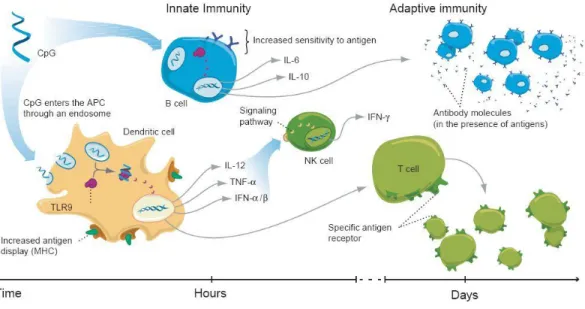
Unmethylated CpG is recognized by toll-like receptor 9 (TLR9). It is an intracellular membrane receptor expressed almost exclusively on immune cells. TLR9 is thought to exist as a homodimer, and the majority is found in the endoplasmic reticulum.(Latz, Verma et al., 2007) When cells are exposed to CpG, TLR9 is localized to the endosomal membranes by a currently unknown mechanism,(Latz, Schoenemeyer et al., 2004) whereby proteolytic cleavage coverts the TLR9 into its active form.(Park, Brinkmann et al., 2008) Upon CpG binding, the active form undergoes a conformational change and transmembrane binding of MyD88 occurs.(Latz, Verma et al., 2007) MyD88 signaling eventually leads to activation of NF-κB and release of inflammatory cytokines. These cytokines are able to initiate an innate immune response by activating macrophages and natural killer (NK) cells and also an adaptive response by dendritic cell activation of CD8+ T cells and activation of B cells (Figure 1.1).(Krieg, 2003)
6
1.3 CpG Oligodeoxynucleotides
CpG oligodeoxynucleotides (ODN) are short 6-23 nucleotide DNA molecules that bind to TLR9 and can mimic the effects of bacterial DNA.(Krieg, Yi et al., 1995) Random screening showed the immune response to CpG ODN was sensitive to sequences flanking the CpG: the optimal sequence found for mice was GACGTT, whereas for larger animals, including humans and primates, the optimal sequence was GTCGTT.(Rankin, Pontarollo et al., 2001)
Our bodies are not accustomed to having DNA exist outside the cells and have mechanisms to remove this potential danger. Extracellular nucleases can rapidly cleave DNA into nucleotides; 5’- or 3’-exonucleases cleave DNA at the terminal ends while endonucleases can cleave anywhere along the strand. The majority of nucleases present extracellularly and in plasma are 3’-exonucleases and to a lesser extent endonucleases and 5’-exonucleases. ODN that have a natural phosphodiester (PO) backbone are rapidly metabolized by these nucleases and have poor pharmacokinetics (PK). This instability limits their therapeutic potential.
albumin with a kd ~ 50-300 µM.(Srinivasan, Tewary et al., 1995) This binding is not very tight as evidenced by the rapid distribution of ODN when injected into mice.(Sands, Gorey-Feret et al., 1994) In the physiological setting, PS ODN have affinities to other targets which seem to be collectively greater than that of albumin.
CpG monotherapy has been used in the preclinical setting to treat solid tumors. The general consensus is that the CpG must be injected near the site of the tumor to elicit a response.(Heckelsmiller, Rall et al., 2002; Nierkens, den Brok et al., 2009) If it is injected systemically, the response is not sufficient enough to have clinical effects. This is no doubt a consequence of the poor PK of ODN. When they are injected systemically, only a small percentage of the dose arrives at the tumor, whereas if the dose is injected near the tumor, a higher amount of the drug is in the tumor vicinity before it diffuses out. This suggests delivery to the tumor is a limiting step in the therapeutic potential of systemically-injected ODN.
Our lab has previously shown that IgG antibodies can be used as a carrier for CpG to increase its systemic effectiveness.(Palma and Cho, 2007) CpG was derivatized with a 2,4-Dintriophenol (DNP) hapten and were injected into mice that had been previously immunized against DNP and therefore expressed a high anti-DNP IgG titer in their circulation. The anti-DNP IgG were able to form monomeric immune conjugates with the DNP-CpG and increase the plasma half-life and tumor accumulation. Consequently, the DNP-CpG was considerably more efficacious than underivatized CpG in tumor suppression.
8
the amount of IgG present.(Cheung and Cho, 2010) The average concentration of IgG in normal adults is 12 mg/ml. This corresponds to the total amount of IgG for all antigens, but only a small fraction of the IgG will be specific for a single antigen. This could be overcome by creating an active immune response prior to CpG-hapten therapy, but that would require extra time and more procedures. Because of these limitations, we decided to investigate the potential of using a different endogenous carrier for CpG ODN that would be abundant and accessible.
1.4 Serum Albumin
The most abundant protein in our blood is serum albumin. It has a molecular weight of 67 kDa and bears a net -19 charge at physiological pH. In humans, the plasma concentration is approximately 40 mg/ml and the interstitial concentration is approximately 20 mg/ml. This concentration difference gives rise to oncotic pressure which balances blood pressure and potentiates osmotic pressure. Albumin has several other functions in the body: it acts as a carrier for fatty acids, bilirubin, and numerous other endogenous ligands.(Peters, 1996) It also provides binding sites for numerous insoluble therapeutic agents. The amount of albumin in the average human is approximately 350 g, and its half-life in healthy individuals is estimated to be approximately 20 days.
preventing unwarranted catabolism, a recycling receptor, FcRn, is expressed on endothelial cells and other phagocytic cells.(Akilesh, Christianson et al., 2007) FcRn has a higher affinity for albumin at low pH, such as that found in the endosome, and lower affinity at neutral pH. This pH-dependent binding is related to protonation of His residues of FcRn.(Andersen, Dee Qian et al., 2006; Andersen and Sandlie, 2009) This allows the albumin to be recovered from the endosome and recycled back to the surface of the cell, and is responsible for the longer half-life that albumin and IgG have compared to other serum proteins.(Chaudhury, Mehnaz et al., 2003)
There are several other receptors involved in the transport of albumin. Albondin, or gp60, is an endothelial surface protein that can transcytose albumin into the extravascular space.(Schnitzer and Oh, 1994) Other scavenger receptors, gp30 and gp18, have a higher affinity for modified or degraded albumin and lead to degradation, primarily in the liver, rather than transcytosis.(Schnitzer, Sung et al., 1992)
The half-life and distribution of albumin scales with species weight: in mice, the halflife is 1.2 d, rat 2.5 d, rabbits 5.7 d, dogs 8 d, and humans 20 d.(Allison, 1960) Albumin plasma profiles, when introduced via bolus injection, exhibit biphasic behavior. They are indicative of an initial distribution to tissues followed by a slower clearance phase. In humans, this distribution phase takes approximately 3 days to complete and the concentration drops to approximately 40% of the initial value.(Peters, 1996)
10
nm, which approximates the size of albumin and as a result, normal vasculature can restrict the transport of albumin out of the vessels via hydrodynamic sieving.(Rippe, Rosengren et al., 2002) The restriction is not a complete barrier to albumin escape but is sufficient to generate and maintain an albumin concentration gradient across the vessel wall. It is not clearly established if the intercellular leakage implied by Starlings equation is, in fact, the albondin-mediated transcytosis which has since been discovered.
Albumin has 35 Cys residues; 34 of these are engaged in disulfides leaving one free thiol, Cys34. Cys34, albeit located within a hydrophobic cleft of the albumin molecule, is accessible to the surrounding solvent and accounts for up to 85% of the total free thiol content of blood.(Kratz, Warnecke et al., 2002) Approximately 25% of Cys34 exists as a disulfide with small molecular weight thiols such as cysteine or glutathione attached. Despite being located on a large molecule, Cys34 is quite reactive because its pKa~5-6 is much lower than the pKa~8 for most thiols. It thus exists as thiolate anion at physiological pH,(Kratz, Warnecke et al., 2002) much more nucleophilic than a neutral free thiol group.
Several studies have investigated the potential of using albumin as a carrier by modifying drugs with maleimide groups.(Elsadek and Kratz, 2012) When the maleimide-containing drugs are injected into animals they react covalently with Cys34 within minutes.(Kratz, Warnecke et al., 2002) Due to this fast rate, it has been possible to inject maleimide-modified drugs and have them react with circulating albumin without appreciable loss. While a 1:1 conjugate may seem like a poor loading capacity, it avoids the polyvalenancy which is known provoke an immune response and generate antibodies.(Singh, Kaur et al., 2004) This may be even more important when using an immune-activating drug. Additionally, the more that albumin is modified, the more it is recognized as non-native and is subject to rapid clearance.(Stehle, Sinn et al., 1997)
1.5 Albumin and Cancer
12
nutrients to sustain its growth.(Stehle, Sinn et al., 1997) This leads to a loss of body weight and low concentration of serum proteins.
1.6 Proposed Studies
Collectively, those facts and findings introduced above indicate that serum albumin could be used as a carrier of CpG ODN. The proposed sequence of events that may happen in order to lead to a response can be described as follows: (i) upon intravenous injection, the majority of maleimide-modified CpG ODN will covalently bind to albumin thereby limiting their rapid clearance and distribution; (ii) the CpG-albumin conjugate will behave similarly to albumin and will have increased circulation half-life; (iii) the CpG-albumin will extravasate from the tumor endothelium near the tumor periphery to a higher extent due to local increased permeability; (iv) the CpG-albumin will be taken up by highly phagocytic cells, such as macrophages and dendritic cells, in the tumor vicinity; (v) CpG-albumin will bind to TLR9 and cause upregulation of cytokine release, NK cell activation, and silencing of immunosuppressive Treg cells; (vi) antigen-presenting cells (APCs) will sample tumor antigen and become activated, (vii) APCs will travel to lymph nodes to initiate clonal expansion of CD8+ T cells; (viii) CD8+ T cells will infiltrate the tumor and begin to destroy the tumor cells; and (ix) any distant metastases can be identified by the lasting immune response. This scenario appears to be wholly consistent with the scientific knowledge well established thus far and represents the scope of this dissertation.
14
REFERENCES
Akilesh, S., G. J. Christianson, D. C. Roopenian and A. S. Shaw (2007). "Neonatal FcR expression in bone marrow-derived cells functions to protect serum IgG from catabolism." J Immunol 179(7): 4580-4588.
Allison, A. C. (1960). "Turnovers of erythrocytes and plasma proteins in mammals." Nature
188: 37-40.
Andersen, J. T., J. Dee Qian and I. Sandlie (2006). "The conserved histidine 166 residue of the human neonatal Fc receptor heavy chain is critical for the pH-dependent binding to albumin." Eur J Immunol 36(11): 3044-3051.
Andersen, J. T. and I. Sandlie (2009). "The versatile MHC class I-related FcRn protects IgG and albumin from degradation: implications for development of new diagnostics and therapeutics." Drug Metab Pharmacokinet 24(4): 318-332.
Bird, A. P. (1986). "CpG-rich islands and the function of DNA methylation." Nature
321(6067): 209-213.
Chaudhury, C., S. Mehnaz, J. M. Robinson, W. L. Hayton, D. K. Pearl, D. C. Roopenian and C. L. Anderson (2003). "The major histocompatibility complex-related Fc receptor for IgG (FcRn) binds albumin and prolongs its lifespan." J Exp Med 197(3): 315-322. Cheung, R. and M. Cho (2010). "Importance of avidity for an endogenous drug carrier: an
antibody carrier for CpG oligonucleotides." Mol Pharm 7(4): 1338-1341. Crooke, S. T. (1992). "therapeutic applications of oligonucleotides."
Dunn, G. P., L. J. Old and R. D. Schreiber (2004). "The three Es of cancer immunoediting." Annu Rev Immunol 22: 329-360.
Elsadek, B. and F. Kratz (2012). "Impact of albumin on drug delivery - New applications on the horizon." J Control Release 157(1): 4-28.
Enzler, T., S. Gillessen, J. P. Manis, D. Ferguson, J. Fleming, F. W. Alt, M. Mihm and G. Dranoff (2003). "Deficiencies of GM-CSF and interferon gamma link inflammation and cancer." J Exp Med 197(9): 1213-1219.
16
Grulich, A. E., M. T. van Leeuwen, M. O. Falster and C. M. Vajdic (2007). "Incidence of cancers in people with HIV/AIDS compared with immunosuppressed transplant recipients: a meta-analysis." The Lancet 370(9581): 59-67.
Gupta, G. K. and D. K. Agrawal (2010). "CpG oligodeoxynucleotides as TLR9 agonists: therapeutic application in allergy and asthma." BioDrugs 24(4): 225-235.
Hashizume, H., P. Baluk, S. Morikawa, J. W. McLean, G. Thurston, S. Roberge, R. K. Jain and D. M. McDonald (2000). "Openings between defective endothelial cells explain tumor vessel leakiness." Am J Pathol 156(4): 1363-1380.
Heckelsmiller, K., K. Rall, S. Beck, A. Schlamp, J. Seiderer, B. Jahrsdorfer, A. Krug, S. Rothenfusser, S. Endres and G. Hartmann (2002). "Peritumoral CpG DNA elicits a coordinated response of CD8 T cells and innate effectors to cure established tumors in a murine colon carcinoma model." J Immunol 169(7): 3892-3899.
Jain, R. K. and T. Stylianopoulos (2010). "Delivering nanomedicine to solid tumors." Nat Rev Clin Oncol 7(11): 653-664.
Kawai, T. and S. Akira (2010). "The role of pattern-recognition receptors in innate immunity: update on Toll-like receptors." Nat Immunol 11(5): 373-384.
Kratz, F., A. Warnecke, K. Scheuermann, C. Stockmar, J. Schwab, P. Lazar, P. Druckes, N. Esser, J. Drevs, D. Rognan, C. Bissantz, C. Hinderling, G. Folkers, I. Fichtner and C. Unger (2002). "Probing the cysteine-34 position of endogenous serum albumin with thiol-binding doxorubicin derivatives. Improved efficacy of an acid-sensitive
doxorubicin derivative with specific albumin-binding properties compared to that of the parent compound." J Med Chem 45(25): 5523-5533.
Krieg, A. M. (2003). "CpG motifs: the active ingredient in bacterial extracts?" Nat Med 9(7): 831-835.
Krieg, A. M., T. Wu, R. Weeratna, S. M. Efler, L. Love-Homan, L. Yang, A. K. Yi, D. Short and H. L. Davis (1998). "Sequence motifs in adenoviral DNA block immune
activation by stimulatory CpG motifs." Proc Natl Acad Sci U S A 95(21): 12631-12636.
Krieg, A. M., A. K. Yi, S. Matson, T. J. Waldschmidt, G. A. Bishop, R. Teasdale, G. A. Koretzky and D. M. Klinman (1995). "CpG motifs in bacterial DNA trigger direct B-cell activation." Nature 374(6522): 546-549.
Latz, E., A. Schoenemeyer, A. Visintin, K. A. Fitzgerald, B. G. Monks, C. F. Knetter, E. Lien, N. J. Nilsen, T. Espevik and D. T. Golenbock (2004). "TLR9 signals after translocating from the ER to CpG DNA in the lysosome." Nat Immunol 5(2): 190-198.
"Ligand-induced conformational changes allosterically activate Toll-like receptor 9." Nat Immunol 8(7): 772-779.
Lin, E. Y. and J. W. Pollard (2004). "Role of infiltrated leucocytes in tumour growth and spread." Br J Cancer 90(11): 2053-2058.
Matsumura, Y. and H. Maeda (1986). "A new concept for macromolecular therapeutics in cancer chemotherapy: mechanism of tumoritropic accumulation of proteins and the antitumor agent smancs." Cancer Res 46(12 Pt 1): 6387-6392.
Nierkens, S., M. H. den Brok, T. Roelofsen, J. A. Wagenaars, C. G. Figdor, T. J. Ruers and G. J. Adema (2009). "Route of administration of the TLR9 agonist CpG critically determines the efficacy of cancer immunotherapy in mice." PLoS One 4(12): e8368. Norbury, C. J. and I. D. Hickson (2001). "Cellular responses to DNA damage." Annu Rev
Pharmacol Toxicol 41: 367-401.
Palma, E. and M. J. Cho (2007). "Improved systemic pharmacokinetics, biodistribution, and antitumor activity of CpG oligodeoxynucleotides complexed to endogenous
antibodies in vivo." J Control Release 120(1-2): 95-103.
Park, B., M. M. Brinkmann, E. Spooner, C. C. Lee, Y. M. Kim and H. L. Ploegh (2008). "Proteolytic cleavage in an endolysosomal compartment is required for activation of Toll-like receptor 9." Nat Immunol 9(12): 1407-1414.
Peters, T. (1996). All about albumin : biochemistry, genetics, and medical applications. San Diego, Academic Press.
Rabkin, C. S., R. J. Biggar and J. W. Horm (1991). "Increasing incidence of cancers associated with the human immunodeficiency virus epidemic." Int J Cancer 47(5): 692-696.
Rankin, R., R. Pontarollo, X. Ioannou, A. M. Krieg, R. Hecker, L. A. Babiuk, S. van Drunen and L. V. van den Hurk (2001). "CpG motif identification for veterinary and
laboratory species demonstrates that sequence recognition is highly conserved." Antisense Nucleic Acid Drug Dev 11(5): 333-340.
Rippe, B., B. I. Rosengren, O. Carlsson and D. Venturoli (2002). "Transendothelial transport: the vesicle controversy." J Vasc Res 39(5): 375-390.
Sands, H., L. J. Gorey-Feret, A. J. Cocuzza, F. W. Hobbs, D. Chidester and G. L. Trainor (1994). "Biodistribution and metabolism of internally 3H-labeled oligonucleotides. I. Comparison of a phosphodiester and a phosphorothioate." Mol Pharmacol 45(5): 932-943.
18
Schnitzer, J. E., A. Sung, R. Horvat and J. Bravo (1992). "Preferential interaction of albumin-binding proteins, gp30 and gp18, with conformationally modified albumins. Presence in many cells and tissues with a possible role in catabolism." J Biol Chem 267(34): 24544-24553.
Shankaran, V., H. Ikeda, A. T. Bruce, J. M. White, P. E. Swanson, L. J. Old and R. D.
Schreiber (2001). "IFNgamma and lymphocytes prevent primary tumour development and shape tumour immunogenicity." Nature 410(6832): 1107-1111.
Siegel, R., D. Naishadham and A. Jemal (2013). "Cancer statistics, 2013." CA Cancer J Clin
63(1): 11-30.
Singh, K. V., J. Kaur, G. C. Varshney, M. Raje and C. R. Suri (2004). "Synthesis and characterization of hapten-protein conjugates for antibody production against small molecules." Bioconjug Chem 15(1): 168-173.
Society, A. C. (2013). "Cancer Facts & Figures 2013." Atlanta: American Cancer Society. Srinivasan, S. K., H. K. Tewary and P. L. Iversen (1995). "Characterization of binding sites,
extent of binding, and drug interactions of oligonucleotides with albumin." Antisense Res Dev 5(2): 131-139.
Stehle, G., H. Sinn, A. Wunder, H. H. Schrenk, S. Schutt, W. Maier-Borst and D. L. Heene (1997). "The loading rate determines tumor targeting properties of methotrexate-albumin conjugates in rats." Anticancer Drugs 8(7): 677-685.
Stehle, G., H. Sinn, A. Wunder, H. H. Schrenk, J. C. M. Stewart, G. Hartung, W. MaierBorst and D. L. Heene (1997). "Plasma protein (albumin) catabolism by the tumor itself - implications for tumor metabolism and the genesis of cachexia." Critical Reviews in Oncology/Hematology 26(2): 77-100.
Street, S. E., Y. Hayakawa, Y. Zhan, A. M. Lew, D. MacGregor, A. M. Jamieson, A. Diefenbach, H. Yagita, D. I. Godfrey and M. J. Smyth (2004). "Innate immune surveillance of spontaneous B cell lymphomas by natural killer cells and gammadelta T cells." J Exp Med 199(6): 879-884.
Williams, D. L., T. Ha, C. Li, J. H. Kalbfleisch, J. J. Laffan and D. A. Ferguson (1999). "Inhibiting early activation of tissue nuclear factor-κB and nuclear factor interleukin 6 with (1→ 3)-β-d-glucan increases long-term survival in polymicrobial sepsis."
Surgery 126(1): 54-65.
Wolf, A. M., D. Wolf, M. Steurer, G. Gastl, E. Gunsilius and B. Grubeck-Loebenstein (2003). "Increase of regulatory T cells in the peripheral blood of cancer patients." Clin Cancer Res 9(2): 606-612.
Yuan, F., M. Dellian, D. Fukumura, M. Leunig, D. A. Berk, V. P. Torchilin and R. K. Jain (1995). "Vascular permeability in a human tumor xenograft: molecular size
CHAPTER II: SYNTHESIS AND APPLICATION OF A
HETEROTRIFUNCTIONAL CROSSLINKER FOR 124I-BASED PET
IMAGING
2.1 Overview
An efficient method for radioiodinating drug-carrier conjugates where the site of iodination is contained within the crosslinker has been developed. A heterotrifunctional crosslinker was synthesized with terminal maleimide and N-hydroxysuccinimide ester groups for conjugation to cargo and a centrally incorporated Tyr residue to allow facile labeling with 124
I. The crosslinker was applied to amino-modified oligodeoxynucleotides (ODN) in order to measure in vivo conjugation to Cys34 of circulating serum albumin. It was found the in situ reaction was complete within minutes and proceeded quickly enough to dramatically alter the clearance and distribution of the ODN. This labeling strategy could be used as a way to introduce any isotope of radioiodine to various drug-carrier combinations bearing the requisite functional groups.
2.2 Introduction
2011) Additionally, since each animal can provide information at multiple time points, there is a net reduction in the total number of experimental animals needed to obtain an equivalent amount of information. Finally, non-invasive imaging can be translated to a clinical setting. However, current imaging systems require a radioisotope as a label for detection. Incorporation of the desired label into the therapeutic may not be trivial, as often there is not an accessible labeling functionality.
Considerable research is being performed using macromolecular carriers and nanoparticles to enhance the therapeutic potential of drugs, particularly for targeting drugs to solid tumors.(Bae and Park, 2011) In many cases this involves crosslinking drug molecules to larger carrier molecules. In this study, a labeling moiety is incorporated into the crosslinker itself, rendering radiolabeling of the conjugate independent of the drug or carrier. The purpose of these experiments was to investigate the potential of using serum albumin as carrier of ODN. Other groups have shown that modifying therapeutics with a maleimide functional group allows them to covalently react with the Cys34 of circulating albumin.(Elsadek and Kratz, 2012) This approach has been extensively applied to small molecule anticancer drugs(Chung and Kratz, 2006; Kratz, 2007) and various peptides(Leger, Thibaudeau et al., 2004; Thibaudeau, Leger et al., 2005; Xie, Yao et al., 2010). The present work was designed to test whether this approach would also work for ODN and to specifically address whether the in situ reaction was fast enough to prevent the ODN from rapid distribution and excretion.(Lau, Graham et al., 2012)
22
applicable for measuring PK/BD at longer biological t1/2. Iodine has several radioactive isotopes and the 124I isotope (t1/2 = 4.2 d; β+ = 22%) can be used for PET imaging. In spite of some limitations(Pentlow, Graham et al., 1996), 124I is successfully quantified.
2.3 Experimental Procedures
All chemicals, except where noted, were purchased from EMD Sciences or Sigma Aldrich and were ACS reagent grade or higher.
2.3.1 Crosslinker Synthesis (Scheme 2.1)
1. Mal-Tyr(tBu)-OtBu
δ 174.4, 171.2, 154.2, 134.9, 134.2, 133.5, 132.2, 130.2, 129.3, 124.7, 124.2, 123.3, 37.2, 35.3, 28.1, 27.2, 27.1, 26.1, 25.2.
2. Mal-Tyr
To a 25-mL round bottom flask was added 650 mg of 1 and 8 mL of 50% trifluoroacetic acid in DCM. After 10 h at 25°C, the solvent was evaporated under a stream of N2 and the product was recrystallized from acetone/DCM to give 425 mg (86%) of slight yellow crystals. mp: 161-163°C. 1H NMR (400 MHz, (CD3)2CO): δ 1.25 (m, 2H, – CH2CH2CH2–), 1.5-1.6 (m, 4H, –CH2CH2CH2–), 2.2 (t, 2H, –CH2C=O), 2.9-3.1 (m, 2H, CHCH2–), 3.44 (t, 2H, NCH2–), 4.7 (q, 1H, CHCH2–), 6.7 (d, 2H, Ar–H,m), 6.81 (s, 2H, – CH=CH–), 7.08 (d, 2H, Ar–H,o), 7.32 (d, 1H, CONH–). 13C NMR (100 MHz, CD3OD): δ 174.6, 173.8, 171.4, 156.1, 134.8, 134.0, 133.4, 130.4, 129.6, 127.9, 115.6, 115.2, 114.8, 114.4, 37.2, 35.3, 28.0, 25.9, 25.1. ESI-MS (neg, MeOH): m/z 373.1 [M – H]-
3. Mal-Tyr-TEG-COOH
24
acid in 1:9 methanol:DCM to give 465 mg (70%) of a yellow oil. 1H NMR (400 MHz, CDCl3): δ 1.2 (m, 2H, –CH2CH2CH2–), 1.5-1.6 (m, 4H, –CH2CH2CH2–), 2.2 (t, 2H, – CH2C=O), 2.6 (t, 2H, –CH2CH2CO–), 2.9 (m, 2H, CHCH2–), 3.2-3.7 (m, 18H, – CH2CH2O–), 3.79 (t, 2H, NCH2–), 4.68 (q, 1H, CHCH2–), 6.71 (s, 2H, –CH=CH–), 6.76 (d, 2H, Ar–H,m), 7.01 (d, 2H, Ar–H,o), 7.06 (d, 1H, –CONHCH–) 7.38 (t, 1H, –CONHCH2– ). 13C NMR (100 MHz, CDCl3): δ 174.8, 173.5, 172.0, 171.1, 155.8, 134.4, 134.1, 130.6, 130.2, 127.6, 115.8, 115.6, 70.5 70.1, 66.8, 37.8, 36.2, 35.4, 28.5, 26.4, 25.2. ESI-MS (neg, MeOH): m/z 620.2 [M – H]-
4. Mal-Tyr-TEG-NHS
2.3.2 ODN Chemistry
The ODN used in all experiments was a 20mer purchased from either Integrated DNA Technologies (Coralville, IA) or from Girindus America, Inc. (Cincinnati, OH) with a phosphodiester backbone, unless specifically stated. They were supplied as the Na+ salt form. The sequence was TCCATGACGTTCCTGACGTT and contained a commercially available 3’-amino modification.
HPLC Conditions
All HPLC analysis and purification was performed using Shimadzu SCL-10A system controller with two Shimadzu LC-8A pumps connected to a Rainin Dynamax UV-C detector and a Shimadzu C-R6A Chromatopac recorder. Solvent A was 5% acetonitrile in 10 mM triethylammonium acetate buffer; solvent B was 100% acetonitrile. For analytical work an Agilent Zorbax 300SB-C18 4.6 x 150mm analytical column with 5 µm particle size and a total flow rate of 1.0 mL/min was used with the following gradient: t = 0-5 min, %B = 0; t = 5-30 min, %B = 0-25; t = 30-33 min, %B = 25-100. For purification work the same gradient protocol was used with an Agilent Zorbax 300SB-C18 9.4 x 250mm semi-preparative column and a total flow rate of 4 mL/min. All detection was performed at λ = 260 nm.
ODN-mal
26
evaporated in vacuo after addition of excess 3 M sodium acetate to acidify the pH to 5.2. The ODN was then ethanol precipitated from 0.3M sodium acetate at -20°C to give 7 mg (56%) of ODN-mal as a Na+ salt. ESI-MS (neg, H2O) 7194.4 [M], 7211.8 [M + NH4].
ODN-COOH
To a microcentrifuge tube containing 900 µg of ODN-mal was added 200 µL of 50 mM NaOH. After 4 h at 37ºC the ODN was ethanol precipitated from 0.3 M sodium acetate to give 840 µg (93%) of ODN-COOH as a Na+ salt.
Murine Mercaptalbumin
Mouse serum albumin (MSA) Fraction V was purchased from MP Biomedicals (Solon, OH). Reaction with Ellman’s reagent indicated that the free thiol content of this albumin ranged from 0.2-0.3 mol of thiol per mol of MSA.(Janatova, Fuller et al., 1968) Mercaptalbumin was generated by the addition of 3 molar equivalents of DL-Dithiothreitol and incubation for 5 min at 25°C.(Funk, Li et al., 2010) The unquenched reaction was directly applied to a Sephadex® G-25 size exclusion column equilibrated with phosphate buffered saline (PBS). The unretained fractions containing MSA, as measured by UV, were pooled and Ellman’s test indicated the thiol content was 0.9-1.0 mol of thiol per mol of MSA. The MSA was further purified by mini-Q strong anion exchange spin columns (Pierce, Rockford, IL) and eluted with PBS to generate mercaptalbumin in a manner similar to methods previously reported.(Janatova, Fuller et al., 1968)
Radiolabeled ODN-mal was added to 8 equivalents of mercaptalbumin in PBS for 2 h at 25°C. The reaction mixture was loaded onto Q strong anion exchange spin columns and eluted with increasing stepwise NaCl gradient in 20 mM phosphate buffer, pH = 7.4. Unreacted MSA was eluted with 300 mM NaCl and the conjugate was eluted with 400 mM NaCl. The buffer was exchanged to PBS by repetitive ultracentrifugation using 30 kDa molecular weight cut off (MWCO) filters.
2.3.3 Pharmacokinetic Experiments
Iodination of ODN and MSA
The ODN were iodinated using pre-coated Iodogen® tubes (Pierce, Rockford, IL). Na124I was purchased from IBA Molecular (Richmond, VA). The Na124I needed to be regenerated to in order obtain reliable yields. The regeneration process consisted of calculated addition of stock solution containing 1 mg/mL of NaI and 1 mg/mL of NaIO3 in 1 mM NaOH.(Verel, Visser et al., 2004) The general procedure was as follows: The ODN to be labeled was dissolved in 100 µL of 100 mM sodium phosphate buffer at pH 7.4. To a pre-rinsed iodination tube, added were Na124I and a calculated amount of regeneration stock containing 0.9 mol equivalent of total iodine relative to ODN. After 1 min the ODN was added to the tube and the reaction was allowed to progress for 6 min with periodic gentle shaking. The unquenched reaction was directly applied to a Sephadex® G-25 size exclusion column equilibrated with PBS. The fractions containing ODN, as measured by UV, were pooled and concentrated using ultracentrifugation with 3 kDa MWCO filters. MSA was labeled using a similar procedure.
28
All animals were handled in accordance with an approved protocol by UNC Institutional Animal Care and Use Committee. 4T1 cells were purchased from ATCC (Manassas, VA) and grown according to ATCC recommendations. Eight female Balb/c mice, 18-20 g, were orthotopically inoculated with 1 x 10⁵ 4T1 cells in 50 µL PBS by subcutaneous injection into the mammary fad pad. The mice were randomly divided into four groups and imaging experiments began 15 days after tumor inoculation.
PET Image Acquisition
One day prior and throughout the imaging experiments mice were supplied ad libitum drinking water supplemented with 0.1% KI to block thyroid uptake of labeled 124I.(Verel, Visser et al., 2004) All animals were anesthetized using isoflurane and catheterized via tail vein. For each scan, two mice were placed on a cardboard platform on the scanning bed of a GE VISTA eXplore scanner and secured with surgical tape. A heart and breathing rate probe was used to monitor vitals while scanning. The mice were first imaged with a CT scan and then were dynamically imaged with PET for 1 h. The animals were injected with 0.2-0.3 mCi of 124I labeled material corresponding to 100 µg of ODN in 100 µL of sterile 0.22 µm filtered PBS and the catheters were flushed with a minimal volume of normal saline. The amount of activity remaining in the catheter and syringe was measured using a calibrated dose calorimeter (Capintec CRC®-25R, Ramsey, NJ) and subtracted from the initial amount to quantify the amount of injected activity.
Image Processing
were reconstructed using an attenuation correction, scatter correction, and 2D OSEM projection using the supplied manufacturer software (MMWKS Image Software, Laboratorio de Imagen HGUGM, Spain). The images were then loaded into AMIDE for analysis.(Loening and Gambhir, 2003) The images were aligned using fiducial markers placed below the scanning bed. Three-dimensional regions of interest (ROI) were manually drawn around the heart and bladder using the CT images. The amount of PET signal contained within a ROI was calculated and converted to percent of injected dose per mL (%ID/mL) using appropriate conversions to correct for time decay and a cylindrical phantom of known activity.
2.4 Results
2.4.1 Crosslinker Synthesis
30
2.4.2 ODN Preparation and Iodination
The crosslinker 4 was conjugated to ODN-NH2 using standard conditions.(Hermanson, 1996) Non-thiol reactive control ODN-COOH was synthesized from ODN-mal by hydrolysis of the maleimide group under basic conditions.(El-Sagheer, Cheong et al., 2011) The structures of the ODN are shown in Figure 2.2. Thiol reactivity was monitored using a mercaptohexanol HPLC shift assay; ODN-mal underwent a reaction as evidenced by an increase in peak retention time, whereas ODN-COOH showed no change in peak retention time when treated with mercaptohexanol.
Conjugation with the crosslinker enabled the ODN to be iodinated with 124I using commercially available pre-coated Iodogen® tubes in yields ranging from 80-90%. In a control experiment, ODN-NH2 was modified with a crosslinker which did not contain a Tyr residue. These ODN were not iodinated with the same iodination protocol; yields <0.5%. This result suggests the labeling was specific to the Tyr and not due to non-specific labeling of the ODN bases which can occur at elevated temperatures and extended labeling times.(Piatyszek, Jarmolowski et al., 1988)
2.4.3 Image Analysis
PET/CT whole body images, Figure 2.3, show this difference in initial distribution of the different treatments. MSA and MSA-ODN are restricted to the vasculature volume and show high heart signal, whereas the ODN-COOH is rapidly cleared and distributed showing low heart signal. ODN-mal shows a combination of these two patterns.
The heart is classically assumed to be highly perfused with blood; therefore blood concentration was approximated by total heart concentration.(Bading, Horling et al., 2008) The concentrations were dose-normalized and expressed as %ID/mL.
ODN typically have a PK profile that can be characterized by rapid elimination from the plasma(Sands, Gorey-Feret et al., 1994), which is similar to the profile of ODN-COOH in
Figure 2.4. On the other hand, ODN-mal shows a different plasma profile. Initially there is a steep drop, similar to ODN-COOH, which then transitions to a much slower plasma clearance. The transition appears to be complete after approximately 8 min. To confirm Cys34 of albumin is the major reaction product, ODN-mal was preconjugated to MSA ex vivo prior to injection. This curve did not display a rapid plasma clearance indicating there was no significant free ODN contamination. However, there was a delayed disappearance from the plasma and into the bladder. MSA was used as a control in order to determine if there was a difference between the ODN-MSA conjugates and native albumin. Both the MSA and ODN-MSA appear to be restricted to vascular space and do not undergo a rapid distribution.
2.5 Discussion
32
with the oxidation.(Xie, Liang et al., 2012) This observation shows the crosslinker requires compatibility with the drug or carrier and cannot be used indiscriminately. In this case, no attempt was made to iodinate the crosslinker prior to conjugation with the phosphorothioate ODN-NH2.
The cutoff for kidney filtration is approximately 40 kDa and continuous capillary endothelial gaps are approximately 4 nm (Rippe, Rosengren et al., 2002), therefore, there is no a priori reason to suspect any difference in renal clearance and distribution between ODN-COOH and ODN-mal. Immediately upon injection and prior to any thiol reaction, these two ODN should have similar disposition. However, the ODN-mal is able to undergo thiol addition which has the potential to alter disposition. The ODN-mal blood curve should follow the ODN-COOH curve at early timepoints and begin to transition to a new curve as the thiol addition reaction occurs. The majority of free thiol content in the blood is due to Cys34 of albumin(Kratz, Warnecke et al., 2002), therefore, this new curve should behave similar to albumin. The expected biphasic nature of the ODN-mal blood curve is clearly observed in Figure 2.4.
The blood and urine were not directly analyzed for stability of the label or integrity of the conjugate. In preliminary experiments where the mice were not given KI-supplemented drinking water to block thyroid uptake, the amount of thyroid uptake was greater for labeled MSA compared to the ODN-mal (data not shown). Additionally, labeled MSA was excreted predominantly through the urine, whereas all other treatments containing the radiolabeled crosslinker were excreted in the both urine and feces. Taken together these suggest the crosslinker iodination has greater in vivo dehalogenation stability than labeled protein tyrosines, which is in agreement with other reports of prosthetic iodinations.(Vaidyanathan and Zalutsky, 1990)
34
36
A B
C D
38
REFERENCES
Bading, J. R., M. Horling, L. E. Williams, D. Colcher, A. Raubitschek and S. E. Strand (2008). "Quantitative serial imaging of an 124I anti-CEA monoclonal antibody in tumor-bearing mice." Cancer Biother Radiopharm 23(4): 399-409.
Bae, Y. H. and K. Park (2011). "Targeted drug delivery to tumors: myths, reality and possibility." J Control Release 153(3): 198-205.
Chung, D. E. and F. Kratz (2006). "Development of a novel albumin-binding prodrug that is cleaved by urokinase-type-plasminogen activator (uPA)." Bioorg Med Chem Lett
16(19): 5157-5163.
El-Sagheer, A. H., V. V. Cheong and T. Brown (2011). "Rapid chemical ligation of oligonucleotides by the Diels-Alder reaction." Org Biomol Chem 9(1): 232-235. Elsadek, B. and F. Kratz (2012). "Impact of albumin on drug delivery - New applications on
the horizon." J Control Release 157(1): 4-28.
Funk, W. E., H. Li, A. T. Iavarone, E. R. Williams, J. Riby and S. M. Rappaport (2010). "Enrichment of cysteinyl adducts of human serum albumin." Anal Biochem 400(1): 61-68.
Hermanson, G. T. (1996). Bioconjugate techniques. San Diego, Academic Press.
Janatova, J., J. K. Fuller and M. J. Hunter (1968). "The heterogeneity of bovine albumin with respect to sulfhydryl and dimer content." J Biol Chem 243(13): 3612-3622.
Kratz, F. (2007). "DOXO-EMCH (INNO-206): the first albumin-binding prodrug of doxorubicin to enter clinical trials." Expert Opin Investig Drugs 16(6): 855-866. Kratz, F., A. Warnecke, K. Scheuermann, C. Stockmar, J. Schwab, P. Lazar, P. Druckes, N.
Esser, J. Drevs, D. Rognan, C. Bissantz, C. Hinderling, G. Folkers, I. Fichtner and C. Unger (2002). "Probing the cysteine-34 position of endogenous serum albumin with thiol-binding doxorubicin derivatives. Improved efficacy of an acid-sensitive doxorubicin derivative with specific albumin-binding properties compared to that of the parent compound." J Med Chem 45(25): 5523-5533.
Leger, R., K. Thibaudeau, M. Robitaille, O. Quraishi, P. van Wyk, N. Bousquet-Gagnon, J. Carette, J. P. Castaigne and D. P. Bridon (2004). "Identification of CJC-1131-albumin bioconjugate as a stable and bioactive GLP-1(7-36) analog." Bioorg Med Chem Lett
14(17): 4395-4398.
Loening, A. M. and S. S. Gambhir (2003). "AMIDE: a free software tool for multimodality medical image analysis." Mol Imaging 2(3): 131-137.
Pentlow, K. S., M. C. Graham, R. M. Lambrecht, F. Daghighian, S. L. Bacharach, B. Bendriem, R. D. Finn, K. Jordan, H. Kalaigian, J. S. Karp, W. R. Robeson and S. M. Larson (1996). "Quantitative imaging of iodine-124 with PET." J Nucl Med 37(9): 1557-1562.
Piatyszek, M. A., A. Jarmolowski and J. Augustyniak (1988). "Iodo-Gen-mediated radioiodination of nucleic acids." Anal Biochem 172(2): 356-359.
Rippe, B., B. I. Rosengren, O. Carlsson and D. Venturoli (2002). "Transendothelial transport: the vesicle controversy." J Vasc Res 39(5): 375-390.
Rygh, C. B., S. Qin, J. W. Seo, L. M. Mahakian, H. Zhang, R. Adamson, J. Q. Chen, A. D. Borowsky, R. D. Cardiff, R. K. Reed, F. R. Curry and K. W. Ferrara (2011). "Longitudinal investigation of permeability and distribution of macromolecules in mouse malignant transformation using PET." Clin Cancer Res 17(3): 550-559.
Sands, H., L. J. Gorey-Feret, A. J. Cocuzza, F. W. Hobbs, D. Chidester and G. L. Trainor (1994). "Biodistribution and metabolism of internally 3H-labeled oligonucleotides. I. Comparison of a phosphodiester and a phosphorothioate." Mol Pharmacol 45(5): 932-943.
Schnitzer, J. E. and J. Bravo (1993). "High affinity binding, endocytosis, and degradation of conformationally modified albumins. Potential role of gp30 and gp18 as novel scavenger receptors." J Biol Chem 268(10): 7562-7570.
Thibaudeau, K., R. Leger, X. Huang, M. Robitaille, O. Quraishi, C. Soucy, N. Bousquet-Gagnon, P. van Wyk, V. Paradis, J. P. Castaigne and D. Bridon (2005). "Synthesis and evaluation of insulin-human serum albumin conjugates." Bioconjug Chem 16(4): 1000-1008.
Vaidyanathan, G. and M. R. Zalutsky (1990). "Protein radiohalogenation: observations on the design of N-succinimidyl ester acylation agents." Bioconjug Chem 1(4): 269-273. Verel, I., G. W. Visser, M. J. Vosjan, R. Finn, R. Boellaard and G. A. van Dongen (2004).
40
Xie, D., C. Yao, L. Wang, W. Min, J. Xu, J. Xiao, M. Huang, B. Chen, B. Liu, X. Li and H. Jiang (2010). "An albumin-conjugated peptide exhibits potent anti-HIV activity and long in vivo half-life." Antimicrob Agents Chemother 54(1): 191-196.
CHAPTER III: PHARMACOKINETICS/BIODISTRIBUTION AND PHARMACODYNAMICS OF MALEMIDE DERIVATIZED
OLIGODEOXYNUCLEOTIDES WITH PHOSPHODIESTER BACKBONE IN TUMOR BEARING MICE
3.1 Overview
A 20-mer CpG oligodeoxynucleotide (ODN) with a phosphodiester backbone was derivatized with maleimide (CpG-mal) at 3’-end to promote their reaction with Cys34 of serum albumin. In vitro plasma stability indicated albumin conjugation could partially protect the CpG from nuclease degradation. Pharmacokinetics (PK) and biodistribution (BD) were measured by PET/CT imaging of [124I]-labeled CpG-mal. Plasma and tumor exposure of CpG-mal was increased 70- and 30-fold, respectively, compared to control CpG. In vivo efficacy was measured in an orthotopic 4T1 murine breast carcinoma model. No difference was observed in tumor growth, post-resection survival, or number of lung metastasis for any treatments compared to negative control. In vitro macrophage activation was assessed by IL-6 and IL-12 production in J774 cells. The lack of antitumor response is explained by the weak agonist properties of CpG with phosphodiester backbone.
3.2 Introduction
42
the low uptake by dendritic cells and macrophages in the tumor vicinity due to the poor plasma PK and tumor accumulation of CpG. A method of increasing the plasma halflife of systemically-administered CpG should allow more opportunity for the CpG to interact with the tumor tissue and increase the tumor exposure, thereby improving efficacy.(Palma and Cho, 2007) Using serum albumin as a carrier of anticancer drugs is an attractive way to increase the plasma half-life and tumor exposure.(Elsadek and Kratz, 2012) Derivatizing therapeutics with maleimide groups to allow them to covalently react with Cys34 of serum albumin.(Kratz, Warnecke et al., 2002) Maleimide-modified ODN were able to covalently bind with circulating albumin within minutes which dramatically reduced the initial distribution of the ODN compared to control ODN which was otherwise rapidly cleared from the blood (Figure 2.4). Importantly, the study showed that albumin could be accessed by CpG-mal quickly enough to outcompete the rapid clearance of free CpG-mal so that ex vivo albumin conjugation was unnecessary.
The purpose of this study is to determine the PK and BD of CpG-mal and to test the efficacy in tumor-bearing mice. A rationale for exploring CpG immunotherapies lies in the premise that they can lead to both an innate and an adaptive immune response that causes primary tumor regression, prevent tumor reoccurrence, and treat distant metastases.(Kawarada, Ganss et al., 2001; Kunikata, Sano et al., 2004) Advances in surgery have led to a good prognosis for non-metastatic primary tumors; however, prognosis is considerably worse after the presence of metastases. Therefore treatment of metastasis is of paramount clinical importance.
xenograph models may not sufficiently replicate this delicate balance. For these reasons we have chosen to use a most aggressive orthotopic breast cancer model that is capable of metastasis in order to test the efficacy of the CpG therapy.
3.3 Experimental Procedures
All chemicals, except where noted, were purchased from EMD Sciences or Sigma Aldrich and were ACS reagent grade or higher.
3.3.1 CpG ODN Chemistry
Unless specifically stated, the CpG used in all experiments were purchased from either Integrated DNA Technologies (Coralville, IA) or from Girindus America, Inc. (Cincinnati, OH) with a phosphodiester backbone. They are supplied as the Na+ salt form. The CpG-NH2 sequence was CpG1826: TCCATGACGTTCCTGACGTT and contained a commercially available 3’-amino modification; non-stimulatory GpC-NH2 sequence CpG1982: TCCAGGACTTCTCTCAGGTT was also purchased with a commercially available 3’-amino modification. Unmodified CpG1826 (CpG) was also purchased.
HPLC Conditions
44
5-30 min, %B = 0-25; and t = 30-33 min, %B = 25-100. For purification work the same gradient protocol was used but with an Agilent Zorbax 300SB-C18 9.4 x 250 mm semi-preparative column and a total flow rate of 4 mL/min. All detection was performed at λ = 260 nm.
CpG-mal
To a microcentrifuge tube containing CpG-NH2 in 100 mM sodium phosphate buffer at pH = 7.4 was added 20 equivalents of N-(ε-Maeimidocaproyloxy)succinimide ester (EMCS) (Pierce, Rockford, IL) in acetonitrile. After 90 min at 25°C the reaction was judged complete by analytical HPLC. The mixture was concentrated under a stream of N2 and purified using semi-preparative HPLC. The desired peak was manually collected and concentrated in vacuo after acidification to pH = 5.2 by the addition of excess 3M sodium acetate buffer. The CpG-mal was ethanol precipitated from 0.3 M sodium acetate to give the Na+ salt; yields: 55-70%. ESI-MS (neg, H2O) 6461.2 [M]
CpG-COOH
To a microcentrifuge tube containing CpG-mal was added 50 mM NaOH. After 2h at 37°C the reaction was judged complete by analytical HPLC. The CpG-COOH was ethanol precipitated from 0.3 M sodium acetate at -20°C to give the Na+ salt; yield: 97%.
CpG-MSA Conjugation
for 5 min at 25°C to generate mercaptalbumin and separated on a Sephadex® G-25 size exclusion column equilibrated with phosphate buffered saline (PBS). The void fractions containing MSA were pooled and loaded onto a mini-Q strong anion exchange column (Pierce, Rockford, IL) and eluted with PBS. The MSA was added in 8-fold excess to CpG-mal in PBS. After 2 h at 25°C the reaction mixture was loaded onto a mini-Q strong anion exchange column and eluted with increasing stepwise NaCl gradient in 20 mM sodium phosphate buffer pH = 7.4. Unreacted MSA was eluted with 300 mM NaCl, CpG-albumin conjugate was eluted with 400 mM NaCl, and unreacted CpG-mal was eluted with 500 mM NaCl. When phosphorothioate CpG-mal was used a stronger gradient was needed: 500 mM to elute unreacted MSA, 1 M NaCl to elute CpG-MSA, and 2 M NaCl to elute unreacted CpG-mal. Buffer exchange to PBS was performed using 30 kDa molecular weight cut off (MWCO) ultracentrifugation (Millipore, Billerica, MA); yield: 50-70%.
3.3.2 Pharmacokinetics and Biodistribution
[124I] Radiolabeling
46
CpG. After 1 min the CpG was added to the tube and the reaction was allowed to progress for 6 min at 25°C with periodic gentle shaking. The unquenched reaction was directly applied to a Sephadex® G-25 size exclusion column equilibrated with PBS. The fractions containing CpG ODN, as measured by UV, were pooled and concentrated using ultracentrifugation with 3 kDa MWCO filters. MSA was labeled using a similar procedure. Ex vivo conjugated CpG-MSA was prepared by using 124I labeled CpG-mal in the same manner as previously described.
PET Image Acquisition
Immediately after the final image acquisition the mice were sacrificed by exsanguination by cardiac puncture under isoflurane anesthesia. In order to remove any residual organ blood, each mouse was perfused for 4 min with fetal bovine serum containing 100 IU/mL of heparin sodium using a peristaltic pump (Rainin Rabbit Plus, Woburn MA) at a flow rate of 1.5 mL/min by inserting and clamping a blunt needle into the left ventricle and cutting the vena cava. The heart, lungs, liver, kidneys, spleen, and tumor were harvested, rinsed in PBS, blotted dry and inserted into preweighed microfuge vials. The organs were stored at -20°C until the radioactivity was measured using a well gamma counter (PerkinElmer 2470 WIZARD2, Waltham, MA).
Image Processing
48
3.3.3 In Vitro Plasma Stability
[3H] Radiolabeling
CpG-NH2 was radiolabelled using the procedure of Graham et al.(Graham, Freier et al., 1993) Briefly, 2 mg of CpG-NH2 was lyophilized from 200 µL of 0.1 M sodium phosphate buffer, pH = 7.8, containing 0.2 mM EDTA in microfuge tube. To the dry powder was added 200 µL of T2O (5 Ci/g, Moravek Biochemicals; Brea, CA) and 8.3 µL of β-mercaptoethanol. After 6h at 90℃ the reaction mixture was subjected to repeated
ultracentrifugation using 5kDa MWCO filters to remove the bulk of excess T2O. Exchangeable tritium was removed by several rounds of suspension in 1 mL H2O, incubation at 25°C for 1 h, and lyophilization. The [3H]-CpG-NH2 (1.5 mg; 75% yield) was stored as a dry power at -20℃ until use. Analytical HPLC indicated no degradation had occurred during
the procedure and the [3H]-CpG-NH2 was processed with EMCS as above.
Plasma Incubation


![Figure 3.1. In vitro plasma stability of PO [ 3 H]-CpG. The difference in rate of degradation in plasma versus PBS buffer indicates enzymatic degradation](https://thumb-us.123doks.com/thumbv2/123dok_us/8257971.2187866/71.918.140.788.125.495/figure-stability-difference-degradation-plasma-indicates-enzymatic-degradation.webp)

![Table 3-1. Plasma and tumor exposure of the [ 124 I]-labeled CpG.](https://thumb-us.123doks.com/thumbv2/123dok_us/8257971.2187866/73.918.127.882.235.375/table-plasma-tumor-exposure-i-labeled-cpg.webp)


