EROSIVE TOOTH WEAR:
AN INVESTIGATION INTO KNOWLEDGE AND PREVALENCE
KRISTI ELLEN ERICKSON
A thesis submitted to the faculty of the University of North Carolina at Chapel Hill in partial fulfillment of the requirements for the degree of Masters of Science in the School of
Dentistry (Operative).
Chapel Hill
2013
Approved by
Terry Donovan
Harald Heymann
Nicholas Shaheen
ii © 2013
iii ABSTRACT
Kristi Ellen Erickson: Erosive Tooth Wear: An Investigation in Knowledge and Prevalence (Under the direction of Dr. Terry Donovan)
The purposes of this study were 1) to determine the knowledge of dental
practitioners on the clinical signs, etiology, and treatment of dental erosion, 2) to
determine the prevalence of dental erosion in a population of subjects with
Gastroesophageal Reflux Disease (GERD) as compared to a control population, and
3) to determine the association of number of acidic challenges, number of
medications, age, salivary buffering capacity, initial salivary pH, and salivary flow
rate to the erosive tooth wear (ETW) present. Results indicate that 1) while dental
practitioners can identify the signs of dental erosion with a 36% accuracy, various
etiologies and preventive treatment options are being overlooked, 2) the prevalence of
erosion in a population of GERD subjects is 40% as compared to 15% in a control
population, and 3) the only association seen is an increase of ETW with an increase in
iv
This thesis is dedicated to Stephen Michael Nedzel.
For 18 years you were in my life bringing laughter and music.
You made sure that I always would remember what gingivitis was.
Lemon chills will always be consumed after their alternate use has been told.
Not one musical can I sit through without thinking of you.
The Cubs will someday win the World Series and I will hear you cheering.
Thanks for all the memories.
You are always in my heart.
I will miss you always.
Let the band play on.
v
ACKNOWLEDGEMENTS
Thank you to Drs. Terry Donovan, Nicholas Shaheen, Evan Dellon and Harald Heymann, your guidance and assistance in the development, facilitation and writing of this thesis was
invaluable.
To my parents Nancy and Larry, my sister and brother-in- law, Brita and Jason, my niece and nephew Novalie and Barrett, you helped me to remember I could do this and there is life
outside of school.
To my fellow operative residents thanks for putting me up with me in the mornings and helping me keep my sanity through three years.
To the UNC Operative Dentistry Department thank you for sharing your knowledge and friendships, I am so glad to be part of the UNC Operative family.
To the US Navy and Operative Specialty thank you for allowing me the opportunity to continue my education at the University of North Carolina Chapel Hill. I hope I set a high
standard for those to follow.
vi
TABLE OF CONTENTS
Page
LIST OF TABLES ……….………...…viii
LIST OF FIGURES……….………..…...ix
LIST OF ABBREVIATIONS ………...…x
INTRODUCTION ………1
CHAPTER I: A Survey of Dentists Regarding Erosion……….13
A. Introduction……….………...13
B. Materials and Methods………...15
C. Results………17
D. Discussion………..19
E. Conclusion………30
II: The Prevalence of Erosive Tooth Wear in GERD Patients as Measured by the Basic Erosive Wear Exam ………...41
A. Introduction………41
B. Materials and Methods………...…43
C. Results………....46
D. Discussion………..46
E. Conclusion……….50
vii
A. Erosion Survey………56
B. Diet Diary……….58
REERENCES………...64
viii
LIST OF TABLES
Table 1: Eccles Classification of Dental Erosion………...………..9
Table 2: Smith and Knight Tooth Wear Index…..………..10
Table 3: Simplified Tooth Wear Index – Bardsley………11
Table 4: Basic Erosive Wear Exam Scoring Definitions and Risk Levels………..….12, 51 Table 5: Survey Respondent Demographics………...………31
Table 6: Confidence Level Discussing Erosion ……….36
Table 7: Restorative Treatments Utilized by Respondents………...…………..38
Table 8: Salivary Risk Categories………...52
Table 9: Bivariate Results………...53
Table 10: Logistic Regression………54
ix
LIST OF FIGURES
Figure 1: Frequency of Dental Erosion
(Erosive Tooth Wear)………..32
Figure 2: Diet Analysis Usage Frequency………..33
Figure 3: Primary Care Physician Referral……….34
Figure 4: Correct Identification of Erosion As Respondents
Needed to Select……….……….……35
Figure 5: Frequency of Preventive Measures for Mild
Erosion………..………...………37
Figure 6: Positive Etiologic Agents Selected by
Respondents as Positive Agents………...39
Figure 7: Negative Etiologic Agents Selected by
x
LIST OF ABBREVIATIONS
ACP-CPP Amorphous Calcium Phosphate
ADA The American Dental Association
ADR The 29th Annual Dental Review
BEWE Basic Erosive Wear Exam
CDA California Dental Association
CE Continuing Education
CEJ Cemento- Enamel Junction
DE Dental Erosion
ETW Erosive Tooth Wear
GERD Gastroesophageal Reflux Disease
GI Gastro Intestinal
IRB Institutional Review Board
NC North Carolina
NCCL Non Carious Cervical Lesion
NHANES National Health and Nutrition Examinations Survey
OTC Over the Counter
RDA Relative Dentin Abrasivity
STWI Simplified Tooth Wear Index
TWI Smith and Knight Tooth Wear Index
UK The United Kingdom
xi
US The United States of America
Introduction
As the lifespan has been increasing and the caries rate continues decreasing, we as a
population have been keeping our teeth longer. Along with a change to the modern diet a
resulting increase in non-carious tooth loss has been noted.1 This non-carious loss of tooth
structure may also be called Erosive Tooth Wear (ETW).
Erosive Tooth Wear:
ETW has multiple etiologies, including attrition, abrasion, abfraction and erosion.
Rarely is only one of these entities solely responsible.2-5 The term ETW attempts to
encompass this multifactorial origin and the acceleration of the other etiologies by acid.6-10
Often the terms tooth wear and erosive tooth wear are interchangeable in the literature.11,12
Attrition is defined as loss of tooth structure from tooth to tooth wear from normal
aging and parafunctional habits, such as bruxing, grinding and clenching.5,8 The clinical
appearance of attrition often shows matching polished wear facets on occlusal or incisal
surfaces.8
Abrasion is the loss of tooth structure through an abnormal mechanical process.5
This may be caused by a third source such as a toothbrush and toothpaste, holding nails with
ones’ teeth or chewing on pens.5,8
Abrasion may also occur when porcelain opposes natural
tooth structure. Clinically, abrasion usually appears as cervical concavities, often on
2
Abfraction is often considered more theoretical and results in wedge shaped lesions at
the cementoenamel junction (CEJ).5,8 There are multiple theories to the origin of these
lesions, but it is generally believed they originate due to cervical tooth flexure from tensile
forces generated from occlusal stresses which cause microfractures to occur at the CEJ that
progress with time.5,8,13 Stress concentrations focused in the cervical region also are thought
to weaken the tooth structure in this area making it more susceptible to abrasion and erosion.
Dental erosion is defined as the loss of hard tooth structure due to acid not bacterial in
origin. The primary sources of the acid are dietary acids and gastric juice.14 The concept of
dental erosion has been well accepted in Europe but has been slower to gain acceptance in
the United States as an etiology of tooth wear.2,15 The locations of erosive lesions are
dependent on the origin of the acid. Dental erosion seems to be increasing in frequency.16
Dental Erosion:
Dental erosion is currently estimated to occur in 2-56% of the population, varying
depending on the age and location of the sampled population.17-19 Erosion contributes to
dental wear through external and internal source of acids.4,5,16,20 Initial clinical signs of
erosion present as the loss of enamel texture, silky glossy appearance and sometimes a
dulling of the surface gloss, also known as the “whipped clay effect”.16,21
Initial enamel and
dentin lesions can be extremely difficult to diagnose, often being difficult to differentiate
from abrasive lesions.4,12,16 Other common signs of erosion are cupping of the cusp tips and
incisal edges and restorations “standing proud” above the neighboring tooth structure.4,8,16,22
Intrinsic erosion results from endogenous acid, more specifically gastric acid,
3
reach the oral cavity.23 Anorexics, bulimics, alcoholics, pregnant women and patients with
various GI disorders may be at greatest risk for intrinsic dental erosion. 5
Extrinsic acids include those from sources such as fruits and fruit juices, sports drinks
and sodas, energy drinks, pickled foods, as well as alcoholic drinks and herbal teas.8,15,24
Ethnic diets also are potential sources of dietary acids. For example, ceviche utilizes lemon
and lime juice while Filipino adobo dishes stew meat in vinegar.25 Environmental factors
may also be sources of external erosion; before tighter workplace regulation factory workers
in battery plants had high levels of erosion. Competitive swimmers and frequent swimmers,
especially when the pH of the pool is incorrectly monitored may also experience erosion.26
Medications also have the potential to be acidic and cause erosion.2
Erosion occurs by the demineralization of hydroxyapatite or fluorapatite crystals in
the enamel. The less organized and well formed the apatite crystals are the more prone they
are to acid demineralization.27 Once the dentin is reached, demineralization begins with
apatite crystals at the interface between intertubular and peritubular dentin. The rate of
demineralization decreases as the amount of collagen increases. 28 Unlike caries which is a
slower demineralization-remineralization progression of a subsurface lesion erosion appears
to progress more rapidly and as a surface lesion.27
High risk populations for erosion are teenage males and females, patients with
GERD and the elderly on multiple medications. 8,26 Teenage males are at risk due to their
consumption of acid beverages, sports and energy drinks, while teenage females are at risk
due to anorexia and bulimia.8,22,26 Patients with GERD are at particularly high risk due to the
4
changes in the elderly may lead to decreased salivary flow allowing the encountered acids to
have a greater effect.
Early detection is essential to being able to manage erosion, but is difficult because it
rarely presents with symptoms.2-4,16,21,29 The locations of erosive lesions may help to
determine the source of the acid but should not be used as the sole factor in the
determination.14 Erosion from internal sources tends to show signs of tooth loss on the
anterior maxillary palatal surfaces, posterior maxillary and mandibular occlusal surfaces, and
posterior mandibular buccal surfaces.5,8,15,26 Extrinsic sources of erosion tend to show tooth
wear on the labial surfaces of anterior teeth, the buccal surfaces of posterior teeth and the
occlusal surfaces of the posterior mandibular teeth.8
If the etiology is determined to be intrinsic due to anorexia or bulimia, psychological
counseling is needed. Suspected GERD patients should be referred to primary care
physicians for further diagnosis and treatment.
If an extrinsic source of erosion is suspected, a written diet analysis should be
conducted. It is recommended that two weekdays and the weekend be recorded and
counseling should follow the analysis focusing on diet modifications.8,16,26 Frequent
consumption of acidic foods and drinks, and various oral habits like swishing or holding
drinks in the mouth all may exacerbate erosive potentials. 2,5,16,22,23 Diet modifications
should involve the manner in which food is consumed (chewed, sucked, dissolved),
eliminating certain foods or decreasing contact time (use of a straw); referral to a registered
5
Determining the erosive potential of a food is not as simple as determining the pH,
and the method of contact. The titratable acidity of a food or beverage also must be
considered.2 Additionally the type of the acid, the calcium chelating properties, the calcium,
phosphate and fluoride ion concentrations present, the adherence to enamel, the ability to
stimulate salivary flow, and the temperature may all impact the erosive potential of an acid in
a beverage, food or medicine.27
Not everyone who eats an acidic diet or has GERD presents with erosion. There are
biological factors that can protect the teeth from erosion. The pellicle and saliva are two
protective factors. The pellicle provides a physical layer which the acid must permeate.26
The pellicle’s composition can be influenced by age and degeneration of the salivary glands
which may influence its permeability. 27 Saliva impacts the progression of erosion through
the salivary flow rate, buffering capacity, composition and volume. 5,27 Decreased salivary
flow resulting in dry mouth or xerostomia can be caused as a result of side effects of
medications, loss of function of the salivary glands and/or dehydration.16,30 If a patient
consumes more than three medications per day it has been shown that xerostomia is a likely
side effect, even if the medications do not have a xerostomatic side effect on their own.31 In
addition to a good flow rate to clear any acidic challenge, the saliva also functions to buffer
or neutralize the acid; in 30 seconds with a normal flow rate saliva can buffer an acid with a
pH of 3.5 up to 6.1.27
Treatment goals for erosion should be to decrease the risk and impact of acids. Acid
intake should be reduced, the acidity of the oral environment should be reduced, the salivary
flow rate needs to be increased, remineralization of the erosive lesions should occur, a
6
habits and restorations as needed.2,8,21,26,29,32 These goals can be achieved through a variety
of techniques and treatments. To reduce the acid intake a diet analysis should be performed
and referrals should be made as needed for control of anorexia, bulimia or GERD. The
acidity of the oral environment can be decreased by rinsing immediately after an acid
challenge with water or sodium bicarbonate.21,33-35 To increase salivary flow, sugar free or
xylitol mints and gums may be used in addition to pilocarpine.34 Remineralization of
erosive lesions should occur by increasing the levels of fluoride present either by the
application of fluoride varnish or prescription toothpastes.2 Decreasing abrasive challenges
can be achieved by avoiding brushing for 30 minutes after an acid challenge and using a low
abrasive toothpaste.32,33 Filled dentin bonding agents or sealants should be applied over
exposed dentin when the erosive lesion does not compromise the existing tooth structure.2
The damage caused by parafunctional habits can be reduced through the use of an occlusal
guard. Restorations should be conservative and additive in nature especially in mild and
moderate erosion.34 Once erosion has reached a more severe stage, further into dentin, more
aggressive treatment may be indicated, perhaps restoring at an increased vertical dimension
to replace lost tooth structure.29 These full mouth rehabilitations are complex and take large
amounts of time and money. Prevention and early intervention are much more cost
effective.22
Monitoring the progression of the wear is important in determining the proper
treatment plan and being able to decide when intervention is needed. Photographs and study
casts should be taken once erosion is suspected and repeated periodically so progression can
be accurately assessed.2,16,21 Wear indices are available for dentists to screen for erosive
7
wear indices available though most were developed for research. Eccles developed a
classification for assessment of dental erosion of non-industrial origin; three classes of
lesions are assigned to four tooth surfaces. 4 Table 1 Smith and Knight then introduced the
Tooth Wear Index where four visible surfaces of all teeth are scored for wear.36 Table 2
This was then modified by Bardsley in 2004 to the Simplified Tooth Wear Index .37 Table 3
Most indices are modified for each specific study they are used in and are not easily
comparable in meta-analyses; additionally inter and intra-examiner reliability is an area of
concern.12 Many of these indices are looking for a specific etiology of wear and may lead to
confusion as it has been determined tooth wear is multifactorial.12,38 A weakness of many
indices is that in identifying exposed dentin an accurate assessment by percentage often is
inconsistent between examiners.39
The basic erosive wear exam (BEWE) was developed as a tool for general dentists to
quickly screen for ETW and for its progression. 40,41 Table 4 It is a sextant based exam
which allows a provider to quickly select the tooth in a sextant with the worst wear and grade
it.41 A cumulative score is calculated which allows a risk for erosive wear to be determined.
The etiology of the wear is not assigned during this exam.
While wear indices are often used more modern techniques utilizing scanning and 3D
technology are increasing in use.42 The difficulty in these methods of assessment especially
over a longitudinal period is the absence of stable reproducible reference points.43
The purpose of this thesis was to assess the knowledge of American dentists
8
In Chapter I, the primary aim was to assess the knowledge of American dentists
regarding the clinical signs, etiology and treatment of dental erosion. Secondary aims
examined the frequency erosion (ETW) as seen in their practice, the frequency of their use of
diet analysis and the frequency of referrals to physicians.
In Chapter II, the primary aim was to determine the prevalence of ETW in an adult
American population. To accomplish this task a population of subjects diagnosed with
GERD were assessed for ETW and compared to a control population. A secondary aim was
to evaluate associations between the number of acidic challenges, number of medications,
9 Table 1: Eccles Classification of Dental Erosion
Class I Superficial lesions- involving enamel only Class II Localized lesion—involving dentin for less
than one third of the surface
Class III Generalized lesions – involving dentin for more than one third of the surface
a. Facial surfaces
10 Table 2: Smith and Knight Tooth Wear Index
Score Surface Criterion 0 B/L/O/I
C
No loss of enamel surface characteristics No change of contour
1 B/L/O/I C
Loss of enamel surface characteristics Minimal loss of contour
2 B/L/O
I C
Loss of enamel exposing dentine for less than one-third of the surface
Loss of enamel just exposing dentine Defect less than 1mm deep
3 B/L/O
I
C
Loss of enamel exposing dentine for more than one-third of the surface
Loss of enamel and substantial loss of dentine, but not exposing pulp or secondary dentine
Defect 1-2 mm deep
4 B/L/O
I C
Complete loss of enamel, or pulp exposure, or exposure of secondary dentine
Pulp exposure or exposure of secondary dentine
11 Table 3: Simplified Tooth Wear Index – Bardsley
Score Criteria
0 No wear into dentin
1 Dentin just visible (including cupping) or dentin exposed for less than 1/3 of surface
12
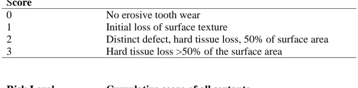
Table 4: Basic Erosive Wear Exam Scoring Definitions and Risk Levels
Score
0 No erosive tooth wear
1 Initial loss of surface texture
2 Distinct defect, hard tissue loss, 50% of surface area 3 Hard tissue loss >50% of the surface area
Risk Level Cumulative score of all sextants
None Less than or equal to 2
Low Between 3 and 8
Medium Between 9 and 13
CHAPTER I
Introduction:
Erosive tooth wear (ETW), which includes attrition, abrasion, abfraction and erosion
appears to be increasing in incidence. Attrition and abrasion are attributed to be the major
etiologies of tooth wear in the United States (US), while in Europe the primary etiology of
tooth wear appears to be erosion.15 Whether this observation is because of the direction of
research or in actual differences in etiologies is difficult to determine. The National Health
and Nutrition Examinations Survey (NHANES) for 2003-2004 was the first cycle with a
measurement of “tooth wear” [erosion] in children ages 13-19 in the United Kingdom
(UK).11 Although the measure of tooth wear is used in conjunction with and interchangeably
with dental erosion in NHANES 2003-2004, the best term is actually erosive tooth wear.17
Differences in the beverages sold in the US and UK were found in Murrell’s 2009 study
examining the pH and erosive potentials.44 While the pH and erosive potentials are
different, it does not fully explain the differences in the erosive patterns seen between the two
countries.44 Perhaps, it is not that the ETW patterns are different between the two countries,
but that the diagnosis and etiology are attributed differently.15 This conclusion would then
suggest that perhaps the US dental education system is not imparting a good understanding of
ETW. A 2010 study conducted in a Brazilian dental school found that the understanding of
dental erosion was not good among their students and faculty.45 Hygiene and dental students
are well educated at the University of North Carolina School of Dentistry first in the
14
the second year dental students presenting the majority of Class V lesions to faculty as
toothbrush abrasion lesions rather than a more general non carious cervical lesion (NCCL).
Once abfraction and erosion are presented to the third year dental students’ diagnoses of the
etiology of NCCLs begin to diversify.
It is suggested that the difficulty with early diagnosis and management of dental
erosion is because the topic is not emphasized in dental curriculums and is not a desirable
continuing education topic.29 Others suggest that dental professionals worldwide are
confused by the signs and symptoms of erosion, especially regarding the similarities and
differences of it to other sources of ETW due to the complex interaction of the etiologic
agents.1 In April of 2011 the California Dental Association (CDA) dedicated an entire issue
of the CDA Journal with the goal of ensuring “that dentists can more effectively recognize
this condition[dental erosion], educate patients, and manage dental erosion and tooth wear
problems early and in a conservative manner.”46
Assessing the knowledge of dentists regarding ETW is rare. In 2003 a survey on
erosion was distributed to general dentists and 12 year old children in Leicestershire, UK
assessing their awareness of erosion.18 A survey was sent out to UK and overseas
prosthodontists in 2008 to examine the management of tooth wear.47 In 2010 a Brazilian
dental school surveyed their students, faculty and patients regarding their knowledge of
erosion.45 In 2011 a survey on dental erosive wear was sent to all dentists who were part of
the Norwegian Public Dental Health Service.48 These four surveys constitute our basis of
what dentists know regarding erosion and only select US prosthodontists represent the
15
The assumption exists that American trained dentists do not have a strong
understanding of the etiology or clinical signs of dental erosion. 15,29 The purpose of this
study was to assess the knowledge of dentists in the United States on the clinical signs,
etiology, and treatment of dental erosion. The frequency of erosion seen in patients, use of
diet analyses, and referrals to physicians were also examined.
The hypothesis was that American dentists are able to identify the clinical signs and
etiologies of erosion and are following the recommend treatments for dental erosion.
Materials and Methods:
In order to assess American dentists’ knowledge and treatment of dental erosion a
survey was developed. Participants were asked to identify the clinical signs that indicated
erosion. Questions addressing the frequency of encounters with erosion (ETW), use of diet
analysis, and referrals to physicians were asked. Preventive and restorative treatments for
erosion were evaluated as were possible etiologies of erosion. The remaining questions dealt
with provider demographics. See Appendix A This study was reviewed by the University of
North Carolina (UNC) Biomedical Institutional Review Board of the Office of Human
Research Ethics and declared exempt (IRB, study # 12-1103, 12-1801, 12-1802).
Survey Development. The survey instrument (Appendix A) was developed with the
assistance of The Odum Institute at The University of North Carolina at Chapel Hill and Dr.
Ceib Phillips, the University of North Carolina School of Dentistry. The survey was created
in Qualtrics Software (Qualtrics, Provo, Utah) and a duplicate paper survey was created
16
Three sample populations were surveyed as part of this study. A convenience sample
was drawn from a continuing education meeting, the 29th Annual Dental Review (ADR) in
Myrtle Beach, SC. A random sample was obtained from the general dentists in North
Carolina. A master list of active dentists was requested from the North Carolina Board of
Dental Examiners. A random sample of 1735 active general dentists was created from this
list. The final sample was all 1083 active duty US Navy Dentists.
Survey Distribution: The survey was distributed in packets given to all registrants
at the ADR. Each morning a verbal invitation was issued to complete the survey; the
meeting ran three days.
A mixed mode distribution method was used for the distribution of surveys to NC
dentists. All NC dentists who provided their email address to the NC Dental Board of
Examiners were emailed a letter explaining the survey and an invitation to complete the
survey electronically through the Qualtrics system. Three electronic reminders were sent to
non-respondents every two weeks after the initial distribution. For those who did not provide
an email address, hard copies of the invitation, letter, and survey were mailed with a postage
paid return envelope. A final hard copy was sent to all non-respondents, whether they
received an initial electronic or hard copy survey. Each mailing contained a cover letter
explaining the survey and postage paid return envelope. Six weeks after the final mailing no
further surveys were counted.
All US Navy Dentists were invited to participate in the survey via the Weekly Dental
Update (WDU), an electronic newsletter sent to US Navy Dental leaders and distributed to
17
electronic link to the Qualtrics survey ran in the WDU. The Navy Qualtrics survey was shut
down after ten weeks.
Results
Respondents were excluded if they did not return a survey, returned an illegible
survey or the survey was returned after the cutoff date.
A master data set was formed from the downloaded data from Qualtrics and the
Teleform responses.
Statistical Analysis. All three distributions of this study were combined into one
data pool for analysis. Descriptive statistics were conducted on all questions. Weighted
values were assigned to determine the ability of providers to correctly identify the clinical
signs of erosion.
Response Rate. A total of 69 surveys were received during the ADR, 180
participants were registered resulting a response rate of 38.3%. A total of 744 out of 1735
surveys were returned from NC general dentists, a response rate of 42.9%. Five surveys
were returned by the post office as undeliverable. One hundred forth five out of 1153 Navy
dentists (70 retirees plus 1083 active duty dentists) responded to the survey invitation for a
response rate of 12.6%. A total of 958 surveys were returned for analysis.
Demographic data is shown in Table 5.
Results of the surveyed dentists over 70.3% saw a patient with erosion or ETW once
a week, 41.2% reported encountering a patient with erosion daily. Seven and a half percent
of the surveyed dentists do not encounter erosion more frequently than once every 6 months.
18
Forty three and seven tenths of a percent of the survey dentists have not used a diet
analysis in their practice in the last year. Figure 2
Fifty eight percent of the surveyed dentists have not referred any patients to their
primary care physicians in the past year. Figure 3
Only 30.5% of the surveyed dentists could correctly identify all the clinical signs of
erosion. Figure 4
Eighty six percent of the surveyed dentists feel competent in recognizing the clinical
signs of dental erosion and discussing erosion with their patients. Eighty one percent of the
dentists feel competent discussing the etiology of erosion with their patients, while only
76.8% feel competent treating patients who have tooth loss due to erosion. Table 6
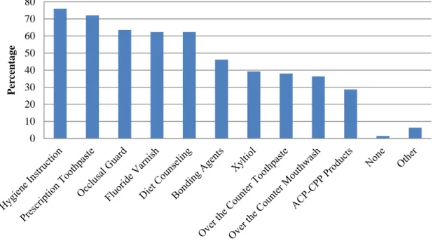
The preventive treatments utilized for patients with mild erosion were most
frequently hygiene instruction (75.8%) and prescription toothpaste (72.1%). Figure5 shows
the frequency of other preventive measures for mild erosion.
Restorative treatments utilized by the surveyed dentists are shown in Table 7. Full
coverage restorations in patients with severe erosion were the most common treatment at
58%. Occlusal guards were the next most common treatment. Fifty one and one tenth
percent of the surveyed dentists use occlusal guards for those with moderate erosion and
45.9% for those with severe erosion.
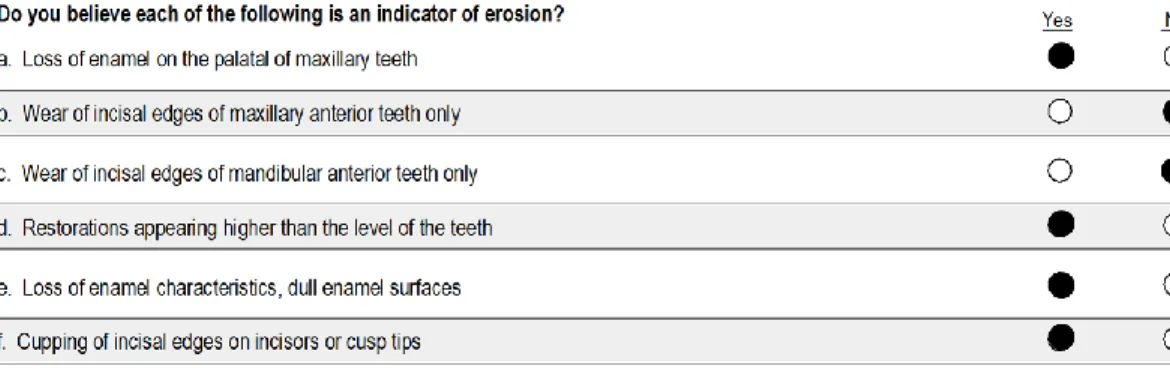
Identification of the etiologic factors of erosion was highly variable. Figure 6 reflects
those that may be considered etiologic factors while Figure7 shows identified agents that are
not etiologic factors for erosion. The highest positively identified etiologic agents were
19
Discussion:
The aim of this study was to determine the knowledge of dentists in the US on the
etiology, clinical signs and treatment of dental erosion. Surveys regarding erosion are fairly
infrequent. Four studies were found on erosion and all but one looked only at dentists
outside of the US.18,45,47,48 The majority of dentists who participated in this study were
general dentists from the state of North Carolina and are not necessarily a representation of
the knowledge of dentists across the US. The inclusion of the US Navy dentists widens the
geographic range and includes more than general dentists.
The low rate, 38%, of response at the CE meeting may be from self-exclusion.
Offices may have brought hygienists, assistants and office staff to the meeting and the
auxiliaries would have been counted as one of the 180 registered attendees. As the survey
targeted dentists, the auxiliaries most likely did not complete or turn in the survey.
Additionally, as the meeting is sponsored by the UNC Department of Operative Dentistry,
faculty and staff were registered and counted among the 180 attendees but did not respond as
they were a biased sample. A response rate of 43% from the random sampling of NC general
dentists is lower than the other comparable studies on erosion but is an average response rate
for a mixed mode survey.18,45,47,48 The 12.6% response rate from the electronically
distributed survey to US Navy dentists may be due to operational issues that will not allow
them to complete the web based survey due to limited band width or blocked access to the
Qualtrics website. The topic of erosion may also not have been applicable to some of the
specialists such as oral surgeons and endodontists.
No definitions of erosion were provided nor any definition for what is considered
20
to be assessed it may also mean a variety of definitions and levels of erosion exist among the
surveyed dentists.
Frequency
Dentists were asked to recall their frequency of encounters with patients with erosion
and their treatments utilized. Forty one and nine tenth percent of the providers saw erosion
or ETW on a daily basis, higher than Dugmore’s 2003 survey of general dentists in
Leicestershire where 36% saw erosion often or very frequently. 18 Dugmore did not define
what is often or frequent and in both studies the memory of the providers is being relied on
rather than tracking patients seen with erosion. Additionally, if it is considered that only
30% of the surveyed providers could correctly identify all the signs of erosion perhaps a high
frequency of erosion is not being seen but a higher frequency of ETW in general.
Referral
Fifty eight percent of the surveyed dentists did not refer their patients to their primary
care physician due to their erosion. Referral to a physician would most likely be due to the
ETW etiology being GERD, anorexia or bulimia. Such a high non referral rate may be due
to patients already having a diagnosis of GERD, anorexia or bulimia and already being under
a physician’s care. Studies find a prevalence of erosion in the range of 18-58% for patients
with GERD.49-51
Identification of Clinical Signs
Only 30.4% of the surveyed dentists were able to correctly identify the signs of
erosion, but 86% feel confident that they can recognize the clinical signs of erosion. Perhaps
if they saw the erosion they would be able to identify the signs but in this survey no clinical
21
of erosion were given. The two additional clinical signs listed were specific to attrition and
normal physiologic wear. The misidentification of the signs could be because of the
difficulty in separating ETW, multifactorial tooth wear, from erosion specifically. Forty nine
and nine tenths percent of the surveyed dentists included at least one clinical sign other than
those characteristic of erosion in their answer. It seems dentists can identify ETW but not
necessarily the primary etiology of the wear. This inability to correctly identify the etiology
of the ETW may lead to improper prevention and treatment of the lesion as it is dependent on
determining the etiology.
Confidence
Over 80% of the surveyed dentists report they feel confident in discussing and
treating erosion. This finding is similar to that found in Hermont’s study at a Brazilian dental
school (50-80%) which included dental students and faculty.45,48 In truth we see in this study
the majority (70%) of dentists were not able to identify the clinical signs for erosion, so
evidently their confidence is misplaced.
Diet Analysis
Diet analysis is underutilized, though providers seem to realize food and beverages
can lead to erosion. Only 14.1% use a diet analysis more than once a month and 42.8% see
an erosion or ETW patient daily. In Mulic’s study only half of the dentists occasionally
recorded the diet history of their erosion patients.48 The recommend technique for a diet
analysis is a four day consecutive record including at least one weekend day; patients should
be instructed to record all food, drink and medications consumed along with the time of
consumption and quantity.34,52 A Cochrane review of one to one dietary interventions in a
22
direct dietary counseling such as found when doing a detailed diet analysis.53 When
reviewing a patient’s diet analysis it is important to discuss the manner in which foods or
drinks are consumed, sucking on citrus fruits or swishing drinks can increase the erosive
potential of foods.8,25 Bartlett et al calculated the odds ratios of foods and beverages risk of
causing erosion and saw the frequency and method of consumption seems to increase the
amount of ETW seen.19
Preventive Techniques
Though diet counseling is used by 62% of the surveyed dentists as a preventive
measure for erosion it could be the hygienist providing this information not the dentist and is
not the same as conducting a diet analysis. The American Dental Association (ADA) code
D1310 for nutritional counseling is defined as “counseling on food selection and dietary
habits as part of treatment and control of periodontal disease and caries” with no mention of
erosion. Diet counseling for erosion should specifically address the frequency and manner of
consumption of food and beverages which have an erosive potential.
Remineralization either by various techniques is advocated to slow the progression of
erosion.8,10,34,54 The lower use of the surveyed providers of amorphous calcium phosphate
(ACP-CPP) (28.7%) than fluoride varnish (62.3%) appears to be supported by the studies on
reduction of ETW. Fluoroapatite is more difficult to erode than hydroxyapatite, so the
application of fluoride varnish is recommended.54 In in vitro studies Ranjitkar et al
demonstrated that ACP-CPP reduces ETW.55,56 Wegehaupt et al though have shown in
another in vitro study that ACP-CPP provides no significant protection against ETW; two
23
Over the counter (OTC) toothpaste, while advertised highly in the media for the
treatment of erosion, in vitro studies are inconclusive if they are effective at preventing
ETW.58 As prescription toothpastes are more frequently (72%) recommended by the
surveyed providers than OTC toothpastes (38%), perhaps providers are focusing on
increasing the fluoride and fluoroapatite concentration rather than the lower relative dentin
abrasivity (RDA) that some toothpastes targeting erosion may offer. OTC toothpastes’
abrasive particle size may or may not play a direct role. Some studies show lower RDA is
relative to the amount of dentin loss seen as related to how soon after an acid exposure the
brushing occurs.59 OTC toothpastes were not specified in the survey so dentists may also
recommended OTC toothpastes focusing on desensitization, sometimes a symptom of ETW.
OTC mouthwashes such as ACT (Chattem, Chattanooga, TN) can be utilized as part
of a protocol to neutralize acid exposure and then shift the balance to remineralization, but
are not frequently recommended by the surveyed population.34 As specifics were not given
regarding the OTC mouthwashes, Biotene (GlaxoSmithKline, RTP, NC) or Boost (Oral
BioTech, Albany, OR), mouth lubricating mouthwashes may also be options that were
recommend to patients. They help to fight xerostomia which may exacerbate ETW.
Bonding agents, especially filled dentin adhesives should be an effective short term
dentin protection and can be reapplied as needed.34,43,54 Forty six and one tenth percent of
the surveyed dentists utilize bonding agents as a preventive method but it decreases as a
desired treatment as the severity of erosion increases. Sabahipour’s survey of prosthodontists
in the UK and overseas showed prosthodontists are most likely to cover erosive lesions with
24
Hygiene instructions, which over 75% of the surveyed providers report they use for
the prevention of erosion, need to be specific and not the basic instructions given on
frequency and technique of brushing and flossing. It is impossible to determine from this
survey if dentists are giving the appropriate hygiene instructions regarding erosion.
Specifically, patients should be instructed to rinse immediately with water or sodium
bicarbonate after an acid challenge, delay brushing until 30 minutes after an acid challenge,
and utilize a fluoride mouthwash.34,54
About 40% of the surveyed providers recommend xylitol gum to their patients to help
prevent erosion. While xylitol gum or any chewing gum as a method to increase salivary
flow to clear the acid is suggested by some authors, others do not recommend chewing gum
as a preventive technique. There may exist an increased risk of abrasion to the acid
challenged tooth surface from the tongue and buccal tissues during the gum chewing.810,54 If
enamel is still present then recommending xylitol gum may not be a bad preventive
technique, but should be used with caution as dentin exposure increases.
Sixty three percent of the surveyed dentists use an occlusal guard as a preventive
measure for erosion. This method should be used if signs of parafunctional wear are present
not solely erosion.34,54 ETW is multifactorial and the presence of acid can potentiate
abrasion and attrition. 10,29 The use of an occlusal guard as a preventive treatment for
erosion again returns to the realization that while the surveyed population is poor at
identifying erosion but better at identifying ETW. As ETW is multifactorial, treating ETW
with an occlusal guard to reduce abrasion and abfraction is not wrong but is not specifically
25
Preventive techniques, while theoretically good have not been proven effective at
stopping ETW in vivo. The key to prevention of ETW is early diagnosis and identification of
the etiologic agent.20,29,54 In a review of the literature Holbrook et al found that clinicians
have a hard time classifying erosion severity especially when looking at exposed dentin so
catching erosion early can be difficult.39 The goal of preventive treatment for ETW should
be the reduction in acid exposure, the reduction of abrasion, and the increase in
remineralization.29,34,54,60
Restorative Treatment
With the high use of occlusal guards (27% mild, 51% moderate, 46% severe) as a
restorative treatment it can be inferred that providers either are treating erosion as
parafunctional wear or they are recognizing the multifactorial nature of erosive tooth wear
and are trying to address various possible etiologies. Due to the lack of use of diet analyses,
the former would be indicated. Additionally, the inability to correctly identify the clinical
signs of erosion leads to the belief that providers are attributing the ETW to attrition
specifically parafunction. Dental erosion does weaken the teeth through demineralization
and then permits greater damage to occur when parafunctional habits such as bruxing or
clenching occur or when abrasion is present through tooth brushing or opposing porcelain
surfaces.9 It seems that the confusion over the multifactorial nature of erosion is not unique
to the surveyed providers. In Dugmore’s study a hard toothbrush was attributed as a cause of
erosion in 9% of the surveyed dentists.18 While occlusal guards may be a reasonable
treatment for some erosive tooth wear, their frequent use as a preventive and treatment
measure gives support to Bartlett’s theory that US dentists attribute ETW to attrition and
26
Restorative treatments need to be focused on additive techniques and early
conservative intervention. Composite resins, glass ionomers or resin modified glass
ionomers are best indicated for mild to moderate ETW.29
Onlays, in particular ceramic bonded onlays, should be considered more than they are
used by the surveyed providers; 53% do not use onlays as a restorative treatment option.
Onlays are a conservative method to add to a tooth without destroying as much of the
remaining healthy tooth structure. Providers may be hesitant to treat patient with onlays,
only 16.7% use in the case of severe erosion, because it is not a commonly taught procedure
in dental school and may be more technically involved than a full crown preparation.
Full coverage restorations may do more harm than good depending on how much
tooth structure is removed. In severe cases of ETW, the occlusal vertical dimension may
need to be increased and full coverage may be the only treatment available, though again
onlays may be a reasonable alternative or even direct composite restorations.29,61 Full
coverage is used by over 58% of the surveyed dentists when severe erosion is identified
possibly dueto the loss of vertical dimension and due to familiarity with the needed
preparation.
Etiologic Factors
Etiologic factors contributing to erosion were not narrowly defined in this survey. A
wide range of erosive potential can exist for several of them. Tea for example can be neutral
such as in the case of chamomile or highly acidic and having a high erosive potential when
citrus fruits are part of it.62-64 An increased risk of erosion is different than a low or acidic
27
Also, it must be remembered that the presence of an etiologic risk factor alone does not mean
erosion will occur.
Diet soda (75.7%) was selected less than regular soda (83%) as an etiologic in this
study. Diet sodas are just as likely to cause erosion as regular sodas.66 While patients switch
to diet beverages believing they are making a positive change for their teeth and diet, their
frequency of consumption may be greater leading to an increased risk of erosion. Jarvinen
saw an increased risk of erosion if more than 4-6 soft drinks were consumed weekly.30
Unlike dentists in Dugmore’s study, in this study GERD (87%), anorexia and bulimia
(88.3%) are well recognized by study participants as etiologic factors of erosion.18,20
Dugmore’s providers, while also general dentists, were asked to consider erosion specifically
in their 12 year old patients. Mulic’s survey of Norwegian dentists showed that GERD,
anorexia and bulimia were considered uncommon (8%) causes of erosion, while consumption
carbonated beverages [sodas] were the most common cause.48 Mulic’s patient population
was 18-30 year olds. This is the population usually considered at risk for anorexia and
bulimia, especially in females. Jarvinen et al showed an increased risk of erosion from 10
times greater if heartburn and other gastric symptoms occurred at least weekly and if
vomiting occurred at least weekly there was a 31 times greater risk of erosion.30 These
differences in opinions seem to support the differences that Bartlett spoke of between
European and American dentists as to the cause of ETW. Even when considering erosion
specifically the surveyed dentists were more certain of the intrinsic sources of erosion while
their European counterparts more often identify extrinsic sources.
Frequent consumption of sports drinks are a possible etiologic agent of erosion due to
28
an etiologic risk for erosion. Jarvinen determined weekly consumption of sports drinks to
increase the risk of erosion.30 Teenage boys are a the most common population effected by
erosion due to high sport drink consumption and should be counseled on the risk of frequent
consumption.34
Seventy four percent of the surveyed dentists agreed fruit juice is an erosive etiologic
factor. Multiple studies agree with this as a risk factor.4,20,63,68-70 Fruit juice consumption
was shown by Okunseri et al to increase the risk for ETW.71 Frequency of apple juice gave
the highest risk of ETW.71
A vegetarian diet may possibly cause erosion. Linkosalo found vegetarians to have a
significantly higher prevalence of erosion than non-vegetarians.72 Herman et al observed a
slight increase in the ETW of those following a vegetarian diet but it was not statistically
significant.73 Al-Dlaigan et al saw no difference in the prevalence of erosion between
vegetarian and non-vegetarian teenagers.74
Fruits were only considered by the study population to be possible etiologic agents of
erosion 60% of the time. This observation may be due to a definition of fruits that is too
broad and inclusive. Citrus fruits are much more likely to be considered erosive due to the
presence of citric acid, but apples, pears and plums may be forgotten.25 Consumption of
citrus fruits more frequently than twice a day was found by Jarvinen to increase the risk of
erosion.30 The frequency of consumption is not the sole risk factor, but the method of
consumption, mulling or sucking of fruits and frequency can also increase the erosive
potential.20,25
White wine has a greater erosive potential than red wine though the surveyed
29
with wine tasters show severe erosion, especially because they swish the wine as part of their
job.77 Beer, while acidic has a low erosive potential so is not considered an etiologic factor
for erosion.65 The surveyed 16% who consider beer a risk factor may be considering the
addition of citrus fruits to beers like an orange slice to Blue Moon (Coors, Golden, CO) or a
lime wedge to Corona (Amheuser-Busch inBev, Leuvenm, Belgium).
Hard candies and mints do not necessarily cause erosion but there is an increased
popularity in sour candies such as jaw breakers which are erosive.78,79 This fact may account
for the 34% of surveyed dentists who consider hard candies and mints a possible etiologic
agent of erosion.
Unflavored carbonated water does not seem to have an erosive impact on the
dentition, but again if flavors are added especially with citric acid the erosive potential of the
beverage increases.63,70,80,81 This may be what the 32.8% are considering.
Dehydration (28%) and xerostomia (50%) increase the risk of erosion by two
methods, by decreasing the salivary flow rate and allowing whatever beverages and food that
are consumed to have a greater impact on the dentition. Jarvinen et al found that if subjects
had a low unstimulated salivary flow rate they had a higher risk of erosion even if they do not
have a high number of acidic challenges.30
In general it is not enough to just assume a food or drink will be erosive due to its pH.
It is a good starting place but does not accurately reflect the damage that it may inflict on
tooth structure.63 Yogurt, though acidic does not have a high erosive potential attributed to
its high calcium and phosphate content.63,82Lussi et al conducted a study on the erosive
effects of various foods, drinks and medications; they showed that pH, buffering capacity,
30
fluoroapatite all contribute to the erosive potential.63 The frequency and method of
consumption of food or beverage must also be considered as it can impact the erosive
potential too.19,67 It is also important to remember as Bartlett points out that most studies
linking dietary acids and erosion are laboratory or associative epidemiological studies, causal
relationships are much harder to prove.14 Most studies on dietary acids also rely on the
subjects self-reporting and recollection of what food and drinks they have consumed.
Competitive or frequent swimming may cause erosion though only 38% of the
surveyed population considered this a possible etiologic agent. This is often seen in case
reports of patients who undergo rapid loss of tooth structure.83,84 The frequency and duration
of the swimming and the pH of the swimming pool are the main factors determining if dental
erosion is seen in the patient.
Further surveys could address a more representative geographical sample of the
American dentist population, or could see if providers record the progression of erosion and
even what is taught in the North American Dental School curriculums.
Conclusions:
-Dentists do not appear to know the signs of erosion but think they do.
-Reported erosion (ETW) is frequent.
-Dietary analysis is underutilized.
-American dentists do appear to treat erosion as a parafunctional wear with occlusal
31 Table 5: Survey Respondent Demographics
Annual Dental Review n= 69
North Carolina n=744 US Navy n=145 Total n=958 (%) Years of Practice
0-10 10 154 73 237 (24.7)
11-20 17 157 24 198 (20.7)
21-30 15 192 23 230 (24.0)
>30 26 228 18 272 (28.4)
No answer 1 13 7 21 (2.2)
Currently practicing dentistry
yes 66 702 126 894 (93.3)
no 3 16 15 34 (3.5)
No answer 0 26 4 30 (3.1)
Specialty
General Dentistry 64 724 99 887 (92.6)
Orthodontics 0 3 2 5 (.5)
Periodontics 1 1 4 6 (.6)
Prosthodontics 2 2 15 19 (2.0)
Other 2 5 16 23 (2.4)
No answer 0 9 9 18 (1.9)
Gender
Female 13 183 34 230 (24.0)
Male 54 543 103 700 (73.1)
No answer 2 18 8 28 (2.9)
Age
25-35 6 103 55 164 (17.1)
36-45 15 169 33 217 (22.7)
46-55 16 163 24 203 (21.2)
>55 30 300 25 355 (37.1)
<25 1 0 0 1 (.1)
32
Figure 1: Frequency of Dental Erosion (Erosive Tooth Wear)
3%
1%
42%
29%
19%
6%
No Answer
Never
Daily
33 Figure 2: Diet Analysis Usage Frequency
5%
44%
27% 10%
14%
No Answer
Never
1-6
7-20
34 Figure 3: Primary Care Physician Referral
58% 33%
9%
No
Yes
35
36 Table 6: Confidence Level Discussing Erosion
Please evaluate your agreement on the following statements
Yes
(n=958) %
I feel competent to recognize the clinical signs of dental erosion 824 86
I feel competent to treat patients whose loss of tooth structure has
occurred from dental erosion 736 76.8
I feel competent discussing the dental erosion with my patients 830 86.6
I feel competent discussing the etiology of dental erosion with my
37
Figure 5: Frequency of Preventive Measures for Mild Erosion
0 10 20 30 40 50 60 70 80
P
er
ce
nta
g
38
Table 7: Restorative Treatments Utilized by Respondents
Bonding Agent %
Flowable Composite %
Glass Ionomer %
Resin Modified Glass Ionomer %
Onlay %
Full Coverage %
Occlusal Guard %
Mild
Erosion 35.7 25.7 9.3 10.3 2.2 2.4 26.9
Moderate
Erosion 23.6 40.9 24.1 28.9 6.7 12.2 51.1
Severe
Erosion 11.6 19.1 17.2 22.9 16.7 58.2 45.8
Do Not
39
Figure 6: Positive Etiologic Agents Selected by Respondents as Positive Agents
0 10 20 30 40 50 60 70 80 90 100
P
er
ce
nta
g
40
Figure 7: Negative Etiologic Agents Selected by Respondents as Positive Agents
0 5 10 15 20 25 30 35 40
P
er
ce
nta
g
Chapter II
Gastroesophageal Reflux Disease and Erosive Tooth Wear
Introduction
As seen in the previous chapter 95% of dental practitioners surveyed feel that
gastroesophageal reflux disease (GERD) is an etiology for dental erosion. Studies show that
erosion does not always occur in a patient though they may have a risk factor for it. There is
a variable range in the prevalence/ frequency of erosion in the population. Seven to fifty-six
percent of the population have been shown to have dental erosion in the UK depending on an
array of variables being used.11 While the association between GERD and dental erosion has
been shown to be plausible, the strength of the association is not clear.85,86
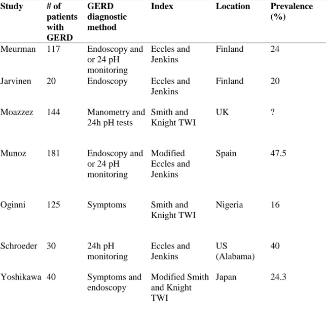
Pace et al conducted a systematic review “to assess the relationship between DE
[dental erosion] and GERD.”49
Their results showed a median prevalence of 24% erosion in
GERD patients but with a range in prevalence from 5-47.5%. When looking at adult subjects
with erosion there was a median prevalence of 32.5% of GERD. Pace et al concluded there
was a strong association between GERD and dental erosion. The range of prevalence and
strength of association between GERD and dental erosion can be attributed to a number of
variables. First, the diagnostic method used to determine if a subject has GERD may vary.
Symptoms, endoscopy or 24-hour pH manometry all may be used. Second, the tooth wear
index being used to measure the erosion or erosive tooth wear (ETW) present may vary.
Also to be considered is the age of the study population; many erosion studies are conducted
on children or adolescents which may not accurately reflect the prevalence of erosion in an
42
The majority of studies on erosion have been conducted outside of North America.
As discussed in the previous chapter studies on tooth wear in the US have focused on
attrition and abrasion.15 Prevalence studies on erosion are beginning to be seen in the US but
as the US covers such a large geography and diverse population, variations in the prevalence
of erosion may be greater within the US.
Gastroesophageal Reflux Disease
GERD as defined by the Montreal consensus is “a condition which develops when the
reflux of stomach contents causes troublesome symptoms and/or complications.”87
While
symptoms and complications from GERD such as heartburn, esophageal stricture, bleeding,
and Barrett’s esophagus are commonly cited, less likely to be mentioned is dental erosion.88
Silent GERD is the absence of heartburn symptoms in a patient who actually has reflux. In
silent GERD atypical symptoms can present such as hoarseness, throat problems, respiratory
issues and dental erosion, all of which may not lead to diagnosis of the disease.89,90
Over 15 million Americans experience heartburn symptoms each day and 10-20% of
the US adult population is affected by GERD.91 Dental erosion due to GERD is an etiology
that seems likely but is still relatively unknown to the general medical and dental
practitioner.88,92
The purpose of this study was to determine the prevalence of ETW in GERD subjects
compared to that in a control population. The association between the number of acidic
challenges, number of medications, age, salivary flow rate, initial salivary pH and salivary
buffering capacity and the erosive wear present will also be examined.
The null hypotheses tested in this study were that (1) there was no difference in
43
GERD subjects and the control and (2) there was no association between the selected factors
and the ETW present.
MATERIALS AND METHODS:
This cross sectional study was to determine the prevalence of dental erosion in a
population diagnosed with GERD and a control population. Subjects’ dentition’s were
examined using the BEWE. They provided a stimulated salivary sample and took home a
diet diary to complete.
The research protocol was reviewed and approved by the University of North
Carolina (UNC) Biomedical Institutional Review Board (IRB, study #11-2327).
Subject Selection
Subjects were recruited from the Center for Esophageal Disease and Swallowing,
University of North Carolina Hospital Division of Gastroenterology and Hepatology and the
University of North Carolina at Chapel Hill School of Dentistry Operative Dentistry Clinic.
Subjects were asked to participate in the study if they were positively diagnosed with GERD
or no had history of GERD (control).
Inclusion Criteria
-Adults (18 – 85 years old)
-Two natural teeth per sextant (12 teeth total)
-Diagnosis of GERD or no history of GERD (control)
Exclusion Criteria
44 -History of anorexia or bulimia
-Greater than 85 years old
A total of 22 subjects with GERD and 22 control subjects were desired in the study.
Sample size calculations were calculated with alpha=.05 and power= 0.8, assuming the
proportion of dental erosion in the GERD population to be 0.4 and the proportion of dental
erosion in the control group to be 0.05.
Descriptions of the specific tests follow.
Basic Erosive Wear Exam (BEWE)
Each patient was examined by the primary investigator (KEE) using the BEWE. The
BEWE was developed by Bartlett et al.41 It was developed to be a quick screening method to
determine the erosive wear present on the dentition. The mouth is divided into sextants and
the tooth with the greatest wear is rated in each sextant. Scoring is from 0-3. A cumulative
score is calculated and the subject is assigned a risk category for erosive wear.
See Table 4: Basic Erosive Wear Exam Scoring Definitions and Risk Levels
Stimulated Salivary Sample
Subjects were asked to chew on a paraffin wax tablet for 5 minutes. Rather than
swallowing the generated saliva it was expectorated and collected in a sterile container. The
samples were labeled with the subject’s unique numerical identifier and stored on ice for
transportation to the Oral Microbiology Lab at the UNC School of Dentistry. Initial pH,
salivary buffering capacity and salivary flow rate were determined by the lab. Salivary
buffering capacity was determined by the saliva being diluted four-fold in 0.0005N HCl and
45
4.1-4.9 is moderate risk, and less than 4.0 is high risk. All samples were destroyed after
testing. See Table 8: Salivary Risk Categories
Dietary Analysis
Subjects were given a diet diary to take home and record all food, drink and
medications consumed during the next Thursday through Sunday period. Instructions were
reviewed with the subjects and a sample sheet was included on their diet diary; a stamped
addressed envelope was given to the subjects to return the diaries to the primary investigator
(KEE). The subject’s unique numerical identifier was placed on the diet diary. If the diary
was returned with the subject’s name on it the sheet was removed and shredded. Daily acidic
challenges were counted. If the subject was ambiguous to the specifics of a potential acidic
food or drink (eg tea v chamomile tea) the decision was made to assume it was acidic. The
average daily number of acidic challenges was calculated for each subject. If subjects had
not returned the diet diary after 3 weeks, a reminder letter, new diet diary and postage paid
return envelope was sent to the participant. If still after an additional 3 weeks no diet diary
was received a final reminder, diet diary and envelope were sent.
Statistical Analysis
Frequency and bivariate analysis were performed. A logistic regression was
performed to identify significant explanatory variables for the outcome of BEWE adjusting
for group. A forward selection was used with an entry at a significant level .05. All analyses
46
Results
A total of 67 subjects were recruited to participate in the study. Eleven subjects did
not follow through with any portion of the exam. The remaining 56 completed the exam and
salivary sample. Forty nine out of the 56 returned their diet analysis (25 GERD and 24
controls). Table 9: Bivariate Results
The prevalence of ETW in GERD subjects is not statistically different from the
prevalence of erosion in the control subjects. 40% of the GERD subjects were at medium
risk for ETW as compared to 15.4% of the control subjects. 50% of the GERD subjects were
at low risk as compared to 73.1% of the control subjects.
The association of selected factors and ETW was only statistically significant for age.
Table 10 Logistic Regression
Discussion
The prevalence rate found 40% of the GERD subjects to be at moderate risk of ETW
versus 15.4% of the control subjects. This difference is not statistically significant. The
sample size of this study was small and may contribute to the prevalence rate not being
statistically significant.
This prevalence is only truly applicable to the region studied; subjects came from
North Carolina, South Carolina and Virginia and were all adults. Children prevalence studies
exhibit a great range in prevalence 14-87% in Pace’s systematic review but this 40%
prevalence falls into Pace’s range of prevalence in adult studies.49
47
Differences in the observed prevalence and prevalence in previous studies may be due
to the difference in methods being utilized to diagnosis GERD. This study used subjects who
had been diagnosed with GERD through either a positive endoscopy or positive 24 hr pH
manometry (monitoring).
Differences in the observed prevalence and in previous studies may also be due to the
different wear indices being used. Though all have similar methods of evaluating wear, the
differences between them may increase the difficulty of comparing the results. The BEWE is
a newer exam and while its validity and sensitivity and specificity have been tested, there
exist very few published studies using it as a tooth wear index.38,40,93
Only one examiner (KEE) performed the BEWE so in addition to possible bias from
this and not being blinded to the subject populations that were being examined there was also
a learning curve on the exam. As tooth surface area increased such as from recession, more
surface area is exposed, such as the root surfaces, so the amount of tooth loss may decrease
overall. The BEWE is based on the percentage of surface area worn and molar cervical wear
may give a higher score than localized incisal wear. The exam was also performed under
less than ideal conditions on the most of the GERD subjects. There was no air/ water
syringe, only drying of the teeth with gauze, and the patients were in office chairs not dental
exam chairs so lighting was not ideal. This may have led to difficulty assessing enamel
versus dentin wear and could have led to underestimating the ETW present.
Some subjects only posterior uncrowned teeth were second or third molars, which can
be difficult to assess when not in a dental chair. High numbers of full coverage restorations