THE EFFECTS OF ADIPOSITY AND TYPE II DIABETES ON THE IMMUNE RESPONSE TO INFLUENZA VIRUS IN ADULTS
Heather A. Paich
A dissertation submitted to the faculty of the University of North Carolina at Chapel Hill in partial fulfillment of the requirements for the degree of Doctor of Philosophy in the
Department of Nutrition, Gillings School of Global Public Health.
Chapel Hill 2013
Approved by:
ABSTRACT
HEATHER ANN PAICH: The Effects of Adiposity and Type II Diabetes on the Immune Response to Influenza Virus in Adults
(Under the direction of Melinda A. Beck)
There are very limited data on mechanisms that mediate the obesity-associated and type II diabetes-associated impairments in immune function against viral infections, such as with the influenza virus. The purpose of this dissertation was to assess the humoral and cellular immune responses to the influenza virus, as well as to examine the effects of type II diabetes on T cell metabolism. This dissertation followed three aims. Aim 1 utilized a
Although markers of dendritic cell activation and function remained intact, markers of T cell activation and function were significantly impaired in overweight and obese, compared to healthy weight adults. Together these data suggest that there would be significant deficiencies in the activation and cytotoxic function of CD8+ T cells, as well as in the activation and helper function of CD4+ T cells resulting from overweight and obesity. Aim 3 was an exploratory aim designed to generate preliminary data towards answering the question of how obesity with or without type II diabetes will affect T cell glucose
metabolism. The data suggests that there are differences in glucose metabolism in
ACKNOWLEDGEMENTS
I gratefully acknowledge the incredible support I have received from all the
members of my dissertation committee: Melinda A. Back (chair), Jean Handy, Ilona Jaspers, Liza Makowski, and Patricia A. Sheridan. Dr. Beck has taught me a tremendous amount about how to be an effective scientist and her unwavering encouragement and guidance have allowed me the freedom to think creatively about approaching and solving scientific problems.
I would also like to thank my entire family, including parents, grandparents, and sister, for always supporting my educational endeavors. I would like to thank my friends and fellow graduate students who have enhanced my doctoral training experience (Dori
TABLE OF CONTENTS
LIST OF TABLES ... xii
LIST OF FIGURES ... xiii
LIST OF ABBREVIATIONS ... xv
CHAPTER I OVERVIEW AND SPECIFIC AIMS ... 1
Overview ... 1
Specific Aims ... 2
CHAPTER II BACKGROUND AND SIGNIFICANCE ... 4
Introduction ... 4
The Obesity Epidemic ... 6
Obesity and Immune Function in Animal Models ... 8
Obesity and Immune Function in Humans ... 9
Conditions and Disease States Associated with Obesity ... 10
Obesity and the Response to Vaccination ... 11
Obesity and PBMC Populations ... 12
The Pathogenesis of Type II Diabetes ... 16
Type II Diabetes and the Response to Influenza Infection and Vaccination ... 18
Type II Diabetes and PBMC Populations ... 20
Influenza ... 21
Seasonal Influenza Epidemics ... 22
Pandemic Influenza Outbreaks ... 23
Influenza Virus Biology ... 23
Antigenic Drift and Shift of the Influenza Virus ... 25
Influenza Infection ... 27
Influenza Virus Recognition and Host Cell Signaling ... 29
Influenza Vaccination ... 31
Humoral Immune Response to Influenza ... 33
Signaling in the Humoral Immune Response ... 34
Role of Dendritic Cells and CD4+ T Cells in the Humoral Immune Response ... 36
B Cell Class Switching ... 37
Antibody Function in the Humoral Response ... 39
Humoral Immune Response to Influenza Vaccination ... 40
Cellular Immune Response to Influenza ... 41
Dendritic Cells in Cellular Immunity ... 43
CD4+ and CD8+ T Cells in Cellular Immunity ... 45
Effector CD4+ T Cells ... 46
Memory T Cells ... 49
Humoral and Cellular Responses to Influenza ... 50
T Cell Metabolism ... 50
Significance ... 53
CHAPTER III OBESITY IS ASSOCIATED WITH IMPAIRED IMMUNE RESPONSE TO INFLUENZA VACCINATION IN HUMANS ... 55
Overview ... 55
Introduction ... 56
Methods ... 57
Results ... 61
Discussion ... 64
Tables and Figures ... 67
CHAPTER IV OVEREWEIGHT AND OBESE ADULT HUMANS HAVE A DEFECTIVE CELLULAR IMMUNE RESPONSE TO PANDEMIC H1N1 INFLUENZA A VIRUS ... 71
Overview ... 71
Introduction ... 72
Methods and Procedures ... 73
Results ... 77
Discussion ... 81
Tables and Figures ... 87
Introduction ... 103
Materials and Methods ... 104
Results ... 108
Discussion ... 110
Tables and Figures ... 113
Future Directions ... 114
CHAPTER VI SYNTHESIS ... 124
Overview of Research Findings ... 124
Potential Mechanism ... 125
Implications of Research Findings and Public Health Significance ... 131
Recommendations for Future Research ... 133
Conclusions ... 136
APPENDIX CD8+ T CELLS FROM OBESE SHOW IMPAIRMENTS IN ACTIVATION AND FUNCTIONAL MARKERS TO PH1N1, DESPITE INCREASED PBMC PROLIFERATION AT 30 DAYS POST-VACCINATION ... 138
Introduction ... 138
Materials and Methods ... 139
Results ... 140
Discussion ... 141
Tables and Figures ... 142
LIST OF TABLES
Table 3.1 Demographic Characteristics of 2009-2010 and 2010-2011 Returning Study Participants ... 67
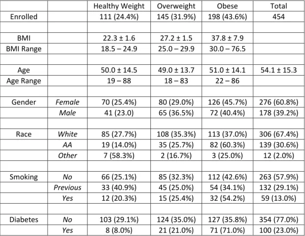
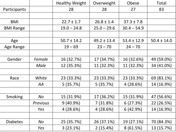
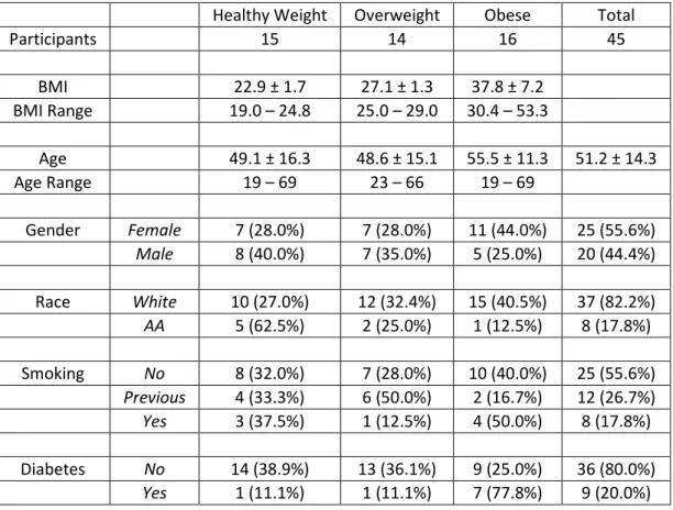
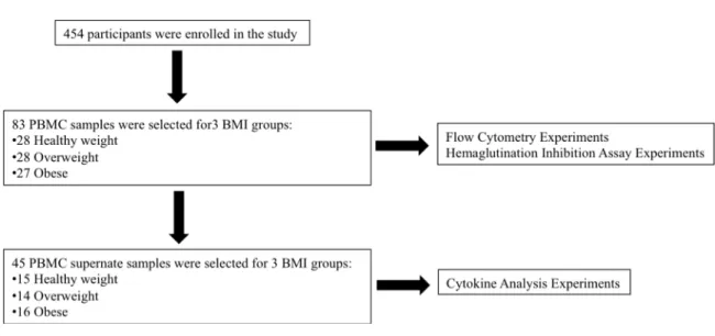
Table 4.1 Demographic Characteristics and Study Overview ... 87
Table 4.2 Fluorochrome-conjugated Antibodies Used for T Cell and Dendritic Cell FACS Panels ... 91
Table 5.1 Demographic Characteristics of the Individuals in the Study
Population ... 114
Table 5.2 Demographic Characteristics of the Study Population ... 115
LIST OF FIGURES
Figure 2.1 Signaling Pathways Initiated with Influenza Infection ... 31
Figure 2.2 Dendritic Cell Presentation of Influenza Peptides to B Cells and CD4+ T Cell-induced Class Switching ... 38
Figure 2.3 Dendritic Cell Presentation of Influenza Peptides to CD4+ and CD8+ T Cells ... 44
Figure 2.4 PI3K/Akt Signaling Pathway in Activated T Cells ... 53
Figure 3.1 Obese Participants Do Not Have an Impaired Initial Response to Influenza Vaccination ... 68
Figure 3.2 Obesity Results in a Greater Decline of Influenza Antibodies ... 69
Figure 3.3 Obesity Results in Defective CD8+ T Cell Activation and Production of the Functional Proteins Granzyme B and IFNγ by
Influenza-stimulated PBMCs ... 70
Figure 4.1 Activation and Function of Dendritic Cells Remain Intact in PBMCs from Overweight and Obese Individuals ... 92
Figure 4.2 Activation and Function of CD4+ T Cells are Impaired in PBMCs from Overweight and Obese Individuals ... 93
Figure 4.3 Activation and Function of CD8+ T Cells are Impaired in PBMCs from Overweight and Obese Individuals ... 95
Figure 4.6 Dendritic Cell Antigen Presentation to CD4+ and CD8+ T Cells ... 99
Figure 4.7 Representative Example of the Gating Strategy Used to Analyze CD4+ T Cells ... 100
Figure 4.8 PBMC Cytokine Secretion from Healthy weight, Overweight, and Obese Individuals ... 101
Figure 4.9 HAI Titers Measured in Serum from Healthy Weight, Overweight, and Obese Individuals ... 102
Figure 5.1 Age, BMI, and HbA1c Levels ... 116
Figure 5.2 Hypothesized Results for ECAR and OCR in Non-Diabetic Stimulated T Cells ... 117
Figure 5.3 ECAR in Unstimulated T Cells With and Without Insulin ... 118
Figure 5.4 ECAR in Stimulated T Cells With and Without Insulin ... 119
Figure 5.5 OCR in Unstimulated T Cells With and Without Insulin ... 121
Figure 5.6 OCR in Stimulated T Cells With and Without Insulin ... 122
Figure A.1 Fresh, Cryopreserved, Stimulated, and Unstimulated PBMC Populations ... 143
Figure A.2 CD3+CD8+ T Cell Populations in Healthy Weight and Obese ... 144
LIST OF ABBREVIATIONS
AID activation-induced deaminase AP-1 activator protein-1
APRIL a proliferation-inducing ligand BAFF B-cell activating factor
Bax Bcl-2–associated X protein Bcl-2 B-cell lymphoma-2
BCR B cell receptor
Bid BH3 interacting-domain
BMI body mass index
CCR7 C-C-C chemokine receptor type 7 CD cluster of differentiation
CD40L CD40 ligand
CD62L CD62 ligand
CDC Centers for Disease Control and Prevention CXCR5 chemokine (C-X-C motif) receptor 5
CXCL13 C-X-C motif chemokine 13
ERK extracellular signal-regulated kinase GLUT glucose transporter
GM-CSF granulocyte/macrophage-colony-stimulating factor
HA hemagglutinin
HAI hemagglutination inhibition IFNα interferon-α
IFNβ interferon-β
IFNγ interferon-γ
IgA immunoglobulin
IL2 interleukin
IL7R interluekin-7 receptor IL12R interluekin-12 receptor IL15R interluekin-15 receptor IPS-I IFNβ promoter stimulator-1 IRF-3 IFN response factor-3
JAK2 janus kinse-2
JNK c-Jun NH(2)-terminal protein kinase
M1 matrix protein
M2 ion channel protein
MAPK mitogen-activated protein kinase MCP monocyte chemoattractant protein MH major histocompatibility complex MyD88 myeloid differentiation factor 88
NA neuraminidase
NFAT nuclear factor of activated T-cells NF-κB nuclear factor kappa-B
NHANES National Health and Nutrition Examination Survey
NP nucleoprotein
NS1 non-structural protein-1
PBMC peripheral blood mononuclear cell
pH1N1 2009-2010 pandemic influenza virus A H1N1 PI3K phosphoinositol-3-kinase
PKCγ protein kinase C-γ
RIG-I RLR retinoic-acid-inducible gene I
RLR retinoic-acid-inducible gene I-like receptor
RNP ribonucleoprotein
Ser473 serine 473
STAT4 signal transducer and activator of transcription-4 TBK-I tank-binding kinase-I
TCR T cell receptor
TGF-β transforming growth factor-β
Thr308 threonine 308
TIV trivalent influenza vaccine
TLR toll-like receptor
TNFα tumor necrosis factor-α
TRIF toll/IL-I receptor domain-containing adaptor
US United States
CHAPTER I
OVERVIEW AND SPECIFIC AIMS
Overview
analyze differences in the cellular immune response to influenza vaccination. In addition, T cells isolated from the PBMCs obtained from obese individuals with and without type II diabetes were analyzed to determine differences in cellular metabolism under stimulated and unstimulated conditions. There are widespread outbreaks of influenza throughout the world each year and with the dramatic increases in obesity and type II diabetes this is an especially significant and urgent global health challenge. The data generated in this
dissertation provides important information about the biochemical mechanisms underlying this phenomenon and will inform strategies to provide the most effective prevention and treatment for influenza for all individuals.
Specific Aims
Specific Aim 1: To determine if increasing body mass index (BMI) will alter the humoral immune response to influenza vaccination.
Hypothesis: Overweight and obesity will result in an impaired humoral immune response to influenza vaccination, characterized by lower levels of influenza-specific antibodies.
Hypothesis: Overweight and obesity will result in an impaired cellular immune response to influenza vaccination, characterized by deficient activation and function of dendritic cells and cluster of differentiation (CD) 4+ and CD8+ effector T cells.
Exploratory Specific Aim 3: To generate preliminary data towards answering the question of if obesity in conjunction with type II diabetes will impair T cell glucose metabolism,
compared to obesity without the presence of type II diabetes.
Hypothesis: Obesity in conjunction with type II diabetes will impair T cell glucose
CHAPTER II
BACKGROUND AND SIGNIFICANCE
Introduction
Obesity is a serious health concern of throughout the world (1). Obesity is associated with numerous health conditions, including insulin resistance, type II diabetes,
cardiovascular disease, and hyperlipidemia (2). In addition to these co-morbidities, obesity itself is considered an immunosuppressive condition (3-5). However, only recently has a correlation between obesity and influenza been suggested. During the pandemic of
influenza A H1N1 (pH1N1), obesity was recognized for the first time as an independent risk factor for increased influenza morbidity and mortality (6) and a recently published large epidemiology study examining records over 12 flu seasons found that obese individuals were more likely to have a respiratory hospitalization during the influenza season than healthy weight individuals (7).
million cases of severe illness and 250,000 to 500,000 deaths each year, even in the absence of a major pandemic (8).
Although annual vaccination is the primary strategy available for decreasing the impact of influenza infection, no studies have examined how obesity may affect the
response to influenza vaccination in humans. Obesity is associated with decreased antibody response to hepatitis B vaccine (9) and to tetanus toxoid (10). Diet-induced obese mice were found to have greater mortality following influenza infection and impaired innate immune responses (11), as well as an impaired CD8+ T cell memory response that increased morbidity and mortality from a secondary influenza challenge (12). However, the effects of obesity on the humoral and cellular immune responses to influenza vaccine have not been characterized in humans. In addition, it is not known how the obesity-associated type II diabetes may affect circulating immune cells in humans,
The Obesity Epidemic
associated with numerous health problems (19). Body mass index (BMI) is calculated using both height and weight measurements with the formula BMI = (mass in kilograms)/((height in meters)2) and is generally accepted to correlate to the amount of body fat in most people. The Centers for Disease Control and Prevention (CDC) defines four categories of BMI: underweight (BMI<18.5), healthy weight (BMI 18.5-24.9), overweight (BMI 25.0-29.9), and obese (BMI>30) (19). Obesity is associated with a large number of health conditions and diseases, including insulin resistance, glucose intolerance, type II diabetes, hypertension, cardiovascular disease, arthritis, sleep apnea and some cancers (2, 19-23). It is thought that many of the detrimental health consequences associated with obesity are due to the hormonal and metabolic changes related to both increased adipose tissue mass and
Obesity and Immune Function in Animal Models
Although obesity is a recognized risk factor for increased morbidity and mortality, its specific effects immune function, both humoral and cellular, are as of yet incompletely understood. A previously published study from our lab investigated the effects of obesity on the immune response to influenza virus (11). Diet-induced obese and lean control mice were infected with a mouse-adapted strain of influenza A and the immune responses were monitored. The diet-induced obese mice had a dramatically higher mortality rate than the lean control mice. At 9 days after infection, only 50% on the diet-induced obese mice were still alive compared to over 95% of the lean control mice. In addition, the diet-induced obese mice had elevated lung pathology, altered antiviral and proinflammatory cytokine expression in the lungs, and substantial reductions in natural killer cell cytotoxicity. This groundbreaking study shows that obesity impairs the ability to respond to the influenza virus and suggests that obesity may result in greater morbidity and mortality from influenza infection (11). In a similar study from our lab (32), dendritic cell numbers from diet-induced obese mice were lower overall and had impaired antigen presentation, although antigen uptake and migration appeared to be intact. In addition, IL2, IL12, and IL6 were altered in the diet-induced obese mice, thereby affecting the number and frequency of CD3+ and CD8+ T cells in the lung. These data suggest that obesity impairs the cellular immune responses to influenza virus infection (32).
both from our lab (12, 33). The first indicates that following a secondary influenza viral challenge, obese mice display reduced levels of influenza-specific CD8+ effector memory T cell in the lung, compared to lean mice, and the influenza-specific CD8+ effector memory T cells in the obese mice express less CD122, the IL2β receptor, compared to cells from the lean mice (12). Obese mice also had lower levels of the cytokine IL7 in lungs following the primary influenza infection (12). Another study following a secondary influenza viral challenge indicated that obese mice had a 25% higher mortality rate and increased lung pathology and viral lung titers, compared to lean mice (33). Obese mice also had lower levels of interferon (IFN)-γ expression and IFNγ-producing influenza-specific CD8+ T cells than lean mice. Even when memory CD8+ T cells from obese mice were stimulated with influenza-pulsed dendritic cells from lean mice, IFNγ expression was lower (33). A recently published study, also utilizing an obese mouse model, showed that obese mice had higher mortality and higher initial viral titers in the lungs following infection with pH1N1 than lean control mice, possibly due to dysregulated cytokine production associated with obesity (34).
Obesity and Immune Function in Humans
with the pH1N1 pandemic, the highest influenza-associated mortality was seen in adults 25 to 49 years of age, followed by adults 50 to 64 years of age, and then individuals who were 5 to 24 years of age (35, 36). Interestingly, a recent study showed that obesity was
associated with more severe pH1N1 infection in children hospitalized for influenza (37) and in non-hospitalized school age children (38). As was expected, severe infection with pH1N1 was seen in populations with underlying risk factors, including cardiovascular disease, chronic lung disease, hypertension, diabetes and pregnancy (39, 40). However,
epidemiological evidence of the pH1N1 pandemic showed, for the first time, that obesity was an independent risk factor for morbidity and mortality from influenza infection (41). In fact, in adults over 20 years of age obesity was identified as the major risk factor associated with pH1N1 death, in the absence of other risk factors, and in adults with morbid obesity, there was a very strong association with death and hospitalization from pH1N1 (41). Another study found that in adults admitted to the intensive care unit during the first wave of the pH1N1 pandemic, the obese and morbidly obese patients with severe influenza infection had a longer duration of stay and a higher risk of developing pneumonia than healthy weight patients (42).
Conditions and Disease States Associated with Obesity
as respiratory infections, including community-related respiratory tract infections (46), such as pneumonia (47). In addition, obese individuals are more likely to develop nosocomial infections; specifically obese individuals who have undergone surgery have a higher
incidence of nosocomial infections including infection of wounds, pneumonia, bacteraemia, and Clostridium difficile colitis, compared to healthy weight individuals (48, 49). Obese individuals who have undergone surgery also have a higher risk of developing an infection at the surgical site, compared with healthy weight individuals (50). Other infections of the skin have also been found to be increased in obese individuals, compared to healthy weight individuals, which include candidiasis, cellulitis, erythrasma, folliculitis, intertrigo, and even necrotising fasciitis (51, 52). The latter may be related to the higher incidence of
Staphylococcus aureus living in the nasal passages of obese individuals, which is also a risk
factor for surgical-site infections (53). In addition to skin infections and surgical site infections, obese individuals also exhibit impaired wound healing following surgery or trauma (54, 55). Finally, obesity is known to accelerate thymic aging in both mice, who display replacement of lymphostromal thymic zones with adipose tissue, and in humans, who show decreased numbers of naive T cells that originate from the thymus (56).
Obesity and the Response to Vaccination
tetanus antibody titers in overweight children, compared to healthy weight children (10). Another such study found that following three doses of hepatitis B virus plasma vaccine, obese individuals had a much lower levels of detectable antibody to hepatitis B surface antigen in serum, compared to healthy weight individuals (9). These data were confirmed by a number of other studies (57, 58). However, in a subsequent study, it was shown that obese adolescents who were immunized against hepatitis B with a 1.5-inch needle achieved significantly higher antibody titers than obese adolescents immunized with a 1-inch needle (59). As such, a 1.5-inch needle was utilized for the vaccination studies presented in this dissertation.
Importantly, using an obese mouse model, it was shown that following immunization to pH1N1, obese mice had lower antibody titers, and following a post-vaccination infection, had higher pulmonary virus titers, higher levels of inflammation and proinflammatory cytokines in the lungs, and a higher mortality rate, compared to lean control mice (60), although there are no data specifically examining the response to pH1N1 vaccination in a human population.
Obesity and PBMC Populations
overall circulating T cell numbers (56, 61), increases decreases overall circulating T cell numbers (62), and that obesity has no effect on T cell numbers (63). Some studies provide evidence that although overall PBMC numbers or T cell numbers are not changed by
obesity, the subpopulations may be altered. One such study that demonstrated there was a reduced frequency of CD8+ T cells, but an increased frequency of CD4+ T cells (64), while a different study indicated that obesity promotes CD4+ T cell differentiation to the TH17 subtype (65). A study in females from Saudi Arabia found that there was a positive
correlation between BMI and CD4+ T cell numbers, but not CD8+ T cell numbers (66), while a similar study in women from the US found that morbid obesity was associated with
increased percentages of both CD4+ and CD8+ T cell numbers (67). More recently, a study showed that CD4+ T cells from morbidly obese individuals were skewed towards a T
H 2-dominated phenotype (68). Another such study indicated that obesity was associated with increased numbers of leukocyte and altered lymphocyte subset numbers (69). Although it is unknown how obesity affects numbers of circulating PBMCs, it does appear to impair function of certain cell populations. Natural killer cells from obese adult have been shown to exhibit decreased overall numbers, impaired anti-tumor activity, and increased
showed an impaired ability to produce the antiviral cytokines IFNα and IFNβ, compared to PBMCs from healthy weight adults (75), which is similar to data generated in diet-induced obese mouse studies (11). With morbid obesity, dendritic cell numbers were found to be lower numbers and function was found to be impaired (76). Another study showed that overweight and obesity were associated with increased expression in PBMCs of TLR2, TLR4, and myeloid differentiation factor 88 (MyD88), and an even higher increase in expression of those markers with type II diabetes (77). Similarly, it was found that there was elevated TLR2 expression in PBMCs obtained from pregnant women with gestational diabetes compared to pregnant women with normal glucose tolerance (78). Interestingly, there is some evidence suggesting that weight loss may correct impairments in immune function. The production of monocyte chemoattractant protein (MCP) and IFNγ following stimulation were measured in morbidly obese adults with an average BMI of 62.4 before and one year after gastric bypass surgery. The weight loss completely normalized the ability to produce MCP and IFNγ, to levels similar to those seen in healthy weight adults (79).
The Type II Diabetes Epidemic
people throughout the world died from consequences of hyperglycemia (80). Reports from the CDC indicate that the risk for death among people with diabetes is approximately twice that of people without diabetes of similar age within the US (81). Appropriate treatment can drastically improve the outcome of type II diabetes and access to treatment is certainly influenced by economics, as more than 80% of diabetes deaths occur in low- and middle-income countries (80). Obesity is an independent risk factor for type II diabetes, however some individuals develop type II diabetes who are not obese (82). Adults with a BMI equal to or greater than 30 have a significantly higher risk (60 to 80 times higher risk) of
The Pathogenesis of Type II Diabetes
Type II diabetes is associated with and characterized by insulin resistance, glucose intolerance, hyperinsulinemia, and hyperglycemia (85). Obesity is hypothesized to induce insulin resistance, leading to type II diabetes, in two ways, via increased levels of lipid
saturated fatty acids, but not cis-monounsaturated fatty acids, may increase insulin resistance by increasing production of detrimental lipid metabolites or by stimulating inflammatory signaling (91, 92). This suggests that it may be the increase in intracellular content of lipid metabolites or the alterations in fatty acid residues in the lipid metabolites, or perhaps both, that lead to impairments in insulin sensitivity. As adipose tissue
accumulates large quantities of triacylgylcerol, there is a concurrent increase in the production and release of proinflammatory adipocytokines, such as TNFα (93). TNFα may circulate through the body in the bloodstream and elicit harmful effects on non-adipose tissues. It is thought that TNFα directly antagonizes insulin’s effects by initiating cascades that result in inappropriate phosphorylation of insulin receptor substrate, rendering it inactive (94). In addition, TNFα is proinflammatory cytokine and has important roles in innate immunity. It is one of the primary mediators of the acute inflammatory response to host cell infection with influenza virus. TNFα receptors are present on most cell types in the body; TNFα receptor binding initiates signaling cascades that include activation of caspase-8, which initiates apoptotic pathways, or association with TNF receptor-associated factors (TRAF), followed by activation of transcription factors such as nuclear factor kappa-B
(NF-κB) and activator protein-1 (AP-1) (95). As part of normal local inflammatory responses to viral infection, TNFα signaling induces endothelial cells and macrophages to secrete
sialyltransferases enzymes, which catalyze the addition of sialic acid residue to the terminal sugar groups of glycoproteins and glycolipids (96). These are the sialic acid containing molecules that are found in the plasma membrane of nasal epithelial cells, where they act as receptors for the influenza virus (97). TNFα has important roles in innate immunity in the respiratory mucosa; the increased expression induces neutrophil and eosinophil
recruitment to the site (98). It is interesting to hypothesize that increased numbers of receptors for influenza virus could lead to an increased amount of virus taken up into the cell, increased rate of viral replication, and poorer overall health outcomes. A simple way to begin assessing this possibility would be to test expression of TNFα in respiratory epithelia cells.
Type II Diabetes and the Response to Influenza Infection and Vaccination
In addition, there is evidence that links type II diabetes with impaired responses to both influenza infection and influenza vaccination. After the pH1N1 pandemic of 2009, it was determined that patients with diabetes were three times more likely to be hospitalized following a pH1N1 infection and four times more likely to be admitted to the intensive care unit for complications with the infection (99). A study that analyzed the outcomes of diabetic patients with diabetes who w3ere hospitalized with pH1N1 infection showed that although more of the diabetic patients died and were admitted to the intensive care unit, compared with non-diabetic patients, the worse outcomes may be due to existing
can impair the immune response to other viral infections. In genetically-induced obese/type II diabetes mice infected with West Nile virus, there was impaired viral clearance, increased mortality, delayed induction of IFNα, and lower levels of West Nile virus-specific IgM and IgG antibodies, compared with lean, non-diabetic control mice (106). While this study provides information about the combined effects of obesity and type II diabetes, it would be interesting to separate the effects of obese and type II diabetes, perhaps by adding a genetic mouse model of obesity without alterations in glucose or insulin metabolism.
Type II Diabetes and PBMC Populations
In addition, as one of the hallmark symptoms of type II diabetes is elevated blood glucose, the populations and function of PBMCs circulating in the blood may be altered. When PBMCs from adults with and without type II diabetes were stimulated with a
mitogen, there was a great percentage of total CD8+IFNγ+ T cells from the adults with type II diabetes, but the cells had lower overall levels of IFNγ+, as measured by mean fluorescence intensity (107). Gene expression studies of PBMCs from adults with and without type II diabetes showed that a number of genes in the c-Jun NH(2)-terminal protein kinase (JNK) signaling pathway were upregulated and multiple genes involved in the mitochondrial pathways of oxidative phosphorylation and the electron transport chain were
suggesting higher levels of oxidative stress (109). Following stimulation with a mitogen and with a bacterial antigen, PBMCs from adults with type II diabetes had much higher
intracellular levels of IL12 and IFNγ, compared to from adults without type II diabetes, respectively (110, 111). Lymphocytes from patients with polycystic ovarian disease, acanthosis nigricans, and type II diabetes activated with a mitogen showed impaired pyruvate dehydrogenase responsiveness to both low and high levels of insulin stimulation, compared to non-diabetic patients (112). Finally, T cells from patients with type II diabetes showed decreased glucose uptake compared to T cells from patients without type II diabetes, suggesting impairments in glucose metabolism (113).
Influenza
Influenza is an acute viral infection, primarily characterized by respiratory illness, which is caused by an influenza virus (8). Symptoms include sudden onset of high fever, cough, headache, muscle and joint pain, severe malaise, sore throat, and runny nose (8). Influenza viruses are highly contagious and can be spread through airborne droplet
Seasonal Influenza Epidemics
There are widespread outbreaks of influenza virus infection throughout the world each year. The WHO estimates that worldwide, seasonal influenza epidemics result in three to five million cases of severe illness and 250,000 to 500,000 deaths each year, even in the absence of a major pandemic (8). Within the US, it is estimated that 5% to 20% of the population gets the flu each year, of which over 200,000 people are hospitalized due to flu-related complications and approximately 36,000 people die from flu-flu-related causes (7, 115). In 2010, the CDC modified the method by which estimates of deaths associated with
influenza were reported, indicating that the influenza-associated deaths from respiratory and circulatory causes ranged from 3,349 to 48,614 per year (116). In addition, deaths associated with influenza infection are thought to be the seventh leading cause of death in the US(117).
Pandemic Influenza Outbreaks
In addition to seasonal influenza virus epidemic outbreaks, worldwide influenza virus pandemic outbreaks can occur. There were three influenza virus pandemics that occurred in the 20th century, the Spanish influenza pandemic in 1918-1919, the Asian pandemic in 1957, and the Hong Kong in 1968-1969 pandemic, each of which caused millions of deaths
worldwide (118-120). The first pandemic of the 21st century occurred in 2009 when a novel H1N1 influenza A virus circulated around the globe. This H1N1 strain is hypothesized to have formed from reassortment of swine influenza A viruses from Europe, Asia, and North America (121). In April of 2009, the US declared a public health emergency due to the high pathogenicity and rapid spread of this influenza virus (122). The CDC estimated that
between April 2009 and April 2010, there were 61 million clinical cases of influenza and that 274,000 persons were hospitalized and 12,500 died due to influenza-related complications within the US (123). In addition, as mentioned above, epidemiological data collected during the pH1N1 pandemic identified obesity an independent risk factor for morbidity and
mortality from influenza infection for the first time (41).
Influenza Virus Biology
three types of influenza viruses: A, B, and C, based on differences in expression of the internal proteins NP and matrix protein (M1) (8). Influenza viruses B and C are not classified by subtype, however influenza virus A is further classified by strains (8). The influenza virus A genome is comprised of 8 RNA segments that code for 11 different proteins, two of which are the two cell surface glycoproteins hemagglutinin (HA) and neuraminidase (NA).
protein-1 (NS1), and non-structural protein-2 (NEP) (127). PA, PB1, and PB2 together comprise the RNA polymerase complex, which mediate viral genome replication (127). PB1-F2 is theorized to play a role in initiating cell death in infected host cells (127). M1 is an internal protein that provides structure to the virus particle and acts as a link between the lipid membrane and the RNP complexes, while M2 functions as an ion channel and is essential for effective viral entry into host cells, viral assembly, and viral budding (127). NS1 is involved in evasion of the influenza virus from host cell defenses, while NS2 is important for the translocation of the newly formed RNP complexes to the lipid membrane during replication (127).
Antigenic Drift and Shift of the Influenza Virus
seasonal epidemics from year to year (128, 129). While antigenic drift is constantly
occurring, another type of mutation occurs only rarely and at random intervals (128, 129). This second type of mutation of the influenza virus, antigenic shift, is much more dramatic and results in the introduction of a completely novel influenza A subtype into the human population, which may have unique HA and NA proteins (128, 129). Antigenic shift can occur in two ways, either from reassortment between two different influenza viruses in a cell infected with two or more strains of influenza virus, which can sometimes occur in an intermediate host such as swine, or from the direct transfer of a new avian influenza virus to humans (128, 129). If the human population is immunologically naive to this new
influenza virus, a pandemic can occur. The three influenza virus pandemics that occurred in the 20th century, the Spanish influenza pandemic in 1918-1919, the Asian pandemic in 1957, and the Hong Kong in 1968-1969 pandemic, are theorized to have resulted from dramatic antigenic shifts of the influenza A virus (118-121, 130). The influenza virus that caused the recent 2009 pH1N1 pandemic theorized to have formed by antigenic shift from
Influenza Infection
In humans, influenza can infect any cells that express the α-2,6-linked sialic acid residues (131), including nasal mucosal, tonsil, trachea and lung cells (132), and can be contracted via airborne, droplet, or contact transmission (131). Following the interaction between the influenza viral HA protein and α-2,6-linked sialic acid residues, the bound virus is taken up into the cell via receptor-mediated endocytosis (96). The low pH of the virus-containing endosome causes a change in HA conformation, which promotes fusion with the endosomal membrane and the release of the viral genome-containing nucleocapsid into the cytosol (96, 133).
After the nucleocapsid is released into the cytoplasm, it is transported into the nucleus of the host cell, where replication and transcription occur (133). Within the nucleus, viral RNA polymerases associate with the negative sense single-stranded viral RNA, which is transcribed to make complementary positive sense RNA (133). In addition to the replication of the viral genome, the influenza virus also promotes the transcription and translation of HA and NA proteins, both of which localize to the host cell plasma membrane and
consequently the viral envelope, in addition to M1, matrix protein, and M2, an ion channel protein (134). Since the influenza virus assembles and buds from the plasma membrane, all the viral components synthesized within the host cells must be directed to the apical domain of the plasma membrane (134). Both HA and NA possess the biochemical
and NA proteins are synthesized in the same compartment of membrane-bound
polyribosomes and are transported together from the endoplasmic reticulum through the Golgi network to the plasma membrane, while M1 and M2 are synthesized on free cytosolic polyribosomes and then move to the plasma membrane (134). As the infection progresses, more and more viral proteins accumulate on the plasma membrane indicating that the release of influenza virus progeny from the host cell is imminent (134).
In the later part of the infection cycle, the viral RNA genome, polymerase subunits, and nucleoprotein will form RNP complexes, which will then be exported from the nucleus to the cell membrane to form new virions, which will exit the host cell via budding,
enveloped in a lipid coating obtained from the host cell plasma membrane (133). RNP complexes congregate near the areas of the plasma membrane with the highest
separate from the host plasma membrane and be released into the extracellular environment (134).
Influenza Virus Recognition and Host Cell Signaling
When viral entry occurs, the infected host cell is able to detect viral genomes at initial entry or after replication by pattern recognition receptors, which include TLRs and retinoic-acid-inducible gene I-like receptors (RLRs) (135). TLRs and RLRs activate the infected cell’s immune response via complex (136) signaling pathways, which result in the induction of antiviral proteins, including IFNα and IFNβ (136). IFNα and IFNβ are essential for the early antiviral response of target epithelial cells by interfering with viral replication and spread (137), and have even been suggested to be important factors in the impaired response to influenza with obesity (138). In addition, IFNα and IFNβ regulate innate and adaptive immunity by mediating hundreds of IFN stimulated genes, many with antiviral effects (137). Although the TLR signaling pathways are under continued investigation, it is thought that TLR3, which is expressed on intracellular endosomes of target cells, responds to the presence of double stranded RNA influenza virus, present in the cell following replication, by binding with TRIF (Toll/IL-I receptor domain-containing adaptor). This leads to the activation of tank-binding kinase-I (TBK-I), which phosphorylates IFN response factor (IRF)-3 that dimerizes, translocates into the nucleus, and upregulates expression of IFNβ
IFNα and IFNβ (137), while the other results in activation of phosphoinositol-3-kinase (PI3K) and Akt, which phosphorylate and activate IRF-3, resulting in the upregulation of IFNβ
Figure 2.1 Signaling Pathways Initiated with Influenza Infection
Figure 2.1: TLR3 is activated by double-stranded influenza viral RNA, while TLR7 and RIG-I are activated by single-stranded influenza viral RNA. The signaling pathways upregulate expression of the antiviral cytokines IFNα and IFNβ.
Influenza Vaccination
influenza strains and generates protective lymphocytes that can respond to cross-reacting influenza strains in future encounters and will elicit a rapid, amplified, and effective immune response (141, 142).
The most commonly used influenza vaccine in the US is the inactivated subunit TIV (141, 143). To create the TIV, viruses are grown in embryonated chicken eggs and are then inactivated and purified (144). Because influenza has a segmented genome, the virus strain selected for the upcoming vaccine can be grown in eggs in conjunction with a donor virus, influenza A/Puerto Rico/8/34, which has a very high growth rate in eggs (145). It is a subunit formulation designed to contain primarily the purified surface glycoproteins HA and NA, although suggest that small amounts of other viral proteins, including internal influenza proteins, are present even after inactivation and purification (146-150). The level of
protection conferred by vaccination is dependent on the antigenic match between the virus used to create the vaccine and the virus circulating in the population during the influenza season and the quantity and quality of the humoral and cellular immune responses in each individual (115, 141). Vaccination is especially recommended for young children and older adults, as well as for individuals who have a medical condition that could be highly
exacerbated by an influenza infection, such as people with chronic cardiovascular or
pulmonary disease, pregnant women, and immunosuppressed patients (115, 141). Although there is no definite level of serum antibodies produced in response to vaccination that will ensure protective immunity, many articles indicate that a ≥1:40 serum HAI
151, 152), as that level of serum HAI titer is associated with a low frequency of influenza infections following vaccination (153, 154).
There are some questions about the effectiveness of the influenza vaccine preventing influenza infection in certain groups of people. In healthy younger adults, seasonal influenza vaccination is estimated to be 70% to 90% effective in preventing influenza infection in any given year (155). In adults aged 65 year or older the effectiveness of the seasonal influenza vaccination is thought to be closer to 60% in preventing influenza infection in any given year, due to age-related decreased in immunity, although it does reduce influenza-associated mortality by 80% in this population (155).
Humoral Immune Response to Influenza
Humoral immunity is the primary defense against extracellular microbes and their toxins and is mediated by antibodies produced by B cells. B cells recognize extracellular and cell surface antigens, become activated, differentiate into plasma B cells, and initiate production of antibodies specific to the antigen (142, 156). Antibodies recognize microbial antigens and bind to them, thereby neutralizing the infectivity of the microbes and marking them for elimination (142, 156). Antibodies are able to recognize and bind both soluble and cell-associated antigens (142, 156). B cells differ from T cells in that they are able to
peptide antigens (157-159).
The B cell response to influenza virus is mediated via CD4+ helper T cell-dependent antibody responses to influenza virus protein antigens (157, 158). Both B cells and CD4+ T cells must be activated by antigen before the B cell-CD4+ T cell interaction can occur, which mediates the subsequent CD4+ T cell induced activation of the B cell (157, 158, 160). Activated B cells begin expressing CD40, which can bind CD40 ligand (CD40L) on T cells, thereby creating an interaction that is necessary for optimal B cell antibody production and isotype switching (161). Naive B cells are present in and circulate amongst the follicles of the nodes of the peripheral lymphoid organs (157). B cell receptor (BCR) complexes are present in the membranes of naive B cells, and are comprised of IgM or IgD molecules and the noncovalently associated Igα and Igβ molecules, which are linked to each other via disulfide bonds (157, 162). Antigen activation of the BCR complex can occur by three routes. Influenza virus protein antigens can be captured by dendritic cells and displayed on the cell surface in an intact form, where antigen-specific B cells can access them (163). Influenza virus protein antigens can also be transported into B cell follicles by the complement C3 protein and its receptors (164). In addition, it is possible for soluble influenza virus protein antigens to enter follicles and be recognized by B cells (163).
Signaling in the Humoral Immune Response
the Igα and Igβ molecules, which contain tyrosine residues that can be phosphorylated by the nearby tyrosine kinases of the Src family, including Lyn, Fyn, and Blk (165). Following the phosphorylation of the Igα and Igβ molecules, Syk is phosphorylated and activated, which result in the activation of multiple downstream signaling pathways (165, 166). These pathways include the protein kinase C-γ (PKCγ) pathway, which results in increases in the second messengers diacylglycerol and Ca2+, which activate PKC and other enzymes (167), in addition to the Ras-MAPK pathway which activates extracellular signal-regulated kinase (ERK) and JNK (168). These BCR complex signaling pathways lead to the activation of transcription factors such as NFAT, NK-κB, and AP-1, which regulate the proliferation and differentiation of B cells (169, 170). BCR complex signaling also results in the uptake of the antigen via receptor-mediated endocytosis; the antigen is then processed in an
Role of Dendritic Cells and CD4+ T Cells in the Humoral Immune Response
Dendritic cells internalize and process the influenza virus antigen proteins, move to lymph nodes, and then present them in association with class II MHC molecules to naive CD4+ T cells, thereby activating them (174-176). CD4+ T cell activation is required for a robust B cell response (159, 160). The dendritic cells are also induced to express B7-1 and B7-2, also known as CD80 and CD86, together forming B7, which provide second signals for CD4+ T cell activation (177, 178). The activated CD4+ T cells begin to proliferate, upregulate expression of CD40L (176), and increase secretion of cytokines (160). They also
B Cell Class Switching
Following the B cell-CD4+ T cell interaction at the interface between the follicle and the T cell zone, activated B cells migrate deeper into the lymphoid follicle, where germinal center formation occurs, which is the site for heavy chain isotype (class) switching, B cell affinity maturation, and memory cell generation (185, 186). Class switching occurs within the germinal center (186). Without cytokine input from CD4+ T cells, B cells will only
produce antibodies of the IgM and IgD classes (159, 186). CD4+ T cells are able to direct the humoral immune response that will best counteract the specific invading microbe through the types of cytokines they produce and secrete (159), as shown in Figure 2.2. One of the key enzymes involved in class switching is activation-induced deaminase (AID), which is upregulated via CD40 activation (187, 188). The influenza virus particularly activates TH1 cell subset of CD4+ T cells, which produce high levels of IFNγ (159, 189, 190). This cytokine is a potent inducer of B cell class switching to IgG antibodies, the primary antibody found in the serum and of great importance in the humoral response to the influenza virus (191).
subtype antibodies in patients with severe pH1N1 infection (194).
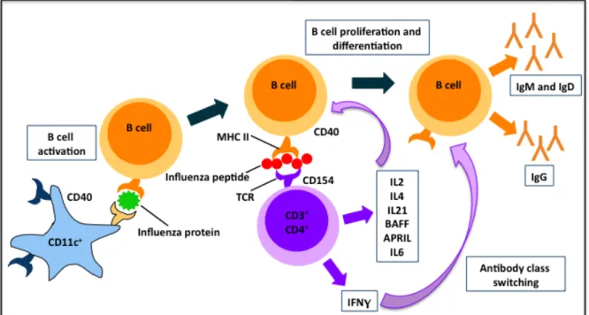
Figure 2.2 Dendritic Cell Presentation of Influenza Peptides to B Cells and CD4+ T Cell-induced Class Switching
Figure 2.2: Once activated, CD11c+ dendritic cells present influenza proteins to B cells, thereby activating them and inducing their differentiation and proliferation, as well as upregulating antibody production. CD4+ T cells are able to secrete numerous cytokines that are able to bind their receptors on the B cell in close proximity, including IL2, IL4, IL21, BAFF, and APRIL which mediate B cell proliferation and differentiation, and IL6, which acts as a B cell growth factor. CD4+ T cells also produce high levels of IFNγ, which is a potent inducer of B cell class switching to IgG antibodies, the primary antibody found in the serum and the most important antibody in the humoral response to the influenza virus.
microbes (196). This is important for influenza virus infection, which often is initiated in the upper respiratory mucosa (196). B cell affinity maturation also occurs in germinal centers, where antigen-specific B cells rapidly proliferate and experience high levels of somatic mutations in the Ig genes, resulting in the production of B cells that produce slightly
different antibodies with varying levels of affinities to the antigen (197). Follicular dendritic cells display the antigen and the B cells producing the antibodies with the highest affinity to the antigen are selected to survive (197). The cytokines IL2, IL4, and IL6 produced by CD4+ T cell further promote and regulate antibody synthesis and secretion by the selected B cells (181, 182). Antibody secreting plasma B cells can migrate from the germinal center to the bone marrow where they can reside for long periods of time and still produce the antigen-specific antibodies (198). In addition, some antibody-secreting B cells can become memory cells, which are able to survive in the periphery for long periods of time, even in the
absence of antigen (199). If the antigen is encountered in the future, the memory B cells will be able to act to mediate a faster and more effective humoral immune response for many years (199).
Antibody Function in the Humoral Response
initiating the opsonization process that marks the influenza virus particle for phagocytosis (196, 200). In addition, IgG antibody binding to HA and NA on the influenza virus can also trigger complement activation, which results in the formation of the membrane attack complex that destroys the virus (196, 200). IgG antibodies can also initiate
antibody-dependent cell-mediated cytotoxcity, which marks an influenza-infected cell for destruction by natural killer cells (196, 200).
Humoral Immune Response to Influenza Vaccination
IgG antibodies specific to viral proteins provide protection via the serum in the lower respiratory tract (203). Antibodies specific to influenza virus proteins can be detected in the peripheral blood within 10-14 days after infection initiation; IgG antibody levels peak 4-6 weeks after the start of the infection, while IgA and IgM antibodies reach their highest levels at about 2 weeks following the start of the infection (204). The inactivated TIV is injected intramuscularly in the deltoid muscle of the arm and the humoral immune response proceeds as described above, primarily directed towards the influenza virus membrane glycoproteins HA and NA (205). The humoral response to vaccination is rapid, as increases in influenza-specific antibodies can be seen in serum 2-6 days following
vaccination (205, 206). The predominant antibody detected in the serum in response to influenza vaccination is IgG, with IgA and IgM antibodies detected at lower levels (205, 207, 208). Even though subunit vaccines are purified formulations that primarily contain the surface glycoproteins HA and NA, vaccination with subunit vaccines can stimulate antibody production specific to other internal viral proteins, such as M1 and NP (209), which is interesting to note when interpreting data from our studies.
Cellular Immune Response to Influenza
CD4+ helper T cells and CD8+ cytotoxic T cells. CD4+ helper T cells produce cytokines, which function to promote inflammation, proliferation, and differentiation of T cells, as well as activation other immune cells such as macrophages and B cells; CD8+ cytotoxic T cells directly destroy infected cells. Both types of T cells have a restricted specificity for antigens, recognizing only peptide antigens that are bound to MHC molecules on the surface of other cells. There are two main types of MHC molecules: class I MHC molecules (MHC-I) and class II MHC molecules (MHC-II). Class I MHC molecules are expressed on all nucleated cells, while class II MHC molecules are expressed only on limited types of cells, including dendritic cells, B cells, and macrophages (211). In addition, the two types of MHC molecules function to sample different pools of peptide antigens. Class I MHC molecules access peptide
Dendritic Cells in Cellular Immunity
Dendritic cells are specialized or “professional” antigen-presenting cells and process microbial antigens from the external environment; they are found in many areas of the body including lymphoid organs, as well as the epithelium of the skin, gastrointestinal, and respiratory tracts (174, 175). Immature dendritic cells express CD209, a receptor that binds the mannose-type carbohydrates typically found on viruses, such as the influenza virus (213). After binding, dendritic cells can internalize and process antigen protein (174, 175). Activated dendritic cells localize to draining lymph nodes where they present antigen peptide-MHC complexes to naive T cells, initiating the adaptive immune response (174, 175). CD40, a transmembrane surface receptor,is constitutively expressed at low levels on non-activated dendritic cells; its expression is substantially upregulated when dendritic cells process antigen and is therefore used as marker of activation (214). CD40 signaling
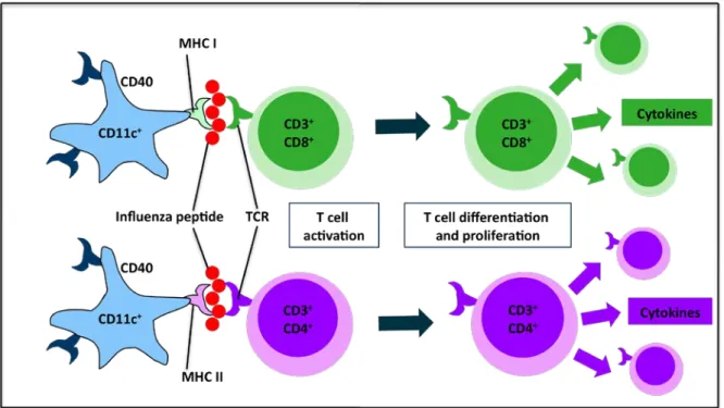
ingest infected cells and present extracellular antigens derived from these cells in
association with class I MHC molecules to CD8+ T cells, in addition to the more conventional route of presentation, in which the extracellular antigens are associated with class II MHC molecules and presented to CD4+ T cells (212), as shown in Figure 2.3. This is especially important with influenza viral infections to ensure that CD8+ cytotoxic T cell responses can be initiated despite the fact that the influenza virus is an intracellular microbe.
Figure 2.3 Dendritic Cell Presentation of Influenza Peptides to CD4+ and CD8+ T Cells
CD4+ and CD8+ T Cells in Cellular Immunity
The CD4 and CD8 molecules are transmembrane glycoproteins and coreceptors; they function to bind with MHC molecules and to enhance TCR signaling. CD4+ T cells and CD8+ T cells recognize and interact with class II MHC-peptide complexes and class I MHC-peptide complexes, respectively (211). CD4+ T cells and CD8+ T cells both express CD3, a
transmembrane protein with intracellular and extracellular domains (220). The T cell-specific Src family tyrosine kinase Lck is closely associated with the cytoplasmic regions of CD4 and CD8 and when activated will function to phosphorylate the tyrosine residues in CD3 on the T cells, thereby stimulating the T cell activation cascade (221). Many CD4+ and CD8+ T cells also express CD28, a costimulatory receptor that binds with B7 on dendritic cells and functions to initiate a signaling cascade that promotes activation of T cells (222). Activation of CD4 and CD8 results in proliferation and expansion of antigen-specific T cells and differentiation into effector and memory T cells. Dendritic cells further support expansion and differentiation of the T cell pool via the secretion of IL12 (223). Activated CD4+ and CD8+ T cells both produce and secrete IL2, which has autocrine functions to promote expansion and differentiation (224). T cells can migrate to the location of infection where they act to eliminate the source of infection.
cytotoxic function of CD8+ T cells and the activation and antibody production of B cells. It is primarily the TH1 subset of CD4+ T cells that mediates the immune response to influenza (189, 190). The main functions of CD8+ T cells are to kill influenza-infected cells, thereby limiting the spread and severity of infection, and also to secrete cytokines that act to regulate the immune response. The higher risk for influenza-related complications in obese individuals could be caused by defects in the CD4+ and CD8+ T cell responses to the virus.
The activation of CD4+ and CD8+ T cells is a highly coordinated process. CD69 is a T cell activation marker and its expression increases substantially and early in the activation process (225). CD28, the receptor for the costimulatory molecules CD80 and CD86, also increases with T cell activation and signals to promote proliferation, expansion, sensitivity to antigen, and survival of T cells (226). CD40L expression is upregulated in mature T cells and it binds and activates CD40 on dendritic cells (227). In addition, the IL12 receptor (IL12R) becomes enriched in the cell surface of activated T cells (228).
Effector CD4+ T Cells
differentiation on CD8+ T cells (229). CD4+ T cells can also activate macrophages to destroy the microbes that they have taken up by phagocytosis (230). CD4+ T cells can be further divided into a number of subsets, including TH1, and TH2 cells. Differentiation of CD4+ T cells into CD4+ TH1 cells is primarily mediated by IL12, which signals via the janus kinse-2-signal transducer and activator of transcription-4 (JAK2-STAT4) pathway (231-233), and by IFNγ, which signals via STAT1 (234, 235). One of the most important functions of differentiated CD4+ T cells is to produce numerous cytokines; T
H1 cells produce high amounts of IFNγ, particularly in response to influenza (189, 236), and lesser amounts of IL3, IL10, and granulocyte/macrophage-colony-stimulating factor (GM-CSF) (236-238), while TH2 cells produce large amounts of IL4, IL5, and IL13 and lesser amounts of IL3 and GM-CSF (237, 238). IL5, secreted predominantly by the TH2 subset of CD4+ T cells, is more closely
associated with allergic responses rather than viral pathogens (239). It is primarily the TH1 cells that mediate the immune response to influenza infection; in addition, TH1 cells promote the CD8+ T cell-mediated responses to influenza vaccination and help to maintain the vaccination-specific memory CD8+ T cell population (240).
Effector CD8+ T Cells
(241). Signals from CD4+ T cells may be required for differentiation of CD8+ T cells, such as the binding of CD154 on CD4+ T cells to CD40 on antigen-presenting cells, making them more efficient at stimulating differentiation of CD8+ T cells (242). The primary function of differentiated CD8+ T cells is to act to destroy cells that have been infected with intracellular microbes. CD8+ T cells form an immunological synapse with the infected antigen-expressing target cell (243). CD8+ T cells have granules in their cytoplasm containing substances that function to destroy target cells. Upon cytoskeleton rearrangement the granules are translocated to the immunological synapse, where they fuse with the plasma membrane and releases their contents (243). The granules contain perforin and granzymes. Perforin forms a pore in the target cell, which allows the granzymes to move into the infected cell (243), a process called degranulating, of which the marker CD107a is an indicator (244). One of the most potent granzymes is GrB, which executes the cytotoxic function of CD8+ T cells. GrB is a serine protease enzyme that cleaves peptides after aspartyl residues and activates both the caspase and Bcl-XL inhibitory protein BH3 interacting-domain (Bid) signaling cascades, resulting in the fragmentation of nuclear and mitochondrial DNA, respectively (245). GrB activates caspase-3 and the Bcl-2/Bid, which triggers the mitochondrial pathway of apoptosis (245). Bcl-2 is a mitochondrial protein and an inhibitor of the mitochondrial pathway of apoptosis (245). Bid can bind to Bcl-2 and inactivate it, thereby decreasing its apoptotic inhibition (245). Bid can also bind and opens the mitochondrial membrane Bax channel, which allows the mitochondrial protein cytochrome C to move into the cytoplasma where it helps to activate caspase-9 (245). CD8+ T cells also express the Fas ligand
initiates a signaling pathway that activates caspase-8 (245). Caspases are powerful
proteolytic enzymes that cleave proteins at aspartate residues, leading to destruction of the infected target cell (245). Another function of differentiated CD8+ T cells is to produce cytokines, including IFNγ, lymphotoxin, and TNFα, which promote inflammation and activate phagocytic cells (246).
Memory T Cells
Humoral and Cellular Responses to Influenza
The scope of the cellular response to influenza is dictated by the antigenic peptides that are presented to CD4+ cells and CD8+ T cells in the lymph nodes by dendritic cells or other antigen-presenting cells. The cellular response to influenza infection and vaccination is different than the humoral response as it is focused on the destruction of virus-infected cells, rather than infection prevention. The cellular response to infection is primarily mediated via the helping functions of CD4+ T cells through production of cytokines and via the cytotoxic functions of CD8+ T cells which directly kill infected cells. Infection with influenza virus activates a significant CD4+ T cell response, particularly the T
H1 CD4+ T cell response, which functions to upregulate B cell influenza-specific antibody production (159, 186). During infection, T cells specific to the influenza virus have been found in the
peripheral blood and lower respiratory tract secretions in humans (250). Numbers of influenza-specific CD8+ T cells can be measured in peripheral blood 6-14 days following initiation of infection or vaccinated vaccination in humans (251).
T Cell Metabolism
which includes the Kreb’s cycle, the electron transport chain, and fatty acid β-oxidation (254). After activation, T cells rapidly switch from obtaining most of their ATP from oxidative phosphorylation to almost entirely deriving ATP from aerobic glycolysis (255), in addition to the pentose phosphate pathway and glutaminolytic pathways (256, 257). This tremendous flux through aerobic glycolysis results in the production of very high levels of lactate from pyruvate, surprisingly even under conditions of adequate oxygen (258-260), similar to the Warburg effect in cancer cells (261). Therefore the major metabolic byproduct from activated T cells is lactate (254, 262); as much as 85% of glucose consumed by activated T cells is converted to lactate (263), and much of that is then secreted from the T cells. If glucose uptake or flux through the glycolytic pathway is suboptimal, T cells cannot synthesize cytokines, such as IFNγ (264), cannot proliferate effectively (265), and
proapoptotic proteins are upregulated, thereby increasing T cell death (265). Even under conditions of an excess of alternative energy sources, glucose limitations can prevent T cell proliferation and survival (264). Studies show that there is a direct positive correlation between proliferation and survival of T cells and increasing glucose concentrations (265). Furthermore, resting T cells express very low levels of the glucose transporter (GLUT) 1 (265), and the insulin receptor (266); however upon activation, expression of both GLUT1 and the insulin receptor is rapidly and significantly upregulated (265, 266). In addition, insulin may have anti-inflammatory effects on circulating immune cells and may drive T cell differentiation towards an anti-inflammatory TH2 phenotype, which may suppress the pro-inflammatory TH1 phenotype (267). Full activation of T cells requires two signals:
costimulation, which increases GLUT1 trafficking to the cell surface through PI3K/Akt
signaling pathway (268). Stimulation of T cells with both anti-CD3 and anti-CD28 antibodies, but with neither alone, resulted in increase GLUT1 translocation to the cell surface and significant increases in glucose uptake rate (268). In fact, increases in glucose utilization into T cells are seen as soon as one hour after stimulation with anti-CD3 and anti-CD28
antibodies (262, 269, 270). Increases in GLUT1 gene transcription, GLUT1 protein
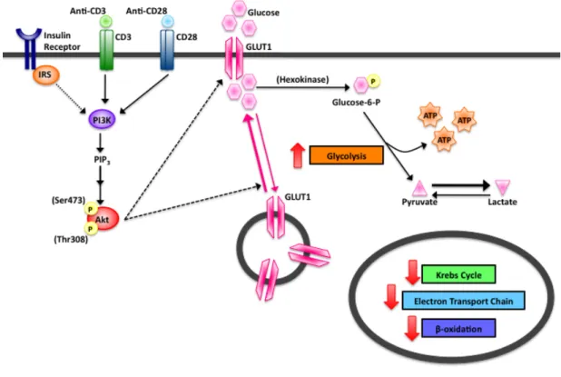
Figure 2.4 PI3K/Akt Signaling Pathway in Activated T Cells
Figure 2.4: In order to accommodate the increased glucose needs of the activated T cell, expression of the glucose transporter GLUT1 and the insulin receptor are rapidly
upregulated. Signaling through the PI3K/Akt pathway results in increased GLUT1 transcription, translation, translocation, and activity. Full activation of Akt requires phosphorylation at both the threonine 308 (Thr308) and serine 473 (Ser473) amino acid residues.
Significance
both. Potential humoral response mechanisms include defective maintenance of antibody-producing B cells or the production of less effective antibodies. Potential cellular response mechanisms include modifications to the proliferation, differentiation, or function of dendritic cells, effector CD4+ T cells, or effector CD8+ T cells or to the generation or maintenance of memory CD4+ T cells, or CD8+ T cells. Impaired humoral or cellular responses to influenza vaccination or influenza infection, resulting in increased
CHAPTER III
OBESITY IS ASSOCIATED WITH IMPAIRED IMMUNE RESPONSE TO INFLUENZA VACCINATION IN HUMANS1
Overview
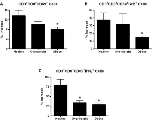
Obesity is an independent risk factor for morbidity and mortality from pandemic influenza H1N1. Influenza is a significant public health threat, killing an estimated 250,000 to 500,000 worldwide each year. More than one in ten of the world’s adult population is obese and more than two-thirds of the US adult population is overweight or obese. No studies have compared humoral or cellular immune responses to influenza vaccination in healthy weight, overweight and obese populations despite clear public health importance. The study employed a convenience sample to determine the antibody response to the 2009-2010 inactivated TIV in healthy weight, overweight and obese participants at one and 11 months post vaccination. In addition, activation of CD8+ T cells and expression of IFNγ and GrB were measured in influenza-stimulated PBMC cultures. BMI correlated positively with higher initial fold increase in IgG antibodies detected by ELISA to TIV, confirmed by HAI
1Previously published: Patricia A. Sheridan and Heather A. Paich (co-first authors), Jean
antibody in a subset study. However, eleven months post vaccination, higher BMI was associated with a greater decline in influenza antibody titers. PBMC’s challenged ex vivo with vaccine strain virus demonstrated that obese individuals had decreased CD8+ T cell activation and decreased expression of functional proteins compared with healthy weight individuals. These results suggest obesity may impair the ability to mount a protective immune response to influenza virus.
Introduction
Influenza causes some three to five million cases of severe illness and 250,000 to 500,000 deaths every year around the world, even in the absence of a major pandemic (8). Obesity is a growing health concern of epidemic proportions in many countries (1) ; more than one in ten of the world’s adult population is obese (1) and more than two-thirds of the US adult population is overweight or obese (14). In addition to co-morbidities such as cardiovascular disease and diabetes, obesity itself is an immunosuppressive condition (3-5). During the recent pandemic of influenza A/H1N1/2009 (pH1N1), obesity was recognized for the first time as an independent risk factor for increased influenza morbidity and mortality (6, 41, 274-276).