DO SIMILAR NEURAL PROFILES UNDERLIE SOCIAL COGNITIVE DEFICITS IN SCHZOPHRENIA AND HIGH-FUNCTIONING AUTISM?
Amy E. Pinkham
A dissertation submitted to the faculty of the University of North Carolina at Chapel Hill in partial fulfillment of the requirements for the degree of Doctor of Philosophy in the
Department of Psychology (Clinical Psychology).
Chapel Hill 2006
Approved by
Advisor: David Penn, Ph.D. Reader: Joseph Hopfinger, Ph.D.
ABSTRACT
AMY PINKHAM: Do Similar Neural Profiles Underlie Social Cognitive Deficits in Schizophrenia and High-functioning Autism?
(Under the direction of David Penn, Ph.D.)
Previous research suggests that schizophrenia and autism share similar behavioral deficits in social cognition. This study investigated both neural activation and behavioral performance during a task of complex social cognition in healthy controls, individuals with high-functioning autism, and individuals with schizophrenia. Event-related functional magnetic resonance imaging was utilized as individuals viewed faces and made ratings of trustworthiness. It was hypothesized that both clinical groups would show reduced activation in discrete brain regions comprising a social cognitive neural circuit, which included the amygdala, the fusiform face area (FFA) of the fusiform gyrus, and the superior temporal sulcus (STS). Activation in the ventrolateral prefrontal cortex (VLPFC) was also examined as this area has been implicated in the process of making evaluative judgments. Results largely confirmed the main study hypothesis: both clinical groups showed significant reductions in neural activation while making complex social judgments compared to non-clinical controls. Significant reductions for both non-clinical groups were evident in the right amygdala and FFA and left VLPFC, and no differences in neural activation were evident between the clinical groups. Behavioral performance on the Trustworthiness task
ACKNOWLEDGEMENTS
TABLE OF CONTENTS
Page
LIST OF TABLES……….viii
LIST OF FIGURES………..ix
Chapter I INTRODUCTION………...1
Social Cognition………...4
Social Cognition in Schizophrenia………...6
Social Cognitive Deficits and Biases in Schizophrenia………...6
Theory of Mind………..6
Attributional Style………..8
Social Perception………...9
The Functional Significance of Social Cognition in Schizophrenia………...12
Social Cognition in Autism………..14
Social Cognitive Deficits in Autism………14
Theory of Mind………15
Attributional Style………17
Social Perception……….17
The Neurobiology of Social Cognition………21
The Frontal Cortices and Theory of Mind………...22
The Fusiform Gyrus and Superior Temporal Sulcus in Face Processing………24
The Amygdala and Complex Social Perception………..25
The Neural Correlates of Social Cognition in Schizophrenia and Autism………...28
The FG, STS, and Amygdala in Schizophrenia………...29
Fusiform Gyrus………29
Superior Temporal Sulcus………30
Amygdala……….30
The FG, STS, and Amygdala in Autism………..32
Fusifrom Gyrus………32
Superior Temporal Sulcus………34
Amygdala……….34
Integration………..35
Unanswered Questions………...36
II METHOD………40
Participants………...40
Imaging Stimuli and fMRI Experiment………...42
Behavioral Tasks/Measures……….43
Cognitive Assessments………44
Symptomatology………..44
Image Acquisition………45
Spatial Preprocessing………...46
Data Analysis………...46
Imaging Data………47
Masks Defining Regions of Interest………48
Supplemental Analysis of ROI Time Courses……….49
Behavioral Data………...49
III RESULTS………51
Imaging Data………51
Within-group Comparisons………..51
Between-group Comparisons………...52
Behavioral Data………...54
Behavioral Data during Scanning: Trustworthiness Task…………...54
Social Functioning………...55
Cognitive Functioning and Symptomatology………..55
Correlational Analyses: BOLD Signal Change and Social Functioning………..56
IV DISCUSSION………..58
FIGURE CAPTIONS………..74
LIST OF TABLES
Table Page
1. Descriptive Statistics for Sample Characteristics………...70 2. Cerebral Foci of Activation within Each ROI in Response to the
Trustworthiness Task………..71 3. Descriptive Statistics for Behavioral Measures………..72 4. Correlations between Amygdala Activation and Social Fucntioning
LIST OF FIGURES
Figure Page
1. Social Cognition in Schizophrenia and Autism………..76
2. Unique Response of VLPFC and STS in Controls for Untrustworthy Faces……….77
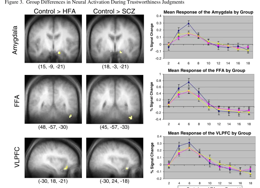
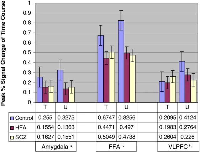
3. Group Differences in Neural Activation during Trustworthiness Judgments………78
4. Interaction between Trust Rating and Group………..79
CHAPTER I INTRODUCTION
The direct comparison of schizophrenia and autism enjoys a varied history in which the focus has primarily been differentiating the two disorders diagnostically. Recently however, investigators have shifted their attention to similarities between the disorders that suggest similar underlying pathologies. For instance, Goldstein and colleagues (2002) found similar cognitive profiles for individuals with high functioning autism and individuals with schizophrenia, and Sheitman, Kraus, Bodfish, and Carmel (2004) demonstrated that autistic symptoms can be present in schizophrenia and that these symptoms covary with negative symptoms. Similarly, Konstantareas and Hewitt (2001) noted that half of the autistic individuals in their sample met criteria for disorganized schizophrenia and that individuals with autism are likely to have several negative symptoms such as affective flattening, alogia, and asociality that are present in schizophrenia. These cognitive and symptom similarities provide a firm foundation for continued comparisons and open the door for evaluations across several other domains that may also speak to shared mechanisms underlying the disorders. Two such related areas are social functioning and the cognitive processes that subserve social functioning (i.e. social cognition).
Impairments in social functioning are characteristic of several psychological disorders; but nowhere are they more pronounced than in schizophrenia and autism.
(DSM- IV; APA, 1994), and individuals with schizophrenia often experience difficulties in multiple areas of social functioning such as interpersonal relationships, work and personal achievement, and self-care (Corrigan & Penn, 2001). Such deficits are present throughout the course of the disorder, including the first-episode, and are often resistant to psychiatric intervention (Addington & Addington, 2000). Additionally, social dysfunction has been found to precede the onset of psychosis and has been identified in individuals with a biological parent who has schizophrenia, both of which suggest that social impairments are vulnerability markers for developing schizophrenia (Davidson et al., 1999; Hans, Auerbach, Asarnow, Styr, & Marcus, 2000). In addition to a contributing role in the development of schizophrenia, poor social functioning has been linked to an increased rate of relapse (Perlick, Stastny, Mattis, & Teresi, 1992). Thus, it appears that impairments in social functioning represent a core behavioral feature of schizophrenia.
Similarly, social dysfunction is primary to autism and Asperger’s Syndrome. As with schizophrenia, impairments in social interaction are among the diagnostic criteria for both disorders (DSM-IV; APA, 1994), and adults with autism spectrum disorders show
impairments in social relationships, competency at work, independence, and social
autism spectrum disorders.
In an effort to better understand the processes underlying social dysfunction in both schizophrenia and autism, attention has been given to the role of neurocognitive abilities (e.g. executive function) in supporting social behavior. Although these theories have merit, some findings suggest that they are incomplete. For instance, reviews of the literature do support an association, both cross-sectional and longitudinal, between neurocognition and
psychosocial dysfunction in schizophrenia (Green, Kern et al., 2000; Penn et al., 1997); however, this relationship is only modest (Penn et al., 1997). Likewise, neurocognitive theories of autism fall short in explaining how some individuals, in particular, those with HFA or Asperger’s, can have intact cognitive abilities but still be socially impaired (Green, Kern et al., 2000). Because of these modest associations and inconsistencies, investigators have more recently sought to examine specific and unique aspects of cognition that underlie social function and that may be distinct from traditional neurocognitive domains. One such aspect that has been targeted in both disorders is social cognition.
possibility of amygdala dysfunction in schizophrenia and autism. Finally, conclusions regarding potential social cognitive and neural similarities between autism and schizophrenia will be discussed, and hypotheses for the proposed study will be provided.
Social Cognition
Social cognition refers broadly to the cognitive processes involved in how individuals perceive, interpret, and process social information. Definitions of social cognition vary widely in complexity and specificity, however two primary definitions include “the mental operations underlying social interactions, which include the human ability to perceive the intentions and dispositions of others” (Brothers, 1990, p. 28) and “the processes that subserve behavior in response to conspicifics, and in particular, to those higher cognitive processes suberving the extremely diverse and flexible social behaviors that are seen in primates” (Adolphs, 1999a). These definitions, and others, firmly link social cognition to social behavior and highlight the potential role that deficits in social cognition may play in social dysfunction. Further, several different specific skills comprise the domain of social cognition and thus support a multidimensional view of the construct. These skills include emotion recognition, social cue perception, theory of mind (ToM), and attributional style.
Evidence for the relative independence of social cognition from traditional
social functioning regardless of normative levels of cognitive intelligence (Bar-On, Tranel, Denburg, & Bechara, 2003). The fact that social cognition can become selectively impaired while sparing nonsocial cognition suggests that unique neural circuits subserve social
cognition. Additionally, lesions to ventromedial occipital cortex can result in prosopagnosia, a condition in which individuals show selective impairments in the perception of faces but preserved perception for nonsocial stimuli. Such findings have led some to suggest that facial processing is the result of domain specific neural mechanisms (Kanwisher, 2000).
This conclusion is bolstered by studies of clinical populations, specifically individuals with Williams' syndrome (WS) and individuals with autism, that show a dissociation between social cognitive and nonsocial cognitive skills in these groups. Individuals with WS tend to be outgoing and social, despite having below normal intelligence (Jones et al., 2000), and these individuals appear to have relatively preserved basic social cognitive skills (i.e., facial processing and simple Theory of Mind abilities, Karmiloff-Smith, 2000), despite having deficits in spatial cognition (Karmiloff-Smith, Klima, Bellugi, Grant, & Baron-Cohen, 1995; Tager-Flushberg, Boshart, & Baron-Cohen, 1998). Recent work examining specific neural structures in individuals with WS has attempted to explore this dissociation. Reiss and colleagues (2004) used structural neuroimaging to examine volumetric abnormalities in neural structures included in the visual-spatial system and the neural structures most
of social and nonsocial cognitive systems.
The partial preservation of social cognition seen in WS is in direct contrast to persons with High Functioning Autism and Asperger's syndrome, who show specific impairments in social cognition and social behavior that may not be related to general cognitive abilities (Heavey, Phillips, Baron-Cohen, & Rutter, 2000; Klin, 2000). As with WS, these findings lend support for the hypothesis that specific neural modules exist that are devoted to the processing of social information, a hypothesis that has also been maintained in the areas of evolutionary biology and primatology (Adolphs, 2001; Cosmides & Tooby, 1994; Frith & Frith, 1999; Penn et al., 1997; Phillips, Drevets, Rauch, & Lane, 2003a; 2003b).
Social cognition has been studied widely in both schizophrenia and autism. The following sections will present a brief review of work exploring social cognitive deficits in schizophrenia and the functional significance of these deficits. The autism literature will then be reviewed using a parallel structure.
Social Cognition in Schizophrenia Social Cognitive Deficits and Biases in Schizophrenia
The study of social cognition in schizophrenia has generally focused on three primary domains of social functioning: theory of mind, attributional style, and social perception, which includes emotion recognition (Penn, Addington, & Pinkham, 2006). In recent years, each of these three domains has received considerable attention; however, in keeping with the focus of this proposal, the majority of this review will be devoted to social perception whereas theory of mind and attributional style will be covered only briefly.
Theory of Mind
successful on simple ToM tasks but impaired on more complicated tasks, and other studies have found that the first-degree relatives of individuals with schizophrenia perform worse than non-clinical controls on ToM tasks (Janssen, Krabbendam, Jolles, & van Os, 2003; Wykes, Hamid, & Wagstaff, 2001) and that children who later develop schizophrenia perform poorly on measures assessing components of ToM (Schiffman et al., 2004), all of which lend support to a trait hypothesis.
Attributional Style
The majority of work on attributions in schizophrenia has focused on investigating attributional style in individuals with paranoia or persecutory delusions. From this work, two attributional biases have most commonly been observed: a self-serving bias and a
personalizing bias. Bentall and colleages (2001) have argued that individuals with persecutory delusions tend to show an exaggerated self-serving bias in which negative outcomes are attributed to others and positive outcomes to one's own actions (for partial failures to replicate this finding, see Kristev, Jackson, & Maude, 1999 and Martin & Penn, 2002), although this effect may be stronger for attributing negative outcomes to others, rather than taking credit for success (Garety & Freeman, 1999). A personalizing bias is evidenced by individuals with paranoia in that for negative interpersonal events, these individuals are more likely to blame others, rather than the situation or circumstances, relative to persons without paranoia and/or persecutory delusions (Bentall, 2001; Craig, Hatton, Craig, &
Social Perception
Studies of social perception in schizophrenia can be broken down into two general areas: facial affect recognition and social cue perception. Reviews of the literature on facial affect recognition (i.e., Edwards, Jackson, & Pattison, 2002; Hellewell & Whittaker, 1998; Kohler & Brennan, 2004; Mandal, Pandey, & Prasad, 1998; Penn et al. 1997) suggest the following conclusions. First, individuals with schizophrenia have deficits in facial affect perception compared to non-clinical control participants. Second, these deficits are present relative to individuals with other psychiatric disorders such as depressive disorder (Wenger, Lange, Ruther, & Irle, 2004); however, results are inconsistent when compared to disorders that include psychotic features such as bipolar disorder. Third, greater impairment is evident for the perception of negative emotional displays compared to positive displays, with perhaps the greatest impairment for the perception of fear (Edwards, Pattison, Jackson, & Wales, 2001; Evangeli & Broks, 2000; Kohler et al., 2003). Fourth, longitudinal studies support a stable deficit in emotion perception (Addington & Addington, 1998; Gaebel & Wolwer, 1992; Kee, Green, Mintz, & Brekke, 2003; Kucharska-Pietura & Klimkowski, 2002); although, there is some evidence that individuals whose symptoms are in remission may perform better on affect perception tasks than individuals in an acute phase of the disorder (Gessler, Cutting, Frith, & Weinman, 1989; Penn et al., 2000). Fifth, there is some evidence that individuals with paranoid schizophrenia are better at facial affect perception than
Loughland, Gordon, & Davidson, 1999; Williams, Loughland, Green, Harris, & Gordon, 2003) and spend less time examining salient features of the face (Loughland, Williams, & Gordon, 2002b; Phillips & David, 1997; Phillips & David, 1998; Williams et al., 1999), which may contribute to poor performance (Loughland et al., 2002a; 2002b; Williams et al., 1999). And finally, the jury is still out regarding whether facial affect perception deficits are part of a generalized performance deficit (Baudouin, Martin, Tiberghien, Verlut, & Franck, 2002; Bellack, Blanchard, & Mueser, 1996; Kerr & Neale, 1993; Mueser et al., 1996; Sachs, Steger-Wuchse, Kryspin-Exner, Gur, & Katschnig, 2004; Salem, Kring, & Kerr, 1996) or specific to decoding only facial emotions (e.g., Heimberg, Gur, Erwin, Shatasel, & Gur, 1992; Penn et al., 2000).
Unlike facial affect recognition stimuli, tasks that assess social cue perception utilize more dynamic stimuli that require multiple sensory modalities, and consistent with work on facial affect perception, individuals with schizophrenia are generally impaired in general social perception (Archer, Hay, & Young, 1994). Bell, Bryson, and Lysaker (1997)
evaluated the performance of individuals with schizophrenia on a task of emotion recognition in which an actor portrayed a basic emotion through facial expression, verbal tone, and upper-body movements while reciting one of three standardized monologues. They found that individuals in the schizophrenia sample performed significantly worse than individuals with substance abuse and healthy control participants. Corrigan, Davies-Farmer, and Stolley (1990) also found that individuals with nonparanoid schizophrenia were impaired in
accurately recognizing social cues from vignettes of social interactions.
actor’s behavior (e.g., “what is she doing?”) and characteristics (e.g., “what is she wearing?”) whereas abstract cues consist of inferences of affect and goals. In a series of studies,
Corrigan and colleagues have found that individuals with schizophrenia are more sensitive to, and better able to recognize, concrete social cues rather than abstract ones (Corrigan, Garman, & Nelson, 1996; Corrigan & Green, 1993a; Corrigan & Green, 1993b; Corrigan & Nelson, 1998; Corrigan, Silverman, Stephenson, Nugent-Hirschbeck, & Buican, 1996), a finding consistent with what we would expect from individuals who have difficulties in discerning the intentions of others (i.e., Theory of Mind skills).
A third category of social perception that is just recently receiving attention is the perception of stimuli requiring complex social judgments. As knowledge of basic emotion perception abilities has grown, researchers have become increasingly interested in more complex, real-life social cognitive situations. Thus, assessment methods and stimuli have become more sophisticated, requiring participants to draw upon several social cognitive abilities within the same task. For example, the Eyes Test (Baron-Cohen, Wheelwright, Hill, Raste, & Plumb, 2001) incorporates both emotion perception and ToM abilities. This task consists of still frames of the eye-region of faces depicting various social emotions and complex mental states; therefore, in order to perform well on the task, participants must be able to identify social emotions as well as make inferences about the intentions of the individual shown. A similar task that requires participants to make complex mental
judgments is the Trustworthiness/Approachability Task developed by Ralph Adolphs (1998). Here, participants are shown black and white photographs of individuals’ faces, and they are asked to rate how trustworthy and approachable they perceive each individual to be.
into the literature. No studies have investigated the performance of individuals with
schizophrenia on the Trustworthiness/Approachability Task, and only two have utilized the Eyes Task. These studies are however largely consistent with the general findings for social perception. Specifically, Craig et al. (2004) found that individuals with paranoid
schizophrenia performed significantly worse than healthy controls on the Eyes Task and that this effect remained when controlling for IQ. Likewise, Oguz and colleagues (2003) found similar performance deficits for individuals with schizophrenia and schizoaffective disorder that were not correlated with IQ. Finally, in a later study, Oguz et al. (2005) also found poor performance on the Eyes Task was associated with the severity of negative symptoms. The Functional Significance of Social Cognition in Schizophrenia
As noted previously, investigators turned their attention to social cognition in the hopes of identifying factors that underlie social functioning, and thus far, several studies have confirmed that a relationship does exist between social cognition and social functioning. To begin, relationships with various aspects of social functioning have been clearly elucidated for affect recognition. For instance, Kee et al. (2003) reported that emotion perception was related to work functioning and independent living, and Poole, Tobias, and Vinogradov (2000) found that errors in affect recognition were correlated with lower quality of life and impoverished interpersonal relations. Hooker and Park (2002) also reported that emotion perception abilities were associated with communication and occupational abilities. Another study found that, for inpatients, affect perception was related to social competence as
(clear enunciation of speech) and conversation involvement as assessed by a role play test. Finally, facial affect perception has also been associated with adaptive ward functioning, particularly hygiene and grooming (Penn, Spaulding, Reed, & Sullivan, 1996).
While sparse, work attempting to link social cue perception to social functioning has also supported a relationship between the two. Corrigan and Toomey (1995) found that sensitivity to social cues was associated with interpersonal problem-solving skills; however, a similar study by Ihnen et al. (1998) that attempted to link social cue perception to social skill found only a weak association. This latter finding is somewhat contradictory to the findings of a study by Bellack and colleagues (1992). Bellack et al. found that social perception was correlated with overall social skill but that this relationship held only in situations involving negative affect. This caveat may explain the discrepancy between these two findings since Ihnen et al. did not examine negative affect situations. Thus, there is some, albeit limited evidence, that social cue perception is related to social functioning.
In addition, there is evidence that social knowledge and general social perception are related to social functioning among persons with schizophrenia. Specifically, the ability to identify the sequence of behavioral steps used in social situations and to place them in the correct order was associated with less irritability on the ward among chronically ill patients (Penn et al., 1996) and persons recovering from an acute psychotic episode (Penn, Ritchie, Francis, Combs, & Martin, 2002). Moreover, Appelo and colleagues (1992) reported that knowledge of social situations accounted for more variance in ward functioning than symptoms.
In Brune (2005), ToM was found to account for 24% of the variance in severe social
behavioral problems, and Roncone et al. (2002) found that ToM was related to global social functioning and that this relationship remained when controlling for IQ. Roncone and colleagues also noted that ToM abilities accounted for more variance in social functioning than cognitive factors such as verbal fluency, memory, and executive function. Similarly, Pinkham and Penn (in press) reported that performance on measures of ToM was related to better overall social skill.
Thus, there is growing evidence that social cognition is related to social impairments in schizophrenia, and the foregoing provides strong evidence for the functional significance of social cognition in schizophrenia. Perhaps most impressive is that in a few studies (Corrigan & Toomey, 1995; Penn et al., 1999; Penn et al., 1996; Roncone et al., 2002), the association between social cognition and social functioning could not be accounted for by cognitive deficits. These findings lend support to the hypothesis that social cognition contributes independent variance to functional outcomes beyond non-social cognition alone.
Social Cognition in Autism Social Cognitive Deficits in Autism
Theory of Mind
In autism, ToM deficits span the developmental period and remain present in adulthood, a finding which has prompted some to hypothesize that ToM impairments are a core feature of the disorder and the primary deficit in autism (Cohen, 1989; Baron-Cohen, Leslie, & Frith, 1985; Kleinman, Marciano, & Ault, 2001; Leslie & Frith, 1988; Ozonoff, Pennington, & Rogers, 1991). These deficits are manifest in young children with autism as absent or delayed joint attention behaviors (Dawson et al., 2004; Morgan,
Maybery, & Durkin, 2003) and as difficulty with pretend play (Jarrold, 2003; Rutherford & Rogers, 2003). Later, children with autism spectrum disorders show difficulty with false belief tasks (Baron-Cohen, 1995; Yirmiya, et al., 1998) and other more complex ToM abilities such as deception and faux pas (Baron-Cohen, O’Riordan, Stone, Jones, & Plaisted, 1999; Brent, Rios, Happe, & Charman, 2004; Pilowsky et al., 2000). Although ToM abilities remain impaired as compared to age matched typically developing individuals, some
improvement over time does occur such that higher functioning adolescents and adults become able to pass first- and second-order false belief tasks (Bowler, 1992; Dahlgren & Trillingsgaard, 1996; Steele, Joseph, & Tager-Flushberg, 2003).
Given that most adults with autism spectrum disorders are able to pass false belief tasks, more complicated tasks have been utilized to examine ToM abilities in older
ability to make complex social judgments and include multiple social cognitive processes. As noted earlier, the Eyes task incorporates elements of emotion perception as well as ToM, and the SAT necessitates the attribution of biological motion as well as ToM. Despite these complications, studies using these tasks will still be reviewed under the current ToM heading in order to remain consistent with the conclusions put forth by the authors of these studies.
On the whole, studies of ToM abilities in adults with autism spectrum disorders have shown considerable deficits as compared to healthy controls. A series of studies by Baron-Cohen and colleagues has consistently shown that high functioning individuals with autism and Asperger’s syndrome are impaired on the Eyes Task (Baron-Cohen, Jolliffe, Mortimore, & Robertson, 1997; Baron-Cohen et al., 2001; Baron-Cohen, Wheelwright, & Jolliffe, 1997). One of these, Baron-Cohen, Jolliffe et al. (1997), also utilized a clinical control group,
individuals with Tourettes Syndrome, and still found impairments in ToM. Likewise, other groups have used the Eyes Task in conjunction with other ToM tasks to demonstrate that ToM impairments in autism are not limited to the visual modality (Kleinman et al., 2001) and that these impairments extend beyond attributing a mental state to a face; they are also
apparent when trying to infer the true meaning of a hint (Craig et al., 2004). These overall deficits are confirmed by meta-analyses of studies examining ToM and appear to be present not only as compared to healthy individuals but to individuals with mental retardation as well (Yirmiya et al., 1998).
elements of the story and to use ToM or affective terms in their narrations. Moreover, he found that performance was not related to age or verbal ability. These findings not only supported a general deficit in ToM abilities, but also that individuals with autism spectrum disorders may not naturally seek social meaning in the environment.
Attributional Style
Although attributional style is under-researched in autism spectrum disorders, a few studies have attempted to apply the attributional model of paranoia to Asperger’s syndrome. Increased rates of paranoia are often seen clinically in individuals with Asperger’s syndrome (Hare 1997; Wing 1996), and given this similarity to psychotic disorders, two studies have investigated whether the attributional biases seen in schizophrenia are also be present in individuals with Asperger syndrome (Blackshaw, Kinderman, Hare, & Hatton, 2001; Craig et al., 2004). Both studies found increased rates of paranoia as compared to healthy controls, however both also failed to find evidence of attributional biases. Thus from these limited data, it appears that individuals with Asperger syndrome do not display any attributional abnormalities and that the paranoia seen in such individuals likely stems from mechanisms that differ from those of schizophrenia.
Social Perception
The ability to perceive biological motion is most commonly assessed using point-light displays of human figures (i.e. Grossman & Blake, 1999), but other methods such as moving animated characters (Pelphrey, Morris, & McCarthy, 2004) and geometrical shapes moving in conjunction to mimic walking (Pelphrey, Mitchell, et al., 2003) have also been used. To date, only point light displays have been utilized with an autistic sample, and the two studies that have done so have employed children. Moore, Hobson, and Lee (1997) found that children with autism consistently performed more poorly than control participants in differentiating biological motion from the motion of inanimate objects. A similar study confirmed this deficit in perceiving biological motion and found a positive correlation between degree of autistic symptomatology and impairment in detecting biological motion (Blake, Turner, Smoski, Pozdol, & Stone, 2003). Unfortunately, it would be premature to conclude that these deficits will persist into adulthood, but these studies do provide a foundation upon which such a hypothesis could be based.
Beyond perceiving biological motion, investigators have also examined how
Lahaie et al., 2006 for evidence of enhanced processing of facial features in autism). Examination of the visual scanpaths of individuals with autism also confirms an abnormal featural processing strategy. Klin and colleagues (2002) demonstrated that when viewing social scenes, individuals with autism spend less time examining the eyes of actors in the scene and more time looking at the mouths of the actors or objects in the scene. Pelphrey et al. (2002) also found that individuals with autism devote more time to viewing non-salient features of the face (i.e. the ear or chin) than core features of the face such as the eyes, nose, and mouth. Interestingly, Pelphrey et al. monitored these visual scanpaths within the context of an emotion recognition task, and the behavioral results of this study indicated that
individuals with autism correctly identified fewer emotions than controls. Together, these results support the hypothesis that individuals with autism utilize abnormal strategies to process faces and that these strategies may contribute to impairments in face and emotion processing.
Grossman, Klin, Carter, & Volkmar, 2000; Pelphrey et al., 2002). Further, Loveland et al. (1997) demonstrated that high functioning individuals performed better on emotion
recognition tasks than lower functioning individuals, which has prompted some to conclude that high functioning individuals may be able use compensatory strategies to mask deficits when processing basic emotions (Teunisse & de Gelder, 2001). Third, and as alluded to above, greater impairment is evident for more complex emotions such as fear, surprise, and complex social emotions (e.g. distrustful, accusing, and friendly) (Baron-Cohen et al., 1993; Baron-Cohen et al., 2001; Capps et al., 1992; Pelphrey et al., 2002). Fourth, the evidence is mixed regarding whether this is a specific deficit (Celani, Battacchi, Arcidiacono, & Di-Domenico, 1999; Hobson, 1986a; Hobson, 1986b; Hobson, Ouston, & Lee, 1988b; Weeks & Hobson, 1987) or if it can be accounted for by more generalized deficits in face perception (Critchley, Daly, Bullmore, et al., 2000) or cognitive abilities (Ozonoff, Pennington, & Rogers, 1990).
The last area of social perception is the processing of complex social stimuli. Similar to the current trend in schizophrenia research, autism investigators have recently begun exploring the ability of individuals with autism to process stimuli in a manner that requires complex social judgments. As can be recalled from the ToM review, several studies have utilized the Eyes Task to demonstrate that individuals with autism show deficits in
processes necessary for higher-level social cognition and that they may fail to link social judgments to the perception of a face. Studies such as these are the first of their kind to use tasks that are sensitive to, and that target, subtle impairments in social cognition.
The Functional Significance of Social Cognition in Autism
As compared to schizophrenia, less emphasis has been placed on understanding the impact of social cognition on functioning in autism; however, the studies that have examined this question do support a relationship between social cognitive abilities and social
functioning. Specifically, ToM abilities have been linked to social behavior in children with autism (Frith, Happe, & Siddons, 1994; Hughes, Soares-Boucaud, Hochmann, & Frith, 1997), and various elements of social perception have also been shown to correlate with social functioning in individuals with autism and individuals with pervasive developmental disorders (PDD). In the Klin et al. (2002) study examining the visual fixation patterns of individuals with autism while viewing social scenes, the authors found that more time fixating on objects in the scene instead of the actors was related to poorer social adjustment. Additionally, Fein and colleagues (1992) noted that emotion perception abilities were
correlated with level of social skill in individuals with PDD, and a similar study by Braverman and colleagues (1989) also reported that deficits in emotion perception were related to greater social impairment.
The Neurobiology of Social Cognition
2000; Baron-Cohen et al., 1994; Stone, Baron-Cohen, & Knight, 1998), as well as several others that may play secondary roles (i.e. the right parietal cortex, the insular cortex, the basal ganglia, (Adolphs, 2002) and the temporal-parietal junction at the top of the superior temporal gyrus, and the temporal poles, (Frith, 2001)). Although these neural structures also subserve other cognitive functions (e.g., problem solving; conceptual reasoning), they, and not other neural structures, tend to be most consistently activated in response to social stimuli, thus underscoring their role in neural models of social cognition. In what follows, I will briefly describe the major neural structures and mechanisms that have consistently shown a role in social cognition, particularly those structures implicated in ToM and social perception. An in depth treatment of each brain region would far exceed the scope of this paper; thus, I will limit this discussion to the specific areas proposed by Brothers (1990) and Adolphs (1999a; 2001; 2002) as subserving social cognition: the medial prefrontal cortex, the superior temporal sulcus, the fusiform gyrus, and the amygdala. Finally, in keeping with the focus of this project, research elucidating the role of the amygdala in social perception will be reviewed in the most detail.
The Frontal Cortices and Theory of Mind
There is growing evidence that performance on ToM tasks is associated with
during similar non-ToM tasks. Similarly, Goel, Grafman, Sadato, and Hallett (1995) found selective activation of the left medial frontal cortex (BA 9) throughout a ToM task in which normal participants were asked to infer the thoughts of a contemporary of Christopher Columbus. Thus, results from these early studies indicated that ToM was specific to the medial frontal cortex (see also Calarge, Andreasen, & O’Leary, 2003; Stuss, Gallup, & Alexander, 2001 for more recent support of the role of the medial frontal cortex in ToM).
More recent studies have used both verbal and nonverbal tasks in their experimental design, and have supported the role of the prefrontal cortex (McCabe, Houser, Ryan, Smith, & Trouard, 2001; Vogeley et al., 2001), specifically the medial prefrontal cortex, including portions of BA 8 and 9, in ToM skills. Gallagher and colleagues (2000) used functional magnetic resonance imaging (fMRI) to assess brain activity while participants read and answered theory of mind questions about a verbal passage, and interpreted and explained the meaning of cartoons that required theory of mind skills. Relative to control conditions, there was unique activation of the medial prefrontal cortex during the ToM tasks. Similar results were found using only a cartoon task (Brunet, Sarfati, Hardy-Bayle, & Decety, 2000). Additionally, the medial prefrontal cortex has also been implicated in ToM tasks that use non-human stimuli and tasks that do not explicitly ask participants to interpret the mental states of others. For example, Castelli and colleagues (2000; 2002) found that the medial prefrontal cortex was selectively activated when the movement patterns of geometric shapes evoked mental state attribution but not during simple action description. German, Niehaus, Roarty, Giesbrecht, and Miller (2004) also found increased activity in the medial prefrontal cortex when subjects viewed pretend actions as opposed to real actions (recall that
Additional studies have also implicated the orbito-frontal cortex in ToM skills. Baron-Cohen et al. (1994) used single photon emission computerized tomography (SPECT) to identify areas of activation during performance on a mental state terms task (a ToM task). They found increased cerebral blood flow in the right orbito-frontal cortex of healthy
participants during a ToM task, but not during a control task. Lesion studies also lend support to this pattern of findings. Stone et al. (1998) found that individuals with bilateral orbito-frontal lesions performed similarly to individuals with Asperger’s syndrome on a task requiring the recognition of a faux pas, a task that requires social reasoning as well as theory of mind. And, Mah, Arnold, and Grafman (2004) found that individuals with lesions to the orbitofrontal/anterior cingulate cortex were impaired in the detection of lies, also a process that requires ToM. Collectively, the these studies suggest that activation of the medial prefrontal cortex, and to some extent, the orbito-frontal cortex, is critical to being able to infer the mental states of others (see Frith, 2001, for two additional brain regions that are activated during ToM tasks but that are not within the scope of this paper).
The Fusiform Gyrus and Superior Temporal Sulcus in Face Processing
Gauthier, Curran, Curby, & Collins, 2003; Gauthier, Skudlarski, Gore, & Anderson, 2000). Additionally, the FG activates strongly during tasks focusing on identity and appears to be most involved in the processing of non-changeable, static aspects of the face (Haxby, Hoffmann, & Gobbini, 2000; Winston, Henson, Fine-Goulden, & Dolan, 2004).
In contrast, the STS is more strongly activated during tasks focusing on visual gaze shifts (Kingstone, Tipper, Ristic, & Ngan, 2004; Pelphrey, Singerman, Allison, & McCarthy, 2003; Pelphrey, Viola, & McCarthy, 2004) and is involved in processing the changeable aspects of the face such as the eyes and the mouth (Haxby et al., 2000; Winston et al., 2004). Such a distinction between the static and dynamic features of the face is important because it is the dynamic features that provide the most social information. Changes in the direction of gaze indicate the focus of one’s attention, and changes in the shape of the eyes and mouth facilitate emotional expression, and indicate emotions such as happiness and aggression. The Amygdala and Complex Social Perception
Both lesion and imaging studies have consistently supported the role of the amygdala in detecting threat, recognizing emotions, and making complex social judgments.
recognizing the intensity of fearful expressions (Adolphs, Tranel, Damasio, & Damasio, 1994). Although these studies and others (Anderson & Phelps, 2000; Broks et al., 1998) support a specific impairment in the recognition of fear, several more recent studies have found similar deficits for other negatively valanced emotions such as sadness and anger. Schmolck and Squire (2001) noted that patients with bilateral amygdala lesions had difficulty discriminating negative emotions and often mistook fear for surprise or anger and sadness for disgust or anger. Similarly, in two different studies, Adolphs and Tranel also found that individuals with bilateral lesions show a specific impairment in recognizing sadness as opposed to happiness (2004), and that individuals with bilateral amygdala damage are more likely to falsely identify anger as happiness (2003).
Functional neuroimaging studies utilizing healthy participants have largely confirmed and extended the findings from lesion studies. Using PET, Morris et al. (1996) found a differential response in the amygdala to fear and happiness. Amygdalar activation was much more pronounced when participants viewed photographs of fearful faces, and there appeared to be an interaction between level of activation and intensity of emotion such that the more fearful a face looked, the greater the level of activation. Using fMRI, Phillips et al. (1997) explored the differential amygdalar response to fear by comparing activation in response to fearful and disgusted facial expression whereas Breiter and colleagues (1996) compared fearful and neutral expressions. Results from both studies indicated that the amygdala was only activated when viewing fearful faces and not when viewing disgusted and neutral faces, respectively. Similarly, Whalen et al. (2004) found significantly greater activation of the amygdala in response to fearful eye whites as compared to happy eye whites.
demonstrating that it is maximally engaged during automatic or rapid processing of faces. Whalen et al. (1998) presented photographs of happy and fearful expressions in a backward masking procedure that resulted in the majority of participants being unaware of seeing fearful and happy expressions, and despite lack of conscious awareness, significantly greater amygdalar activation was noted in response to fearful faces. Likewise, Critchley, Daly, Phillips et al. (2000) compared explicit and implicit facial emotion processing and found that implicit processing activated the amygdala to a greater extent than explicit processing. Indeed, Hariri et al. (2000) noted that asking participants to actively label emotional expressions might actually result in deactivation of the amygdala, a finding that has been mirrored in investigations of automatic and controlled social evaluation (Cunningham et al., 2004). Overall, findings from both lesion and imaging studies clearly indicate that the amygdala is important for emotion recognition and suggest that the amygdala may play a disproportionate role in the automatic processing of negative or threatening stimuli (Adophs et al., 1999).
significance (Adolphs, Baron-Cohen, & Tranel, 2002). An interesting line of research examining the role of the amygdala in evaluating trustworthiness supports this view; both lesion and imaging studies have established that the amygdala is integral to making such social judgments. First, Adolphs et al. (1998) asked three individuals with complete bilateral amygdala damage and seven individuals with unilateral amygdala damage to rate faces for approachability and trustworthiness. All three bilateral participants judged the faces to be more approachable and trustworthy than control participants, and this was most notable for faces that the control participants rated the least approachable and trustworthy. In contrast, individuals with unilateral lesions performed comparably to control participants. Taken together, these results seem to imply that only unilateral activation of the amygdala, as opposed to bilateral activation, is necessary for accurate processing of trustworthiness stimuli and that this processing is not highly lateralized. A functional imaging study of healthy individuals lends credence to this conclusion. Winston and colleagues (2002) found bilateral activation of the amygdala in response to untrustworthy faces, and interestingly, faces rated as the most untrustworthy evoked greater amygdalar response. Thus, as can be seen from these studies, the amygdala is not limited to the processing of basic emotions but also plays a critical role in complex social judgment.
consistent with the focus of this project, only those structures primarily involved in social perception will be reviewed, however, please see Pinkham, Penn, Perkins, and Leiberman (2003) for a review of the frontal cortices and ventromedial prefrontal cortex in
schizophrenia.
The FG, STS, and Amygdala in Schizophrenia Fusiform Gyrus
In general, the fusiform gyrus shows decreased volume and abnormal activation in persons with schizophrenia. First, both McDonald et al. (2000) and Paillere-Martinot et al. (2001) found decreased regional gray matter in the left fusiform gyrus of individuals with schizophrenia as compared to healthy controls, and their findings have been replicated in both chronic (Onitsuka et al., 2003) and first-episode samples (Lee et al., 2002; although see Pinkham et al., 2005 for a failure to replicate). Second, several studies show reduced
activation of the FG while viewing faces (see Hempel et al., 2003 for conflicting results). Both Streit et al. (2001) and Quintana et al. (2003) reported that individuals with
schizophrenia show reduced activation of the right lateral FG during emotion perception tasks as compared to healthy individuals. Moreover, a study by Williams et al., (2004) found reduced activation of the FG bilaterally as compared to healthy individuals. It is also
interesting to note that Malaspina and colleagues (1999) report increased rCBF in the right fusiform gyrus during a visual fixation task. Although the latter finding may initially seem counterintuitive, one must consider that increased rCBF was not present in healthy control participants and in this respect, may indicate an abnormality in individuals with
suggest that the FG of individuals with schizophrenia may not be specialized for social stimuli.
Superior Temporal Sulcus
Unfortunately, very little work has spoken to either STS volume or functioning in schizophrenia, and the only study that has done so utilized a ToM task instead of a social perception measure. In this study, Brunet, Sarfati, Hardy-Bayle, and Decety (2003) asked participants to choose concluding frames for comic strips that required the understanding of intentions or physical causality. Despite the absence of moving figures or real people, the authors still found activation of the right STS in individuals with schizophrenia during the processing of cartoons that included human figures. This activation was comparable to healthy controls. Thus, from this very limited evidence, it appears that individuals with schizophrenia may display intact STS functioning.
Amygdala
2005). Additionally, the findings of Sumich et al. also suggest that reductions in amygdala volume may not be consistent across symptom profiles or schizophrenia subtypes.
Specifically, in this study, individuals with paranoid schizophrenia were found to have significantly smaller left amygdala volumes than non-paranoid individuals. Thus, the exact nature of these volume reductions may be more subtle than originally thought.
There is also evidence that amygdalar activation is abnormal in individuals with schizophrenia, particularly when negative affect is involved. Schneider and colleagues (1998) used mood induction in both persons with schizophrenia and normal controls and showed that persons with schizophrenia had reduced amygdalar activation during sadness, despite self-ratings of sadness that were comparable to controls. In addition, Taylor, Liberzon, Decker, and Koeppe (2002) found reduced activation of the amygdala in
individuals with schizophrenia while viewing non-aversive, but emotionally salient stimuli, and Takahashi et al. (2004) found reduced activation of the right amygdala in individuals with schizophrenia as compared to controls while viewing emotionally unpleasant stimuli despite similar behavioral ratings of the photos. Moreover, a study utilizing an emotion labeling task found significantly less activation of the amygdala bilaterally in individuals with schizophrenia as compared to control participants (Hempel et al., 2003), and a similar study found reduced activation of the left amygdala in patients while discriminating emotional valance (Gur et al., 2002; see Kosaka et al., 2002 for evidence of increased activation when identifying happiness).
Finally, a few innovative studies have begun to examine whether amygdala activation may vary within schizophrenia sub-type. One such study evaluated individuals with
that relative to healthy control participants, both groups of individuals with schizophrenia were not only less accurate in identifying emotions, but they also displayed no amygdalar activation to fearful expressions (Phillips et al., 1999). An elegant study combined fMRI and skin conductance arousal measures to further explore potential differences between paranoid and non-paranoid patients. Via this methodology, Williams and colleagues (2004) found that individuals with paranoid schizophrenia reacted with greater levels of arousal to fearful faces than either controls or non-paranoid patients and that despite this increased arousal, these individuals showed reduced amygdala activity. Interestingly, non-paranoid subjects did not differ from controls in level of amygdala activation. Collectively, these studies and those reviewed earlier provide considerable evidence of reduced amygdala activity in
schizophrenia although no definitive conclusions can yet be drawn concerning the consistency of this abnormality across schizophrenia sub-types.
The FG, STS, and Amygdala in Autism Fusiform Gyrus
Given the considerable evidence for face processing deficits in autism and the relative specificity of the FG, this neural structure became an early target for investigators. Schultz and colleagues (2000) were among the first to compare FG activation in individuals with autism to typically developing individuals. In this study, Schultz et al. first identified
Several subsequent studies have complemented these findings by also reporting a lack of FG activation during face perception (Hubl et al., 2003; Pierce, Muller, Ambrose, Allen, & Courchesne, 2001), explicit processing of facial emotions (Critchley et al., 2000), and affect perception (Hall et al., 2003; Piggot et al., 2004).
and a visual stimulus in which a participant with autism had a special interest, namely “Digimon” cartoon characters. By visually presenting these stimuli during functional neuroimaging, the authors demonstrated FG activation in response to Digimon stimuli but not to familiar or unfamiliar faces. Here, FG activation required a stimulus in which the participant had a special interest, or expertise, but it is worthwhile to note that FG activation was achieved.
Superior Temporal Sulcus
As with schizophrenia, there is a general lack of information addressing STS volume and functioning in autism, however, the information that is available does suggest both volumetric and functional abnormalities in autism. First, a structural MRI study of children with autism revealed decreased volumes of grey matter in the STS as compared to typically developing children (Boddaert et al., 2004), and second, both functional imaging studies that examined the STS found reduced activation as compared to control participants (Castelli et al., 2002; Pierce et al., 2001). In Castelli and colleagues, reduced activation was found in response to animations of geometrical shapes moving in socially relevant patterns, and Pierce et al. noted reduced activation during face perception.
Amygdala
Reviews examining amygdala volume in autism provide mixed results with some studies showing increased volume as compared to controls whereas others show no
differences or reductions in volume (see Brambilla et al., 2003 and Sweeten, Posey, Skekhar, & McDougle, 2002 for reviews). Functional studies, on the other hand, present more
Analysis of this data revealed no activation of the amygdala in individuals with autism. Similarly, Pierce et al. (2001) failed to find amygdala activation in individuals with autism during a task of face processing, and Critchley, Daly, Phillips et al. (2000) reported a lack of activation of the left amygdala during implicit processing of facial expressions as compared to control participants. Thus far, only one study has failed to find differences in amygdala activation in individuals with autism (Piggot et al., 2004), and although these results are not consistent with previous studies, it is possible that task demands and participant
characteristics can account for the null finding. Specifically, this study included only high-functioning individuals with autism and asked them to process only basic emotions. Given that high-functioning individuals often do not show impairments in identifying basic emotions, these results would be consistent with behavioral data. Likewise, these results pose interesting questions about variations in level of amygdala impairment across the autism spectrum and variations in amygdala functioning based on difficulty of social cognitive task.
Integration
Although, as noted earlier, reduced FG activation in autism may be secondary to inappropriate selective attention.
Likewise, both groups show reduced, or absent, amygdala activation while processing emotion and making social judgments. In schizophrenia, it is unclear whether reductions in activation are unilateral or bilateral, and it is also unclear whether amygdala activation may vary by schizophrenia subtype. For autism, a lack of activation is apparent in both explicit and implicit processing of emotions, and the extent of the deficit remains unclear in that one study found intact activation during the processing of basic emotions (Piggot et al., 2004). Combining these literatures would suggest that individuals with schizophrenia should also show reduced amygdala activation during both explicit and implicit processing of social information and that across both groups, the degree of amygdala activation should negatively correlate with task complexity. Therefore, when processing complex social stimuli, both groups should show reduced amygdala activation. Moreover, this synthesis would also suggest that reduced amygdala activation in schizophrenia and autism should be reflected in abnormal behavioral performances on complex tasks of social perception. If borne out, these speculations would not only clarify similarities between schizophrenia and autism, but could also provide compelling evidence for the hypothesis that neural abnormalities underlie, and are responsible for, deficits in social cognition. Such conclusions would have a considerable impact on intervention techniques for both disorders and could potentially contribute to the development of interventions that target both behavioral and neural functioning.
Unanswered Questions
two disorders. Thus far, only three studies have directly compared the social cognitive abilities of individuals with schizophrenia to those of autism. Pilowski et al., (2000) found that children with autism performed worse on a deception task than children with
schizophrenia but that both clinical groups performed worse than controls on a false belief task. Similarly, Craig et al. (2004) compared ToM abilities in adults with schizophrenia and autism. Using both a hinting task and the Eyes Task, Craig et al. found comparable deficits in each group as compared to healthy controls. Finally, Bolte and Poustka (2003) have been the only investigators to compare these groups on a measure of social perception. In their study, only individuals with autism showed impaired performance on a test of facial affect recognition whereas control participants and individuals with schizophrenia did not differ from each other.
Examined as a whole, these preliminary results suggest an intriguing pattern in which individuals with schizophrenia may outperform individuals with autism on tasks of basic social cognition (i.e. basic emotion perception) but perform similarly to individuals with autism on tasks that require higher levels of social cognitive skill (i.e. the Eyes Task). This raises the interesting question of whether this behavioral pattern remains manifest across a different social perception task that requires complex social judgments (i.e. the
Trustworthiness Task) and more importantly, whether these similarities would be reflected in comparable patterns of neural activation. To our knowledge, no studies have used functional imaging to examine neural activation during the Trustworthiness Task in clinical populations, and no study has directly compared the neural profiles of schizophrenia and autism.
profile underlies social cognitive abnormalities in both disorders and to clarify the means by which two disorders with different developmental pathways can have similar social cognitive outcomes. This information may not only shed light on the developmental neural
mechanisms of social cognition, but also offers the opportunity to inform our understanding of abnormalities at the level of brain-behavior interactions in schizophrenia and autism.
Thus, the specific hypotheses and goals of this study are as follows. First, based on previous research, it is predicted that individuals with schizophrenia and individuals with autism will show reduced neural activation in key components of the “social brain” during complex social judgments as compared to healthy control participants. Reductions in activation should be apparent for both individuals with autism and individuals with schizophrenia in the amygdala and the fusiform gyrus, and also in the superior temporal sulcus for individuals with autism only. Additionally, the discrepancy between activation levels of the amygdala in the clinical groups and the healthy participants should be most pronounced when stimulus faces are perceived as untrustworthy.
Next, in regard to behavioral performance on the Trustworthiness Task, two competing hypotheses will be tested. First, the work of Adolphs and colleagues (2001) suggests that individuals with autism should rate more faces as trustworthy and that this should occur as a function of hypoactivtion of the amygdala. Work demonstrating
someone, some organization, or some force or power is trying to harm oneself in some way” (Wing, Cooper, & Sartorius, 1974, p. 170). Given this definition, it is likely that individuals with paranoia will perceive more stimuli as threatening or untrustworthy. Therefore, the competing hypothesis predicts that both clinical groups should rate fewer faces as trustworthy due to increased paranoia.
CHAPTER II METHOD
Participants
Participants were individuals recruited from three groups: non-clinical control participants (n=12), individuals with schizophrenia or schizoaffective disorder (SCZ: n=12), and individuals with high-functioning autism or Asperger’s disorder (HFA: n=12). To be included in the study, all participants had to be male, between the ages of 18 and 35, be free of neurological impairment, have no metal in his body, be right-handed, have a visual acuity of at least 20/70, and could not meet current criteria for substance abuse or dependence.
Non-clinical control participants were recruited via informational emails soliciting participation in research and from other research studies conducted in our lab. All control participants were screened for personal and family history of psychopathology to ensure that they did not meet past or present criteria for schizophrenia, schizoaffective disorder, or any autism spectrum disorder and that they did not have any first- degree relatives with a psychotic, affective, or developmental disorder.
Individuals in the schizophrenia group were recruited from the Schizophrenia Treatment and Evaluation Program (STEP) at the University of North Carolina
1997; Wing 1996), an attempt was made to recruit individuals who were actively experiencing symptoms of paranoia, so as to match the two clinical groups as closely as possible.
Of the 12 participants in the SCZ group, 8 individuals had a diagnosis of
schizophrenia and 4 of schizoaffective disorder. Severity of symptoms was assessed with the Positive and Negative Syndrome Scale (PANSS: Kay, Opler, & Fiszbein, 1992), a semi-structured clinical interview designed to measure the full range of psychotic
symptomatology. The PANSS was administered by research assistants who had been trained to adequate reliability (ICC of > .80 with a gold standard rater), and symptom severity was rated on a scale from 1 (absent) to 7 (extremely severe). All participants were experiencing significant symptoms of paranoia at the time of testing, scoring at least a 4 or above on the suspiciousness/persecution item, and overall symptom totals were as follows: positive symptom total: M=18.08 (SD = 4.34); negative symptom total: M= 11.83 (SD = 6.16); and general symptom total: M=31.00 (SD = 6.55). Additionally, at the time of testing, all individuals in this group had been adhering to a stable regimen of atypical antipsychotic medications for at least four weeks, and as based on Woods (2003), the mean
Chlorpromazine equivalent dose was 404.86 mg/day (SD=249.2).
Finally, individuals with autism spectrum disorders were recruited through the University of North Carolina STAART (Studies to Advance Autism Research and
Observation Schedule (ADOS; Lord, Rutter, DiLavore, & Risi, 1999) or chart review. Chi-square tests and a multivariate analysis of variance (MANOVA) were conducted on the demographic variables (see Table 1 for demographic information). No differences existed between the three groups on ethnicity ( 2=2.4, p=.663) or marital status ( 2=.3.6, p=.165); however, the groups did significantly differ on the combined variables of age and
years of education (Wilks’ =.587, F(4, 64)=4.886, p=.002). Univariate analyses revealed that the multivariate effect was driven by a significant difference between the groups in years of education (F(2, 33)=10.118, p<.001) but that the groups did not significantly differ on age (F(2, 33)=1.174, p=.322). Overall, the control group had completed more years of education than both the SCZ and HFA comparison groups (p<.001 and p=.001, respectively) who did not differ from one another (p=.819).
Imaging Stimuli and fMRI Experiment
To examine neural activation, functional magnetic resonance imaging (fMRI) was utilized while individuals completed the abbreviated Trustworthiness/Approachability Task (Adolphs et al., 1998). In this task, individuals were asked to view 42 grayscale frontal images of faces and to make a dichotomous decision regarding the trustworthiness of the individual in each photo. It should be noted that the original version of this task requires participants to rate each face on a Likert scale from -3 (not at all trustworthy) to +3 (very trustworthy); however, in order to integrate this task with an imaging paradigm, the rating scale was replaced with a forced choice of trustworthiness. Thus, participants rated each face as either trustworthy or untrustworthy. This procedure was based on Winston et al. (2000). Participants responded by pushing a button corresponding to their rating, and these
performance on this task.
In further adapting this task to an imaging paradigm, it was divided into two
functional runs, each containing 21 photographs, in which each photograph was displayed for 2 seconds with 16 seconds between each face presentation. While not viewing a face,
participants were instructed to keep their eyes focused on a white fixation cross that was presented in the middle of the viewing area. This was done in order to control for visual fixation during the task.
Following the Trustworthiness/Approachability Task, individuals participated in a localizer session designed to isolate the face responsive region of the fusiform gyrus, or fusiform face area. In this task, individuals passively viewed series of grayscale photos of either faces or tools that were presented in six 30 second blocks with a 20 second interval between blocks. Stimulus epochs alternated between the two different conditions (i.e. faces only vs. tools only) and began with a block of face stimuli. During each stimulus epoch, 45 different photos were presented at the rate of one every 670 msec (with the stimulus on for 500 msec and off for 170 msec). Between stimulus block presentations, participants were asked to focus their gaze on a fixation cross to control for visual fixation, and this task was completed in one functional run. This procedure is based on that of Kanwisher, McDermott, & Chun (1997).
Behavioral Tasks/Measures
were used to explore potential impairments in both clinical groups and to explore
relationships between neural activation and functioning. Finally, tasks assessing cognitive abilities and symptomatology were utilized to assess “third variable” factors that may have influence performance and contributed to group differences on the social perception and social functioning measures.
Cognitive Assessments
General cognitive ability was assessed with the Wechsler Abbreviated Scale of Intelligence (WASI). This measure provides an estimate of full-scale IQ as well as estimates of verbal and performance IQs. Additionally, an assessment of reading ability was obtained with the reading scale of the Wide Range Achievement Test-III (WRAT-III; Wilkinson, 1993). Previous research has demonstrated that reading ability can be considered a gross estimate of premorbid IQ (Dalby & Williams, 1986; Griffin, Rivera-Mindt, Rankin, Ritchie, & Scott, 2002; Johnstone & Wilhelm, 1996), and thus, the use of this measure allows a comparison of intellectual functioning between groups somewhat independently of clinical status.
Symptomatology
(Combs & Penn, 2004) and to correlate well with clinical ratings of paranoia in psychotic populations (Smari, Stefansson, & Thorgilsson, 1994).
Social Functioning
Social functioning was assessed with the Social Functioning Scale (SFS; Birchwood, Smith, Cochrane, Wetton, & Copestake, 1990). The SFS assesses strengths and weaknesses in seven areas of functioning: social engagement, interpersonal communication, pro-social activities, recreation, independence-competence, independence-performance, and
employment/occupation. Responses in each section are scored from 3-0, 3 being the highest social functioning rating and 0 being the lowest. Performance is indexed as the percentage achieved of the maximum possible score on each subscale such that higher percentages indicate better social functioning.
Image Acquisition
seconds per run. During the localizer task, 165 images were collected with an interleaved sequence resulting in a total acquisition time of 324 seconds. Each functional run was preceded by two volumes that were discarded to allow for equilibration effects. Following the three functional runs, a structural scan sequence (MPRAGE) was also conducted to obtain a T1 weighted anatomical image (128 slices, voxel size 1x1x1 mm, matrix=256x256, FOV=208x256, TR=1520 ms, TE=4.38 ms) for co-registration and display of functional data. The acquisition time for this scan was 318 seconds. Throughout all MRI data collection, cushioned head restraints were used to control for movement.
Spatial Preprocessing
All images were spatially preprocessed using SPM2 (Wellcome Department of
Cognitive Neurology, Queen Square, London, United Kingdom). Data was first corrected for slice-acquisition time and motion. Images were then normalized to an EPI template
corresponding to standard MNI (Montreal Neurological Institute) space and smoothed using an 8-mm FWHM (full width at half maximum) Gaussian kernel. In-plane anatomical images were then co-registered to the functional images.
Data Analysis
As the hypotheses of this study included both neural and behavioral outcomes, it should be clarified that imaging data were utilized to test the first hypothesis which stated that HFA individuals would show reduced neural activation in the amygdala, FFA, and STS compared to controls, and that SCZ individuals would show reduced activation in the amygdala and FFA compared to controls. Behavioral data were used for the second hypothesis predicting that both clinical groups would differ from controls on the