USING THE MOUSE RETINA TO MODEL THE ROLE OF SOX2 IN NEURAL INDUCTION
Whitney E. Heavner
A dissertation submitted to the faculty of the University of North Carolina at Chapel Hill in partial fulfillment of the requirements for a degree of Doctor of Philosophy in the Program in
Genetics and Molecular Biology
Chapel Hill 2013
Approved by
Robert Duronio, Ph.D.
Stephen Crews, Ph.D.
Norman Sharpless, M.D.
Ellen Weiss, Ph.D.
ABSTRACT
WHITNEY E. HEAVNER: Using the Mouse Retina to Model the Role of SOX2 in
Neural Induction
(Under the direction of Dr. Larysa Pevny)
Neural competence is the ability of a progenitor cell to generate a neuron. The eye
is one of the few tissues derived from the neural ectoderm that contains both neurogenic
and non-neurogenic cells, all of which arise from a common progenitor pool. Therefore,
the eye is a particularly useful model to study the molecular mechanisms that confer
neural competence. Moreover, this cell fate dichotomy is highly reminiscent of the earlier
process of neural induction, or the decision of an ectoderm precursor cell to become
neural plate or epidermis. The HMG-box transcription factor SOX2 is crucial for both of
these processes. Little is known about the role of SOX2 in neural induction, and what is
known has been worked out primarily in lower vertebrates. Humans and mice with
mutations in
SOX2
exhibit a range of neural defects; therefore, from the perspective of
human health, it is important to understand SOX2’s function in mammalian
neuroepithelium. This project takes advantage of the accessibility of the embryonic
mouse eye to identify pathways that SOX2 regulates to maintain neural competence.
mammalian eye development and the similarities between optic cup regionalization and
neural induction are described.
Chapter II investigates one potential mechanism of how SOX2 specifies neural
fate: antagonism of
Pax6
. Using mouse genetics, we show that ablation of
Sox2
in the OC
leads to increased
Pax6
expression and loss of neural competence. This phenotype is
partially rescued by lowering
Pax6
in
Sox2
cond/cond;
P0
CreiresGFPcells. Chapter III
investigates the increased WNT signaling activity observed upon SOX2 ablation. Using
mouse genetics, we show that removal of
-Catenin
from
Sox2
-negative cells rescues
some aspects of the
Sox2
-mutant phenotype.
Chapter IV offers some ideas of how these studies potentially relate to the
ACKNOWLEDGMENTS
Scientific inquiry is often a solitary endeavor. Discoveries made in isolation can
be thrilling and deeply satisfying. However, science by nature is collaborative, and its
aims, ideally, are selfless. The work presented here stands on the shoulders of intellectual
giants. Indeed, it often seems that eye development has not gone untouched by any major
development biologist in the past few decades. Although I could never do adequate
justice to those who came before me, directly contributed to this project or shared in the
trials and joys associated with my six years in graduate school, I hope this work does
some honor to their efforts and accomplishments.
I am also particularly indebted to Dr. Scott Hutton, who took on much more
responsibility than any senior graduate student or post-doc should ever have to. And if he
ever resented this fact, he never showed it. From how to deal with lab safety compliance
to running qPCR experiments and culturing neurospheres, if Scott didn’t know how to do
it, then no one did. But most of all, Scott stepped in as an unofficial advisor when I
needed it the most. For that I am extremely grateful.
I never cease to be amazed by the amount of support one can receive in a time of
need. For the help and encouragement coming from members of the Neuroscience Center
and my committee, I am particularly thankful. Dr. Bill Snider, Dr. Eva Anton, Dr. Steve
Crews and Dr. Bob Duronio were all instrumental in helping me finish the thesis and find
a post-doc. I am also eternally thankful for the support and enthusiasm of Dr. Raluca
Dumitru. If I was ever unsure about something, Raluca would give me the confidence and
positive energy to go for it. And for the many belays and good advice from Dr. JrGang
Cheng.
I am also indebted to Dr. Scott Magness for reagents and technical expertise, Dr.
Vladimir Ghukasyan for knowing more about confocal microscopy than I could ever
hope to learn, Dr. Chandri Yandava for bioinformatics help, and Megumi Aita for all the
For community, colleagues and training I owe the Fundamental Issues in Vision
Research course supported by the NEI and the UNC Curriculum in Genetics and
Molecular biology.
Lastly and most of all, I am grateful and truly lucky to have been a student of
Larysa’s. It’s very rare in life to meet someone who not only meets your expectations but
constantly defies them. In her humble demeanor and quiet perseverance, Larysa was full
of surprises. She didn’t speak often, but when she spoke, it was captivating and
meticulous.
Larysa welcomed me into her lab with enthusiasm and warmth. I knew beyond a
doubt that I had found my lab home when I hadn’t even finished my third rotation and
she had already cleared off a bench for me. Her lab and she were just perfect. In fact, I
knew before even meeting her that she is who I wanted to work for. Her name on my list
of possible rotations had asterisks and circles around it. I joined her lab with an
exclamation point and was not disappointed. The science was fascinating, the lab was fun
and the PI was fantastic. She encouraged me, a timid but endlessly curious young
graduate student, to speak at international conferences, and she would work with me
tirelessly until my talks were perfect. She was genuinely excited to see me do well, like a
coach for an athlete, and this was the best encouragement I could receive.
tube or an antique Erlenmeyer flask. This was the type of excitement that Larysa
embodied. And she would get frustrated when others didn’t appreciate what she saw. Her
science world was rich and full, and often no one appreciated it more than she did.
I don’t say that Larysa was caring in passing. I think it is quite rare for someone to
be as empathetic as she was, in science or elsewhere. Her passion for her work was so
pure, almost dutiful. It seems like during the last months of her life, she felt extra
pressure to pursue her ideas in the lab. Like she only had so much time left to paint this
beautiful picture for the world. Like she had to get it out there, what she saw under the
microscope. And I would get angry with her for working so hard when her body was
obviously so weak. But she had so much drive, and nothing would make her happier than
to be in the lab. This is the most visible irony of Larysa’s life: that she put so much effort
into the health of her cells and her mice and her lab while in the end, no one could do
anything to save her. But also how typical of Larysa’s life; she gave everything she could
and never asked for anything in return. You couldn’t help but love her.
Larysa excelled as a scientist. To watch her at her best was awe-inspiring. She
also excelled at making me feel better about the world. She had a firm belief in the beauty
of the embryo, the possibilities of science and the sanctity of truth. Larysa has probably
been the most influential figure in my adult life, and I am lucky for that.
Work in the Pevny laboratory is supported by the NIH (NIMH – R01MH071822
and NEI – R01EY018261). Additional support comes from a UNC Neuroscience
in situ
PREFACE
The section on eye development in Chapter I is largely from a review that Larysa
and I published with the following citation:
Heavner, W. and Pevny, L. (2012). Eye development and retinogenesis.
Cold
Spring Harb Perspect Biol
4.
Although the text and figures are of my own creation (with the exception of
Figure 1-7), Larysa made significant contributions to the sections on Muller Glia and
Retinogenesis. Permission to use this review in a PhD thesis was granted according to
our copyright transfer agreement. The section on gene therapy in Chapter IV is also from
this review.
Chapter II was published prior to the writing of this thesis with the following citation:
Matsushima, D., Heavner, W. and Pevny, L. H. (2011). Combinatorial regulation
of optic cup progenitor cell fate by SOX2 and PAX6.
Development
138, 443-54.
TABLE OF CONTENTS
LIST OF FIGURES ... xvi
LIST OF ABBREVIATIONS ... xx
CHAPTER I: INTRODUCTION ... 1
Overview ... 1
Eye Development in Mammals ... 1
The eye field: establishing the optic primordia ... 2
Eye field transcription factors ... 4
Division of the eye field ... 6
Establishing boundaries in the optic vesicle ... 6
Neural retina versus retinal pigment epithelium versus optic stalk ... 7
Signaling networks in the optic vesicle ... 9
Optic cup morphogenesis ... 10
Lens and cornea ... 12
Axes in the neural retina ... 13
Ciliary body and iris ... 15
Retinal neurogenesis ... 16
Neural Induction ... 19
Neural development in mice ... 19
The default model ... 20
WNT signaling and neural induction... 22
The roles of SOXB1 factors in neural induction ... 23
The roles of SOXB1 factors in neural progenitor cell fate ... 26
SOXB1 transcriptional targets in neural progenitor cells ... 28
Neural ectoderm/neural retina is morphologically distinct from
epidermis/ciliary epithelium ... 32
Proliferation of neurogenic progenitor cells differs from that of non-neurogenic progenitor cells ... 33
Neural retina and ciliary epithelium are specified by the same pathways that specify neural plate and epidermis ... 35
Summary and scope ... 38
CHAPTER II: SOX2 ANTAGONIZES PAX6 TO MAINTAIN NEURAL
RETINAL FATE ... 50
Overview ... 50
Introduction ... 50
Materials and Methods ... 54
Mouse Breeding ... 54
Tissue preparation, Immunohistochemistry, and in situ hybridization ... 54
BrdU labeling ... 55
PAX6 immunofluoresence intensity analysis ... 56
Results ... 57
The central NR and the peripheral optic cup margin are defined by an inverse gradient of SOX2 and PAX6 ... 57
Ablation of SOX2 results in neurogenic to non-neurogenic cell fate conversion ... 58
Optic cup progenitor transcription factors PAX6, CHX10 and RAX are not sufficient to maintain neuronal differentiation capacity in the absence of SOX2 ... 62
Fate mapping of Sox2 mutant cells depicts loss of neural progenitor capacity in the retina ... 64
Sox2 and Pax6 genes interact to coordinate eye development ... 64
Discussion ... 67
SOX2 maintains neuorgenic fate of proliferating neuorepithelial progenitor cells ... 67
SOX2 ablation initiates gradual cell fate conversion from NR to CE ... 67
CHAPTER III: SOX2 AND WNT SIGNALING COORDINATE THE
NEUROGENIC BOUNDARY OF THE RETINA ... 87
Overview ... 87
Introduction ... 88
Materials and Methods ... 91
Mouse Breeding ... 91
Tissue Preparation, immunohistochemistry and in situ hybridization ... 91
BrdU and IdU labeling ... 92
Cell Counting, S-phase and Cell Cycle Measurements ... 92
Whole-genome expression analysis... 93
Electrophoretic Mobility Shift Assay (EMSA) ... 93
Results ... 94
Ablation of Sox2 in the optic cup causes proliferation defects ... 94
Sox2-ablated OCPCs gradually increase cell cycle time ... 96
CyclinD1 protein is up-regulated in SOX2-ablated cells ... 97
Canonical WNT signaling is activated in Sox2-mutant optic cups ... 99
Genetic ablation of canonical WNT signaling partially rescues the Sox2-mutant phenotype ... 102
Genetic ablation of canonical WNT signaling does not rescue Cyclin D1 upregulation .... 104
SOX2 antagonizes Pax6 signaling independently of -catenin ... 106
Genetic reduction of Pax6 does not rescue Cyclin D1 upregulation ... 108
Trophic pathways are activated in the Sox2-mutant OC ... 109
Discussion ... 110
SOX2 and -Catenin co-regulate the neurogenic/non-neurogenic boundary of the retina ... 110
SOX2 regulates Cyclin D1 independently of PAX6, SHH and
canonical Wnt signaling ... 113
The Sox2-deficient OC provides a resource to identify genes important for CE development and glaucoma pathogenesis ... 115
Chapter IV: DISCUSSION AND FUTURE DIRECTIONS ... 134
Re-analysis of the default model of neural induction ... 134
The optic cup can serve as a model of SOX2 function in neural induction ... 134
Neural retina is the ground state of the optic vesicle ... 138
Re-defining default ... 138
The role of WNT signaling in OC development is consistent with its putative role in non-neural fate specification in the mouse epiblast ... 139
SOX2 is the neural determinant in the mouse optic cup ... 140
Clinical Significance ... 140
Is there a “Factor X” that rescues neurogenesis in a Sox2-deficient retina? ... 140
A resource for identifying glaucoma-associated genes ... 143
The current state of using gene therapy to treat eye disease... 144
The developing optic cup is an accessible model for understanding SOX2 and disease .... 148
LIST OF FIGURES
Figure 1-1 Anatomy of embryonic mouse forebrain and eyes ... 39
Figure 1-2 Network of transcription factors that establish the eye field ... 40
Figure 1-3 Signaling networks establish boundaries in the optic vesicle ... 41
Figure 1-4 Regulation of Pax6 expression via its eye-specific enhancer during the early stages of eye development ... 42
Figure 1-5 Mechanics of lens placode formation ... 43
Figure 1-6 Axes in the Optic Cup ... 44
Figure 1-7 Temporal progression of retinogenesis in the mouse ... 46
Figure 1-8 Variations on SOX2, being regulated by neural inducing signals, as a marker of neural ectoderm in Xenopus ... 46
Figure 1-9 SOX2 expression in neural progenitor cells is highly conserved ... 47
Figure 1-10 Circular dichroism spectrum of purified full-length recombinant SOX2 ... 48
Figure 1-11 The morphology of OC progenitor cells is reminiscent of that of neural tube cell. ... 49
Figure 2-1 The neural retina and optic cup margin are defined by an inverse gradient of SOX2 and PAX6 ... 71
Figure 2-2 SOX2 is expressed in the adult mouse NR but is absent from the ciliary body ... 72
Figure 2-3 Expression profiles of the central NR and peripheral OCM at E13.5 reflect the inverse expression patterns of SOX2 and PAX6 ... 73
Figure 2-4 Ablation of SOX2 in the peripheral OC results in loss of neural characteristics and central expansion of the OCM ... 74
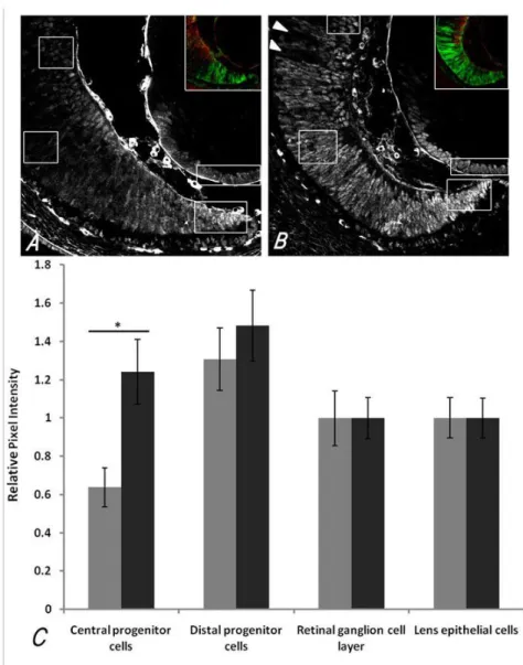
Figure 2-5 PAX6 IF intensity per cell in different regions of the E14.5 OC is increased in central OC progenitor cells of mutants ... 75
Figure 2-6 WNT signaling is expanded in Sox2-mutant eyes at E16.5 ... 76
Figure 2-7 Ablation of SOX2 by αP0CREiresGFP results in cell fate conversion of the NR to CE ... 77
Figure 2-9 Mosaic ablation of SOX2 in neural progenitors results in
loss of neuronal fate and central expansion of the OCM ... 79
Figure 2-10 At P0 mosaic regions of SOX2 ablation indicate cell fate conversion of NR to CE ... ..80
Figure 2-11 Fate mapping Sox2-mutant progenitor cells ... 81
Figure 2-12 Pax6-heterozygosity significantly rescues the Sox2-mutant NR ... 82
Figure 2-13 The NR is rescued in Sox2 Pax6 double mutant eyes ... 83
Figure 2-14 Model of how SOX2 expression in the central OC antagonizes Pax6 ... 84
Figure 3-1 Sox2-ablated OCPCs gradually decrease proliferation from the center to the periphery at E16.5. ... 116
Figure 3-2 The Sox2-ablated OC shows a graded increase in the lengths of S-phase and cell cycle at E16.5 ... 117
Figure 3-3 Many Sox2-deficient OCPCs prematurely exit the cell cycle by P0 ... 118
Figure 3-4 D-type Cyclins are aberrantly expressed in Sox2-ablated OCPCs at E16.5 ... 119
Figure 3-5 P27Kip1 is absent from Sox2-ablated OCPCs at E16.5... 120
Figure 3-6 Genome-wide expression analysis of SOX2-ablated OCs reveals functional characteristics of the peripheral and central OC and indicates increased WNT activity at E16.5 ... 121
Figure 3-7 WNT activity is expanded in the Sox2-ablated OC at E16.5 and P1 ... 122
Figure 3-8 SOX2 and -CATENIN can be efficiently ablated from the OC using Chx10CreGFP without causing increased cell death ... 123
Figure 3-9 Deletion of Ctnnb1 in Sox2-ablated OCPCs rescues the ectopic expression of CE genes ... 124
Figure 3-10 Deletion of Ctnnb1 in Sox2-ablated OCPCs does not rescue NR identity... 125
Figure 3-11 Deletion of Ctnnb1 in Sox2-ablated OCPCs does not rescue increased Cyclin D1 at E13.5 ... 126
Figure 3-12 Deletion of Ctnnb1 in Sox2-ablated OCPCs does not rescue the aberrant expression of in D Cyclins observed in Sox2-deficient cells at P0 ... 127
Figure 3-14 SOX2 directly antagonizes Pax6, but reduction of Pax6 in
Sox2-ablated OCPCs does not rescue increased Cyclin D1 at E14.5 ... 129
Figure 3-15 CD spectra of SOX2 with DNA versus and DNA ... 130 Figure 3-16 Model of how SOX2 and canonical WNT signaling regulate
the neurogenic boundary of the OC ... 131
Figure 4-1 Expression of positive and negative cell cycle regulators are
changed consistent with a cell fate conversion from NR to CE ... 150
LIST OF TABLES
Table 2-1 PCR primers and protocols ... 85
Table 2-2 Expression profiles of the prospective NR and CE ... 86
Table 3-1 PCR primers and genotyping protocols ... 132
Table 3-2 Working dilutions of antibodies for immunohistochemistry ... 133
Table 4-1 Up-regulated transcripts in the Sox2-ablated OC that contain a consensus SOX2/POU binding site(s) in the proximal regulatory region ... 152
Table 4-2 Down-regulated transcripts in the Sox2-ablated OC that contain a SOX2/POU consensus binding site(s) in the proximal regulatory region ... 154
LIST OF ABBREVIATIONS
ANP – Anterior neural plate AVE – Anterior visceral endoderm
bHLH – basic helix loop helix transcription factor BMP – Bone morphogenetic protein
BrdU - Bromodeoxyuridine CB – Ciliary body
CD – Circular dichroism
Chx10 – C. elegans ceh-10 homeodomain containing homolog (see also VSX2) CE – Ciliary epithelium
CNS – Central nervous system E – Embryonic day
EFTF – Eye field transcription factor EGF – Epidermal growth factor
EMSA – Electrophoretic mobility shift assay ESC – Embryonic stem cell
FGF – Fibroblast growth factor IdU - Iododeoxyuridine IF - Immunofluorescence IHC - Immunohistochemistry ILM – Inner limiting membrane INL – Inner nuclear layer IOP – Intraocular pressure IPL – Inner plexiform layer
LCA – Leber’s congenital amourosis
LEF1 – Lymphoid enhancer binding factor 1 LHX2 – LIM homeobox protein 2
MATH – Mouse atonal homolog MASH – Mouse achaete-scute homolog NPC – Neural progenitor cell
NR – Neural retina OC – Optic cup
OCM – Optic cup margin
OCPC – Optic cup progenitor cell
OCPTF – Optic cup progenitor transcription factor OCT4 – POU domain-containing transcription factor 4 ON – Optic nerve
ONL – Outer nuclear layer OPL – Outer plexiform layer OS – Optic stalk
OV – Optic vesicle P – Postnatal day
PAX6 – Paired box gene 6
POAG – Primary open angle glaucoma PNR – Peripheral neural retina
PR – Photoreceptor
RAX – Retina and anterior neural fold homeobox RGC – Retinal ganglion cell
Sey – Small eye
SOX2 – SRY-box containing gene 2 SRY – Sex-determining region Y
TCF – T-Cell specific transcription factor family member TEM – Transmission electron microscopy
TGF - Transforming growth factor TSS – Transcription start site
VSX2 – Visual system homeobox 2
CHAPTER I: INTRODUCTION
Overview
This chapter summarizes what is known about eye development in mammals and
describes the classical studies that inform the current questions surrounding neural induction.
The role of the SOXB1 family of transcription factors in these processes is referenced
throughout. The function of the SOXB1 family member SOX2 as a transcriptional activator, and
sometimes a transcriptional repressor, is described in terms of neural development. Finally, the
usefulness of the optic cup as a model of neural induction is explained.
Eye Development in Mammals
For generations of biologists, the eye has offered an accessible model for investigating
the mechanisms that coordinate the development and morphogenesis of diverse cell types. At
the beginning of the twentieth century, embryologists studying eye development in amphibians
described the concept of induction for the first time after discovering that tissues of different
origins must interact in order to generate a lens (Spemann, 1901). A century later, with
significant advances in molecular biology, the eye has provided a system for studying gene
interactions. Because vision is not essential for life, eye malformations have revealed that a
single mutation can lead to disease. Currently, the ability to profile gene expression in single
retinal cells, which has revealed extensive heterogeneity among retinal progenitors, enables the
study of eye development at the systems level (Byerly and Blackshaw, 2009; Kim et al., 2008;
Roesch et al., 2008; Trimarchi et al., 2008). Genes that coordinate eye development are highly
development, the underlying cellular and molecular paradigms can be applied to lower
vertebrates as well. Indeed, the eye development field thrives on a rich history of
developmental biologists working in organisms from flies to humans.
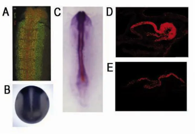
Briefly, the visible stages of mouse eye development, which are described in detail
below, include optic sulci (E8.0), optic vesicle evagination (E9.0), optic cup invagination (E10.0),
and retinal neurogenesis (E11.0 – P7).
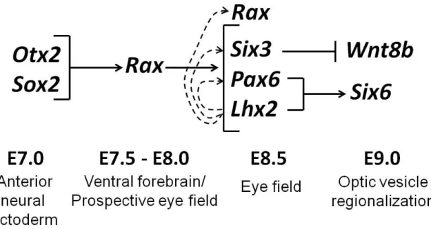
The eye field: establishing the optic primordia
Shortly after gastrulation, the eye primordium, or eye field, is specified in the medial
anterior neural plate and contains all the progenitors of the neural-derived eye structures (Li et
al., 1997; Wilson and Houart, 2004; Zaghloul et al., 2005). In mice, the first morphological sign of
the eye field is the formation of bilateral indentations in the prospective forebrain termed the
optic sulci, or optic pits, at embryonic (E) day 8.0 (Adelmann, 1929; Li et al., 1997; Wilson and
Houart, 2004) (Figure 1-1A). Molecularly, the eye field can be identified by the overlapping
expression domains of a set of highly conserved genes termed the eye field transcription factors
(EFTFs) (Lee et al., 2006; Moore et al., 2004; Zaghloul et al., 2005) (Byerly and Blackshaw, 2009;
Zuber et al., 2003). In mammals, the EFTFs, which include Pax6, Rax, Six3 and Lhx2,constitute a
regulatory network required for eye development (Byerly and Blackshaw, 2009; Zuber et al.,
2003). The identification of the upstream signaling pathways that specify the eye field is
challenging given that these genes also play a role in forebrain development (Hagglund et al.,
2011).
A recent study identified an 11kb genomic region in the Lhx2 promoter that specifically
activity of this Lhx2 regulatory element may reveal specific pathways required for eye field
specification. In fact, the conditional inactivation of Lhx2 in this cell population has no effect on
the activity of the Lhx2 eye field enhancer, suggesting that Lhx2 itself is not essential for eye
field priming (Hagglund et al., 2011). However, the finding that eye development is arrested in
mice lacking Lhx2 corroborates a study in which ectopic expression of eye field transcription
factors can generate eyes in Xenopus only when endogenous Lhx2 expression is induced.
(Cavodeassi et al., 2005; Fuhrmann, 2008; Rasmussen et al., 2001; Zuber et al., 2003). Moreover,
this study demonstrated that OTX2, a transcription factor essential for forebrain development,
and Noggin, a BMP antagonist, potentiate EFTF expression in the anterior neural plate (Zuber et
al., 2003). Similarly, in vitro data suggest that OTX2 cooperates with SOX2 to activate Rax
expression, even though Otx2 becomes down-regulated in the Rax expression domain of the
early eye field (Andreazzoli et al., 1999; Danno et al., 2008; Zuber et al., 2003). SOX2 in
coordination with a POU-factor may directly antagonize OTX2 in this region (see Discussion).
These data demonstrate that SOX2 plays an integral role in the earliest stages of eye
development and support a model of “progressive induction” (Zuber et al., 2003). This model
predicts that the anterior forebrain must be primed for eye field formation such that SOX2 and
OTX2 activate EFTF expression, and EFTFs then work in a feedback network to maintain the eye
field (Figure 1-2).
Heterozygous mutations in human OTX2 can cause a range of ocular phenotypes from
bilateral anophthalmia to retinal dystrophy (Ragge et al., 2005a). In contrast to SOX2 mutations
(discussed below), OTX2 mutations are commonly associated with impaired retinal function,
perhaps owing to the role of OTX2 in RPE development (Chase, 1944; Grindley et al., 1995;
2009; Ragge et al., 2005a; Tabata et al., 2004; Tucker et al., 2001; Voronina et al., 2004;
Wawersik and Maas, 2000; Zhang et al., 2000)
Eye field transcription factors
Genetic studies in flies (Wawersik and Maas, 2000), mice and humans illustrate that the
EFTFs are essential for proper eye development and function in a conserved network to regulate
eye formation.
a. Pax6 and Lhx2
An initial genetic demonstration of EFTF function was the identification of heterozygous
mutations in the Pax6 locus that cause the mouse small eye(Sey) haploinsufficient phenotype
(Hill et al., 1991; Hogan et al., 1988). Pax6 is a member of the evolutionarily conserved family of
paired domain-containing transcription factors (further discussed in Chaper II) (Walther and
Gruss, 1991). Humans with mutations in one copy of PAX6 often have aniridia, a severe ocular
malformation characterized by abnormal iris development and, more rarely, microphthalmia,
corneal cataracts and macular and foveal hypoplasia (Glaser et al., 1994; Glaser et al., 1992;
Hever et al., 2006). Mice with one copy of the Sey allele exhibit reduced eye size and variable
abnormal development of the retina, iris, lens and/or cornea (Hever et al., 2006; Hill et al.,
1991). Homozygous loss of Pax6 function in humans and mice causes anophthalmia, or the
complete lack of eyes (Glaser et al., 1994; Hill et al., 1991).
Like Pax6Sey/Sey mouse mutants, Lhx2-/- embryos generate optic vesicles but never form
optic cups (Porter et al., 1997). Conditional inactivation of Lhx2 in the eye field leads to
developmental arrest of the optic vesicle just prior to optic cup formation, but the expression of
The maintenance of Pax6 in Lhx2-/- mutants, and the maintenance of Lhx2 in Pax6Sey/Sey mutants,
suggests that these two EFTFs are independently essential but separately insufficient for proper
eye development (Porter et al., 1997). Moreover, Pax6 may cooperate with Lhx2 to induce the
expression of Six6, a retinal determinant gene, in the optic vesicle (Tetreault et al., 2009).
b. Rax
Like the Sey mouse line, the spontaneous mutant mouse strain eyeless, first discovered
in the 1940s, carries a hypomorphic mutation in the Rax (retina and anterior neural fold
homeobox) locus (Chase, 1944; Tucker et al., 2001). Mutations in human RAX are associated
with anophthalmia (Hanson et al., 1993; Voronina et al., 2004). The role of RAX has been
further elucidated using mouse model systems where it was shown that Rax-/- mice fail to
upregulate EFTF expression in the presumptive eye field and do not develop optic vesicles
(Grindley et al., 1995; Mathers et al., 1997; Zhang et al., 2000). In chimeric mice containing
wild-type and Rax-/- cells, the Rax-negative cells segregate together and are never found in eye
field-derived tissues. This result suggests that RAX is involved in the sorting of cells to form a distinct
eye territory, perhaps through the action of cell surface molecules (Medina-Martinez et al.,
2009). Conversely, overexpression of Rax in mouse embryonic stem cells co-cultured with a host
retina promotes retinal cell fates (Tabata et al., 2004).
c. Six3
Six3 encodes a homeobox-containing transcription factor homologous to the Drosophila
sine oculis gene (Oliver et al., 1995). Genetic inactivation of Six3 in presumptive eye tissue has
demonstrated that it is essential for eye development in mammals (Liu et al., 2010; Marquardt
et al., 2001). Conditional inactivation of Six3 in the eye field abrogates neural retina
causes ectopic optic vesicles (Lagutin et al., 2001; Liu et al., 2010). Mutations in human SIX3 are
associated with holoprosencephaly, or a failure of the cerebral hemispheres to separate
(described below) (Geng et al., 2008).
These studies collectively illustrate the importance of the EFTFs in regulating the
network of events that control early eye development, from the specification of the eye field to
the maintenance of progenitor multipotency.
Division of the eye field
Developmental biologists working in the 1920s observed that both eyes arise from a
single eye field that is divided into bilateral hemispheres (Adelmann, 1929; Li et al., 1997;
Mangold, 1931). At least two molecules have been clearly demonstrated to be involved in this
morphogenetic process. The first is sonic hedgehog (Shh), which is expressed in the ventral
forebrain and prechordal mesoderm (Echelard et al., 1993). Targeted disruption of Shh in mice
results in the failure of the eye field to split, resulting in cyclopia and a single Pax6-positive optic
vesicle (Chiang et al., 1996). The second player is Six3, which is expressed throughout the
anterior neural ectoderm before becoming restricted to the ventral forebrain and eye field
(Oliver et al., 1995). In humans, loss-of-function mutations in either SHH or SIX3 result in midline
defects that frequently include cyclopia (Belloni et al., 1996; Muenke and Cohen, 2000; Roessler
et al., 1996). In fact, SIX3 was shown to regulate Shh expression in the ventral midline of the
rostral diencephalon via an upstream enhancer element (Geng et al., 2008; Jeong et al., 2008).
Establishing boundaries in the optic vesicle
Ocular development begins with the formation of the optic vesicles. From E8.5-9.0 of
close contact with the surface ectoderm (Figure 1-1D) where the lens placode is formed. Each
optic vesicle (OV) consists of the retinal stem cells (RSCs) that give rise to all
neuroectoderm-derived cells of the eye. RSC patterning occurs along the dorso-distal/proximal-ventral axis of
the OV prior optic cup formation. Regions along this axis correspond to the presumptive neural
retina (NR -- distal OV), retinal pigment epithelium (RPE -- dorsal/proximal OV) and optic stalk
(OS -- ventral/proximal OV) (Figure 1-3, Figure 1-4A).
Several cell-intrinsic signaling pathways are involved in patterning the OV. Each
compartment of the OV expresses a specific set of transcription factors that are important for
the development of the cell type in which they are expressed. The presumptive neural retina
specifically expresses the homeodomain protein Vsx2 (formerly Chx10), the future RPE
expresses the basic helix-loop-helix transcription factor Mitf and the prospective OS expresses
the paired domain protein Pax2 (Hodgkinson et al., 1993; Liu et al., 1994; Nornes et al., 1990).
SOX2 is maintained in the prospective NR and OS but is down-regulated in the prospective RPE.
There is evidence to suggest that OTX2 directly represses SOX2 in the RPE (Nishihara et al.,
2012). Many studies have revealed that Vsx2, Mitf and Pax2, in combination with the EFTFs,
have cell intrinsic roles in compartmentalizing the future optic cup, often through reciprocal
transcriptional repression of one another (Figure 1-3). Collectively, these studies also suggest
that the RSCs of the OV are competent to become NR, RPE or OS when provided with the
appropriate combination of signals.
Neural retina versus retinal pigment epithelium versus optic stalk
Mutual antagonism between Vsx2 and Mitf serves to establish the boundary between
the future NR and RPE (Horsford et al., 2005; Nguyen and Arnheiter, 2000). Mitf is initially
of Vsx2 (Nguyen and Arnheiter, 2000). The function of Mitf in boundary formation is supported
by the observation that mice with loss of function mutations in Mitf exhibit a conversion of RPE
to NR (Bumsted and Barnstable, 2000; Nguyen and Arnheiter, 2000). A similar RPE-to-NR
conversion phenotype occurs in mice that are deficient in both Otx1 and Otx2, transcription
factors that are normally expressed in the dorsal OV/presumptive RPE (Martinez-Morales et al.,
2001). Conversely, mice with loss-of-function mutations in Vsx2, termed orJ or ocular
retardation mice, exhibit ectopic expression of Mitf and Mitf target genes in the NR. Fate
mapping analyses suggest that this phenotype is a direct transdifferentiation of the neural retina
to RPE (Horsford et al., 2005; Rowan et al., 2004).
A similar antagonistic relationship exists between Pax2 and Pax6. Pax2-/- mice exhibit
ventral expansion of the Pax6 expression domain and subsequent expansion of the NR and RPE
at the expense of the OS (Schwarz et al., 2000). Conversely, Pax6-deficient mice exhibit a dorsal
expansion of the Pax2 expression domain and fail to develop a neural retina or RPE, maintaining
only the Pax2-positive optic stalk. This reciprocally repressive relationship appears to involve a
direct molecular interaction, as PAX2 can bind the Pax6 retina-specific enhancer, , (Figure
1-4A), and PAX6 can bind the Pax2 OS-specific enhancer (Schwarz et al., 2000). Similarly, humans
with PAX2 mutations have optic nerve coloboma caused by the failure of the ventral optic
fissure to properly close during development (Torres et al., 1996). Therefore, Pax2 and Pax6
likely establish the boundary between the optic stalk and the neural retina through mutual
repression of one another.
The EFTF Lhx2 appears to act upstream of the above described genetic interactions. In
Lhx2 loss-of-function mutants, the OV fails to become regionalized, exhibiting ventral expansion
ablation of Lhx2 in the eye field, Pax2 expression persists in the ventral optic vesicle, but Mitf is
downregulated, and Vsx2 is absent (Hagglund et al., 2011).
Signaling networks in the optic vesicle
Adding complexity to the system of early eye development is the understanding that
these cell intrinsic transcription factors modulate extrinsic signals to functionally
compartmentalize the OV. The extrinsic pathways involved in OV patterning include BMP, FGF,
Wnt and Sonic Hedgehog signaling. Early Lhx2 activity, for instance, is required to transduce
BMP7 to activate Pax2 expression in the ventral OV, while laterin development, Lhx2 is required
to maintain BMP4 expression in the OV (Yun et al., 2009). Similarly, FGF1 or FGF2 from the
surface ectoderm activates Vsx2 in the presumptive NR, which in turn represses Mitf (Horsford
et al., 2005; Nguyen and Arnheiter, 2000).
FGF9, which is normally confined to the distal OV, promotes NR fate when ectopically
expressed in the presumptive RPE, and mice with targeted deletion of Fgf9 exhibit expansion of
the RPE into the NR domain (Zhao et al., 2001). Moreover, OV-specific deletion of the protein
phospatase Shp2, which mediates the FGF signaling cascade via sustained activation of Ras,
causes a cell fate conversion from NR to RPE (Cai et al., 2010). Conversely, inactivation of
canonical Wnt signaling in the presumptive RPE causes it to transdifferentiate to NR
(Westenskow et al., 2009). Lastly, in addition to its role in splitting the eye field,
midline-secreted Shh plays an additional role in ventralizing the OV. The optic vesicle of Shh mutant
mice shows expanded Pax6 expression at the expense of Pax2, while Otx2, a presumptive RPE
(dorsal) marker, persists in the cyclopic Shh mutant eye (Chiang et al., 1996).
Humans with mutations in some of these genes exhibit ocular malformations. Mutations
coloboma and retinal dystrophy (Hayashi et al., 2008). Similarly, mutations in VSX2 are
associated with microphthalmia, iris abnormalities, coloboma and retinal dystrophy (Ferda
Percin et al., 2000; Iseri et al., 2010). Although rare in comparison to cyclopia,
anopthalmia/microphthalmia and coloboma can result from mutations in human SHH (Bakrania
et al., 2010).
Optic cup morphogenesis
After the formation of the OV, a coordinated invagination of the lens placode and the
OV forms the lens vesicle and the bi-layered optic cup (OC) (Figure 1-1B, C, E, F). The OV folds
into itself creating two nested cups; the distal OV becomes the inner layer of the OC -- the
presumptive NR -- while the proximal OV becomes the outer layer of the OC -- the presumptive
RPE (Figure 1-1F, Figure 4B). An additional invagination occurs in the ventral OV where the optic
stalk meets the ventral retina to generate the optic or choroidal fissure (Figure 1-4B). The optic
fissure provides an exit for retinal axons and an entrance for the hyaloid artery, which supplies
blood to the retina (Saint-Geniez and D'Amore, 2004). The OC grows circumferentially until it
closes over the optic fissure (Figure 1-4C).
Crosstalk between the presumptive lens and retina may be necessary for the proper
invagination of these tissues in vivo (Ashery-Padan et al., 2000; Bassett et al., 2010; Grindley et
al., 1995; Smith et al., 2009). SOX2 is expressed in both of these tissues and is necessary for their
specification (discussed further in Chapter 3). The optic vesicle and lens placode are tightly
apposed at the initiation of OV invagination. However, the necessity of the presence of the lens
placode for the formation of the two-walled optic cup remains unclear. A recent study used
three-dimensional culture of mouse embryonic stem cell aggregates to derive hollowed spheres
invaginated, in the absence of a lens or ectodermal tissues, to form the optic cup (Eiraku et al.,
2011). This autonomous morphogenetic process appeared to be driven by forces within the
retinal anlage, suggesting that, at least in vitro, the OV can form the OC without instruction from
other structures (Eiraku et al., 2011). This finding is relevant to the cell-autonomous role of
SOX2 in patterning the OC independently of lens-derived signals (discussed in Chapter II).
Conversely, the apposition of the lens placode and the OV appears to be important for
placode invagination. The LP arises from the preplacodal region (PPR), an ectoderm-derived
bilateral structure that forms discrete thickenings called placodes, the precursors to various
vertebrate sensory structures (Streit, 2007). The LP is identified as the group of thickened
columnar cells in the head surface ectoderm that arises in response to OV proximity. The LP and
OV become physically tethered through cytoplasmic processes, or filopodia, originating from the
base of the lens (Chauhan et al., 2009).The LP invaginates to form the lens cup or lens pit which
eventually separates from the surface ectoderm to form the lens vesicle (Graw, 2010).
Placode thickening is associated with local changes in cell shape without associated
changes in cell volume. LP formation appears to be mediated by adhesion between the optic
vesicle and the surface ectoderm, where lens precursors continue to proliferate but cannot
expand beyond the region of adhesion (Hendrix and Zwaan, 1975; Huang et al., 2011). The
increase in cell number in this fixed area is sufficient to account for the increase in cell length
observed during placode formation (Hendrix and Zwaan, 1975). Adhesion between the optic
vesicle and the surface ectoderm is mediated by the extracellular matrix protein fibronectin1
(fn1) (Huang et al., 2011). Indeed, fn1 expression is lost when Pax6, a master regulator of lens
Lens and cornea
In addition to Pax6, several other transcriptional regulators are important for lens
specification. Six3 and Sox2, for example, are both essential for proper lens development. Six3
expression precedes that of Pax6 in the presumptive lens ectoderm and activates Pax6
transcription (Liu et al., 2006b). While Sox2 expression in the LP is induced by signals from the
OV, it may also be mediated by SIX3 in the surface ectoderm (Furuta and Hogan, 1998; Kamachi
et al., 1998). Thus, much like the cell-intrinsic regulation of OV patterning, transcription factors
involved in lens induction transduce signaling cascades to activate downstream targets. One
such signal is BMP4 from the OV, which may activate Sox2 but not Pax6 expression in the LP.
Another is BMP7 in the head ectoderm, which may activate Pax6 expression in the LP (Furuta
and Hogan, 1998; Wawersik et al., 1999).
SOX2 and PAX6 also function in cross- and self- regulatory feedback loops in lens
development. Sustained Sox2 expression appears to depend on PAX6 only after lens placode
stages (Ashery-Padan et al., 2000; Smith et al., 2009). Moreover, SOX2 cooperates with PAX6 in
its own upregulation in lens precursor cells via the N3 enhancer upstream of the Sox2 coding
sequence (Inoue et al., 2007). Likewise, PAX6 and SOX2 synergistically activate Pax6 expression
via the head surface ectoderm enhancer element LE9 (Aota et al., 2003). SOX2 and PAX6 may
also co-regulate other genes important for lens development, such as those encoding lens
crystallins (Kamachi et al., 1998; Kamachi et al., 2001). The relationship between SOX2 and PAX6
in lens development is consistent with a strong genetic interaction in optic cup development, as
described in detail in Chapter II.
The lens vesicle remains transiently attached to the surface ectoderm via the lens stalk.
eventually giving rise to the corneal epithelium. The corneal endothelium is composed of
migrated forebrain and midbrain neural crest-derived mesenchymal cells (Kanakubo et al., 2006;
Trainor and Tam, 1995). These cells invade the newly formed space between the lens vesicle
and the surface ectoderm and condense into a multilayered structure connected by a loose
extracellular matrix. The cells between the corneal epithelium and endothelium differentiate
into karatocytes and make up the corneal stroma (Cvekl and Tamm, 2004). The space between
the future cornea and lens, called the anterior chamber, becomes fluid filled. A second group of
mesenchymal cells migrates into the angle between the presumptive cornea and peripheral
edge of the optic cup to become the stroma of the iris and ciliary body (described below).
Around the same time that mesenchymal cells migrate into the future cornea (E11.5), other
POM cells invade the space between the lens and the retina, called the hyaloid, giving rise to the
hyaloid vasculature (Gage et al., 2005)
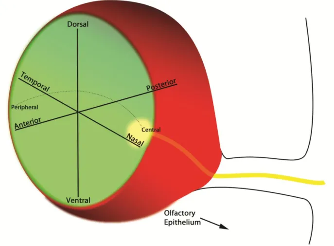
Axes in the neural retina
The inner layer of the OC, which gives rise to the neural retina, is patterned along its
dorsal-ventral (D-V) and nasal-temporal (N-T) axes (Figure 1-6). By E10.5, the optic stalk (ventral)
has begun to elongate and will eventually give rise to the optic nerve. The axons of retinal
ganglion cells from the innermost layer of the NR begin to enter the optic stalk around E11.5
and intersperse with the PAX2-positive optic stalk cells, the glial precursors that will develop
into the astrocytes that make up the mature optic nerve (Torres et al., 1996).
Proper optic nerve placement depends on signals that pattern the OC along the D-V axis.
These signals are mediated in part by members of the VAX family of homeodomain transcription
factors. At E13.5, Vax1 is expressed in the optic stalk, while Vax2 exhibits a steep ventralhigh
expressed in the optic stalk and ventral retina from E9.5 to E11.5, which may explain why Vax1-/;
Vax2-/- double mutant mice exhibit severe ventral eye defects. The optic stalk of these mice
becomes dorsalized, developing into RPE and NR instead of optic nerve (Mui et al., 2005). In
vitro and in vivo data suggest that VAX1 and VAX2 cooperatively specify the ventral optic cup
and optic stalk by directly repressing Pax6 expression via the Pax6 retina-specific
enhancer(Mui et al., 2005). Conversely, the T-box transcription factor Tbx5, a BMP4 target, is
normally expressed in the dorsal NR (Behesti et al., 2006). Its expression is lost when Pax6 is
ablated from the developing NR, further demonstrating Pax6’s role in specifying the dorsal OC
(Baumer et al., 2002; Behesti et al., 2006). These genetic studies illustrate the precise regulation
of PAX6 localization in the OC, which in turn helps to establish the dorsal-ventral axis.
N-T patterning of the optic cup ensures that the axons of retinal ganglion cells will
correctly map to their targets in the superior colliculus (SC) and the dorsal lateral geniculate
nucleus (dLGN). In mammals, axons from the nasal retina project to the caudal SC, and axons
from the temporal retina project to the rostral SC. The earliest transcriptional regulators of N-T
identity in the OC are the forkhead transcription factors FOXD1 (BF-2) and FOXG1 (BF-1). Foxg1
is expressed in the nasal retina, but appears to play an additional role in D-V patterning, as
Foxg1-/- mutants lose Shh signaling and have dorsalized optic vesicles (Huh et al., 1999).
However, Foxd1 functions in N-T patterning to specify the retinal ganglion cells (RGCs) of the
temporal retina (Carreres et al., 2011). Indeed, RGCs of Foxd1-/- mutants lose topographic
specificity, mapping indiscriminately to the entire extent of the SC (Carreres et al., 2011).
Expression of both Foxd1 and Foxg1 is lost in Pax6-/- mutants, demonstrating an additional role
Ciliary body and iris
A third axis along which the OC is patterned is the central-peripheral axis (Figure 1-4D,
Figure 1-6). The distal tip of the OC, where the presumptive NR meets the RPE, is termed the
“ciliary margin” in mice and contains non-neurogenic progenitors that give rise to the epithelia
of the ciliary body (CB) and iris (Beebe, 1986; Davis-Silberman and Ashery-Padan, 2008). The CB
epithelia are continuous with the RPE and NR, while the iris epithelia are distal to the CB. The
outer layer of the ciliary margin gives rise to the pigmented epithelium of the CB and the
anterior pigmented layer of the iris, whereas the inner layer gives rise to the non-pigmented
epithelium of the CB (herein referred to as the ciliary epithelium or CE) and the posterior
pigmented layer of the iris. Non-neurogenic progenitor cells of the optic cup margin can be
identified as early as E12.5 by their specific expression of transcription factors, including Msx1
and Otx1, lack of expression of neuronal markers and a slower proliferation rate relative to the
NR (Beebe, 1986; Cho and Cepko, 2006; Martinez-Morales et al., 2001; Monaghan et al., 1991;
Trimarchi et al., 2009). Indeed, CB development is absent in mice that lack Otx1 (Acampora et
al., 1996).
Several cell-intrinsic and extrinsic pathways have been identified to play a role in
specifying the ciliary margin in mammals. CE-specific genes can be induced in embryonic mouse
retina when cultured adjacent to an explanted chick lens (Thut et al., 2001). The inductive power
of the lens may in part involve BMP signaling, as transgenic expression of the BMP antagonist
Noggin in the developing mouse lens abrogates expression of Bmp4 and Bmp7 in the
presumptive CE at postnatal stages (Zhao et al., 2002). Without BMP signaling, the CE-specific
genes Otx1 and Msx1 fail to be expressed, and neural retinal cells develop in place of the CB
The appearance of the NR at the expense of the CE in the BMP-deficient ciliary margin
suggests that progenitor cells at the boundary of the NR and CE remain competent to take on
one fate or the other after these tissues have begun to be specified. Transcriptional control of
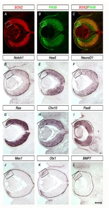
this binary cell fate decision is mediated by SOX2 and PAX6 (Figure 1-4D). This relationship is
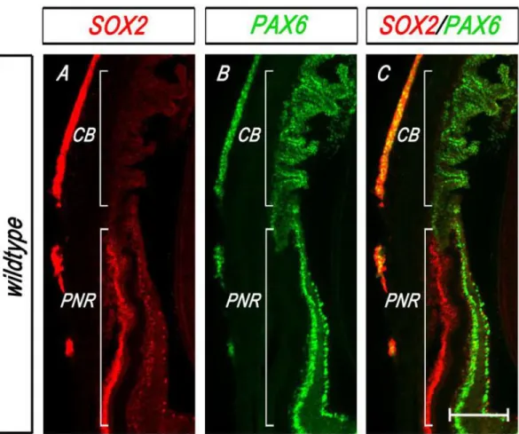
discussed at length in Chapter III. Briefly, in the early optic cup, SOX2 and PAX6 exhibit inverse
gradients of expression, with SOX2 high in the central OC but low in the periphery where PAX6 is
maintained at a high level due in part to its retina-specific enhancer(Baumer et al., 2002;
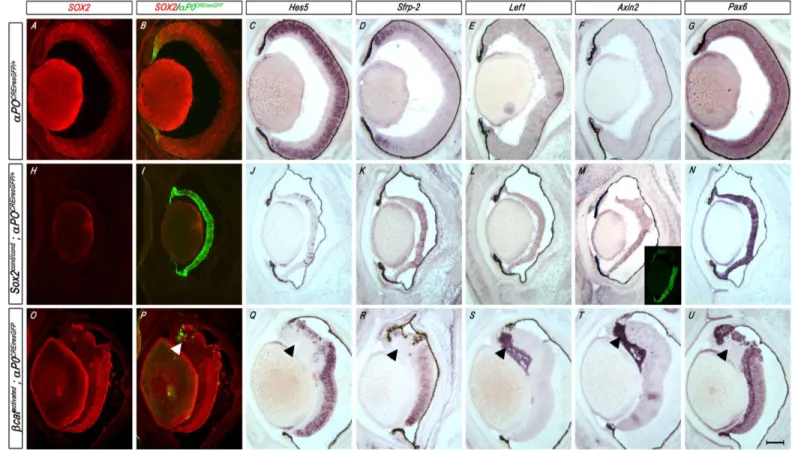
Matsushima et al., 2011). Specific ablation of SOX2 in OC progenitor cells results in elevated
Pax6 expression and cell fate conversion from NR to CE, a phenotype described in detail in
Chapter II. (Matsushima et al., 2011). This cell fate conversion can be partially rescued by
reducing Pax6 (Matsushima et al., 2011). Indeed, several studies have demonstrated the
importance of PAX6 to the development of the ciliary margin. Mice that are haplo-insufficient
for Pax6 exhibit reduced ciliary margin size, and humans with mutations in PAX6 have aniridia
(no iris) and small ciliary bodies (Davis-Silberman et al., 2005; Hanson et al., 1993; Okamoto et
al., 2004). Further evidence suggests that Wnt signaling plays a role in specifying CE fate.
Activated Wnt signaling in the peripheral NR induces expression of CE-specific genes (Liu et al.,
2007b). Moreover, the NR-to-CE cell fate conversion caused by the loss of SOX2 is associated
with centrally expanded Wnt signaling prior to the ectopic expression of CE genes (Matsushima
et al., 2011). Chapter IV focuses on the genetic relationship between SOX2 and Wnt signaling in
OC patterning and CE fate specification.
Retinal neurogenesis
Retinal Progenitor Cells (RPCs) are established in the optic vesicle as early as E9.0, each
Neurogenesis in the retina proceeds in an ordered and predictable fashion, beginning in the
central optic cup and moving toward the periphery. Specification of RPC fate is coordinated with
cell cycle exit to generate the correct numbers and types of neurons that make up the mature
retina. Neuronal differentiation begins at E11.0 and cell types are generated in overlapping
phases, beginning with retinal ganglion cells (RGCs), which are the projection neurons of the
retina. The initiation of RGC production is followed in succession by horizontal interneurons,
cone photoreceptors and amacrine interneurons in an early wave of neurogenesis (E11.0 –
E18.0). A temporal switch in RPC competence then produces rod photoreceptors, bipolar
interneurons and Muller glial cells in a late wave of neurogenesis (P0 – P7) (Young, 1985).
The architecture of the mature retina is as follows: The cell bodies of rod and cone
photoreceptors are localized to the apical side of the retina in a layer abutting the RPE known as
the outer nuclear layer (ONL). Just basal to the ONL is the inner nuclear layer (INL), which
contains the cell bodies of the retinal interneurons -- the bipolar, amacrine and horizontal cells –
as well as the cell bodies of the Muller glia. The ganglion cells are located in the ganglion cell
layer (GCL) on the basal side of the retina, nearest to the lens. Separating the ONL and the INL is
the outer plexiform layer (OPL) where photoreceptors synapse onto horizontal cells and bipolar
cells. The more elaborate inner plexiform layer (IPL) is located between the INL and the GCL and
contains the dendrites of amacrine cells and RGCs (Figure 1-7). All together, this structure
illustrates the first neurological steps of visual processing: the photoreceptors receive light input
and transmit the signal through bipolar cells, whose axons terminate in the IPL, where RGCs
then transmit the signal to visual processing centers in the brain. SOX2 expression remains only
A number of transcription factors that regulate RPC fate decisions have been identified.
These are members of the homeodomain family or bHLH family of transcription factors. (Figure
1-7). The homeodomain transcription factors include those that are expressed in the forebrain
and eye field (OTX2, RAX, SIX3, SIX6, PAX6 and LHX2) as well as those that are expressed early in
the prospective retina (VSX2) or play a role in specifying retinal subtypes (PROX1, CRX, LHX1,
DLX1 and BRN3). The bHLH factors include repressors of neuronal differentiation, HES1 and
HES5, and activators of neuronal differentiation, including the Atonal homologs MATH5, MATH3
and NEUROD and the Acheate-Schute homologs NGN2 and MASH1. RPCs that express one or a
combination of these bHLH factors are biased toward a particular cell fate (Hatakeyama and
Kageyama, 2004). Achaete-Schute homologs generally play dual roles in regulating the onset of
differentiation and cell fate specification. Similarly, atonal homologs are expressed in RPCs at
the onset of neuronal differentiation and often identify the fate of the progenitor cell. MATH5
expression in early RPCs, for example, predicts RGC development, while NEUROD and MATH3
each predict amacrine cell development. Late RPCs are competent to give rise to bipolar cells or
Muller glia. Math3 or Mash1 expression in these cells predicts bipolar fate (Hatakeyama et al.,
2001). Homeodomain and bHLH factors can also act in combination to specify cell fate, with the
former regulating layer specificity and the latter regulating subtype within the layer
Neural Induction
The development of the central nervous system (CNS) in vertebrates begins with neural
induction, or the process in which an ectodermal cell is specified as neural (neural plate) or
non-neural (epidermis). The mechanisms directing this cell fate decision have come to be understood
primarily through experiments using gastrulating frog and chick embryos. Nonetheless, certain
neural induction paradigms hold true for mammals as well: secreted factors, namely BMPs and
FGFs, specify whether an ectoderm progenitor cell maintains neural competence, becoming
neural plate, or acquires epithelial identity, thus losing neurogenic capacity (Delaune et al.,
2005; Hemmati-Brivanlou and Melton, 1997; Kuroda et al., 2005; Linker and Stern, 2004;
Reversade et al., 2005; Stern, 2006; Wawersik et al., 2005; Wills et al., 2010).
Neural development in mice
At E5, the mouse embryo consists of two layers, the primitive endoderm and the
epiblast. The epiblast gives rise to all the cells of the adult mouse. The naïve state of the epiblast
is ectoderm. Gastrulation begins at E6.5 when the ectoderm invaginates to produce the
primitive streak, which moves anteriorly to produce the mesoderm and endoderm. The anterior
end of the primitive streak is the organizing center or node, which secretes signals important for
specifying neural progenitor cells, which arise from the anterior distal tip of the ectoderm
(Zernicka-Goetz, 2002). Nodal signaling from the primitive streak stabilizes the epiblast state,
while Nodal antagonists expressed from the anterior visceral endoderm (AVE), an
extraembryonic tissue, disrupt Nodal signaling. The earliest derivative of the mouse epiblast is
the anterior neural plate (ANP). It is thought that Nodal, a member of the TGFfamily, functions
to specify the AVE, which in turn functions to specify anterior identity in the adjacent epiblast
2001; Perea-Gomez et al., 2001). In the absence of Nodal, epiblasts ectopically express neural
plate markers, including Sox1 and Hesx1, suggesting that Nodal antagonizes neural fate (Camus
et al., 2006).
The default model
The classical model of neural induction predicts that neural is the default or ground
state of the ectoderm (Brivanlou and Melton, 1997; Weinstein and
Hemmati-Brivanlou, 1999). Mouse embryonic stem (ES) cells develop into neural progenitor cells (NPCs)
without the addition of exogenous factors (Ying and Smith, 2003). The anterior ectoderm of the
mouse will express neural genes in the absence of Nodal (Camus et al., 2006). Similarly, cultured
Xenopus ectodermal explants, which express BMP4, develop into epidermis when kept intact,
but become neural when dissociated. Non-neural fate in these cells can be rescued with the
addition of BMP4. Moreover, intact explants become neural with the addition of BMP inhibitors
(Munoz-Sanjuan and Brivanlou, 2002). These experiments informed the idea that ectoderm
becomes epidermis in response to BMP signals and neural plate -- “by default” -- when BMP
signals are inhibited.
According to this model, non-neural fate must thus be imposed upon the ectoderm.
Nodal-deficient tissues exhibit little-to-no BMP and Wnt activity, suggesting that all three of
these pathways are inhibited during anterior neural fate specification. In other words, Nodal,
BMP and/or WNT must be active for non-neural fate to be specified (Camus et al., 2006). In
Xenopus, BMP signaling through BMP receptor (BMPR)-mediated activation of the transcription
factor SMAD1 promotes non-neural fate. The BMP antagonists Noggin, Chordin and Follistatin
collectively prevent BMPR from phosphorylating SMAD1, thus allowing the ectoderm to
of the dorsal mesoderm that is thought to maintain neural fate in the ectoderm (Reviewed in
(De Robertis and Kuroda, 2004)).
However, numerous studies have suggested that the default model is an
over-simplification of neural induction. For one, it does not fully explain neural fate specification in
avian embryos. In chicks, the organizer is sufficient for neural fate, but BMP inhibition is not;
only cells at the border of neural and non-neural ectoderm are competent to respond to BMP
inhibition, suggesting that these cells are exposed to additional neural-provoking cues (Streit et
al., 1998; Streit and Stern, 1999). Similarly, BMP inhibition is sufficient to suppress epidermis but
insufficient to induce neural fate in regions of the ectoderm distant from the endogenous neural
plate (Delaune et al., 2005). Experiments in Xenopus suggest that FGF and IGF, which are potent
inducers of neural fate, may be required for neural ectoderm specification and are potential
candidates for “neural-inducing cues” (Hardcastle and Papalopulu, 2000; Pera et al., 2001; Streit
et al., 2000; Wilson et al., 2000). These growth factors function through SMAD1 regulation as
well: activation of MAPK by FGF or IGF phosphorylates SMAD1 in a highly conserved linker
region, inhibiting its neural-antagonizing ability. Mutation of the target serines in this linker
region results in a strongly ventralized embryo, indicating loss of neural fate. Conversely, BMPR
phosphorylates the C-terminus of SMAD1. Mutating these C-terminus serine residues in addition
to those in the linker region rescues ventralization, demonstrating that BMPR activity is required
to antagonize neural fate (Pera et al., 2003). Therefore, a fine balance between FGF, IGF and
BMP signaling regulates neural ectoderm development via SMAD1.
The roles of BMP and FGF signaling differ between mammals and lower vertebrates. In
Xenopus, as described above, neural induction requires both BMP inhibition by Chordin and the
neural plate, induce ectopic sensory neurons and suppress epidermal genes while inducing
anterior neural genes. Moreover, constitutive activation of BMPR in a dorsal animal blastomere
of Xenopus embryos suppresses neural fate, resulting in loss of Sox2 and Sox3, and induces
epidermis, expressing cytokeratin81 (Delaune et al., 2005). Likewise, inhibition of FGF signaling
by mutation of the SMAD1 linker region results in loss of neural fate (Pera et al., 2003). In mouse
embryos, however, the role of SMAD1 is more complex. Mutation of the linker region does not
abrogate neural fate. Rather, mice with targeted disruption of these SMAD1 sites exhibit defects
of the gastric epithelium and actin cytoskeleton (Aubin et al., 2004). This relatively mild
phenotype may be due to the compensatory function of SMADs 5 and 8. Nonetheless, the
regulation of neural fate in mammals is exceedingly more complex than in lower vertebrates.
And despite the evolutionary conservation of neural induction genes throughout all vertebrates,
further investigation into the function of these genes in mammals is needed.
WNT signaling and neural induction
The studies described above demonstrate that BMP and FGF signaling converge at the
regulation of SMAD1 activity and have opposing effects on neural fate. In chick embryos,
however, activation of FGF alone or in combination with BMP antagonists is insufficient to
induce neural fate. Indeed, even in Xenopus, complete removal of Smad1 only results in a mild
ventralization phenotype, suggesting that additional pathways are required for neural induction.
A third pathway implicated in neural induction is WNT/-Catenin signaling. WNTs are secreted
glycoproteins that bind transmembrane Frizzled receptors, initiating a signaling cascade that
culminates in the de-repression of -CATENIN, a transcription factor that complexes with
members of the TCF/LEF family of transcription factors to regulate the expression of target
WNTs are generally thought to promote non-neural fate in the ectoderm or epiblast:
WNT signaling must be inhibited for mouse ESCs to become neural, and antagonism of WNT and
Nodal signaling increases generation of telencephalic (neural) precursors (Aubert et al., 2002;
Watanabe et al., 2005). Conversely, WNT1 and lithium chloride, which stimulates WNT signaling,
can inhibit neural fate in mouse ESCs. In the chick epiblast, Wnt signaling can block the ability of
ectoderm cells to respond to FGF, and antagonism of Wnt signaling permits cells to respond to
FGF, placing WNTs at the hinge of BMP and FGF antagonism (Wilson et al., 2001). In contrast to
its role as an antagonist of anterior neural fate, WNT signaling has been demonstrated to
regulate posterior neural plate development in mice through direct activation of the Sox2
enhancer N-1 (Takemoto et al., 2006). This study provides a direct link between WNT signaling
and SOXB1 factors in neural fate specification, and thus begins to piece together the relationship
between cell-extrinsic signaling molecules and cell-intrinsic gene expression.
The roles of SOXB1 factors in neural induction
Little is known about the transcriptional regulation of neural induction. Members of the
SOXB1 family of transcription factors are highly conserved early makers of neural fate in many
species from Drosophila (Ma et al., 1998; Nambu and Nambu, 1996; Soriano and Russell, 1998)
to fish (Vriz et al., 1996) and avians (Uwanogho et al., 1995) (Figure1-9). In chick embryos, SOX2
is expressed in pre-gastrula cells that are competent to undergo neural induction upon node
transplantation (Streit et al., 1997). Similarly, in Xenopus, SOX2 is a specific early readout of
neural identity (Mizuseki et al., 1998). BMP antagonists, including Chordin, induce SOX2
expression, and SOX2 synergizes with FGF signaling to initiate neural fate (Mizuseki et al., 1998).