doi: 10.1093/cercor/bhx065
Advance Access Publication Date: 18 March 2017 Original Article
O R I G I N A L A R T I C L E
Effects of Antenatal Maternal Depressive Symptoms
and Socio-Economic Status on Neonatal Brain
Development are Modulated by Genetic Risk
Anqi Qiu
1,2
, Mojun Shen
2
, Claudia Buss
3,4
, Yap-Seng Chong
2,5
,
Kenneth Kwek
6
, Seang-Mei Saw
7
, Peter D. Gluckman
2
, Pathik D. Wadhwa
4
,
Sonja Entringer
3,4
, Martin Styner
8,9
, Neerja Karnani
2
, Christine M. Heim
3,6
,
Kieran J. O
’
Donnell
10,11
, Joanna D. Holbrook
2
, Marielle V. Fortier
7
,
Michael J. Meaney
2,10,11
and the GUSTO study group
1
Department of Biomedical Engineering and Clinical Imaging Research Center, National University of
Singapore, Singapore 117576, Singapore,
2Singapore Institute for Clinical Sciences, Singapore 117609,
Singapore,
3Departent of Medical Psychology, Charité University Medicine Berlin, Berlin 10117, Germany,
4Development, Health and Disease Research Program, Department of Pediatrics, University of California,
Irvine, CA 92697, USA,
5Department of Obstetrics & Gynaecology, Yong Loo Lin School of Medicine, National
University Health System, Singapore 119228, Singapore,
6Department of Biobehavioral Health, Pennsylvania
State University, University Park, PA 16802, USA,
7Department of Diagnostic and Interventional Imaging, KK
Women
’
s and Children
’
s Hospital (KKH), Singapore 229899, Singapore,
8Department of Computer Science,
University of North Carolina, Chapel Hill, NC 27599, USA,
9Department of Psychiatry, University of North
Carolina, Chapel Hill, NC 27514, USA,
10Ludmer Centre for Neuroinformatics and Mental Health, Douglas
Mental Health University Institute, McGill University, Montréal H4H 1R3, Canada and
11Sackler Program for
Epigenetics & Psychobiology at McGill University, Montréal H4H 1R3, Canada
Address correspondence to Anqi Qiu, Department of Biomedical Engineering, National University of Singapore, 4 Engineering Drive 3, Engineering Block 4, #04-08, Singapore 117583, Singapore. Email: [email protected]
Abstract
This study included 168 and 85 mother–infant dyads from Asian and United States of America cohorts to examine whether a genomic profile risk score for major depressive disorder (GPRSMDD) moderates the association between antenatal maternal
depressive symptoms (or socio-economic status, SES) and fetal neurodevelopment, and to identify candidate biological processes underlying such association. Both cohorts showed a significant interaction between antenatal maternal
depressive symptoms and infant GPRSMDDon the right amygdala volume. The Asian cohort also showed such interaction on
the right hippocampal volume and shape, thickness of the orbitofrontal and ventromedial prefrontal cortex. Likewise, a significant interaction between SES and infant GPRSMDDwas on the right amygdala and hippocampal volumes and shapes.
After controlling for each other, the interaction effect of antenatal maternal depressive symptoms and GPRSMDDwas mainly
© The Author 2017. Published by Oxford University Press.
shown on the right amygdala, while the interaction effect of SES and GPRSMDDwas mainly shown on the right
hippocampus. Bioinformatic analyses suggested neurotransmitter/neurotrophic signaling, SNAp REceptor complex, and glutamate receptor activity as common biological processes underlying the influence of antenatal maternal depressive symptoms on fetal cortico-limbic development. Thesefindings suggest gene–environment interdependence in the fetal development of brain regions implicated in cognitive–emotional function. Candidate biological mechanisms involve a range of brain region-specific signaling pathways that converge on common processes of synaptic development.
Key words:amygdala, cortical thickness, depression, morphological shape, polygenic risk score
Introduction
Antenatal maternal depression, independent of postnatal mater-nal mood, influences fetal neurodevelopment, in particular the amygdala, where alterations associate with affective disorders (Rifkin-Graboi et al. 2013;Qiu et al. 2015a;Wen et al. 2017). Since depression is a familial disorder with a significant estimate of heritability (Sullivan et al. 2000), biological pathways linking maternal mood during pregnancy to offspring neurodevelop-ment are likely to vary as a function of genetic vulnerability for depression. Studies of candidate genes or of family history sug-gest the influence of“depressogenic”environmental conditions, such as childhood maltreatment and socio-economic status (SES), is moderated by genotype (Moffitt and Caspi 2006), refl ect-ing gene–environment interdependence. However, candidate gene approaches bear weaknesses (Duncan and Keller 2011) and gene products operate in networks, such that alterations in neural systems that enhance vulnerability for psychopathology derive from genomic variants at multiple sites and may converge to influence common biological systems. Indeed, the genetic contribution to susceptibility for depression appears to reflect the cumulative influence of multiple genetic variants (Lubke et al. 2012). This idea led to the use of methods of genomic risk profiling to examine the influence of genetic burden as reflected by a set of“risk”alleles for specific psychiatric disorders (Ripke et al. 2013). The risk alleles and effect sizes of single-nucleotide polymorphisms (SNPs) are established from existing genome-wide association studies (GWAS) for depression using relevant “discovery” samples based on their P-values below a defined threshold. A genomic profile risk score (GPRS) is calculated for each individual in the target sample as the sum of the count of risk alleles weighted by the effect size in the discovery sample. The GPRS for major depressive disorder (GPRSMDD) predicts the
risk for depression (P<10−6) (Ripke et al. 2013) and moderates
the influence of childhood maltreatment on depressive symp-toms (Peyrot et al. 2014). The target phenotype can also differ from that examined in the discovery sample allowing cross-phenotype analyses and studies of individual differences in endophenotypes. For example, a higher GPRSMDDassociates with
cortical thinning in the medial prefrontal cortex (Holmes et al. 2012), a region strongly implicated in the pathophysiology of depression (Kupfer et al. 2012).
Given the evidence on the relationship between antenatal maternal depression and the amygdala development during the fetal period (Rifkin-Graboi et al. 2013;Qiu et al. 2015a), we exam-ined whether such relationship was moderated by infant GPRSMDD,
controlling for maternal GPRSMDDusing a longitudinal Asian birth
cohort sample. We then identified candidate genetic pathways and biological processes that might mediate the influence of envir-onment conditions associated with the genetic risk for MDD on the amygdala. We repeated the analysis on the amygdala using a US cohort of mother–infant dyads with measures of antenatal maternal depressive symptoms, neonatal neuroimaging data, and
measures of GPRSMDD(see the description of the US cohort in the
Supplementary Material). In this study, we computed GPRSMDD
based on the SNPs selected from a Caucasian GWAS (Ripke et al. 2013). Given the fact on population differences in genotype fre-quency and risk genetic variants for MDD (Woo et al. 2004;Serretti et al. 2007;Porcelli et al. 2012;Sheikh et al. 2013;Ming et al. 2013, 2015), we noted that there may be a potential limitation of using a Caucasian sample as a discovery sample for studying the GPRSMDD
in the Asian sample. Nevertheless, to the best of our knowledge, the discovery sample used in this study is from one of the most robust GWAS for MDD with the largest sample size (Ripke et al. 2013) at the current stage. Despite its limitation, it is informative in the sense that individual SNPs have opposing effects between Asian and Caucasian populations but they are still risk SNPs for MDD. For instance, Caucasians withSallele of the serotonin trans-porter polymorphism (5-HTTPLR) show higher amygdala activa-tion in response to emoactiva-tional stimuli (e.g.,Hariri et al. 2002,2005; Murphy et al. 2013), whereas in Asians,Lallele is associated with amygdala hyperactivation (Li et al. 2012). Hence, we expected that if Asian neonates with high GPRSMDDshow the positive association
between antenatal maternal depressive symptoms and the amyg-dala volume, whereas Caucasian neonates with high GPRSMDD
show the opposite association or vice versa.
We further performed an exploratory analysis on whole brain morphology, including cortical thickness and hippocampal morphology, and examined whether GPRSMDDalso modulates the
association of antenatal maternal depression and other brain regions, particularly in the cortico-limbic system. Due to the com-patibility in the magnetic resonance imaging (MRI) acquisition and the anatomical definitions of the brain structures, this study only performed the exploratory analysis using the Asian cohort. In par-ticular, we employed large deformation diffeomorphic metric mapping (LDDMM) (Du et al. 2011) to perform detailed analysis of the morphological shape of cortical and subcortical brain regions. This approach allows for precise anatomical examination of these structures, well beyond that examined in brain volumetric studies, and reveals specific-regional shape changes associated with dis-ease status (Qiu et al. 2009,2010). Based on genetic contribution to MDD (Lubke et al. 2012) and the evidence that antenatal maternal depression influences fetal cortico-limbic development (Qiu et al. 2015a), we hypothesized that neonates with high GPRSMDDmay
show the negative relationship of antenatal maternal depression with fetal development of the cortico-limbic structures, including the hippocampus and prefrontal cortex. To further explore the moderating influence of the GPRSMDD, we examined the effects of
SES, which influences the risk for a broad range of mental disor-ders, including MDD (e.g.,Gilman et al. 2003), and cortico-limbic development (Farah et al. 2010;Noble et al. 2015).
This study, to the best of our knowledge, is thefirst kind to provide a strategy for the analysis of gene–environment inter-dependence and identify candidate biological processes that might mediate the influence of antenatal maternal depressive
symptoms on fetal brain development. Because of challenges in imaging neonates shortly after birth, the sample size of this study was modest for the study of gene–environment inter-action and hence ourfindings are preliminary but may suggest biologically plausible candidate processes for transgenerational transmission of depression from mother to child.
Materials and Methods
Subjects
One-hundred and eighty-nine infants of mothers who partici-pated in the prospective Growing Up in Singapore Towards healthy Outcomes (GUSTO) birth cohort study (Soh et al. 2012) were recruited for neuroimaging (Asian cohort). The Asian cohort recruited pregnant women attending an antenatal ultra-sound scan clinic who were Singapore citizens or Permanent Residents of Chinese, Malay, or Indian ethnic background, and had no history of psychotropic medication.
We included the 168 neonates with genetic and neuroimaging data, gestational age at birth≥35 weeks, birth weight>2500 g, and a 5-min Appearance, Pulse, Grimace,Activity, andRespiration score≥9 and whose mothers had genetic data and returned the questionnaires stated below. The Asian study was approved by the National Healthcare Group Domain Specific Review Board (NHG DSRB) and the Sing Health Centralized Institutional Review Board (CIRB). Written consent was obtained from mothers.
Scales of Maternal Depressive Symptoms and SES
In GUSTO, the Edinburgh Postnatal Depression Scale (EPDS) ques-tionnaire was administered to mothers at 26 weeks of pregnancy to quantify depressive symptomatology. The EPDS (Cox et al. 1987) is a widely used 10-item self-report scale designed as a screening instrument for postnatal depression and valid for use in gestation. Each item of the EPDS is scored on a 4-point scale (0–3), and items 3 and 5–10 are reverse scored. The reliability of the EPDS was 0.82 assessed using Cronbach’s analysis for our cohort. SES was mea-sured using monthly household income (Table1) and was col-lected at 26 weeks gestation via questionnaire.
MRI Acquisition and Analysis
In the Asian cohort, axial fast spin-echo T2-weighted MRI (repeti-tion time=3500 ms; echo time=110 ms;field of view=256 mm×
256 mm; matrix size=256×256; 50 axial slices with 2.0 mm thick-ness) were acquired in infants 5–14 days of age using a 1.5-Tesla general electric scanner at the Department of Diagnostic and Interventional Imaging of the KK Women’s and Children’s Hospital. The detailed acquisition and image quality check proced-ure were previously reported (Qiu et al. 2013,2015b). No sedation was used and precautions taken to reduce exposure to the MRI scanner noise. A neonatologist was present during scanning. A pulse oximeter was used to monitor heart rate and oxygen satur-ation throughout the entire scans. All brain scans were reviewed by a neuroradiologist (M.V.F). To determine image motion, all axial slices of the T2-weighted MRI data were visually inspected to ensure no cross-slice motion and checkerboard patterns.
Within individual subjects, when possible, 2 T2-weighted MRI acquisitions were first rigidly aligned and averaged to increase signal-to-noise ratio. In cases where only one scan was acquired, data from one scan were used in lieu of the aver-age axial imaver-age. The skull of the averaver-aged axial imaver-age was removed using Brain Extraction Tool (Smith 2002). A Markov random field model was used to automatically delineate
amygdala, hippocampus, cortical gray matter, white matter, and cerebrospinal fluid (CSF) from the neonatal T2-weighted MRI data. The validation of this method in our sample was reported elsewhere (Qiu et al. 2013,2015b;Rifkin-Graboi et al. 2013). Against the manual segmentation of the 20 brain images randomly selected in our data set, the segmentation accuracy rates of the amygdala, hippocampus, cortical gray matter, and white matter were respectively 0.75, 0.71, 0.79, and 0.86.
Based on the above tissue segmentation, a cortical surface was constructed at the boundary between gray and white mat-ter using a graph-based topology correction algorithm. Cortical thickness was measured as the distance between cortical sur-faces at the boundary between gray matter and CSF and at the boundary of gray matter and white matter. We employed a LDDMM algorithm to align individual cortical surfaces to the Asian atlas that was generated based on the cortical anatomy of the 20 subjects of this study and transferred the thickness of each individual subject to the atlas (Bai et al. 2012). The cortical thickness was smoothed using the Laplace–Beltrami basis func-tions on the cortical surface (Qiu et al. 2006).
Amygdala and hippocampal morphological shapes were represented using surfaces contoured at the structure boundary. The LDDMM-surface mapping (Qiu and Miller 2008;Zhong and Qiu 2010;Tan and Qiu 2016) was then used to take surfaces as objects and provided transformation that deformed the Asian atlas to be similar to subjects (Bai et al. 2012). Shape variations of individual subjects relative to the Asian atlas were character-ized by the Jacobian determinant of the transformation in the logarithmic scale, which represents the ratio of structural vol-ume to the atlas volvol-ume. Positive values correspond to expan-sion, while negative values correspond to compression of the structure relative to the atlas at each anatomical location.
SNP Genotyping and GPRS Computation for MDD
In the Asian cohort, genomic DNA was extracted from frozen umbilical cord specimens for infants and from blood for mothers according to standard procedures. The samples were genotyped Table 1Demographics of the Asian cohort
Mean SD
Gestational age (week) 38.69 1.13
Birth weight (gram) 3117.85 391.76
Birth weight adjusted by gestational age (gram) 3118.14 379.01
Postconceptual age at the MRI visit (week) 40.08 1.21
Antenatal maternal depression 8.59 4.46
N %
Sex, %
Male 168 53.6
Ethnicity, %
Chinese 168 44.0
Malay 39.3
Indian 16.7
Monthly household income (US$), %
≤768 168 3.0
769–1538 13.7
1538–3076 39.3
3077–4615 23.2
≥4616 13.7
Upreported 7.1
Note: SD, standard deviation.
on Illumina Omni express arrays, shown to perform well and with better coverage than alternatives in Asian populations (Jiang et al. 2013), and on Illumina Exome arrays, following the manufacturer’s instructions (Expression Analysis Inc). Further quality control on the genotyping calls were previously described (Qiu et al. 2015b). SNPs were verified for a genotyping rate≥95% and no deviation from Hardy–Weinberg equilibrium (P<0.001), and minor allele frequency≥0.05, in PLINK (Purcell et al. 2007).
The GPRSMDDof infants and mothers was created using PLINK
based on their SNPs and meta-analysis results from the Psychiatric Genomics Consortium (PGC; European ancestry, 9240 MDD cases, and 9519 controls) (Ripke et al. 2013). The GPRSMDDwas a
cumula-tive summary score computed as the sum across SNPs of the num-ber of risk alleles (0, 1, and 2) weighted by odd ratio. Odd ratios were obtained from the meta-analysis on MDD from the Psychiatric Genomics Consortium website (http://www.med.unc. edu/pgc) (Ripke et al. 2013) and represented the allele-specific risk for MDD. We selected the SNPs with low linkage disequilibrium to each other (r2<0.25 within 500 kb window,filtering for signifi
-cance; PLINK-command -clump-p1 1 -clump-p2 1 -clump-r2 0.25 -clump-kb 500) (Peyrot et al. 2014) and that survived atP-value thresholds (0.05, 0.1, and 0.2) to incorporate the proportion of vari-ation in disease risk explained through their additive effects (Purcell et al. 2009;Holmes et al. 2012;Peyrot et al. 2014). The result-ing numbers of selected SNPs for infant GPRSMDDwere 2766, 5081,
and 8935. The numbers of SNPs for maternal GPRSMDDwere 2769,
5078, and 8902. This GPRSMDDwas then standardized to a mean of
zero and standard deviation of one to aid interpretation of results. Assume that genetic risks for rheumatoid arthritis (RA) should not contribute to the risk for MDD and hence should not modulate the relationship between maternal depressive symp-toms and neonatal morphology. In this study, we computed GPRS scores for RA (GPRSRA) as a negative control. We
antici-pated that GPRSRA would not modulate the relationship
between GPRSMDDand neonatal brain morphology.
Statistical Analysis
Linear regressions were used to examine interaction effects of infant GPRSMDD with antenatal maternal depressive symptoms
(or SES) on amygdala and hippocampus volumes and shapes, as well as cortical thickness of the whole brain in the Asian sample. The EPDS scores (or SES) and the GPRSMDD of the infant were
included as main factors, which were entered into thefirst block of linear regressions along with covariates of gender, maternal ethnicity, birth weight adjusted by gestational age, postconcep-tual age on the MRI day. The interaction of the 2 main factors (EPDS score/SES and GPRSMDD) was then entered into the second
block of the regression. For statistical analysis on shapes and thickness, the data were smoothed on the atlas surfaces using a heat kernel with 30-mm full-width-at-half-maximum (Chung et al. 2005). Statistical results were illustrated withP-value<0.001 at the vertex level and with the cluster level of significance (P<
0.05) after correction for multiple comparisons based on random field theory (Chung et al. 2005,2010). The correction of multiple comparisons was applied to one interaction model at a time, but not across genomic polygenic risk scores (GPRSMDDand GPRSRA)
and different brain measures (volume, shape, and thickness). If the interaction of the EPDS score (or SES) and GPRSMDDwas
significant, we performed post hoc analyses where the sample was then divided into 2 groups, one with a GPRSMDD greater
than its mean value, 0 (high GPRSMDDgroup) and the other≥0
(low GPRSMDD group). We examined associations of antenatal
maternal depressive symptoms (or SES) and brain measures in
each GPRSMDDgroup using regression with EPDS (or SES) as a
main factor and covariates of gender, maternal ethnicity, adjusted birth weight, and postconceptual age on the MRI day. The brain measures used in this post hoc analysis were com-puted as average values of thickness, or shape deformation of the brain regions (colored regions shown in Fig.2Aand Fig.3A) with the significant interaction of GPRSMDD with antenatal
maternal depressive symptoms (or SES). The same analysis was repeated while additionally controlling for maternal GPRSMDD.
For the interaction of GPRSMDDand antenatal maternal
depres-sive symptoms, the same analysis was also repeated while add-itionally controlling for SES and vice versa.
Genetic Pathway Annotation Analysis
Genetic pathway enrichment was used to explore the underlying biological processes associated with genes involved in the GPRSMDDcomputation. Wefirst selected the top SNPs that
moder-ated the relationship of antenatal maternal depressive symptoms with individual brain regions. These SNPs were mapped to genes using the batch query function in UCSC Genome Bioinformatics (http://genome.ucsc.edu). PANTHER (http://pantherdb.org) was then used to identify pathways that were statistically over-represented against the reference genes involved in the same genetic pathways based on fold enrichment analysis.
We further explored biological functions of each genetic path-way using GeneMania (Mostafavi et al. 2008: http://Genemania. org). GeneMania was developed as a heuristic algorithm, derived from ridge regression, to integrate multiple functional association networks and predict biological functions from a single process-specific pathway. GeneMania was used to assess candidate bio-logical functions associated with the genes in the pathways identi-fied by PANTHER using multiple functional association data sets, including co-expression analysis, protein–protein interactions, co-localization, gene–gene interactions and protein domain similarity.
Results
Demographics
Table 1 and Supplementary Table S1 list the demographic infor-mation of the Asian and US cohorts. In the Asian cohort, ante-natal maternal depressive symptoms did not vary as a function of infant gender (t166=0.11,P=0.92) and ethnicity (F2165=2.26,
P=0.11; Table1). Antenatal maternal depressive symptoms did not vary as a function of infant or maternal GPRSMDDbased on
the 3 thresholds (allP>0.2). But, SES was significantly corre-lated with antenatal maternal depressive symptoms (r =
−0.273;P=0.001) and significantly differed among the 3 mater-nal ethnicity (F2155=4.726,P=0.01).
Among 168 neonates, 85 had 1 good T2-weighted MRI scan
and 83 had 2 T2-weighted MRI scans. Between these 2 groups,
we did not observe differences in birth weight (t166=−0.833,P=
0.406), postconceptual age on the MRI day (t166 =0.180, P =
0.857), gender (χ =12 1.908, P = 0.216), maternal ethnicity
(χ =22 0.825,
P=0.662), and antenatal maternal depressive symp-toms (t166=1.140,P=0.256).
Interaction of GPRSMDDwith Antenatal Maternal Depressive Symptoms on Neonatal Brain Morphology in the Asian cohort
Regression analysis revealed a significant interaction effect between antenatal maternal depressive symptoms and infant GPRSMDDon the right, but not left amygdala and hippocampal
volumes (Table2) after adjusting for gender, maternal ethnicity, adjusted birth weight, and postconceptual age on the MRI day. The interaction effect was apparent at all 3 threshold levels of the GPRSMDD, with the largest interaction effect found using the
infant GPRSMDDatP=0.2. Thesefindings remained significant
when additionally controlling for the mother GPRSMDD (the
Supplementary Material; Table2) and also the finding for the right amygdala remained significant when additionally control-ling for SES (the Supplementary Material). Subsequent post hoc analysis revealed a significant positive association between ante-natal maternal depressive symptoms and right hippocampal vol-ume (t78 =2.04, P =0.045; Fig. 1B) and the marginal, positive
association between antenatal maternal depressive symptoms and right amygdala volume (t78=1.80,P=0.076; Fig.1A) in
neo-nates with high GPRSMDD (>0). No associations were found
between antenatal maternal depressive symptoms and right amygdala volume (t80=−1.62,P=0.109) and between antenatal
maternal depressive symptoms and right hippocampal volume (t80=−1.62,P=0.109) in neonates with low GPRSMDD(<0).
Similarly, we found significant interaction effects between infant GPRSMDDand antenatal maternal depressive symptoms on
the right amygdala shape (corrected clusterP=7.86e-5; Fig.2A) and anterior right hippocampal shape (corrected cluster P = 0.016; Fig.2A), with the largest interaction effect at GPRSMDDofP=0.2
among the 3 thresholds (0.05, 0.1, and 0.2). This interaction effect was apparent for GPRSMDDof all 3 threshold levels. Thesefindings
remained significant when controlling for maternal GPRSMDDand
the samefinding on the right amygdala shape remained significant
when controlling for SES (the Supplementary Material). The post hoc analysis revealed a significant positive association between antenatal maternal depressive symptoms and right anterior hippo-campal shape (t78=2.03,P=0.046; Fig.2C) and a marginal, positive
association between antenatal maternal depressive symptoms and right amygdala shape (t78=1.84,P=0.070; Fig.2B) in neonates with
high infant GPRSMDD(>0). No associations were found between
antenatal maternal depressive symptoms and right amygdala shape (t80 = −1.77, P =0.08) and between antenatal maternal
depressive symptoms and right anterior hippocampal shape (t80=
−1.63,P=0.108) in neonates with low infant GPRSMDD(<0).
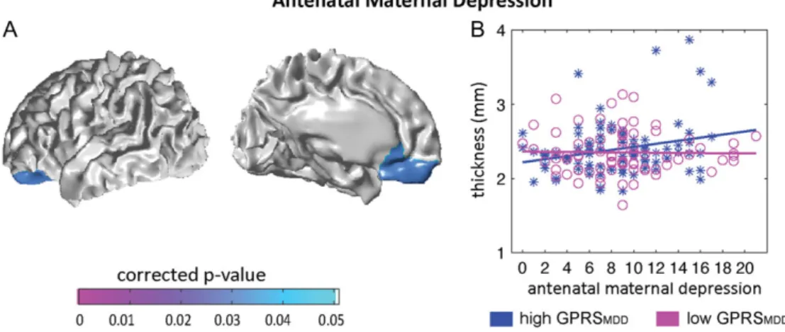
We found a significant interaction effect between antenatal maternal depressive symptoms and infant GPRSMDDon thickness
of left orbitofrontal cortex (OFC) and ventromedial prefrontal cor-tex (vmPFC) (corrected clusterP=0.031; Fig.3A), with the largest interaction effect using the GPRSMDD atP = 0.1 among the 3
thresholds (0.05, 0.1, and 0.2); the interaction was also apparent using the GPRSMDDatP=0.2. Thesefindings remained significant
when additionally controlling for the maternal GPRSMDD (the
Supplementary Material). Thesefindings became a trend of signifi -cance after additionally controlling for SES (the Supplementary Material). The post hoc analysis further revealed the significant positive association between antenatal maternal depressive symptoms and cortical thickness of left OFC and vmPFC (t85=
2.63,P=0.010) in neonates with high infant GPRSMDD(>0),
sug-gesting that a greater maternal depressive score was associated with thicker cortex in left OFC and vmPFC (see Fig.3B). No associ-ation was found between antenatal maternal depressive symp-toms and cortical thickness of left OFC and vmPFC (t80=−0.054,P
=0.957) in neonates with low infant GPRSMDD(<0).
In contrast to the GPRSMDD, GPRSRA did not moderate the
association of antenatal maternal depressive symptoms with the right amygdala and hippocampus at all the 3 thresholds (0.05, 0.1, and 0.2) of the GPRSRA computation (Table 3).
Moreover, the GPRSRA did not affect the association between
cortical thickness in the vmPFC and OFC and antenatal mater-nal depressive symptoms (clusterP-value>0.05).
Interaction of GPRSMDDwith SES on Neonatal Brain Morphology in the Asian Cohort
Likewise, a significant interaction effect between SES and infant GPRSMDDon the right, but not left amygdala and hippocampal
Table 2Statistical results for amygdala and hippocampal volumes in the Asian sample
Structure Mean (SD) (mm3) Interaction effects
Left amygdala 211.41 (34.48) F1161=2.96,P=0.087
Right amygdala 187.09 (33.77) F1161=6.88*,a,P=0.009
Left hippocampus 779.40 (108.18) F1161=0.002,P=0.964
Right hippocampus 781.76 (109.11) F1161=4.15*,a,P=0.043
Note:F-values indicate the interaction effects of infant GPRSMDDwith antenatal
maternal depressive symptoms on the structural volumes. *P-value<0.05.
aThe result remains significant when maternal GPRS
MDDwas entered in
regres-sion analysis as an additional covariate.
Figure 1.Interaction of the infant’s GPRSMDDwith antenatal maternal depressive symptoms on the amygdala and hippocampal volumes. Scatter plots in panels (A)
and (B) illustrate the relationship of antenatal maternal depressive symptoms with right amygdala volume and right hippocampal volume in neonates with high GPRS for major depressive disorder (GPRSMDD, blue asterisks) and those with low GPRSMDD(magenta circles).
volumes and shapes (Supplementary Table S2) after adjusting for gender, maternal ethnicity, adjusted birth weight, and postcon-ceptual age on the MRI day. Such interaction effects remained sig-nificant for the right hippocampus but not the right amygdala when additionally controlling for antenatal maternal depressive symptoms. No interaction effect between SES and infant GPRSMDDwas observed on cortical thickness.
Brain Region-Specific Gene Networks and Biological Processes
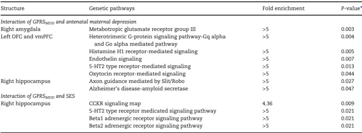
Among the SNPs involved in the GPRSMDD, the top 1551 showed
the most significant interaction with antenatal maternal depres-sive symptoms (P<0.2) on the right amygdala, 1170 SNPs on the right hippocampus, and 1236 SNPs on the left OFC and vmPFC thickness, which were mapped to 629, 508, and 503 genes, respectively. PANTHER analysis revealed biological pathways for the 3 structures. These pathways were statistically over-representative in relation to those in the PANTHER pathway database (Table4). For the right amygdala, 78 genes could not be found in the PANTHER database. The remaining 551 genes formed one statistically over-represented pathway (P =0.003)
labeled by PANTHER as“metabotropic glutamate receptor group III”. For the left OFC and vmPFC, 61 genes could not be found in the PANTHER database. The remaining 442 genes formed 5 stat-istically over-represented pathways, including “heterotrimeric G-protein signaling pathway-Gq alpha and Go alpha”(P=0.004), “endothelin signaling” (P = 0.007), “histamine H1 receptor” (P=0.005), 5-HT2 type receptor (P=0.013), and“oxytocin recep-tor” (P = 0.044). For the right hippocampus, 69 genes could not be found in the PANTHER database. The remaining Table 3Statistical results for amygdala and hippocampal volumes i-n the Asiai-n sample.
Threshold for the
GPRSRAcomputation
Right amygdala Right hippocampus
Antenatal maternal depression
P<0.05 F1159=0.867,P=0.353 F1159=1.457,P=0.229
P<0.1 F1159=1.662,P=0.199 F1159=1.267,P=0.262
P<0.2 F1159=1.662,P=0.199 F1159=1.267,P=0.262
Note:F-values indicate the interaction effects of the infant’s GPRSRAwith
ante-natal maternal depression on the structural volumes.
Figure 2.Statistical maps of the interaction effects of the infant GPRSMDDwith antenatal maternal depressive symptoms (A) on the amygdala and hippocampal
shapes. Panels (B,C) illustrate the relationship of antenatal maternal depressive symptoms with right amygdala shape (left panel) and right hippocampal shape (right panel) in neonates with high GPRS for major depressive disorder (GPRSMDD, blue astericks) and those with low GPRSMDD(magenta circles). Antenatal maternal
depres-sive symptoms are represented by the score of EPDS, with a higher score indicating more severe of depresdepres-sive symptoms.
Figure 3.Statistical maps for interaction of the infant GPRSMDDwith antenatal maternal depressive symptoms (A) on cortical thickness. Panel (B) illustrates the
relation-ship of antenatal maternal depressive symptoms with thickness of left OFC and vmPFC in neonates with high GPRSMDD, (blue asterisks) and those with low GPRSMDD
(magenta circles). Antenatal maternal depressive symptoms are represented by the score of EPDS, with a higher score indicating more severe of depressive symptoms.
439 genes formed 2 statistically over-represented genetic path-ways, including “axon guidance mediated by Slit/Robo” (P =
0.027) and“Alzheimer’s disease-amyloid secretase”(P=0.047). Supplementary Table S3 lists the genes involved in each pathway.
The same analysis for SNPs in infant GPRSMDDthat nominally
moderate the association between SES and fetal neurodevelop-ment did not reveal any significantly enriched pathways for the right amygdala. For the right hippocampus, 40 genes could not be found in the PANTHER database and the remaining 311 genes formed 4 statistically over-represented pathways (Table4), includ-ing“CCK receptor signaling”(P=0.009),“Beta1”(P=0.021) and “Beta2 (P=0.021) adrenergic receptor signaling”, and“5-HT2 type receptor”mediated signaling pathways (P=0.021). Supplementary Table S4 lists the genes involved in each pathway.
We then entered the genes from the individual pathways identified by PANTHER into GeneMania and identified candi-date biological processes (Table 5). GeneMania identified “Glutamate receptor activity” as the primary function of the metabotropic glutamate receptor group III pathway, which included genes that encode for subunits of the N -methyl-D-aspartate (GRIN1, GRIN2A, GRIN2B, GRIN2C, and GRIN2D), Kainate (GRK1-5), andα -amino-3-hydroxy-5-methyl-4-isoxazo-lepropionic acid (GRIA1-4) receptors, as well as for the metabo-tropic receptors (GRM1-8). This same pathway was also significantly associated with the SNAp REceptor (SNARE) com-plex function. Likewise, the SNARE comcom-plex emerged as the primary biological function associated with both the 5-HT2-type receptor- and the oxytocin receptor-mediated signaling pathway identified as candidate moderators of the effect of antenatal maternal depressive symptoms on cortical thickness (Table5).
The Findings from the US Cohort
We also examined the neonatal amygdala and hippocampus structures using the US sample of 85 dyads with available neo-natal brain imaging data of the amygdala and hippocampal volumes, SNP data genotyped using the Illumina Omni express arrays, and measures of maternal depressive symptoms assessed using the Center for Epidemiologic Studies Depression
scale during the third trimester (the Supplementary Material). We used the same statistical model as that employed with the Asian cohort data and found a statistically significant interaction effect between infant GPRSMDDand antenatal maternal
depres-sive symptoms on the right (F1,77=4.09,P=0.047), but not left
amygdala volume (F1,77=1.21,P=0.275), with the largest
inter-action effect at GPRSMDDofP=0.1 among the 3 thresholds (0.05,
0.1, and 0.2). Interestingly, the US cohort showed the significant positive association between antenatal maternal depressive symptoms and right amygdala volume (t36=2.21,P=0.034) in
neonates with low infant GPRSMDD(<0) and the marginal
nega-tive association between antenatal maternal depressive symp-toms and right amygdala volume (t37 = −1.66, P = 0.105) in
neonates with high infant GPRSMDD(>0), which is opposite to
that seen in the Asian sample. This could be due to differences in populations, which has been reported in other studies on Asian and Caucasian populations based on individual SNPs (Sheikh et al. 2013) (Woo et al. 2004;Serretti et al. 2007;Porcelli et al. 2012; Ming et al. 2013,2015). Nevertheless, interaction effects between infant GPRSMDD and antenatal maternal depressive symptoms
were not observed on bilateral hippocampal volumes (left:F1,77=
0.064,P=0.801; right:F1,77=0.903,P=0.345).
Among the SNPs involved in the GPRSMDDfor the US sample,
the top 747 SNPs with the most significant interaction with antenatal maternal depressive symptoms (P<0.1) on the right amygdala volume were mapped to 594 genes. For the right amygdala, 65 genes could not be found in the PANTHER data-base. The remaining 529 genes formed 2 statistically over-represented pathway labeled by PANTHER as “metabotropic glutamate receptor group III”(P=0.002) and“metabotropic glu-tamate receptor group I”(P=0.003). Thus, despite the differ-ences in sample size and ethnicity, these biological pathways were strikingly similar to those identified in the Asian cohort for the interaction between antenatal maternal depressive symptoms and GPRSMDDon the right amygdala volume.
Discussion
This study provides evidence for the role of infant genotype as a moderator of the association of antenatal maternal mental health with fetal development of cortico-limbic structures,
Table 4Genetic pathways associated with interaction of GPRSMDDand antenatal maternal depressive symptoms in the Asian sample
Structure Genetic pathways Fold enrichment P-valuea
Interaction of GPRSMDDand antenatal maternal depression
Right amygdala Metabotropic glutamate receptor group III >5 0.003
Left OFC and vmPFC Heterotrimeric G-protein signaling pathway-Gq alpha
and Go alpha mediated pathway
>5 0.004
Histamine H1 receptor-mediated signaling >5 0.005
Endothelin signaling >5 0.007
5-HT2 type receptor-mediated signaling >5 0.013
Oxytocin receptor-mediated signaling >5 0.044
Right hippocampus Axon guidance mediated by Slit/Robo >5 0.027
Alzheimer’s disease-amyloid secretase >5 0.047
Interaction of GPRSMDDand SES
Right hippocampus CCKR signaling map 4.36 0.009
5-HT2 type receptor medicated signaling pathway >5 0.021
Beta1 adrenergic receptor signaling pathway >5 0.021
Beta2 adrenergic receptor signaling pathway >5 0.021
Notes: The Fold Enrichment is defined as the ratio of the proportion of our input genes involved in a particular genetic pathway to the proportion of the reference genes involved in the same genetic pathway.
aBonferroni corrected
P-value.
including structures such as the amygdala, hippocampus, OFC, and vmPFC that are implicated in affective disorders. These interaction effects were apparent using measures of depressive symptoms across a community sample and remained signifi -cant even after controlling for maternal GPRSMDDthus revealing
selective moderation by infant genotype. This study further revealed over-representative genetic pathways and biological processes formed by the genes included in the GPRSMDD
com-putation for which variations best contributed to modulating the relationship of antenatal maternal depressive symptoms and the morphology of the aforementioned brain structures.
The amygdala, hippocampus, OFC, and vmPFC are cortico-limbic structures implicated in mood disorders and central to emotion and social regulation. Abnormal morphology and activity in these regions are reported in children (Thomas et al. 2001;Pagliaccio et al. 2014) and adults with MDD (Johnstone et al. 2007). Smaller hippocampal volume and greater activity in response to sad faces predict increased depressive symptoms in children (Suzuki et al. 2013). In depressed children severity correlates with activity of the amygdala–hippocampal complex and medial PFC during sad mood elaboration (Pagliaccio et al. 2012) and amygdala reactivity during emotional face viewing (Gaffrey et al. 2011). Likewise, children with early-life stress show smaller OFC (Hanson et al. 2010). Children of parents with MDD also show altered amygdala activity in the emotional facial expression (Monk et al. 2008). Increased amygdala activa-tion in response to novelty or threatening faces in children is associated with measures of negative emotionality (Perez-Edgar et al. 2007), which in turn predicts a greater risk for mood disorders (Bruder-Costello et al. 2007). Moreover, altered func-tional connectivity between the amygdala and medial PFC, including OFC and vmPFC, is observed in 6-month-old infants born to mothers with increased antenatal maternal depressive symptoms (Qiu et al. 2015a). Thesefindings suggest that the cortico-limbic systems may be particularly vulnerable to mater-nal influences during fetal development and reflect, in part, the neural basis for the familial transmission of phenotypes asso-ciated with depression.
Our study revealed an interaction effect between maternal depressive symptoms and infant GPRSMDDthat was apparent in
the right amygdala and hippocampus. Individual differences in stress reactivity associate with morphology and function of the prefrontal cortex, the hippocampus and the amygdala, as well as the connections between these regions (Pruessner et al. 2008;van Marle et al. 2009) and there is compelling evidence implicating the right hemisphere, notably the right amygdala and right hippocampus in the regulation of stress responses
(Abercrombie et al. 1998; Carrion et al. 2007; Hamilton and Gotlib 2008). The risk for depression is linked to heightened stress reactivity, which associates with a familial risk for depression: individuals with increased genetic vulnerability for depression show greater negative affect in response to daily life stress, which then predicts depression (Wichers et al. 2009). Ourfindings suggest the familial transmission of individual dif-ferences in neural systems that underlie stress reactivity may occur during fetal development.
We explored gene networks and their associated biological processes as candidate mediators of the influence of clinically relevant environmental conditions on fetal neurodevelopment. The gene pathways (metabotropic glutamate receptor group pathways) derived from SNPs that best mediated the interaction of antenatal maternal depressive symptoms and infant GPRSMDD
on the right amygdala in both the Asian and US cohorts were strongly associated with glutamate receptor activity. Multiple approaches, including genome-wide analyses, implicate gluta-matergic synaptic transmission in the etiology of depression (Skolnick et al. 2009; Lee et al. 2012) and patients with MDD exhibit abnormal glutamate concentrations in the amygdala (Michael et al. 2003). Postmortem studies with MDD suggest altered glutamatergic synaptic signaling (Hashimoto et al. 2007) and ketamine, a glutamatergic intervention, is a treatment for MDD (Caddy et al. 2014). The antidepressant, tianeptine, blocks stress-induced increases in glutamate release in the amygdala (Piroli et al. 2013) and moderates behavioral responses to stress. This glutamate receptor activity included the GRM1 andGRM8 genes, which are nominally associated with MDD (Terracciano et al. 2010). Knockout mouse models of genes coding for metabo-tropic glutamate receptor proteins, which modulate postsynaptic glutamate signaling, associate with increased anxiety (Bures et al. 2005). The“G-protein signaling”network also identified as a candidate mediator for the effects of maternal depression on cor-tical structure (Supplementary Table S3) includes the GRM1, GRM4, GRM 7, andGRM8genes.
The genetic pathway analysis in the right amygdala identi-fied the“SNARE complex”biological process, which is a funda-mental component of neural connectivity and neurotransmitter release comprised of syntaxin, SNAP-25, and VAMP. Indeed, per-haps the most compelling finding from our analysis was the broad implication of the SNARE complex as a biological process mediating the impact of antenatal maternal depressive symp-toms on multiple brain structures (Table5). Postmortem studies revealed abnormal expression of SNARE proteins in the hippo-campus and frontal cortex of subjects with MDD (Scarr et al. 2006). Likewise, proteomic analyses implicate synaptic Table 5Biological functions of genetic pathway in the Asian sample
Genetic pathway Biological function FDR correctedP-value Coverage
Metabotropic glutamate receptor group III Glutamate receptor activity 7.3e–46 20/20
SNARE complex 3.2e–18 12/32
G-protein signaling G-protein coupled receptor signaling 1.2e–32 29/125
Histamine H1 receptor-mediated signaling Inositol phosphate metabolic process 6.0e–19 13/55
Endothelin signaling Fibroblast growth factor signaling 1.5e–43 35/170
5-HT2 type receptor-mediated signaling SNARE complex 3.1e–25 15/32
Oxytocin receptor-mediated signaling SNARE complex 5.0e–26 15/32
Axon guidance mediated by Slit/Robo Small GTPase mediated signal transduction 3.9e–8 12/291
Amyloid secretase Neurotrophic signaling 2.4e–20 23/277
Beta1 adrenergic receptor signaling SNARE complex 1.6e–27 15/32
Beta2 adrenergic receptor signaling SNARE complex 1.6e–27 15/32
CCKR signaling Intracellular ligand-gated calcium channel activity 3.1e–14 7/10
dysfunction in MDD and identify altered expression of SNARE complex proteins (Martins-de-Souza et al. 2012). Administration of ketamine as well as a broad range of antidepressant medica-tions alters the accumulation of SNARE complexes in synaptic membranes (Bonanno et al. 2005), which then associates with altered capacity for stimulated glutamate release (Bonanno et al. 2005). Two SNP’s (rs3787283 and rs3746544) in the SNAP-25 gene have been associated with MDD in a Han Chinese sample (Wang et al. 2015), an ethnic group that accounted for the majority of the Asian subjects in our study.
Ourfindings suggest that the influence of antenatal maternal depressive symptoms on the cortico-limbic structure is mediated by brain-specific signaling pathways (oxytocin, 5-HT2, and gluta-mate receptor pathways). These pathways are linked to pro-cesses underlying synaptic development. In addition to association with the glutamate receptor activity, the SNARE com-plex was the top biological process for the oxytocin and 5-HT2 gene networks (Table5). There is strong evidence for the import-ance of serotonin, including 5-HT2 receptor activation, in peri-natal brain structure (seeLesch and Waider 2012for a review). While oxytocin receptor activation is not extensively described in terms of brain development, there is a period of transient neocor-tical expression of oxytocin receptor binding in rodent brain that suggests a developmental influence (Hammock and Levitt 2013). Finally, our analyses identified neurotrophic signaling systems as candidate mediators of the effects of antenatal maternal depres-sion on hippocampus (Table4). The Fibroblast growth factor sig-naling system is particularly interesting to consider in light of the findings with rodent models that early postnatal administration of FGF2 decreases fear behavior in animals genetic predisposed to increased expression of anxiety-like behaviors (Eren-Kocak et al. 2011). Sustained FGF2 effects on genes expression included targets involved in cell survival, an effect that is consistent with the role for neurotrophic signaling pathways as a mediator of the effects of antenatal maternal depression. Neurotrophic signaling pathway was the top biological process emerging from the amyl-oid secretase network and includes genes coding for proteins that mediate the effects of brain-derived neurotrophic factor sig-naling, implicated in the pathophysiology of MDD and associated with the activation of inositol phosphate metabolism (Duman and Voleti 2012), which was also a top biological process identi-fied in our analysis. Taken together, ourfindings are consistent with the hypothesis that environmental conditions that increase the risk for MDD associate with alterations in mechanisms con-trolling synaptic plasticity in cortico-limbic brain regions (Duman and Aghajanian 2012).
Interestingly, the direction of the interaction between mater-nal depressive symptoms and infant genotype on right amygdala volume in the US sample is in the opposite direction to that observed in the Asian sample. This is not surprising as there is now considerable evidence for opposing effects of individual SNPs between Asian and Caucasian populations. For instance, Sheikh et al. 2013found that during childhood Caucasian chil-dren homozygous for the Catechol-O-methyltransferase (COMT) val allele show higher levels of internalizing symptoms com-pared to children with at least one copy of the COMTmetallele. In contrast,Baumann et al. 2013found that in Asians homozyges of the COMTmetallele with adverse childhood experiences show higher anxiety sensitivity. Similarfindings are apparent in stud-ies of the serotonin transporter polomorphism (5-HTTPLR).Ming et al. 2015report a replication of the well-established interaction between the 5-HTTPLR and stressful life events on emotional health in Han Chinese, the principal ethnic group in our Asian sample. However, in this study it was theLand not theSallele
that conferred greater sensitivity to stress; studies with Caucasian samples commonly report that the S allele confers sensitivity to adversity. This study was also a replication of a previous, similarfinding with the 5-HTTPLR variant (Ming et al. 2013). Meta-analyses likewise reveal differential effects of 5-HTTLPR on selective serotonin reuptake inhibitors efficacy between Caucasians and Asians (Serretti et al. 2007;Porcelli et al. 2012). Importantly, functional MRI studies show a link betweenS allele and higher amygdala activation in response to emotional stimuli in Caucasians (e.g.,Hariri et al. 2002,2005;Murphy et al. 2013), whereas in Asians Lallele is associated with amygdala hyperactivation (Li et al. 2012). These findings imply that the same risk allele may operate in the opposite fashion as a func-tion of ethnic background. Allele frequency may be a factor. The frequency of theLallele is much lower than that ofSallele in Chinese population and Japan populations, but higher in Caucasians or African–Americans (Lesch et al. 1996;He et al. 2010). Importantly, despite the directional difference, the pathway ana-lyses revealed the same underlying biological processes for the amygdala effect in both samples. Indeed, PANTHER identified the same primary pathway for both samples using independent ana-lyses. Thesefindings are consistent with the studies noted above, that reveal effects of polymorphisms in the same genetic regions, consistent with common underlying biological processes, but with polymorphisms operating in an ethnicity-specific manner.
The relationship between antenatal maternal depression and children’s outcomes is often observed as a function of gen-der. Recent evidence suggests that girls born to depressed mothers may be more likely to exhibit increased cortisol reactivity in childhood and subsequently altered amygdala– prefrontal cortex activity in adolescence (Burghy et al. 2012). Nevertheless, our study did not observe the gender effect, which may partly because of the age of children in this study. It remains possible that gender differences could be observable at later stages in development.
This study is best considered as exploratory and intended to provide a strategy for the analysis of gene–environment inter-dependence and identify candidate biological processes that might mediate the influence of environment conditions asso-ciated with the risk for MDD on fetal brain development. While preliminary, ourfindings do suggest biologically plausible candi-date processes in two independent cohorts. While the neonatal neuroimaging dataset in this study is highly unique in its timing of acquisition and number of subjects, it is nevertheless a modest sample size for genomic analyses. Due to the time limit for scan-ning of neonates and brain immaturity, the neonatal brain MRI, in general, has relatively low contrast between gray and white mat-ter, which could be challenging for imaging and the brain segmen-tation. The contrast between gray matter and white matter is relatively clear on T2-weighted MRI at the neonatal stage but is
diminished around 4–9 months of age. We reported the brain seg-mentation accuracy in the Materials and Methods section to indi-cate our constraint with the neonatal brain images. An additional limitation is the use of GWAS datasets derived from Caucasians for the definition of a GPRS to study within an Asian sample. While this is an important caveat, we note that theWang et al. (2015)study identified multiple SNPs in the SNAP-25 gene, which is a member of the SNARE complex, in the pathophysiology of MDD in Asian population. Moreover, our assessment of maternal depressive symptoms was based on a common screening tool intended to elicit a subjective report of emotional well-being, but does not constitute clinical assessment. The brain variations in the offspring are thus best considered as being associated with self-reported depressive symptoms and not to clinical depression,
per se. Furthermore, other factors, such as marital discord, pater-nal mental illness, medication during pregnancy, materpater-nal sub-stance use, or breastfeeding, could also influence the fetal brain development over and beyond antenatal maternal depression since the brain develops rapidly in early life (Uematsu et al. 2012). In our study, SES was highly correlated with antenatal maternal depressive symptoms and both showed its interaction with GPRSMDD and influence the right amygdala and hippocampus
(Supplementary Table S2 and Figures S1 and S2). After controlling for each other, the interaction effect of antenatal maternal depres-sion and GPRSMDDwas mainly shown on the right amygdala,
while the interaction effect of SES and GPRSMDD was mainly
shown on the right hippocampus (the Supplementary Material). Thesefindings suggested that environment conditions associated with the risk for MDD interplay with genetic risks for MDD on fetal brain development. Finally, comparing brain anatomical mea-sures across different cohorts, particularly, on neonates, is chal-lenging. This is partly because of different strength of the MRI scanners and imaging acquisition protocols. More importantly, the challenge also lies in the definition of the anatomy of individ-ual brain structures. In this study, we obtained the comparable volume measure for the amygdala from the two cohorts. Hence, this study incorporated the amygdalafindings from both cohorts where we could be certain of comparable anatomical localization. Furthermore, the US cohort was lack of maternal GPRSMDD
infor-mation and hence our reported results for the US cohort could not count for the contribution of maternal GPRSMDD.
Conclusion
We present a hypothesis-generating approach to the study of gene×environment interaction that uses GPRS together with bioinformatic approaches to identify candidate biological pro-cesses mediating the influence of environmental conditions on brain development and function. This approach is considered as hypothesis-generating. For example, in larger samples, with sufficient statistical power, we predict that a multi-gene locus score, reflecting polymorphisms in genes involved in the bio-logical processes of glutamate receptor activity, SNARE com-plex, 5-HT2 receptor signaling, should moderate brain region-specific effects of “depressogenic” environmental conditions during fetal development.
Supplementary Material
Supplementary data is available atCerebral Cortexonline.
Funding
The National Medical Research Council (NMRC; NMRC/TCR/004-NUS/2008 and NMRC/CBRG/0039/2013), the Singapore Ministry of Education Academic Research Fund Tier 2 (MOE2012-T2-2-130) and funding from the Hope for Depression Research Foundation, the Sackler Foundation, the National Institute of Health (R01 MH091351, R01 HD060628), and ERA-NET NEURON 01EW1407A and the Canadian Institutes for Advanced Research.
Notes
We thank the GUSTO study group and all clinical and home vis-it staff involved. The voluntary participation of all participants is greatly appreciated. The GUSTO study group includes Pratibha Agarwal, Arijit Biswas, Choon Looi Bong, Shirong Cai, Jerry Kok Yen Chan, Yiong Huak Chan, Cornelia Yin Ing Chee,
Yin Bun Cheung, Audrey Chia, Amutha Chinnadurai, Chai Kiat Chng, Mary Foong-Fong Chong, Shang Chee Chong, Mei Chien Chua, Chun Ming Ding, Eric Andrew Finkelstein, Doris Fok, Keith M. Godfrey, Anne Eng Neo Goh, Yam Thiam Daniel Goh, Joshua J. Gooley, Wee Meng Han, Mark Hanson, Christiani Jeyakumar Henry, Joanna D. Holbrook, Chin-Ying Hsu, Hazel Inskip, Jeevesh Kapur, Ivy Yee-Man Lau, Bee Wah Lee, Yung Seng Lee, Ngee Lek, Sok Bee Lim, Yen-Ling Low, Iliana Magiati, Lourdes Mary Daniel, Cheryl Ngo, Krishnamoorthy Naiduvaje, Wei Wei Pang, Boon Long Quah, Victor Samuel Rajadurai, Mary Rauff, Salome A. Rebello, Jenny L. Richmond, Lynette Pei-Chi Shek, Allan Sheppard, Borys Shuter, Leher Singh, Shu-E Soh, Walter Stunkel, Lin Lin Su, Kok Hian Tan, Oon Hoe Teoh, Mya Thway Tint, Hugo P S van Bever, Rob M. van Dam, Inez Bik Yun Wong, P. C. Wong, Fabian Yap, George Seow Heong Yeo.Conflict of Interest: None declared.
References
Abercrombie HC, Schaefer SM, Larson CL, Oakes TR, Lindgren KA, Holden JE, Perlman SB, Turski PA, Krahn DD, Benca RM, et al. 1998. Metabolic rate in the right amygdala predicts negative affect in depressed patients. Neuroreport. 9: 3301–3307.
Bai J, Abdul-Rahman MF, Rifkin-Graboi A, Chong YS, Kwek K, Saw SM, Godfrey KM, Gluckman PD, Fortier MV, Meaney MJ, et al. 2012. Population differences in brain morphology and microstructure among Chinese, Malay, and Indian Neonates. PLoS One. 7:e47816.
Baumann C, Klauke B, Weber H, Domschke K, Zwanzger P, Pauli P, Deckert J, Reif A. 2013. The interaction of early life experi-ences with COMT val158met affects anxiety sensitivity. Genes Brain Behav. 12:821–829.
Bonanno G, Giambelli R, Raiteri L, Tiraboschi E, Zappettini S, Musazzi L, Raiteri M, Racagni G, Popoli M. 2005. Chronic antidepressants reduce depolarization-evoked glutamate release and protein interactions favoring formation of SNARE complex in hippocampus. J Neurosci. 25:3270–3279. Bruder-Costello B, Warner V, Talati A, Nomura Y, Bruder G,
Weissman M. 2007. Temperament among offspring at high and low risk for depression. Psychiatry Res. 153:145–151. Bures M, Swanson CJ, Linden A-M, Schoepp DD, Johnson MP,
Monn JA. 2005. Metabotropic glutamate receptors as novel targets for anxiety and stress disorders. Nat Rev Drug Discov. 4:131–144.
Burghy CA, Stodola DE, Ruttle PL, Molloy EK, Armstrong JM, Oler JA, Fox ME, Hayes AS, Kalin NH, Essex MJ, et al. 2012. Developmental pathways to amygdala-prefrontal function and internalizing symptoms in adolescence. Nat Neurosci. 15:1736–1741.
Caddy C, Giaroli G, White TP, Shergill SS, Tracy DK. 2014. Ketamine as the prototype glutamatergic antidepressant: pharmacodynamic actions, and a systematic review and meta-analysis of efficacy. Ther Adv Psychopharmacol. 4:75–99. Carrion VG, Weems CF, Reiss AL. 2007. Stress predicts brain changes in children: a pilot longitudinal study on youth stress, posttraumatic stress disorder, and the hippocampus. Pediatrics. 119:509–516.
Chung MK, Robbins SM, Dalton KM, Davidson RJ, Alexander AL, Evans AC. 2005. Cortical thickness analysis in autism with heat kernel smoothing. Neuroimage. 25:1256–1265.
Chung MK, Worsley KJ, Nacewicz BM, Dalton KM, Davidson RJ. 2010. General multivariate linear modeling of surface shapes using SurfStat. Neuroimage. 53:491–505.
Cox JL, Holden JM, Sagovsky R. 1987. Detection of postnatal depression. Development of the 10-item Edinburgh post-natal depression scale. Br J Psychiatry. 150:782–786.
Du J, Younes L, Qiu A. 2011. Whole brain diffeomorphic metric mapping via integration of sulcal and gyral curves, cortical surfaces, and images. Neuroimage. 56:162–173.
Duman RS, Aghajanian GK. 2012. Synaptic dysfunction in depression: potential therapeutic targets. Science. 338:68–72. Duman RS, Voleti B. 2012. Signaling pathways underlying the pathophysiology and treatment of depression: novel mechan-isms for rapid-acting agents. Trends Neurosci. 35:47–56. Duncan LE, Keller MC. 2011. A critical review of thefirst 10 years
of candidate gene-by-environment interaction research in psychiatry. Am J Psychiatry. 168:1041–1049.
Eren-Kocak E, Turner CA, Watson SJ, Akil H. 2011. Short-hairpin RNA silencing of endogenousfibroblast growth factor 2 in rat hippocampus increases anxiety behavior. Biol Psychiatry. 69:534–540.
Farah MJ, Hackman DA, Meaney MJ. 2010. Socioeconomic status and the brain: mechanistic insights from human and animal research. Nat Rev Neurosci. 11:651–659.
Gaffrey MS, Luby JL, Belden AC, Hirshberg JS, Volsch J, Barch DM. 2011. Association between depression severity and amygdala reactivity during sad face viewing in depressed preschoolers: an fMRI study. J Affect Disord. 129:364–370. Gilman SE, Kawachi I, Fitzmaurice GM, Buka SL. 2003. Family
disruption in childhood and risk of adult depression. Am J Psychiatry. 160:939–946.
Hamilton JP, Gotlib IH. 2008. Neural substrates of increased memory sensitivity for negative stimuli in major depres-sion. Biol Psychiatry. 63:1155–1162.
Hammock EA, Levitt P. 2013. Oxytocin receptor ligand binding in embryonic tissue and postnatal brain development of the C57BL/6 J mouse. Front Behav Neurosci. 7:195.
Hanson JL, Chung MK, Avants BB, Shirtcliff EA, Gee JC, Davidson RJ, Pollak SD. 2010. Early stress is associated with alterations in the orbitofrontal cortex: a tensor-based morphometry investigation of brain structure and behav-ioral risk. J Neurosci. 30:7466–7472.
Hariri AR, Drabant EM, Munoz KE, Kolachana BS, Mattay VS, Egan MF, Weinberger DR. 2005. A susceptibility gene for affective disorders and the response of the human amyg-dala. Arch Gen Psychiatry. 62:146–152.
Hariri AR, Mattay VS, Tessitore A, Kolachana B, Fera F, Goldman D, Egan MF, Weinberger DR. 2002. Serotonin trans-porter genetic variation and the response of the human amygdala. Science. 297:400–403.
Hashimoto K, Sawa A, Iyo M. 2007. Increased levels of gluta-mate in brains from patients with mood disorders. Biol Psychiatry. 62:1310–1316.
He Q, Xue G, Chen C, Lu Z, Dong Q, Lei X, Ding N, Li J, Li H, Chen C, et al. 2010. Serotonin transporter gene-linked polymorphic region (5-HTTLPR) influences decision making under ambigu-ity and risk in a large Chinese sample. Neuropharmacology. 59:518–526.
Holmes AJ, Lee PH, Hollinshead MO, Bakst L, Roffman JL, Smoller JW, Buckner RL. 2012. Individual differences in amygdala-medial prefrontal anatomy link negative affect, impaired social functioning, and polygenic depression risk. J Neurosci. 32:18087–18100.
Jiang L, Willner D, Danoy P, Xu H, Brown MA. 2013. Comparison of the performance of two commercial genome-wide associ-ation study genotyping platforms in Han Chinese samples. G3 (Bethesda). 3:23–29.
Johnstone T, van Reekum CM, Urry HL, Kalin NH, Davidson RJ. 2007. Failure to regulate: counterproductive recruitment of top-down prefrontal-subcortical circuitry in major depres-sion. J Neurosci. 27:8877–8884.
Kupfer DJ, Frank E, Phillips ML. 2012. Major depressive disorder: new clinical, neurobiological, and treatment perspectives. Lancet. 379:1045–1055.
Lee PH, Perlis RH, Jung JY, Byrne EM, Rueckert E, Siburian R, Haddad S, Mayerfeld CE, Heath AC, Pergadia ML, et al. 2012. Multi-locus genome-wide association analysis supports the role of glutamatergic synaptic transmission in the etiology of major depressive disorder. Transl Psychiatry. 2:e184. Lesch K-P, Waider J. 2012. Serotonin in the modulation of
neural plasticity and networks: implications for neurodeve-lopmental disorders. Neuron. 76:175–191.
Lesch KP, Bengel D, Heils A, Sabol SZ, Greenberg BD, Petri S, Benjamin J, Muller CR, Hamer DH, Murphy DL. 1996. Association of anxiety-related traits with a polymorphism in the serotonin transporter gene regulatory region. Science. 274:1527–1531.
Li S, Zou Q, Li J, Li J, Wang D, Yan C, Dong Q, Zang YF. 2012. 5-HTTLPR polymorphism impacts task-evoked and resting-state activities of the amygdala in Han Chinese. PLoS One. 7: e36513.
Lubke GH, Hottenga JJ, Walters R, Laurin C, de Geus EJ, Willemsen G, Smit JH, Middeldorp CM, Penninx BW, Vink JM, et al. 2012. Estimating the genetic variance of major depressive disorder due to all single nucleotide polymorph-isms. Biol Psychiatry. 72:707–709.
Martins-de-Souza D, Guest PC, Harris LW, Vanattou-Saifoudine N, Webster MJ, Rahmoune H, Bahn S. 2012. Identification of proteomic signatures associated with depression and psych-otic depression in post-mortem brains from major depression patients. Transl Psychiatry. 2:e87.
Michael N, Erfurth A, Ohrmann P, Arolt V, Heindel W, Pfleiderer B. 2003. Neurotrophic effects of electroconvulsive therapy: a pro-ton magnetic resonance study of the left amygdalar region in patients with treatment-resistant depression. Neuropsycho-pharmacology. 28:720–725.
Ming Q, Zhang Y, Yi J, Wang X, Zhu X, Yao S. 2015. Serotonin transporter gene polymorphism (5-HTTLPR) L allele interacts with stress to increase anxiety symptoms in Chinese adolescents: a multiwave longitudinal study. BMC Psychiatry. 15:248.
Ming QS, Zhang Y, Chai QL, Chen HY, Hou CJ, Wang MC, Wang YP, Cai L, Zhu XZ, Yi JY, et al. 2013. Interaction between a serotonin transporter gene promoter region polymorphism and stress predicts depressive symptoms in Chinese adolescents: a multi-wave longitudinal study. BMC Psychiatry. 13:142.
Moffitt TE, Caspi A. 2006. Gene-environment interactions in psychiatry: joining forces with neuroscience. Nat Rev Neurosci. 7:583–590.
Monk CS, Klein RG, Telzer EH, Schroth EA, Mannuzza S, Moulton JL 3rd, Guardino M, Masten CL, McClure-Tone EB, Fromm S, et al. 2008. Amygdala and nucleus accumbens activation to emotional facial expressions in children and adolescents at risk for major depression. Am J Psychiatry. 165:90–98.
Mostafavi S, Ray D, Warde-Farley D, Grouios C, Morris Q. 2008. GeneMANIA: a real-time multiple association network inte-gration algorithm for predicting gene function. Genome Biol. 9(Suppl 1):S4. doi:10.1186/gb-2008-9-s1-s4 Epub 2008 Jun 27.
Murphy SE, Norbury R, Godlewska BR, Cowen PJ, Mannie ZM, Harmer CJ, Munafo MR. 2013. The effect of the serotonin transporter polymorphism (5-HTTLPR) on amygdala func-tion: a meta-analysis. Mol Psychiatry. 18:512–520.
Noble KG, Houston SM, Brito NH, Bartsch H, Kan E, Kuperman JM, Akshoomoff N, Amaral DG, Bloss CS, Libiger O, et al. 2015. Family income, parental education and brain structure in children and adolescents. Nat Neurosci. 18:773–778. Pagliaccio D, Luby J, Gaffrey M, Belden A, Botteron K, Gotlib IH,
Barch DM. 2012. Anomalous functional brain activation fol-lowing negative mood induction in children with pre-school onset major depression. Dev Cogn Neurosci. 2:256–267. Pagliaccio D, Luby JL, Luking KR, Belden AC, Barch DM. 2014.
Brain-behavior relationships in the experience and regulation of negative emotion in healthy children: implications for risk for childhood depression. Dev Psychopathol. 26:1289–1303. Perez-Edgar K, Roberson-Nay R, Hardin MG, Poeth K, Guyer AE,
Nelson EE, McClure EB, Henderson HA, Fox NA, Pine DS, et al. 2007. Attention alters neural responses to evocative faces in behaviorally inhibited adolescents. Neuroimage. 35: 1538–1546.
Peyrot WJ, Milaneschi Y, Abdellaoui A, Sullivan PF, Hottenga JJ, Boomsma DI, Penninx BW. 2014. Effect of polygenic risk scores on depression in childhood trauma. Br J Psychiatry. 205:113–119.
Piroli GG, Reznikov LR, Grillo CA, Hagar JM, Fadel JR, Reagan LP. 2013. Tianeptine modulates amygdalar glutamate neuro-chemistry and synaptic proteins in rats subjected to repeated stress. Exp Neurol. 241:184–193.
Porcelli S, Fabbri C, Serretti A. 2012. Meta-analysis of serotonin transporter gene promoter polymorphism (5-HTTLPR) associ-ation with antidepressant efficacy. Eur Neuropsychopharmacol. 22:239–258.
Pruessner JC, Dedovic K, Khalili-Mahani N, Engert V, Pruessner M, Buss C, Renwick R, Dagher A, Meaney MJ, Lupien S. 2008. Deactivation of the limbic system during acute psychosocial stress: evidence from positron emission tomography and functional magnetic resonance imaging studies. Biol Psychiatry. 63:234–240.
Purcell S, Neale B, Todd-Brown K, Thomas L, Ferreira MA, Bender D, Maller J, Sklar P, de Bakker PI, Daly MJ, et al. 2007. PLINK: a tool set for whole-genome association and population-based linkage analyses. Am J Hum Genet. 81:559–575.
Purcell SM, Wray NR, Stone JL, Visscher PM, O’Donovan MC, Sullivan PF, Sklar P. 2009. Common polygenic variation con-tributes to risk of schizophrenia and bipolar disorder. Nature. 460:748–752.
Qiu A, Bitouk D, Miller MI. 2006. Smooth Functional and Structural Maps on the Neocortex via Orthonormal Bases of the Laplace-Beltrami Operator. IEEE Trans Med Imaging. 25(10):1296–1306.
Qiu A, Miller MI. 2008. Multi-Structure Network Shape Analysis via Normal Surface Momentum Maps. NeuroImage. 42(4): 1430–1438.
Qiu A, Adler M, Crocetti D, Miller MI, Mostofsky SH. 2010. Basal ganglia shapes predict social, communication, and motor dysfunctions in boys with autism spectrum disorder. J Am Acad Child Adolesc Psychiatry. 49:539–551 e531-534. Qiu A, Anh TT, Li Y, Chen H, Rifkin-Graboi A, Broekman BF,
Kwek K, Saw SM, Chong YS, Gluckman PD, et al. 2015a. Prenatal maternal depression alters amygdala functional con-nectivity in 6-month-old infants. Transl Psychiatry. 5:e508. Qiu A, Crocetti D, Adler M, Mahone EM, Denckla MB, Miller MI,
Mostofsky SH. 2009. Basal ganglia volume and shape in
children with attention deficit hyperactivity disorder. Am J Psychiatry. 166:74–82.
Qiu A, Rifkin-Graboi A, Chen H, Chong YS, Kwek K, Gluckman PD, Fortier MV, Meaney MJ. 2013. Maternal anxiety and infants’hippocampal development: timing matters. Transl Psychiatry. 3:e306.
Qiu A, Tuan TA, Ong ML, Li Y, Chen H, Rifkin-Graboi A, Broekman BF, Kwek K, Saw SM, Chong YS, et al. 2015b. COMT haplotypes modulate associations of antenatal maternal anxiety and neonatal cortical morphology. Am J Psychiatry. 172:163–172.
Rifkin-Graboi A, Bai J, Chen H, Hameed WB, Sim LW, Tint MT, Leutscher-Broekman B, Chong YS, Gluckman PD, Fortier MV, et al. 2013. Prenatal maternal depression associates with microstructure of right amygdala in neonates at birth. Biol Psychiatry. 74:837–844.
Ripke S, Wray NR, Lewis CM, Hamilton SP, Weissman MM, Breen G, Byrne EM, Blackwood DH, Boomsma DI, Cichon S, et al. 2013. A mega-analysis of genome-wide association studies for major depressive disorder. Mol Psychiatry. 18: 497–511.
Scarr E, Gray L, Keriakous D, Robinson PJ, Dean B. 2006. Increased levels of SNAP-25 and synaptophysin in the dorsolateral prefrontal cortex in bipolar I disorder. Bipolar Disord. 8:133–143.
Serretti A, Kato M, De Ronchi D, Kinoshita T. 2007. Meta-analysis of serotonin transporter gene promoter polymorph-ism (5-HTTLPR) association with selective serotonin reuptake inhibitor efficacy in depressed patients. Mol Psychiatry. 12:247–257.
Sheikh HI, Kryski KR, Smith HJ, Dougherty LR, Klein DN, Bufferd SJ, Singh SM, Hayden EP. 2013. Catechol-O-methyltransferase gene val158met polymorphism and depressive symptoms during early childhood. Am J Med Genet B Neuropsychiatr Genet. 162B:245–252.
Skolnick P, Popik P, Trullas R. 2009. Glutamate-based anti-depressants: 20 years on. Trends Pharmacol Sci. 30: 563–569.
Smith SM. 2002. Fast robust automated brain extraction. Hum Brain Mapp. 17:143–155.
Soh SE, Lee SS, Hoon SW, Tan MY, Goh A, Lee BW, Shek LP, Teoh OH, Kwek K, Saw SM, et al. 2012. The methodology of the GUSTO cohort study: a novel approach in studying pedi-atric allergy. Asia Pac Allergy. 2:144–148.
Sullivan PF, Neale MC, Kendler KS. 2000. Genetic epidemiology of major depression: review and meta-analysis. Am J Psychiatry. 157:1552–1562.
Suzuki H, Botteron KN, Luby JL, Belden AC, Gaffrey MS, Babb CM, Nishino T, Miller MI, Ratnanather JT, Barch DM. 2013. Structural-functional correlations between hippocampal vol-ume and cortico-limbic emotional responses in depressed children. Cogn Affect Behav Neurosci. 13:135–151.
Tan M, Qiu A. 2016. Large Deformation Multiresolution Diffeomorphic Metric Mapping for Multiresolution Cortical Surfaces: A Coarse-to-Fine Approach. IEEE Trans Image Process. 25(9):4061–4074.
Terracciano A, Tanaka T, Sutin AR, Sanna S, Deiana B, Lai S, Uda M, Schlessinger D, Abecasis GR, Ferrucci L, et al. 2010. Genome-wide association scan of trait depression. Biol Psychiatry. 68:811–817.
Thomas KM, Drevets WC, Dahl RE, Ryan ND, Birmaher B, Eccard CH, Axelson D, Whalen PJ, Casey BJ. 2001. Amygdala response to fearful faces in anxious and depressed children. Arch Gen Psychiatry. 58:1057–1063.
Uematsu A, Matsui M, Tanaka C, Takahashi T, Noguchi K, Suzuki M, Nishijo H. 2012. Developmental trajectories of amygdala and hippocampus from infancy to early adult-hood in healthy individuals. PLoS One. 7:e46970.
van Marle HJ, Hermans EJ, Qin S, Fernandez G. 2009. From spe-cificity to sensitivity: how acute stress affects amygdala pro-cessing of biologically salient stimuli. Biol Psychiatry. 66: 649–655.
Wang Q, Wang Y, Ji W, Zhou G, He K, Li Z, Chen J, Li W, Wen Z, Shen J, et al. 2015. SNAP25 is associated with schizophrenia and major depressive disorder in the Han Chinese popula-tion. J Clin Psychiatry. 76:e76–e82.
Wen D, Poh J, Soe N, Chong Y, Chen H, Kwek K, Shek L, Gluckman P, Fortier M, Meaney M, et al. 2017. Influences of
prenatal and postnatal maternal depression on amygdala volume and microstructure in young children. Transl Psychiatry.
Wichers M, Geschwind N, Jacobs N, Kenis G, Peeters F, Derom C, Thiery E, Delespaul P, van Os J. 2009. Transition from stress sensitivity to a depressive state: longitudinal twin study. Br J Psychiatry. 195:498–503.
Woo JM, Yoon KS, Choi YH, Oh KS, Lee YS, Yu BH. 2004. The association between panic disorder and the L/L genotype of catechol-O-methyltransferase. J Psychiatr Res. 38: 365–370.
Zhong J, Qiu A. 2010. Multi-Manifold Diffeomorphic Metric Mapping for Aligning Cortical Hemispheric Surfaces. NeuroImage. 49(1):355–365.