i
THE RELIABILITY AND VALIDITY OF THE MYOTONOMETER AS A MEASURE OF HAMSTRING MUSCLE STIFFNESS
Sarah Elizabeth Bell
A thesis submitted to the faculty of the University of North Carolina at Chapel Hill in partial fulfillment of the requirements for the degree of Master of Arts in the Department of Exercise & Sport Science in the College of Arts & Sciences.
Chapel Hill 2014
iii
The purpose of this study was to determine the reliability and validity of the Myotonometer in measuring hamstring stiffness, while also considering the following factors as predictors of hamstring stiffness when measured with the Myotonometer: tissue compliance, and rate of loading. Thirty-three physically active subjects participated in the study. The subjects’ stiffness was measured using the Oscillation Technique while being tethered to laboratory equipment and a measurement computer as well as measured using The Myotonometer. Our findings reveal that The Myotonometer possesses high intra-session and interrater reliability. However, the results also indicate that The Myotonometer is not a valid measure of hamstring stiffness relative to the Oscillation Technique. Although the Mytonometer does not measure active muscle stiffness as the Oscillation Technique does, it does have high reliability and portability and can be used to measure tissue compliance and the effectiveness of treatments in the clinical setting. Future research should focus on portable and clinically applicable tools to measure active hamstring stiffness in efforts to prevent and monitor injuries.
ABSTRACT
SARAH E. BELL: The Reliability and Validity of the Myotonometer as a Measure of Hamstring Muscle Stiffness
TABLE OF CONTENTS
LIST OF FIGURES ... VI LIST OF TABLES ... VII
CHAPTER I ... 2
INTRODUCTION ... 2
RESEARCH QUESTIONS AND HYPOTHESES ... 5
INDEPENDENT VARIABLES ... 6
DEPENDENT VARIABLES ... 7
OPERATIONAL DEFINITIONS ... 7
ASSUMPTIONS ... 8
LIMITATIONS ... 9
CHAPTER II ... 10
INTRODUCTION ... 10
ANTERIOR CRUCIATE LIGAMENT INJURY ... 10
MUSCULOTENDINOUSSTIFFNESS ... 20
CONCLUSION ... 27
CHAPTER III ... 28
SUBJECTS ... 28
v
STATISTICAL ANALYSIS ... 32
CHAPTER IV... 35
MYOTONOMETER RELIABILITY ... 35
MYOTONOMETER VALIDITY ... 35
MEASUREMENT TECHNIQUE RESULTS ... 36
CHAPTER V ... 37
INTRODUCTION ... 37
RELIABILITY ... 37
VALIDITY ... 38
CONCLUSION ... 41
FIGURES ... 42
TABLES ... 44
LIST OF FIGURES
FIGURE 1: MVIC POSITIONING ... 41
FIGURE 2: OSCILLATION TECHNIQUE WEIGHT APPLICATION ... 41
FIGURE 3: MYOTONOMETER PLACEMENT ... 42
vii
LIST OF TABLES
CHAPTER I INTRODUCTION
INTRODUCTION
Organized and recreational athletic participation increase positive health behaviors and self esteem (Steiner, McQuivey et al. 2000). Although there are proven benefits to physical activity and athletic involvement, injuries are also common.
Approximately 4.5 million injuries occur every year during youth and young adult sport and recreational activities in the United States (Fernandez William 2007). Lower extremity injuries are especially prevalent accounting for 52.8% of all high school injuries (Aarrestad, Williams, Fehrer, Mikhailenok, & Leonard, 2004).
2
2005; Shelbourne 2009) and experience long-term effects including a 50-70% chance of developing knee osteoarthritis (Gillquist and Messner 1999; Griffin, Agel et al. 2000).
Seventy percent of all ACL injuries result from a non-contact mechanism (Boden 2000). The explicit mechanism(s) of ACL injury is (are) unclear. However, sagittal and frontal plane biomechanical factors have received substantial attention in the research literature as potential contributors (Markolf, Burchfield et al. 1995; Griffin, Albohm et al. 2006). In particular, the effects of knee valgus motion and moment, and anterior tibial shear force and translation on ACL loading and injury risk have been evaluated in numerous investigations. Knee valgus directly loads the ACL (Durselen, Claes et al. 1995; Markolf, Burchfield et al. 1995), and peak knee valgus moments and angles prospectively predict ACL injury risk (Hewett, Myer et al. 2005). Anterior tibial shear force and anterior tibial translation (ATT) also directly load the ACL (Markolf,
Burchfield et al. 1995; Rudy, Livesay et al. 1996; Li, Rudy et al. 1999; Withrow, Huston et al. 2006) and are sufficient to rupture the ACL in cadaver models (DeMorat 2004).
hamstring stiffness is associated with less ATT during controlled knee joint perturbations (Blackburn, Norcross et al. 2011), and with lesser anterior tibial shear force and knee valgus moment during landing (Blackburn, Norcross et al. 2012) in healthy subjects. In combination, the results of these studies suggest that greater hamstring stiffness may reduce ACL injury risk by limiting sagittal and frontal plane ACL loading mechanisms.
Hamstring strain injuries are also common among physically athletic populations. These injuries comprise 22% of all injuries during Fall practices in the National Football League (Elliott, Zarins et al. 2011), and 22% of all injuries in professional rugby with an estimated time lost from participation of 14 days (Brooks, Fuller et al. 2006). The
majority of hamstring strain injuries occur when the muscle is at its greatest length during the swing phase of gait and engaged in eccentric action (Worrell and Perrin 1992;
Brockett, Morgan et al.). Hamstring strains are more common in individuals who have suffered previous hamstring injuries (Brooks, Fuller et al.) or knee (Verrall, Slavotinek et al. 2001) and/or have quadriceps-hamstring strength imbalance (Brooks, Fuller et al. 2006; Watsford, Murphy et al. 2010). Additionally, greater hamstring stiffness is associated with a greater risk of hamstring strain injury (Watsford, Murphy et al. 2010). Lesser hamstring stiffness may allow the musculotendinous unit to lengthen without generating as much tensile force compared to a more compliant muscle (Watsford, Murphy et al. 2010).
4
injury risk (Watsford, Murphy et al. 2010). This suggests that an optimal range of hamstring stiffness may exist that can mediate the risk of both of these types of injuries. Accordingly, muscle stiffness, can be modified in efforts to reduce injury risk (Kubo, Morimoto et al. 2007; Kubo, Ikebukuro et al. 2009). Therefore, individuals potentially at heightened risk for ACL and hamstring strain injury may be identified by measuring hamstring stiffness prospectively. Measuring hamstring stiffness throughout the rehabilitation process would also allow clinicians to determine the efficacy and progression of efforts to reduce injury risk and return to activity.
The current gold standard method of measuring muscle stiffness is the damped oscillation technique. Hamstring stiffness measured using this method has been linked to ACL loading mechanisms (Blackburn, Norcross et al. 2011; Blackburn, Norcross et al. 2012) and hamstring strain injury risk (Watsford, Murphy et al. 2010). Though this technique is valid and reliable for measuring hamstring stiffness (Ditroilo, Watsford, & De Vito, 2011), it is laboratory-specific and time-intensive. Therefore, it may not be feasible for use in the clinical setting. This is particularly true in the case of large scale preseason screenings that are commonly used in sports medicine. However, the Myotonometer (Leonard, Deshner et al. 2003) is a commercially available device that measures muscle stiffness by quantifying tissue displacement in response to systematic changes in force. This device possesses high inter-session and inter-tester reliability when measuring the biceps brachii and lateral gastrocnemius muscles (Leonard, Deshner et al. 2003). However no study to our knowledge has used this device to evaluate
The amount of fat overlying a muscle may influence muscle stiffness measurements obtained from the Myotonometer. While some of the displacement measured by the Myotonometer is attributable to deformation of the underlying muscle, some is attributable to deformation of the skin and underlying adipose tissue. Fat
thickness could be readily measured in the clinical setting via calipers and used to correct any influence on stiffness values. Therefore, it is important to determine the extent it influences stiffness measures obtained from the Myotonometer to merit clinical use. It is also important to demonstrate that hamstring stiffness measured via the Myotonometer possesses interrater reliability to ensure that multiple clinicians can derive the same results for a given patient.
Hamstring muscle stiffness likely influences an athlete’s risk of lower extremity injury, particularly those of the ACL and hamstrings. Hamstring stiffness can be modified in an effort to mediate injury risk (Kubo, Morimoto et al. 2007; Kubo, Ikebukuro et al. 2009). However, there is currently no practical, clinical measurement technique that has been validated. Therefore, the purpose of this study was to determine the reliability and validity of the Myotonometer for measuring hamstring stiffness. Subjects completed two testing sessions. During the first session, hamstring stiffness was measured using the oscillation technique and the Myotonometer by two different testers to determine intra-session and interrater reliability. Stiffness was measured again using the Myotonometer during the second session to evaluate inter-session reliability.
RESEARCH QUESTIONS AND HYPOTHESES
6
Hypothesis: There will be good (>0.80) inter-session reliability between Myotonometer stiffness measures between sessions 1 and 2.
2: What is the intra-session reliability of hamstring stiffness using the Myotonometer? Hypothesis: There will be good (>0.80) intra-session reliability between Myotonometer stiffness measures across trials.
3. What is the interrater reliability of hamstring stiffness using the Myotonometer? Hypothesis: There will be good(>0.80) interrater reliability of the Myotonometer stiffness measures between raters.
4. What is the criterion validity of the Myotonometer relative to the damped oscillatory technique when measuring hamstring stiffness?
Hypothesis: The Myotonometer will provide a valid measure of hamstring stiffness relative to the oscillatory technique.
5. What is the relationship between hamstring stiffness obtained from the Myotonometer stiffness and posterior thigh fat thickness?
Hypothesis: There will be no relationship between posterior thigh fat thickness and Myotonometer stiffness.
INDEPENDENT VARIABLES
a. Myotonometer
b. Damped Oscillation technique 2. Session
a. Session 1 b. Session 2 3. Trial
a. Trial 1 b. Trial 2 c. Trial 3 4. Tester
a. Tester 1 b. Tester 2
DEPENDENT VARIABLES
Hamstring stiffness (Myotonometer)
Hamstring stiffness (damped oscillatory technique)
Posterior thigh fat thickness
Myotonometer loading rate
OPERATIONAL DEFINITIONS
Muscle Stiffness: the ratio of change in force to change in muscle length (P J McNair 1992)
Posterior Thigh Fat Thickness: distance between outer skin surface and superficial fascia of the muscle belly measured using a linear array probe (Heckmatt, Pier et al. 1988).
8 Damped Oscillation measurement:
K=4π2
mf2 where k=stiffness, m=summed mass of the shank and foot segment and the applied load, and f=damped frequency of oscillation(1/(t2-t1))(Blackburn, Norcross et al. 2011).
Physically Active: Individuals who participate in physical activity for atleast 30 minutes, three times a week.
Loading Rate: accelerometer affixed to the Myotonometer measuring linear acceleration (∆v/t) v=velocity, t=time.
ASSUMPTIONS
The stiffness of the biceps femoris represents half of the stiffness of the hamstring group, and is indicative of the stiffness of the muscle group. The oscillatory stiffness measurement reflects contribution from both the lateral (biceps femoris) and medial (semimembranosus and semitendinosus) hamstrings, while the
Myotonometer was only used to assess the lateral hamstrings. Therefore, stiffness values collected by the damped oscillation technique were halved for comparisons of the two measurement techniques.
LIMITATIONS
The Myotonometer measures the stiffness of an individual muscle whereas the damped oscillation technique measures the stiffness of the knee flexor group. The stiffness of one muscle may not proportionally reflect the stiffness of the entire knee flexor group.
10 CHAPTER II
REVIEW OF THE LITERATURE
INTRODUCTION
The purpose of this review is to present literature regarding lower extremity injury epidemiology, relevant anatomy, injury risk factors, the impact of muscle stiffness on lower extremity injury, stiffness measurement techniques, and additional measures that will be observed in this project.
ANTERIOR CRUCIATE LIGAMENT INJURY Anterior Cruciate Ligament Injury Epidemiology
Knee ligament injuries are common among athletic injuries. The anterior cruciate ligament(ACL) is the most commonly ruptured ligament in the knee (Johnson 1983). Between 80,000 and 250,000 ACL injuries occur every year (Gottlob, Baker et al. 1999; Griffin, Albohm et al. 2006). The NCAA reports that over 2,000 ACL injuries occur in intercollegiate athletics each year (Hootman 2007).
their male counterparts (Arendt 1995). Studies compiling injury rates of high school athletes in Texas reported that females sustained 3.75 times more ACL injuries than males (Gomez, DeLee et al. 1996; Messina, Farney et al. 1999).
Surgical intervention is typically required to repair the ACL and allow the athlete to return to functional activity. Approximately 100,000 reconstructions are performed every year (Prevention (1996). With each operation costing $17,000, the national health care cost amounts to $1.7billion (Griffin, Albohm et al. 2006). This figure is substantial considering that it does not include the costs of acute and long-term rehabilitation, and the costs associated with future degenerative changes contributing to knee osteoarthritis (Griffin, Agel et al. 2000).
12
Although injury to the ACL causes acute pain, long-term effects have been seen in patients who undergo both surgical and conservative treatments. Joint arthrosis including cartilage damage, bone remodeling, and joint destruction is a result of initial radiographic changes observed after injury. Radiographic changes showing decreased space between tibiofemoral articulation leading to increased compressive forces on the surfaces of the tibia and femur in the knee joint are seen in 15-20% of patients with isolated ACL injury compared to an incidence rate of 1-2% in the uninjured population. The likelihood of having radiographic changes increases to between 50 and 70 percent if the ACL injury occurs in conjunction with a meniscal injury. These changes typically occur 15-20 years following injury (Gillquist and Messner 1999; Griffin, Agel et al. 2000). Surgical repair of the ACL can reduce the likelihood of arthrosis but initial damage to the ACL is the largest determining factor(Gillquist and Messner 1999).
Anterior Cruciate Ligament Anatomy
The ACL runs through the intercondylar notch of the femur anteriorly, medially, and distally from the femur to the tibia in the knee joint (Amis and Dawkins 1991). The ligament originates just anterior to the intercondylar eminence of the tibia and inserts on the posterior aspect of the medial side of the lateral condyle of the femur (Tortora 1999).
posterolateral aspect of the tibial attachment (Amis and Dawkins 1991). The anteromedial bundle becomes lengthened and tightened during flexion as the posterolateral bundle is put on slack (Hollis, Takai et al. 1991).
The ACL resists anterior tibial translation during knee flexion while
simultaneously preventing hyperextension of the leg at the knee joint (Marieb 1997). Isolated quadriceps activation has been shown to cause direct strain on the ACL by producing anterior tibial translation (Beynnon, Fleming et al. 1995).
A secondary function of the ACL is to resist internal rotation of the tibia. When anterior and internal rotation forces are combined, the ACL experiences the greatest load (Duthon, Barea et al. 2006). The ACL also experiences significant load during the combination of anterior and valgus forces. These forces impact the ACL the most when the knee joint is positioned greater than 10 degrees of knee flexion (Markolf, Burchfield et al. 1995). Durselen et al (1995) found that the combination of internal rotation and varus forces causes an increase in ACL strain specifically between 20 and 40 degrees of flexion and between 70 and 80 degrees of flexion.
Anterior Cruciate Ligament Injury Risk Factors
Although many ACL injuries occur every year, a specific causative mechanism is unknown (DeMorat 2004; McLean 2005). Seventy percent of all ACL injuries result from a non-contact mechanism (Boden 2000; Griffin, Agel et al. 2000). Risk factors for ACL injury include hormonal, anatomical, and biomechanical factors.
14
metabolism and collagen synthesis of the ACL. Greater anterior/posterior knee joint laxity have been found during the periovulatory and luteal phases of the menstrual cycle in women (Heitz, Eisenman et al. 1999; Shultz, Sander et al. 2005) and 68% of the variability in anterior/posterior laxity across the menstrual cycle is explained by fluctuations in sex hormone concentrations (Shultz, Sander et al. 2005). Although significant research has been completed to discover the impact of the menstrual cycle on ACL injury risk, hormonal changes in females are a non-modifiable risk factor.
Anatomical risk factors of ACL injury include lesser intercondylar notch size, lesser ACL size, greater quadriceps (Q) angle, and navicular drop. Notch stenosis can predispose athletes to tearing of the ACL (LaPrade and Burnett 1994). In a prospective study, individuals who tore their ACLs had significantly lesser notch size found using radiographic images (Souryal and Freeman 1993). Size of the ACL has been shown to play a role in injury risk. Males have greater ACL size when compared to females (Anderson, Dome et al. 2001). Lesser ACL size is correlated with higher risk of ACL tear (Shelbourne and Kerr 2001). Greater hip width and greater Q angle are also risk factors for lower extremity and ACL injury. If the hip joint is lateral in comparison to the knee joint, the patella will track laterally. Greater Q angle puts the knee joint in a more valgus position (Haycock and Gillette 1976) which contributes to ACL loading.
was associated with noncontact ACL injuries (Loudon, Jenkins et al. 1996). These anatomical factors likely increase the risk of ACL injury but they are non-modifiable factors.
Anterior knee laxity is also a risk factor for ACL injury. Individuals with greater anterior knee laxity are at greater risk for ACL tear. Athletes with unilateral ACL tears have greater anterior knee laxity in the contralateral knee than healthy control subjects (Woodford-Rogers, Cyphert et al. 1994). In a prospective study of 1200 military cadets, anterior knee laxity along with elevated body mass index, and greater femoral notch width explained 28% of the variance in noncontact ACL injury risk (Uhorchak, Scoville et al. 2003).
Altered knee biomechanics during landing and other functional tasks have also been suggested as contributors to ACL injury. Biomechanical factors such as greater knee valgus angles and moments, foot pronation, tibial internal rotation, and lesser hip and knee flexion have all been linked to greater ACL loading (Arnold JA 1979; Gray J 1985; McNair PJ 1990; Arendt 1995; Olsen OE 2004). Biomechanical risk factors are modifiable (Noyes, Dunworth et al. 1996) and several studies have demonstrated that injury prevention programs are successful at decreasing ACL injury risk by modifying biomechanical risk factors (Caraffa, Cerulli et al. 1996; Hewett, Lindenfeld et al. 1999; Heidt, Sweeterman et al. 2000; Kubo, Morimoto et al. 2007).
16
were typically planted on the floor lateral to the knee forcing the knee into valgus (Olsen, Myklebust et al. 2004). Video analysis of noncontact ACL injury in basketball has also identified knee valgus as a common position of injury. Knee valgus angle doubled within 50 milliseconds after initial ground contact, and the collapse appeared to be a
combination of hip internal rotation, knee valgus, and external rotation of the tibia (Krosshaug, Nakamae et al. 2007). Along with the greater injury risk, females perform landing and squat tasks with greater knee valgus. When comparing lower extremity landing mechanics between males and females, females display much greater knee valgus than males (Ford, Myer et al. 2003). Furthermore, greater peak knee valgus angles and moments prospectively predict ACL injury risk in adolescent females (Hewett, Myer et al. 2005).
The ACL serves as the primary restraint to tibial internal rotation. Tibial motion is impacted by motion of the talocrural joint. Therefore foot pronation causes tibial internal rotation (Donatelli 1985). As the ACL resists tibial internal rotation, excessive rotation caused by subtalar joint pronation increases strain on the ACL, potentially leading to an increased risk of injury (Bonci 1999). Prolonged pronation of the subtalar joint can lead to preloading of the ACL by consistently putting the tibia into internal rotation (Beckett, Massie et al. 1992).
an individual is at greater risk for a non-contact ACL injury. This injury is made possible because the anterior shear force of the quadriceps’ often exceeds the hamstrings’ ability to provide posterior shear force. Isolated quadriceps activation displaces the tibia 19mm on average when the knee joint is flexed to 20 degrees and has the ability to rupture the ACL in cadavers (DeMorat 2004). Anterior tibial shear force produced by the quadriceps is greatest during the first 30 degrees of knee flexion (Li, Rudy et al. 1999; Taylor, Terry et al. 2011) thus illustrating the interaction effect of knee flexion angle and anterior tibial shear force on sagittal plane ACL loading.
HAMSTRING STRAIN INJURY Hamstring Strain Injury Epidemiology
Another common lower extremity injury is a hamstring muscle strain. In a study completed by the National Football League(NFL), muscle strains comprised 22.2 percent of all injuries during fall practices (Elliott, Zarins et al. 2011). The NFL as a whole suffers 172 hamstring injuries each year with 52 percent occurring during practices. 22 percent of all professional rugby injuries are hamstring strains which results in approximately 14 days of lost participation (Brooks, Fuller et al. 2006).
The most commonly cited mechanism for injury to the hamstrings is forward running (Worrell and Perrin 1992; Thelen, Chumanov et al. 2005; Brooks, Fuller et al. ; Watsford, Murphy et al. 2010; Elliott, Zarins et al.). The body spends more time off of the ground during over-ground running than during walking, thus shortening the duration of ground contact (Worrell and Perrin 1992; Thelen, Chumanov et al. 2005). The
18
phase. The hamstrings begin to lengthen during the swing phase just before the knee joint starts to reverse direction into extension. The hamstrings reach peak lengths during terminal swing just before ground contact (Thelen, Chumanov et al. 2005). The majority of hamstring strain injuries occur at or near peak length during eccentric action (Worrell and Perrin 1992; Brockett, Morgan et al.).
In addition to the high incidence of hamstring strains, the reinjury rate is also high. The NFL reports that 16.5 percent of hamstring strains are subsequent injuries (Elliott, Zarins et al.). Similarly, the professional rugby union reports that 53 percent of hamstring injuries are reinjuries causing on average 25 days lost from activity, 10 more days than the average time lost due to the initial injury (Brooks, Fuller et al.).
Hamstring Anatomy
The biceps femoris, semimembranosus, and semitendinosus form the hamstring muscle group. The biceps femoris is comprised of two heads. The long head originates on the posterior aspect of the ischial tuberosity and the distal portion of the sacrotuberous ligament. The short head originates from the lateral lip of the linea aspera of the femur, the proximal two-thirds of the suprcondylar line, and the lateral intramuscular septum. The biceps femoris inserts on the lateral side of the knee into the head of the fibula, the lateral condyle of the tibia, and the deep fascia of the lower leg. The semitendinosus originates from the ischial tuberosity and inserts on the medial knee into the medial surface of the tibia and the deep fascia of the lower leg. The semimembranosus
able to perform knee flexion, hip extension, and tibial rotation. The biceps femoris performs external rotation while the semimembranosus and semitendinosus perform internal rotation (Worrell and Perrin 1992; Marieb 1997; Tortora 1999).
Hamstring Strain Injury Risk Factors
20
increase the risk of hamstring strain injury, as sixty percent Australian Rules Football players with previous ACL injuries suffered subsequent hamstring strain injuries (Verrall, Slavotinek et al. 2001).
MUSCULOTENDINOUS STIFFNESS
Stiffness originates from the physics equation known as Hooke’s Law: F=kx. The equation states that the amount of force required to deform an object is correlated with a proportionality constant (k) and the distance the object is moved(x). The proportionality constant is referred to as a spring constant and represents stiffness. The spring works with a specific amount of stiffness that determines how much lengthening will occur in response to a given load. This spring is not permanently changed and moves independent of time and velocity (Butler, Crowell et al. 2003). Muscle stiffness is defined as the ratio of the change in force placed on a muscle and that muscle’s lengthening response (Rack and Westbury 1969; Butler, Crowell et al. 2003).
Muscle Stiffness’s Influence on Lower Extremity Injury Risk
ACL Injury
The musculature that surrounds the knee joint plays a large role in its functional stability. Because anterior tibial shear force, ATT, and knee valgus are risk factors for ACL injury, it is important for musculature to prevent these motions. Hamstring activity can reduce the amount of anterior tibial shear force and translation during cadaveric studies (Li, Rudy et al. 1999) and during jump landing tasks (Withrow, Huston et al. 2008). A cadaveric study by MacWilliams et al. (1999) showed that co-contracting the hamstring and quadriceps, muscle groups reduced the amount of ATT. To reduce the load on the ACL and prevent injury, cocontraction of the hamstrings and quadriceps is recommended to reduce ATT (MacWilliams, Wilson et al.). Hamstring activity can also limit knee valgus loading during cadaveric and functional activity (Zheng, Fleisig et al. 1998; Besier, Lloyd et al. 2001; Dhaher, Tsoumanis et al. 2005).
22
greater hamstring stiffness can reduce ATT, anterior tibial shear force and knee valgus moment reducing injury risk to the ACL. Lesser hamstring stiffness has also been found in females, a population that is at greater risk for ACL injury (Granata, Padua et al. 2002; Granata, Wilson et al. 2002; Blackburn, Riemann et al. 2004; Blackburn, Bell et al. 2009). Females have between 56 and 73 percent hamstring muscle stiffness when compared to males (Granata, Wilson et al. 2002). Additionally, men have higher passive and active stiffness and less extensibility of the hamstrings (Blackburn, Riemann et al. 2004). These studies may explain why females experience a higher rate of ACL injury than males. Hamstring stiffness can reduce ACL loading by decreasing ATT, anterior tibial shear force, and knee valgus moment and therefore lead to a decreased risk of ACL injury.
Hamstring Strain Injury
hamstring strain injury risk. Watsford et al. 2010 reported that subjects who sustained a hamstring strain injury had 11 percent higher bilateral hamstring stiffness and less hamstring flexibility. The authors suggest that high rate of hamstring injury may be linked to an individual’s hamstring stiffness and lesser knee and hip range of motion. The authors suggest that greater hamstring stiffness and lesser hamstring flexibility are comorbid risk factors for hamstring strain injuries.
Stiffness Modification
It appears that increasing hamstring stiffness may reduce the risk of ACL injury. However, other evidence suggests that greater hamstring stiffness may increase the risk for a hamstring strain (Watsford, Murphy et al. 2010). Therefore, there may be an optimal range for hamstring stiffness that reduces the risk of lower extremity injuries.
Moreover, stiffness is a modifiable variable and can be targeted with interventions such as resistance training and flexibility training. Spurrs et al. (2003) demonstrated that six weeks of plyometric training involving jumps and dynamic warm up increased muscle stiffness 12.9 percent. Jogging was determined to increase stiffness more than static stretching (McNair and Stanley 1996). Kubo et al. (2007) demonstrated increases in stiffness of the Achilles tendon through eccentric weight training. Conversely, chronic flexibility training reduces muscle stiffness (Wilson, Elliott et al. 1992).
24
protocols to decrease injury risk. For example, strength training or flexibility exercises could be implemented to achieve the optimal level of stiffness. Hamstring muscle stiffness appears to play a vital role in both ACL and hamstring injury risk. Being able to monitor an athlete’s stiffness could provide the clinician with a way to both prevent initial injury and make specific adjustments to rehabilitation plans to prevent secondary injury. Despite the potential clinical value of measurements of hamstring stiffness, no feasible methods exist for measuring hamstring stiffness in the clinical setting.
Stiffness Measures
Myometer only has construct validity and has not been tested for reliability or validity for the hamstring muscle group.
The current gold standard method of measuring muscle stiffness is the damped oscillation technique. The subject lies prone with the hip and knee in 30 degrees of flexion. A standardized load, typically a percentage of body weight or hamstring MVIC is placed on the subject’s distal leg. The subject maintains the knee in this position via isometric hamstring contraction. An accelerometer is attached to the plantar surface of the subject’s calcaneous. The subject receives a random perturbation to the posterior shank provoking small oscillations measured by the accelerometer. The
musculotendinous unit is modeled as a spring-mass system that causes a damping effect during oscillation. This technique is valid and reliable when measuring knee flexor stiffness (McNair 1992; Granata, Wilson et al. 2002; Blackburn, Riemann et al. 2004; Blackburn, Bell et al. 2009; Blackburn, Norcross et al. 2011; Ditroilo, Watsford et al. 2011) and triceps-surae stiffness (Murphy 2003; Faria, Gabriel et al. 2009; Faria, Gabriel et al. 2010). Although the oscillation technique is valid, it can only be used in a research lab setting and is not readily accessible to clinicians for identifying individuals who may be at a greater risk of ACL and/or hamstring injuries.
26
The handheld probe is held perpendicular to the muscle of interest and pushed into the body. The device has two cylinders. Within the inner cylinder, a force transducer measures the amount of tissue resistance as the probe condenses the underlying tissue. The Myotonometer possesses high inter-session and inter-tester reliability (Leonard, Deshner et al. 2003). However no study to our knowledge has used this device in the hamstrings. Additionally, the Myotonometer has never been validated against the oscillation technique. Validating this device is essential before clinical use can be warranted.
Posterior thigh fat thickness and rate of loading are experimental factors which may influence hamstring muscle stiffness measurements derived from the Myotonometer. Firstly, some of the displacement produced during testing may be attributable to skin and adipose tissue because the Myotonometer measures stiffness by approximating total tissue displacement. Diagnostic ultrasound will be used to measure the distance between the superficial fascia of the muscle and the inner surface of the subject’s skin (Heckmatt, Pier et al. 1988; Nordander, Willner et al. 2003). The biceps femoris muscle will be measured at its midpoint, half of the distance between the ischial tuberosity and the head of the fibula.
Rate of loading may also influence Myotonometer stiffness measurements. Muscle is a viscoelastic tissue and is affected by rate at which it is loaded. A muscle loaded by the Myotonometer quickly will report higher stiffness than a muscle loaded at a slower rate (Taylor, Dalton et al. 1990). It is important to establish the impact of thigh fat thickness and rate of loading on muscle stiffness measures obtained from the
neither of the reliability studies of the Myotonometer accounted for rate of loading or fat thickness (Leonard, Stephens et al. 2001; Leonard, Deshner et al. 2003).
Identifying if loading rate and fat thickness affect stiffness values is important due to the clinical applicability of the Myotonometer. Neither of these measures can be readily obtained in the clinical setting. Therefore, it is important to know how they will affect the stiffness measure to evaluate the utility of the Myotonometer for clinical assessment of hamstring stiffness.
CONCLUSION
28 CHAPTER III METHODOLOGY
The primary purpose of this study was to determine the reliability and validity of the Myotonometer for measuring hamstring stiffness. A secondary purpose was to determine the influence of adipose tissue thickness on stiffness values derived from the Myotonometer. Subjects reported to the Neuromuscular Research Laboratory in Fetzer Hall at the University of North Carolina at Chapel Hill for two testing sessions separated by at least two days but no more than one week.
SUBJECTS
PROCEDURES
Subjects first performed a five minute warm-up on a stationary cycle ergometer at a self-selected pace. A maximal voluntary isometric contraction (MVIC) of the
hamstrings was then recorded to identify standardized loading conditions for the stiffness assessments (i.e. %MVIC). Diagnostic ultrasound was used to measure posterior thigh fat thickness. During the first session, each subject completed the Myotonometer and oscillation measurements of hamstring stiffness in a counterbalanced order. The Myotonometer measurements were recorded by two testers during the first session to evaluate intra-session and interrater reliability. During the second session,
Myotonometer and oscillation measurements were assessed in the same order as in session one by the main tester to assess inter-session reliability. Subjects were instructed to maintain their typical physical activity habits between the two sessions.
Oscillatory Hamstring Stiffness Measurements
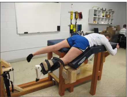
Hamstring stiffness was measured by quantifying the damping effect of the hamstrings on oscillatory knee flexion/extension following perturbation. This technique has been utilized previously to measure hamstring muscle stiffness and is associated with ACL loading mechanisms (McNair 1992; Blackburn, Norcross et al. 2011; Blackburn, Norcross et al. 2012) and hamstring strain injury risk (Watsford, Murphy et al. 2010). The hamstring MVIC was performed to determine the load to be placed on the distal leg during stiffness assessments. The subject was positioned prone with the right hip and knee in 30º degrees of flexion (Figure 1). The foot was secured to a loading device such that the posterior calcaneous was in contact with a load cell to permit
30
which force data were sampled from the load cell. The procedure was performed three times, and the average of the three trials was considered their MVIC.
The distal segment was then freed from the loading device to permit knee flexion/extension, and a load equaling 45% MVIC was secured to the distal leg (Figure 2). The leg was supported by the tester parallel to the floor and the subject was asked to contract the hamstrings isometrically to maintain this position (i.e. 30º of knee flexion). At a random time within 5 seconds, the tester pushed the calcaneous downward, slightly extending the knee (less than 5º). This perturbation produced oscillatory knee
flexion/extension which was captured in the tangential acceleration of the shank recorded by an accelerometer (PCB Piezotronics, Depew, NY, USA) affixed to a splint secured to the ankle/foot complex. The damped frequency of oscillation of the leg was calculated using the time instances of the first two oscillatory peaks (t1 and t2) via the equation f = (
). This value was then used in the stiffness equation where k is stiffness, m is the summed mass of the shank, foot, and applied load, and f is the damped frequency of oscillation. Three trials were performed with 60 seconds of rest between each trial. The leg was supported by the tester between trials to reduce the likelihood of fatigue.
Myotonometer Stiffness Measurements
Within one depression of the probe onto the skin, the Myotonometer calculates 8
different measurements automatically from 0 to 2.0kg of force in 0.25kg increments. The stiffness value was calculated as the ratio of the peak force applied to the total amount of tissue displacement. Each trial consisted of 5 depressions of the probe into the skin.
Subjects were positioned identically as in the oscillatory stiffness assessment and again contracted the hamstrings isometrically against 45% of MVIC to support the knee in 30º of flexion. Within 5 seconds of contraction, the Myotonometer was applied to the midpoint of the biceps femoris muscle measured as 50% of the distance from the ischial tuberosity to the head of the fibula. This process was repeated twice with 60 seconds of rest between trials to reduce the likelihood of fatigue. The leg was supported by the researcher between trials to further reduce the risk of fatigue.
Posterior thigh fat thickness was measured using diagnostic ultrasound (Sonosite M-Turbo; Sonosite, Bothell, WA, USA) with a 4cm width, 5MHz linear-array transducer. Measurements were recorded at the same location as the Myotonometer placement with the transducer placed parallel to the long axis of the muscle. Fat thickness was measured from the deep border of the skin to the superficial border of the muscle fascia.
DATA REDUCTION
32
Antonio, TX, USA). Posterior thigh fat thickness was measured using Image J software (National Institutes of Health, Bethesda, MD).
The oscillatory stiffness measurement reflects contributions from both the lateral (biceps femoris) and medial (semimembranosus and semitendinosus) hamstrings, while the Myotonometer only assessed the lateral hamstrings. Therefore, stiffness values collected by the damped oscillation technique were halved for comparisons of the two measurement techniques. Muscle stiffness was normalized mass prior to analysis.
STATISTICAL ANALYSIS
All statistical analyses were performed using SPSS version 19.0. Univariate descriptive statistics were calculated, and all variables were checked for normality. Reliability was assessed via intraclass correlation coefficients (ICC) derived from means squared produced by relevant ANOVA models. Inter-session reliability of the
Myotonometer was determined by performing one-way repeated measures ANOVA using the mean stiffness values obtained from each session and ICC(2,k). Intra-session reliability of the Myotonometer was determined by performing one-way repeated
Pearson bivariate correlations to characterize the relationship between normalized values for each method. Pearson bivariate correlations were calculated to evaluate the
relationships between the two hamstring stiffness measures, and between Myotonometer stiffness values and posterior thigh fat thickness.
Table 1: Statistical Analysis
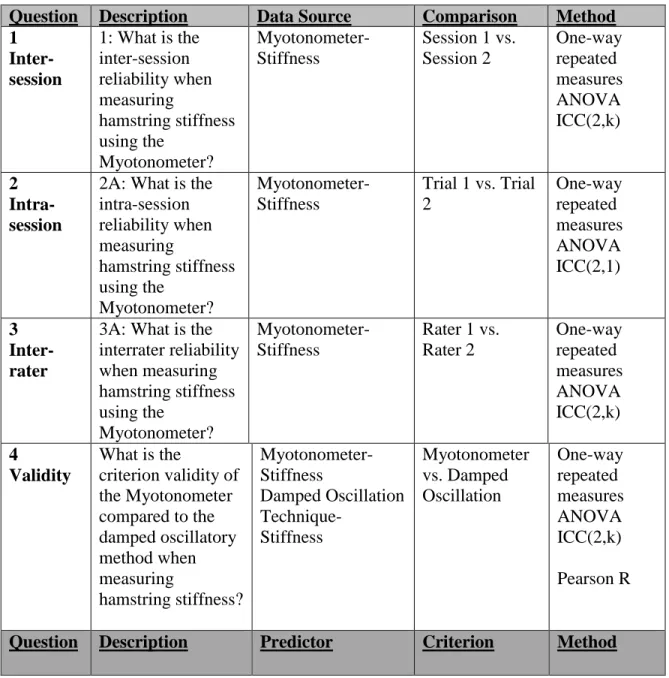
Question Description Data Source Comparison Method 1
Inter-session
1: What is the inter-session reliability when measuring hamstring stiffness using the Myotonometer? Myotonometer- Stiffness
Session 1 vs. Session 2 One-way repeated measures ANOVA ICC(2,k) 2 Intra-session
2A: What is the intra-session reliability when measuring hamstring stiffness using the Myotonometer? Myotonometer- Stiffness
Trial 1 vs. Trial 2 One-way repeated measures ANOVA ICC(2,1) 3 Inter-rater
3A: What is the interrater reliability when measuring hamstring stiffness using the Myotonometer? Myotonometer- Stiffness
Rater 1 vs. Rater 2 One-way repeated measures ANOVA ICC(2,k) 4 Validity
What is the
criterion validity of the Myotonometer compared to the damped oscillatory method when measuring hamstring stiffness? Myotonometer- Stiffness Damped Oscillation Technique- Stiffness Myotonometer vs. Damped Oscillation One-way repeated measures ANOVA ICC(2,k) Pearson R
34
5 What is the
relationship between Myotonometer stiffness and posterior thigh fat thickness?
Posterior thigh fat thickness
Myotonometer- Stiffness
CHAPTER IV RESULTS MYOTONOMETER RELIABILITY
Intra-session reliability of the Myotonometer was high resulting in an ICC of 0.807 (SEM=0.764N/mm). This ICC reveals good correspondence of hamstring stiffness values measured by the Myotonometer between trials during the same session. Inter-rater reliability was also high, resulting in an ICC of 0.830 (SEM=0.705N/mm). This ICC reveals good correspondence of hamstring muscle stiffness values measured by the Myotonometer between raters during the same session. Inter-session reliability was moderate resulting in an ICC of 0.693 (SEM=0.952N/mm). This ICC reveals a moderate correspondence of hamstring muscle stiffness values measured by the Myotonometer between sessions separated by an average of 6.1 days.
MYOTONOMETER VALIDITY
36 MEASUREMENT TECHNIQUE RESULTS
Measurement Technique
Mean ± Standard Deviation
CHAPTER V DISCUSSION
INTRODUCTION
This investigation demonstrated that The Myotonometer possesses high intra-session and inter-rater reliability and moderate inter-intra-session reliability. However, the results indicate that The Myotonometer is not a valid measure of hamstring stiffness relative to the oscillation technique. These findings suggest that these techniques measure two different characteristics. The oscillation technique measures active muscle stiffness that is related to ACL loading biomechanics, and The Myotonometer does not appear to be reflective of the same characteristics.
RELIABILITY
The Myotonometer was proven to have high intra-session reliability similar to the results found in healthy subjects for the biceps brachii and gastrocnemius (Leonard, Deshner et al. 2003). The Myotonometer was also proven to have high inter-rater
reliability. These results are similar to those found in healthy subjects (Leonard, Deshner et al. 2003) and subjects with upper motor neuron lesions (Aarrestad, Williams et al. 2004).
38
pattern observed when measuring passive muscle stiffness of the posterior shoulder in that session reliability is somewhat lower than intra-session reliability. Lower inter-session reliability may be partially explained by the fact that there was no MVIC
procedure performed during the second session. During the first session, the participants were required to complete three maximal contractions of the hamstring muscle, holding each contraction for five seconds. The contractions performed before the stiffness measurements could have led to fatigue in the subsequent trials and, therefore, lower reliability between the two sessions. Although subjects were asked to maintain similar physical activity between sessions, there was no way to monitor or mandate what activity the subjects completed outside of the laboratory. This may have led to decreased
reliability between sessions. It is also likely that there were minor differences in probe placement between sessions that resulted in slightly different values, thus resulting in lower inter-session reliability.
VALIDITY
One of the purposes of this investigation was to determine if The Myotonometer is a clinically relevant tool. The current gold standard technique for measuring active hamstring stiffness, the oscillation technique, requires the athlete to come to the laboratory and undergo approximately 20 minutes of testing. During the testing, the athlete is required to perform a MVIC procedure and then subsequently complete three trials of the perturbation all while being tethered to laboratory equipment and a
measurement computer. The procedures involved with calculating muscle stiffness using the oscillation technique are also more cumbersome than those for The Myotonometer. Conversely, The Myotonometer is a lightweight, portable device that could potentially provide a more convenient and feasible measure of muscle stiffness in the clinical setting. Because muscle stiffness can be modified through intervention, it is important to have a clinically applicable tool to screen for injury risk factors and track changes in muscle stiffness associated with training and rehabilitation. If The Myotonometer had been proven valid relative to the oscillation technique, it would have made hamstring stiffness measurements much more accessible for the clinician and made the idea of full-team screenings much more plausible.
This was the first study, to our knowledge, to evaluate the validity of the Myotonometer for measuring stiffness of the hamstring group. Other studies have demonstrated the validity of The Myotonometer for measuring stiffness of the biceps brachii and the gastrocnemius muscles (Leonard, Deshner et al. 2003) and the plantar flexors (Rydahl SJ 2004). However, these studies compared The Myotonometer against the Modified Ashworth Scale (MAS), a subjective clinician tool used to measure
40
validity in using tissue compliance as a measure of muscular strength (Gubler-Hanna Coral 2007). As such, the validity of the Myotonometer relative to The Oscillatory Technique had not been established prior to our investigation.
The low, non-significant correlation between the two stiffness measurement techniques may be attributable to the different types of stress applied to the hamstrings during testing. The Myotonometer causes displacement by providing a compressive force to the central region of the muscle. This loading scenario results in three-point bending (Figure 4) as compression occurs on the superficial side of the muscle while lengthening occurs on the deep side of the muscle. In contrast, the oscillation technique produces tensile loading via lengthening of the muscle. The collagen fibers of the tendon and the sarcomeres of the muscle fibers are aligned along the long-axis therefore,
providing resistance to tensile/longitudinal loading and deformation (Van Looke M 2006). This anisotropic quality of muscle likely explains why the two measurement techniques were not correlated. Because muscle contributes to dynamic joint stability by actively resisting lengthening, the oscillation technique likely provides a better indication of the muscle’s capacity for doing so, and is a more appropriate method for assessing active muscle stiffness. Therefore, The Myotonometer should not be used to measure active hamstring muscle stiffness.
does not correspond with active stiffness measured via the oscillation technique, it may provide a passive stiffness measure of muscle tissue underlying the probe.
CONCLUSION
Even though the Myotonometer demonstrated high inter-session, intra-session, and inter-tester reliability, it was not proven valid when compared to the oscillation technique. Because the tool is not valid, it should not be used clinically to measure active hamstring muscle stiffness. Future research should be conducted to make measuring hamstring muscle stiffness more clinically applicable so that hamstring strain and ACL injuries can be prevented and rehabilitation progress can be more easily monitored.
42 FIGURES
Figure 1: MVIC Positioning
Figure 3: Myotonometer Placement
44 TABLES
Measurement Technique Results
Measurement Technique
Mean ± Standard Deviation
REFERENCES
Aarrestad, D. D., M. D. Williams, et al. (2004). "Intra- and interrater reliabilities of the Myotonometer when assessing the spastic condition of children with cerebral palsy." J Child Neurol 19(11): 894-901.
Amis, A. A. and G. P. Dawkins (1991). "Functional anatomy of the anterior cruciate ligament. Fibre bundle actions related to ligament replacements and
injuries." J Bone Joint Surg Br 73(2): 260-267.
Anderson, A. F., D. C. Dome, et al. (2001). "Correlation of anthropometric
measurements, strength, anterior cruciate ligament size, and intercondylar notch characteristics to sex differences in anterior cruciate ligament tear rates." Am J Sports Med 29(1): 58-66.
Arendt, E. A., Dick R. (1995). "Knee injury patterns among men and women in collegiate basketball and soccer: NCAA data and review of literature." Am J Sports Med 23: 694-701.
Arnold JA, C. T., Heaton LM, Park JP, Harris WD (1979). "Natural history of anterior cruciate tears." Am J Sports Med 7: 305-313.
Baratta, R., M. Solomonow, et al. (1988). "Muscular coactivation. The role of the antagonist musculature in maintaining knee stability." Am J Sports Med 16(2): 113-122.
Beckett, M. E., D. L. Massie, et al. (1992). "Incidence of Hyperpronation in the ACL Injured Knee: A Clinical Perspective." J Athl Train 27(1): 58-62.
46
Beynnon, B. D., B. C. Fleming, et al. (1995). "Anterior cruciate ligament strain behavior during rehabilitation exercises in vivo." Am J Sports Med 23(1): 24-34.
Bizzini, M. and A. F. Mannion (2003). "Reliability of a new, hand-held device for assessing skeletal muscle stiffness." Clin Biomech (Bristol, Avon) 18(5): 459-461.
Blackburn, J. T., D. R. Bell, et al. (2009). "Comparison of hamstring
neuromechanical properties between healthy males and females and the influence of musculotendinous stiffness." J Electromyogr Kinesiol 19(5): e362-369.
Blackburn, J. T., M. F. Norcross, et al. (2012). "The influence of hamstring stiffness on landing biomechanics linked to anterior cruciate ligament loading." Journal of Athletic Training.
Blackburn, J. T., M. F. Norcross, et al. (2011). "Influences of hamstring stiffness and strength on anterior knee joint stability." Clin Biomech (Bristol, Avon) 26(3): 278-283.
Blackburn, J. T., B. L. Riemann, et al. (2004). "Sex comparison of extensibility, passive, and active stiffness of the knee flexors." Clin Biomech (Bristol, Avon) 19(1): 36-43.
Boden, B. P., Griffin LY, Garrett W (2000). "The etiology and prevention of noncontact ACL injuries." physician sports med 28(4): 53-60, 107-108.
Bonci, C. M. (1999). "Assessment and evaluation of predisposing factors to anterior cruciate ligament injury." J Athl Train 34(2): 155-164.
Brockett, C. L., D. L. Morgan, et al. (2004). "Predicting hamstring strain injury in elite athletes." Med Sci Sports Exerc 36(3): 379-387.
Butler, R. J., H. P. Crowell, 3rd, et al. (2003). "Lower extremity stiffness: implications for performance and injury." Clin Biomech (Bristol, Avon) 18(6): 511-517.
Caraffa, A., G. Cerulli, et al. (1996). "Prevention of anterior cruciate ligament injuries in soccer. A prospective controlled study of proprioceptive training." Knee Surg Sports Traumatol Arthrosc 4(1): 19-21.
Chappell, J. D., B. Yu, et al. (2002). "A comparison of knee kinetics between male and female recreational athletes in stop-jump tasks." Am J Sports Med 30(2): 261-267.
Croisier, J. L., S. Ganteaume, et al. (2008). "Strength imbalances and prevention of hamstring injury in professional soccer players: a prospective study." Am J Sports Med 36(8): 1469-1475.
Damiano DL, Q. J., Owen BF, Payne P, Nelson KC, Abel MF (2002). "What does the Ashworth scale really measure and are instrumented measures more valid and precise?" Developmental Medicine and Child Neurology 44: 112-118.
DeMorat, G., Weinhold, P., Blackburn, T., Chudik, S., Garrett, W. (2004). "Aggressive quadriceps loading can induce noncontact anterior cruciate ligament injury." Am J Sports Med 32(2): 477-483.
Dhaher, Y. Y., A. D. Tsoumanis, et al. (2005). "Neuromuscular reflexes contribute to knee stiffness during valgus loading." J Neurophysiol 93(5): 2698-2709.
Ditroilo, M., M. Watsford, et al. (2011). "Validity and inter-day reliability of a free-oscillation test to measure knee extensor and knee flexor musculo-articular stiffness." J Electromyogr Kinesiol 21(3): 492-498.
Donatelli, R. A. (1985). "Normal biomechanics of the foot and ankle." J Orthop Sports Phys Ther 7(3): 91-95.
48
Duthon, V. B., C. Barea, et al. (2006). "Anatomy of the anterior cruciate ligament." Knee Surg Sports Traumatol Arthrosc 14(3): 204-213.
Elliott, M. C., B. Zarins, et al. (2011). "Hamstring muscle strains in professional football players: a 10-year review." Am J Sports Med 39(4): 843-850.
Faria, A., R. Gabriel, et al. (2009). "Triceps-surae musculotendinous stiffness: relative differences between obese and non-obese postmenopausal women." Clin Biomech (Bristol, Avon) 24(10): 866-871.
Faria, A., R. Gabriel, et al. (2010). "The relationship of body mass index, age and triceps-surae musculotendinous stiffness with the foot arch structure of postmenopausal women." Clin Biomech (Bristol, Avon) 25(6): 588-593.
Fernandez William, Y. E., Comstock Dawn (2007). "Epidemiology of Lower
Extremity Injuries among U.S. High School Athletes." Academic Emergency Medicine 14: 641-645.
Ford, K. R., G. D. Myer, et al. (2003). "Valgus knee motion during landing in high school female and male basketball players." Med Sci Sports Exerc 35(10): 1745-1750.
Gillquist, J. and K. Messner (1999). "Anterior cruciate ligament reconstruction and the long-term incidence of gonarthrosis." Sports Med 27(3): 143-156.
Girgis, F. G., J. L. Marshall, et al. (1975). "The cruciate ligaments of the knee joint. Anatomical, functional and experimental analysis." Clin Orthop Relat Res(106): 216-231.
Gomez, E., J. C. DeLee, et al. (1996). "Incidence of injury in Texas girls' high school basketball." Am J Sports Med 24(5): 684-687.
Gottlob, C. A., C. L. Baker, Jr., et al. (1999). "Cost effectiveness of anterior cruciate ligament reconstruction in young adults." Clin Orthop Relat Res(367): 272-282.
Granata, K. P., S. E. Wilson, et al. (2002). "Gender differences in active
musculoskeletal stiffness. Part I. Quantification in controlled measurements of knee joint dynamics." J Electromyogr Kinesiol 12(2): 119-126.
Gray J, T. J., McKenzie DC, Clement DB, McConkey JP, Davison RG (1985). "A survey of injuries to the anterior cruciate ligament of the knee in female basketball players." int J Sports Med 6: 314-316.
Griffin, L. Y., J. Agel, et al. (2000). "Noncontact anterior cruciate ligament injuries: risk factors and prevention strategies." J Am Acad Orthop Surg 8(3): 141-150.
Griffin, L. Y., M. J. Albohm, et al. (2006). "Understanding and preventing
noncontact anterior cruciate ligament injuries: a review of the Hunt Valley II meeting, January 2005." Am J Sports Med 34(9): 1512-1532.
Gubler-Hanna Coral, L. J., Marx Benjamin, Leonard Charles (2007). "Construct validity of myotonometric measurements of muscle compliance as a measure of strength." Physiological Measurement.
Haycock, C. E. and J. V. Gillette (1976). "Susceptibility of women athletes to injury. Myths vs reality." JAMA 236(2): 163-165.
Heckmatt, J. Z., N. Pier, et al. (1988). "Measurement of quadriceps muscle thickness and subcutaneous tissue thickness in normal children by real-time
ultrasound imaging." J Clin Ultrasound 16(3): 171-176.
Heidt, R. S., Jr., L. M. Sweeterman, et al. (2000). "Avoidance of soccer injuries with preseason conditioning." Am J Sports Med 28(5): 659-662.
Heitz, N. A., P. A. Eisenman, et al. (1999). "Hormonal changes throughout the menstrual cycle and increased anterior cruciate ligament laxity in females." J Athl Train 34(2): 144-149.
50
Hewett, T. E., G. D. Myer, et al. (2006). "Anterior cruciate ligament injuries in female athletes: Part 1, mechanisms and risk factors." Am J Sports Med 34(2): 299-311.
Hewett, T. E., G. D. Myer, et al. (2005). "Biomechanical measures of neuromuscular control and valgus loading of the knee predict anterior cruciate ligament injury risk in female athletes: a prospective study." Am J Sports Med 33(4): 492-501.
Hollis, J. M., S. Takai, et al. (1991). "The effects of knee motion and external loading on the length of the anterior cruciate ligament (ACL): a kinematic study." J Biomech Eng 113(2): 208-214.
Hootman, J., Dick R, Agel J (2007). "Epidemiology of collegiate injuries for 15 sports: summary and recommendations for injury prevention initiatives." J Athl Train 42(2).
Horikawa, M., S. Ebihara, et al. (1993). "Non-invasive measurement method for hardness in muscular tissues." Med Biol Eng Comput 31(6): 623-627.
Johnson, R. J. (1983). "The anterior cruciate ligament problem." Clin Orthop Relat Res(172): 14-18.
Kato G, A. P., Sato H (2004). "Reliability and validity of a device to measure muscle hardness." Journal of Mechanics in Medicine and Biology.
Kerins Caitlyn, M. S., Butterfield Timothy, McKeon Patrick, Uhl Timothy (2013). "Reliability of the Myotonometer for Assessment of Posterior Shoulder Tightness." The International Journal of Sports Physical Therapy 8(3).
Krosshaug, T., A. Nakamae, et al. (2007). "Mechanisms of anterior cruciate
ligament injury in basketball: video analysis of 39 cases." Am J Sports Med 35(3): 359-367.
Kubo, K., M. Morimoto, et al. (2007). "Effects of plyometric and weight training on muscle-tendon complex and jump performance." Med Sci Sports Exerc 39(10): 1801-1810.
LaPrade, R. F. and Q. M. Burnett, 2nd (1994). "Femoral intercondylar notch stenosis and correlation to anterior cruciate ligament injuries. A prospective study." Am J Sports Med 22(2): 198-202; discussion 203.
Leonard, C. T., W. P. Deshner, et al. (2003). "Myotonometer intra- and interrater reliabilities." Arch Phys Med Rehabil 84(6): 928-932.
Leonard, C. T., J. U. Stephens, et al. (2001). "Assessing the spastic condition of individuals with upper motoneuron involvement: validity of the
myotonometer." Arch Phys Med Rehabil 82(10): 1416-1420.
Li, G., T. W. Rudy, et al. (1999). "The importance of quadriceps and hamstring muscle loading on knee kinematics and in-situ forces in the ACL." J Biomech 32(4): 395-400.
Loudon, J. K., W. Jenkins, et al. (1996). "The relationship between static posture and ACL injury in female athletes." J Orthop Sports Phys Ther 24(2): 91-97.
MacWilliams, B. A., D. R. Wilson, et al. (1999). "Hamstrings cocontraction reduces internal rotation, anterior translation, and anterior cruciate ligament load in weight-bearing flexion." J Orthop Res 17(6): 817-822.
Malinzak, R. A., S. M. Colby, et al. (2001). "A comparison of knee joint motion patterns between men and women in selected athletic tasks." Clin Biomech (Bristol, Avon) 16(5): 438-445.
Marieb, E., Mallet, J., (1997). Human anatomy, Benjamin Cummings Publishers.
Markolf, K. L., D. M. Burchfield, et al. (1995). "Combined knee loading states that generate high anterior cruciate ligament forces." J Orthop Res 13(6): 930-935.
52
McNair, P., Wood, G., Marshall, R. (1992). "Stiffness of the hamstring muscles and its relationship to function in anterior cruciate ligament deficient
individuals." Clin Biomech (Bristol, Avon) 7(3): 131-137.
McNair PJ, M. R., Matheson JA (1990). "Important associated with acute anterior cruciate ligament injury." N Z Med J 103: 537-539.
McNair, P. J. and S. N. Stanley (1996). "Effect of passive stretching and jogging on the series elastic muscle stiffness and range of motion of the ankle joint." Br J Sports Med 30(4): 313-317, discussion 318.
Messina, D. F., W. C. Farney, et al. (1999). "The incidence of injury in Texas high school basketball. A prospective study among male and female athletes." Am J Sports Med 27(3): 294-299.
Murphy, A. J., Watsford, M., Coutts, A., Pine, M., (2003). "Reliability of a test of musculotendinous stiffness fof the triceps-surae." Physical Therapy in Sport 4: 175-181.
Nordander, C., J. Willner, et al. (2003). "Influence of the subcutaneous fat layer, as measured by ultrasound, skinfold calipers and BMI, on the EMG
amplitude." Eur J Appl Physiol 89(6): 514-519.
Noyes, F. R., L. A. Dunworth, et al. (1996). "Knee hyperextension gait abnormalities in unstable knees. Recognition and preoperative gait retraining." Am J Sports Med 24(1): 35-45.
Olsen OE, M. G., Engretsen L, Bahr R (2004). "Injury mechanisms for anterior cruciate ligament injuries in team handball: a systematic video analysis." Am J Sports Med 32: 1002-1012.
Olsen, O. E., G. Myklebust, et al. (2004). "Injury mechanisms for anterior cruciate ligament injuries in team handball: a systematic video analysis." Am J Sports Med 32(4): 1002-1012.
Prevention, C. f. D. C. a. (1996). National Hospital Discharge Survey. Atlanta GA.
Rack, P. M. and D. R. Westbury (1969). "The effects of length and stimulus rate on tension in the isometric cat soleus muscle." J Physiol 204(2): 443-460.
Rudy, T. W., G. A. Livesay, et al. (1996). "A combined robotic/universal force sensor approach to determine in situ forces of knee ligaments." J Biomech 29(10): 1357-1360.
Rydahl SJ, B. B. (2004). "Ankle Stiffness and tissue compliance in stroke survivors: A validation of Myotonometer measurements." Archives of Physical
Medicine and Rehabilitation.
Salmon, L., V. Russell, et al. (2005). "Incidence and risk factors for graft rupture and contralateral rupture after anterior cruciate ligament reconstruction." Arthroscopy 21(8): 948-957.
Shelbourne, K. D., Gray, T., Haro, M. (2009). "Incidence of subsequent injury to either knee within 5 years after anterior cruciate ligament reconstruction with patellar tendon autograft." Am J Sports Med 24(6): 246-251.
Shelbourne, K. D. and B. Kerr (2001). "The relationship of femoral intercondylar notch width to height, weight, and sex in patients with intact anterior cruciate ligaments." Am J Knee Surg 14(2): 92-96.
Shultz, S. J., T. C. Sander, et al. (2005). "Sex differences in knee joint laxity change across the female menstrual cycle." J Sports Med Phys Fitness 45(4): 594-603.
Souryal, T. O. and T. R. Freeman (1993). "Intercondylar notch size and anterior cruciate ligament injuries in athletes. A prospective study." Am J Sports Med 21(4): 535-539.
Spurrs, R. W., A. J. Murphy, et al. (2003). "The effect of plyometric training on distance running performance." Eur J Appl Physiol 89(1): 1-7.
54
Taylor, D. C., J. D. Dalton, Jr., et al. (1990). "Viscoelastic properties of muscle-tendon units. The biomechanical effects of stretching." Am J Sports Med 18(3): 300-309.
Taylor, K. A., M. E. Terry, et al. (2011). "Measurement of in vivo anterior cruciate ligament strain during dynamic jump landing." J Biomech 44(3): 365-371.
Thelen, D. G., E. S. Chumanov, et al. (2005). "Hamstring muscle kinematics during treadmill sprinting." Med Sci Sports Exerc 37(1): 108-114.
Tortora, G. (1999). Principles of Human Anatomy, John Wiley and Sons.
Trimble, M. H., M. D. Bishop, et al. (2002). "The relationship between clinical measurements of lower extremity posture and tibial translation." Clin Biomech (Bristol, Avon) 17(4): 286-290.
Uhorchak, J. M., C. R. Scoville, et al. (2003). "Risk factors associated with
noncontact injury of the anterior cruciate ligament: a prospective four-year evaluation of 859 West Point cadets." Am J Sports Med 31(6): 831-842.
Van Looke M, L. C., Simms CK (2006). "A validated model of passive muscle in compression." Journal of Biomechanics.
Verrall, G. M., J. P. Slavotinek, et al. (2001). "Clinical risk factors for hamstring muscle strain injury: a prospective study with correlation of injury by magnetic resonance imaging." Br J Sports Med 35(6): 435-439; discussion 440.
Watsford, M. L., A. J. Murphy, et al. (2010). "A prospective study of the relationship between lower body stiffness and hamstring injury in
professional Australian rules footballers." Am J Sports Med 38(10): 2058-2064.
Wilkie, D. R. (1956). "The mechanical properties of muscle." Br Med Bull 12(3): 177-182.
Wilson, G. J., B. C. Elliott, et al. (1992). "Stretch shorten cycle performance
Withrow, T. J., L. J. Huston, et al. (2006). "The relationship between quadriceps muscle force, knee flexion, and anterior cruciate ligament strain in an in vitro simulated jump landing." Am J Sports Med 34(2): 269-274.
Withrow, T. J., L. J. Huston, et al. (2008). "Effect of varying hamstring tension on anterior cruciate ligament strain during in vitro impulsive knee flexion and compression loading." J Bone Joint Surg Am 90(4): 815-823.
Woodford-Rogers, B., L. Cyphert, et al. (1994). "Risk factors for anterior cruciate ligament injury in high school and college athletes." J Athl Train 29(4): 343-346.
Worrell, T. W. and D. H. Perrin (1992). "Hamstring muscle injury: the influence of strength, flexibility, warm-up, and fatigue." J Orthop Sports Phys Ther 16(1): 12-18.
Zheng, N., G. S. Fleisig, et al. (1998). "An analytical model of the knee for
estimation of internal forces during exercise." J Biomech 31(10): 963-967.