INFLUENCE OF VAGINAL LACTOBACILLI ON THE BARRIER PROPERTIES OF
CERVICOVAGINAL MUCUS AGAINST HIV
Kenetta L. Nunn
A thesis submitted to the faculty of the University of North Carolina at Chapel Hill in partial fulfillment of the requirements for the degree of Master in the Department of Biomedical Engineering
Chapel Hill 2014
Approved by: Samuel Lai
ABSTRACT
Kenetta L. Nunn: Influence of vaginal lactobacilli on barrier properties of cervicovaginal mucus against HIV
(Under the direction of Samuel Lai)
The vaginal epithelium is coated with cervicovaginal mucus (CVM), a dense mesh network of mucin fibers that HIV in semen must penetrate in order to reach target cells residing in the epithelium. Trapping HIV in CVM offers the potential to prevent initial HIV infections altogether, and likely accounts in part for the generally low rates of HIV transmission (~1 in 100-1000 coital acts). CVM is populated by a dense microbial community that can differ markedly between women as well as in the same woman over time, and epidemiological studies indicate that the microbiota can significantly impact the risk of acquiring HIV. Nevertheless, little is understood about whether specific microbes, including different species of Lactobacillus, may modulate the barrier properties of CVM against HIV. In this thesis, I decided to investigate whether differences in the vaginal microbiota may result in differential ability of native CVM to immobilize and trap HIV. Using high-resolution particle tracking, I found that HIV mobility across different CVM specimens was not correlated to vaginal pH or total lactic acid levels, and only slightly correlated to Nugent scores. Instead, I found that HIV readily penetrates CVM with low levels of D-lactic acid (D-LA), but is consistently trapped in CVM with high D-LA levels. Based on vaginal microbiome analysis using high throughput 16S rRNA gene sequencing, I confirmed that CVM specimens with low levels of native D-LA are more likely to possess a L.iners dominated microbiota, whereas CVM specimens with high levels of native D-LA are more likely to possess a L. crispatus
dominated microbiota. These results indicate that vaginal Lactobacilli spp. can alter the barrier properties
of CVM against HIV, and that L. iners, rather than solely BV-associated microbes, may be correlated to increased susceptibility to HIV transmission relative to L. crispatus.
ACKNOWLEDGEMENTS
First and foremost, I would like to thank my PI, Dr. Samuel Lai, without whom this project would not have been possible. Three years ago, I made the decision to leave a full-time position as a Research Associate at Affinergy, Inc. in RTP, NC. Wanting to continue my education, I began looking for
positions in academic labs. Dr. Lai was one of the professors that I contacted and he was gracious enough to bring me on as a research assistant and recommend me for the joint graduate program in BME at UNC/NCSU. I have learned a great deal and matured as a researcher under Dr. Lai’s supervision. I am truly grateful to Dr. Lai for giving me the opportunity to work, prove myself, and further my education in his lab. I would also like to thank Dr. Ron Swanstrom and Dr. Brain Button for graciously agreeing to be on my thesis committee.
TABLE OF CONTENTS
ABSTRACT ... iii
LIST OF TABLES ... vii
LIST OF FIGURES ... viii
CHAPTER 1: INTRODUCTION ... 1
1.1 Mucus as a Barrier ... 1
1.2 Innate and Adaptive Barrier Properties of Mucus ... 2
1.3 The Vaginal Microbiota ... 4
1.4 The Link between Vaginal Microbiota and Susceptibility to Sexually Transmitted Infections ... 5
CHAPTER 2: Vaginal L. crispatus but not L. iners reinforces the innate cervicovaginal mucus barrier against HIV ... 10
2.1 INTRODUCTION ... 10
2.2 RESULTS AND DISCUSSION ... 11
2.3 MATERIALS AND METHODS ... 14
2.3.1 Culture and purification of fluorescent HIV-1 ... 14
2.3.3 Multiple particle tracking of HIV-1 and synthetic beads in CVM ... 16
2.3.4 Microbiome Analysis using 16S rRNA gene sequencing ... 16
2.3.5 Statistical analysis ... 18
LIST OF TABLES
Table 2.1 Characterization of CVM samples: pH, menstrual cycle, D-LA, L-LA,
Total LA, Nugent Score………..……….24
LIST OF FIGURES
Figure 1.1 Heatmap of relative abundance of micobial taxa found in the
vaginal bacterial communities of reproductive age women………..……….………....7
Figure 1.2 The distribution of community state types within each ethnic group of
reproductive age women from the study in Figure 1.1……….………...8
Figure 2.1 Fraction of particles or virions classified as mobile in cervicovaginal mucus
specimens……….19
Figure 2.2 The ensemble averaged diffusivity of particles or virions classified as mobile in
cervicovaginal mucus specimens…….………20
Figure 2.3 Fraction of mobile virions for mCherry-GAG tagged HIV virus-like particles
pseudotyped with YU2 envelope in CVM at native (i.e. unaltered) pH………...…..….21
Figure 2.4 Ensemble averaged diffusivities for mCherry-GAG tagged HIV virus-like
particles pseudotyped with YU2 envelope in CVM at native (i.e. unaltered) pH…………..…………...22
Figure 2.5 CVM that traps HIV appear to be L. crispatus- dominant, whereas CVM
CHAPTER 1
INTRODUCTION
1.1 Mucus as a Barrier
Goblet cells secrete long glycoproteins called mucins, which happen to be the most abundant biopolymer in the body. Mucin fibers entangle and crosslink forming a dense mucin matrix with shear-dependent viscoelastic (i.e. mechanical) properties. This dense mucin matrix forms the viscous and elastic gel of mucus, which coats the surfaces of the respiratory, gastrointestinal, and urogenital tracts. This mucus gel serves as the first line of defense against pathogens and infections at mucosal surfaces by acting as a selectively permeable barrier allowing entry of nutrients while blocking the entry of foreign and dangerous substances, including viral and bacterial pathogens [1]; and, as mucus is continuously cleared and renewed, particles trapped in the mucus layer are readily eliminated from the mucosal surface. Furthermore, many protective factors such as defensins and antibodies are constantly secreted into mucus and can enhance its barrier properties [1]. This barrier function of mucus enables the body to breathe, ingest, and reproduce without succumbing to infections.
Mucus coating the female reproductive tract, is predominantly derived from mucin-secreting glands in the cervix [2, 3]. Cervical mucus flows into the vagina by gravitational and/or abdominal pressure [4, 5], becomes exposed to a myriad of microbes (commensals or pathogenic) in the vagina (see
The viscoelastic property of CVM enables it to behave like a gel excluding most infectious virions in semen from directly contacting the vaginal epithelium. Although CVM can behave as a gel in this instance, it can readily shear thin and function as a lubricant when large and rapid shear forces are introduced, thereby preventing physical trauma during sexual intercourse [1, 3]. Furthermore, to ensure proper function, the composition of mucus is constantly changing. For example, the properties of CVM are markedly altered by the menstrual cycle. During non-ovulatory periods, the viscosity of CVM prevents sperm from reaching the upper reproductive tract. However, CVM becomes less viscous during ovulation enabling sperm to penetrate the mucus layer and swim through [1].
1.2 Innate and Adaptive Barrier Properties of Mucus
Since viruses and other intracellular obligate bacteria must reach target cells to initiate infections, the ability of these pathogens to efficiently traverse mucus overlaying the vaginal epithelium represents the first step in the male to female transmission of sexually transmitted infections (STIs). Conversely, the ability to trap pathogens in mucus, and consequently reduce the flux of pathogens arriving to target cells, offer the potential to reduce infections or prevent infections from taking place altogether.
Native mucus can act directly as a diffusional barrier against pathogens, based on either steric
Mucus may also work in conjunction with the adaptive immune system, namely via secreted antibodies, to block the translocation of pathogens. Indeed, there are more antibodies secreted into the mucosa than the blood or lymph. Antibodies can be delivered into mucus by the MHC class I-related neonatal Fc receptor [11]; this was suggested to account for ten-fold higher concentrations of IgG than IgA observed in CVM [12]. Alternatively, secreted antibodies may be produced in the local tissue and secreted into the lumen by resident plasma cells in a process regulated by M cells, which function as sensory checkpoints for the cellular immune system, and leads to B cell expansion and secretion of antigen-specific antibodies [1].
infection, while removing vaginal mucus by gentle lavage abolished protection. These observations suggest IgG-Fc possess a glycan-dependent “muco-trapping” effector function that may provide exceptionally potent protection at mucosal surfaces.
1.3 The Vaginal Microbiota
In a study using 16S rRNA gene profiling of the vaginal microbiome of nearly 400 non-pregnant women, five consistent groupings of the microbiota, termed community state types (CST), were identified [14]. Four of the CSTs were most often dominated by Lactobacillus sp. (CST-I: L. crispatus, CST-II: L. gasseri, CST-III: L. iners, and CST-V: L. jensenii) and represented approximately 73% of the samples, in good agreement with the prevailing view that Lactobacillus spp. are important members of vaginal
microbiota (Figure 1.1). L. iners was the most prevalent species, followed by L. crispatus. Importantly, ethnicity was associated with CST (Figure 1.2). African-American and Hispanic women were more likely
to be categorized as CST-III (L. iners dominant) or CST-IV (low Lactobacillus spp. and/or high
anaerobes, including many BV-associated organisms such as G. vaginalis, Mobiluncus, and A. vaginae); these ethnic groups have been associated with greater risks of acquiring HIV [15-17]. This suggests that the composition and stability of the vaginal microbiota may serve as the physiological pathway relating ethnicity to HIV infection.
acid can produce a low pH environment that precludes vaginal colonization by nonindigenous organisms [18, 20-24], and recent evidence suggests lactic acid possesses increased antimicrobial activity beyond acidity alone [9, 18, 25, 26]. Lactic acid at low pH (<4.5) is a potent bactericide against BV-associated bacteria [18]. This may help explain why L. iners, which secrete more L-LA than D-LA [42], can more readily fend off BV-associated microbes and thus frequently observed in women with recurring episodes of BV. The recent finding by Tachedjian et al. that LA can inactivate HIV further highlight the potential dual roles of LA in enhancing the mucosal defense, by serving as a surrogate marker for a ‘protective’ microbiota, and also directly participating in the inactivation of HIV [25].
Lactic acid may also act as a signaling molecule influencing host gene expression and stimulating the host innate defense system by enhancing cytokine release in response to antigen exposure. For
example, L-lactic acid can stimulate the IL-23/IL-17 T lymphocyte pathway [27, 28], induce the production of pro-inflammatory cytokines by vaginal epithelial cells in the presence of a synthetic viral
RNA [29] and activate lymphocytes [30]. Given that IL-23 is one of the primary cytokines involved in neutrophil recruitment, mobilization, and activation at mucosal surfaces, this may translate to better immune surveillance of mucosal surfaces with high lactic acid levels in vivo [28]. The LA-induced cytokine release is consistent with the findings of Fichorova et al. that colonization of vaginal epithelial cell monolayers with common bacteria such as L. crispatus may regulate the epithelial innate immunity [31].
1.4 The Link between Vaginal Microbiota and Susceptibility to Sexually Transmitted Infections
CHAPTER 2
VAGINAL
L. CRISPATUS
BUT NOT
L. INERS
REINFORCES THE INNATE
CERVICOVAGINAL MUCUS BARRIER AGAINST HIV
2.1 INTRODUCTION
Semen from HIV-infected individuals contains cell-free and cell-associated HIV. Both forms of virions are plausible mediators of male-to-female transmission of HIV in the female reproductive tract, and to be infectious both must penetrate the genital mucus secretions that coats and adheres to vaginal and penile epithelia during coitus. While leukocytes can migrate through pH neutral mucus, they are rapidly immobilized and then killed by mild acidity (pH < 6), and hence are unlikely to survive in the acidic vagina. In contrast, cell-free HIV is only slowly inactivated by mild acidity, and earlier work suggests that native, acidic CVM from women with presumably Lactobacilli-dominated microbiota were able to effectively immobilize HIV [9]. The adhesive interactions between HIV and mucins were only evident at native pH, as virions were not slowed substantially when the pH of CVM was neutralized.
Interestingly, a later study following up on the aforementioned work suggested that CVM trapped HIV virions to a much lesser extent [32]. While that study did not describe the study participants and the method of recruitment, the variability in the native pH of the mucus specimens, including a large fraction
I was thus interested in better understanding the factors that control HIV interactions with the mucosal environment and modulate the native barrier properties of CVM.
2.2 RESULTS AND DISCUSSION
To directly determine distinct mobility of HIV in different CVM specimens, I studied the motion of HIV virus-like particles (VLP) that were internally fluorescently labeled through the incorporation of a mCherry-Gag fusion capsid. For biosafety considerations, the derivative was replication defective and pseudotyped with a YU2, R5-tropic HIV envelope. I mixed the labeled HIV at minimal dilution (~5% [vol/vol]) into fresh, native and undiluted CVM, and observed the translational movements of hundreds of individual HIV virions in each sample using high-resolution multiple-particle tracking.
to trap HIV (Figure 2.3, 2.4). Therefore, I sought to identify other biomarkers that may more accurately predict the barrier properties of CVM.
I noted that only lactic acid producing bacteria can secrete both D- and L- LA (humans can only secrete L-LA, and do not contribute significantly to the total LA found in the vagina), and different species of Lactobacillus may differ in D- vs. L- LA secretion. Therefore, I tested whether D- or L- LA levels in CVM could be a useful biomarker for evaluating the barrier properties of CVM. I found D-LA was a substantially better predictor of Nugent scores than L-LA, pH of CVM or total LA levels (data not shown). More importantly, I found that CVM samples that did not trap HIV possessed significantly lower D-LA levels (Figure 2.3, 2.4) but otherwise little to no difference in L-LA (Figure 2.3, 2.4) and total LA levels (Figure 2.3, 2.4). Exogenous addition of D-LA to specimens with low D-LA did not
facilitate HIV trapping (data not shown). Interestingly, D-LA secretion by Lactobacillus in women with recurrent BV is also ~50% that of women with healthy vaginal microbiota: even in women experiencing
recurring episodes of bacterial vaginosis (BV) with vaginal pH ≥ 5, up to 40% can carry one or more species/strains of Lactobacillus (most typically L. iners). Their ability to make D-lactic acid is, however, low (3.94 ± 0.72 mM/L) compared to the D-lactic acid produced by Lactobacillus strains from healthy
vagina with vaginal pH ∼4 (8.04 ± 1.07 mM/L) (P<0.001) [33]. Taken together, these results suggest D-LA represents a sensitive biomarker of the vaginal microbiota that in turn modulates the barrier properties of CVM against infections.
It should be pointed out that L. iners was frequently associated with transitions to other CST groups, especially CST-IV (BV-associated microbes). Thus, L. iners likely represents a less stable microbial community than the other Lactobacillus-dominant groupings. Unpublished work from Jacques Ravel’s lab in University of Maryland, based on metatranscriptomics analysis (catalogue and different expression of all expressed genes in a microbial community using sequence-based RNAseq approach),
responsible for the distinct barrier properties of CVM observed. To confirm that the CVM samples that possessed both low D-LA and a low Nugent score are actually CVM from women with L. iners
-dominated microflora, we collaborated with Dr. Ravel’s lab at The University of Maryland to perform a broad 16S rRNA gene-based characterization of the vaginal microbial population of six additional CVM samples via high-throughput sequencing of the 16S rRNA gene V3-V4 regions on the Illumina MiSeq platform (250 bp paired end reads). I found that 3 CVM samples that effectively trapped HIV all possessed a L. crispatus dominant microflora, whereas the 2 CVM samples that did not trap HIV both possessed largely L. iners- dominant microflora, and the remaining sample that could not trap HIV exhibited high amounts of G. vaginalis (Figure 2.5).
Traditionally, the primary means of evaluating vaginal mucosal health, in regards to BV, have
been by Amsel’s criteria and by Nugent scoring: a Lactobacillus-dominated microbiota, as reflected by Nugent scores in the 0-3 range and a pH < 4.5, was typically regarded as “healthy”. Although this
approach is useful in rapidly identifying women with “intermediate” microflora or BV microbiota, these methods do not differentiate between the 4 Lactobacillus species (L. crispatus, L. iners, L. jensenii, L. gasseri) that are commonly found in the general population. Furthermore, the scoring is typically conducted only at the beginning of a trial, and thereby fails to account for the potentially rapid shifting nature of the microbiota. Such imperfect categorization of vaginal microbiota may have precluded the field from better understanding the barrier properties of CVM to date. Since L. iners is commonly found in women who have had episodes of BV or intermediate microflora, it is very likely that risks are even higher, as the published analyses may have included a significant population of women with L. iners-dominated (or even intermediate/BV) microflora in the “healthy” vaginal microbiotagroup that may not adequately protect against HIV or other STIs. Thus, the actual risk factor for women with BV or L. iners may be markedly higher (e.g., >5 fold). Correspondingly, women with L. crispatus-dominated
prevalence of BV and increasing the prevalence of L. crispatus one of the most effective protective strategies against HIV transmission.
2.3 MATERIALS AND METHODS
2.3.1 Culture and purification of fluorescent HIV-1
partially purified through 25% wt/vol sucrose in Hepes-NaCl buffer by centrifugation at 36000 × g at 4°C for 2.5 hr. The pellet was resuspended overnight at 4°C in 10% sucrose in Hepes-NaCl buffer and used immediately or stored at -80°C.
2.3.2 Cervicovaginal mucus (CVM) collection and characterization
CVM collection was performed as published previously [8, 9]. Briefly, undiluted CVM
secretions, averaging 0.3 g per sample, were obtained from women of reproductive age, ranging from 20 to 33 years old (24.7 ± 0.74 years, mean ± SEM), by using a self-sampling menstrual collection device following protocols approved by the Institutional Review Board of the University of North Carolina – Chapel Hill. Informed consent of participants was obtained after the nature and possible consequences of the study were explained. The device was inserted into the vagina for at least 30 s, removed, and placed
into a 50 mL centrifuge tube. Samples were centrifuged at 230 x g for 2 min to collect secretions. Aliquots of CVM for lactic acid measurements (diluted 1:5 with 1x PBS and stored at –80°C) and slides
2.3.3 Multiple particle tracking of HIV-1 and synthetic beads in CVM
Control beads consisted of red or green fluorescent 200 nm carboxyl-modified polystyrene particles (Molecular Probes, Eugene, OR), either uncoated (PS; muco-adhesive) or covalently conjugated with low molecular weight (2 kDa), amine-functionalized polyethylene glycol (PEG; Rapp Polymere, Tuebingen, Germany) to produce coated particles (PS-PEG; muco-inert), as previously described [10]. Fluorescent virions or control beads (approximately 108–109 particles/mL) were added at 5% v/v to 20 µL of CVM placed in a custom-made glass chamber, and incubated for 1 hr at 37°C prior to microscopy. The translational motions of the particles were recorded using an EMCCD camera (Evolve 512; Photometrics, Tucson, AZ) mounted on an inverted epifluorescence microscope (AxioObserver D1; Zeiss, Thornwood, NY), equipped with an Alpha Plan-Apo 100x/1.46 NA objective, environmental (temperature and CO2) control chamber and an LED light source (Lumencor Light Engine
DAPI/GFP/543/623/690). Videos (512 x 512, 16-bit image depth) were captured with MetaMorph imaging software (Molecular Devices, Sunnyvale, CA) at a temporal resolution of 66.7 ms and spatial resolution of 10 nm (nominal pixel resolution 0.156 µm/pixel) for 20 s. Particle trajectories were analyzed using a custom developed MatLab routine based on IDL; image contrast was adjusted to
improve particle visibility, but the same contrast level was applied throughout the entire video and did not bias toward any particle population. Trapped particles were defined as those with effective diffusivity (Deff) < 0.01 µm2/s at a time scale (τ) of 1 s (i.e., particles move less than their diameter within 1 s). In a subset of experiments, we confirmed that particles defined as trapped over the course of 20 s based on this criterion remain confined to the same locations over more than 15 min. At least five independent
experiments in CVM from different donors, with n ≥100 particles per experiment, were performed for each condition.
2.3.4 Microbiome Analysis using 16S rRNA gene sequencing
(IGS) at the University of Maryland [14, 34]. The extraction protocol provided between 5 and 10 µg of high quality whole genomic DNA from vaginal swabs measured using the Quant-iT PicoGreen dsDNA assay kit (Invitrogen). Barcoded universal primers 319F and 806R were used for PCR amplification of the V3-V4 hypervariable regions of 16S rRNA genes similar to Srinivasan et al. [35] but adapted for
2.3.5 Statistical analysis
Figure 2.1. Fraction of particles or virions classified as mobile in cervicovaginal mucus specimens from
different donors (N=8). PS-COOH (carboxylated polystyrene nanoparticles, d ~ 200 nm) which were previously shown to be relatively sticky to mucus in pH 7 CVM; PS-PEG (PEG-coated polystyrene nanoparticles, d ~ 200 nm) which were previously shown to be relatively muco-inert in pH 7 CVM; mCherry-GAG tagged HIV virus-like particles pseudotyped with YU2 envelope in CVM at native (i.e. unaltered) pH; mCherry-GAG tagged HIV virus-like particles pseudotyped with YU2 envelope in CVM adjusted to neutral pH with <3% dilution with 3N NaOH. Symbols in blue represents CVM specimens where HIV exhibited substantial mobile fractions at native pH.
0 20 40 60 80
100 PS-COOH PS-PEG
0 20 40 60 80 100
1 2 3 4 5 6 7 8 mCherry HIV pH 4
F rac tio n M ov ing ( % ) Donor ID
1 2 3 4 5 6 7 8
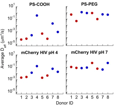
Figure 2.2. The ensemble averaged diffusivity of particles or virions classified as mobile in
cervicovaginal mucus specimens from different donors (N=8). PS-COOH (carboxylated polystyrene nanoparticles, d ~ 200 nm) which were previously shown to be relatively sticky in pH 7 CVM; PS-PEG (PEG-coated polystyrene nanoparticles, d ~ 200 nm) which were previously shown to be relatively muco-inert in pH 7 CVM; mCherry-GAG tagged HIV virus-like particles pseudotyped with YU2 envelope in CVM at native (i.e. unaltered) pH; mCherry-GAG tagged HIV virus-like particles pseudotyped with YU2 envelope in CVM adjusted to neutral pH with <3% dilution with 3N NaOH. Symbols in blue represents CVM specimens where HIV exhibited substantial mobile fractions at native pH (see Figure 2.1C).
10-5
10-3
10-1
101 PS-COOH PS-PEG
10-5 10-3 10-1 101
1 2 3 4 5 6 7 8 mCherry HIV pH 4
A ver ag e D ef f ( µ m 2 /s ) Donor ID
3.5 4.5 5.5 0 20 40 60 80 100 pH (mg/mL) Fra ct io n M o vi n g (% )
0 2 4 6 Nugent
Score
0 2 4 6 8 10 [D-LA]
(g/L)
0 2 4 6 8 10 [L-LA]
(g/L)
0 5 10 15 20 [Total LA]
(g/L)
Figure 2.3. Fraction of mobile virions for mCherry-GAG tagged HIV virus-like particles pseudotyped
with YU2 envelope in CVM at native (i.e. unaltered) pH, correlated to native CVM pH, Nugent Score, endogenous D-lactic acid (D-LA) levels in CVM, endogenous L-LA levels in CVM, and total LA levels
in CVM (N=8). Symbols in blue represents CVM specimens where HIV exhibited substantial mobile fractions at native pH (see Figure 2.1C).
3.5 4.5 5.5 10-3 10-2 10-1 100 pH (mg/mL) D ef f ( µ m 2 /s )
0 2 4 6
Nugent Score
0 2 4 6 8 10 [D-LA]
(g/L)
0 2 4 6 8 10 [L-LA]
(g/L)
0 5 10 15 20 [Total LA]
(g/L) Figure 2.4. Ensemble averaged diffusivities for mCherry-GAG tagged HIV virus-like particles
pseudotyped with YU2 envelope in CVM at native (i.e. unaltered) pH, correlated to native CVM pH, Nugent Score, endogenous D-lactic acid (D-LA) levels in CVM, endogenous L-LA levels in CVM, and total LA levels in CVM (N=8). Symbols in blue represents CVM specimens where HIV exhibited substantial mobile fractions at native pH (see Figure 2.1C).
Figure 2.5: CVM that traps HIV appear to be L. crispatus- dominant, whereas CVM that fails to
trap HIV are either L. iners- dominant, or include substantial G. vaginalis. (A) Representative of the
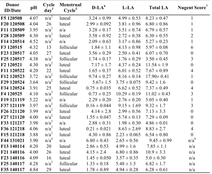
Table 2.1: Characterization of CVM samples: pH, Cycle day, Menstrual cycle phase,
D-LA/L-LA/total LA in g/L, and Nugent Score.
1Cycle day calculated as the number of day from the first of the last menstrual period normalized by the
cycle length to a 29 day cycle. 2N/A = hormonal contraceptive. 3N/A = donor with abnormal cycle, could not calculate cycle day. 4N/A = no menstrual cycle info for donor. 5Cycle phase estimated based on normalized cycle day; no samples were ovulatory based on absence of spinnbarkeit by visual inspection. 6Values are expressed as mean ± SD. 7A Nugent score of 0-3 corresponds to “normal”
(lactobacilli-dominated) microflora, 4-6 to “intermediate”, and 7-10 to “bacterial vaginosis” (BV) – a condition associated with greater risk of STI acquisition. Assessment of Nugent scores was independently
confirmed by the Clinical Microbiology and Immunology Lab at UNC – Chapel Hill. 8Waiting on results. Donor
ID/Date pH
Cycle day1
Menstrual
Cycle5 D-LA6 L-LA Total LA Nugent Score7
F5 120508 4.07 n/a2 luteal 3.24 ± 0.99 4.99 ± 0.53 8.23 ± 0.47 1
F20 120508 4.04 26 luteal 2.99 ± 0.092 3.81 ± 0.96 6.80 ± 0.86 1
F11 120509 3.95 n/a2 n/a 3.28 ± 0.17 3.51 ± 0.74 6.79 ± 0.57 1
F28 120509 4.30 n/a2 luteal 3.58 ± 0.92 2.72 ± 0.38 6.30 ± 0.55 2
F14 120514 4.52 n/a2 n/a 2.09 ± 0.63 3.17 ± 0.86 5.27 ± 0.23 1
F3 120515 4.32 13 follicular 1.84 ± 1.1 4.13 ± 0.98 5.97 ± 0.08 6
F23 120517 4.05 27 luteal 3.56 ± 0.29 2.50 ± 0.41 6.07 ± 0.70 0
F25 120517 4.38 n/a2 follicular 1.74 ± 0.17 1.76 ± 0.29 3.50 ± 0.45 3
F2 120521 4.30 n/a2 luteal 7.17 ± 1.7 4.37 ± 0.24 11.54 ± 1.9 0
F11 120522 4.20 22 luteal 1.65 ± 0.37 6.01 ± 0.52 7.67 ± 0.89 4
F12 120523 3.72 n/a2 follicular 9.74 ± 0.27 8.16 ± 0.14 17.90± 0.41 1
F29 120524 3.64 n/a2 follicular 5.67± 1.5 3.75 ± 0.075 9.42 ± 1.6 0
F34 120524 3.91 25 luteal 0.75 ± 0.035 6.62 ± 0.52 7.37 ± 0.49 4
F4 120525 4.10 n/a2 follicular 0.73 ± 0.25 10.29 ± 0.19 11.02 ± 0.43 3
F19 121119 5.22 n/a2 n/a 2.29 ± 0.20 2.76 ± 0.20 5.05 ± 0.40 1
F37 121119 3.97 n/a2 follicular 0.16 ± 0.044 9.15 ± 1.69 9.32 ± 1.7 3
F26 121120 3.99 n/a2 luteal 4.14 ± 2.8 2.99 ± 0.56 7.13 ± 3.3 0
F27 121120 4.00 n/a2 luteal 1.55 ± 0.047 5.74 ± 0.13 7.29 ± 0.09 0
F33 121217 3.98 n/a3 n/a 2.88 ± 0.31 1.98 ± 0.30 4.86 ± 0.01 0
F36 121218 4.06 n/a2 luteal 0.21 ± 0.021 8.63 ± 2.69 8.83 ± 2.7 4
F15 121218 3.88 n/a2 luteal 4.30 ± 0.86 2.23 ± 0.065 6.54 ± 0.80 0
F44 131021 3.90 n/a4 n/a 6.80 ± 0.43 2.65 ± 0.56 9.45 ± 0.99 n/a8
F13 140114 4.20 20 luteal 2.86 ± 0.53 4.99 ± 1.6 7.85 ± 1.1 n/a
F21 140116 4.00 26 luteal 4.15 ± 2.4 6.80 ± 0.86 10.9 ± 3.3 n/a
F23 140116 4.09 16 luteal 1.45 ± 0.050 3.57 ± 0.35 5.0 ± 0.30 n/a
F25 140117 4.28 n/a2 follicular 1.33 ± 0.18 5.48 ± 1.5 6.82 ± 1.7 n/a
Donor ID/Date Age Race Birth Control F5 120508 26 African American Nuvaring F20 120508 24 African American no F11 120509 25 Caucasian Loestrin F28 120509 20 Caucasian Loestrin 24
F14 120514 32 Caucasian Orthocept
F3 120515 28 Caucasian no
F23 120517 19 Other no
F25 120517 27 Caucasian Lutera
F2 120521 28 Caucasian Loestrin 24 Fe
F11 120522 25 Caucasian no
F12 120523 26 Caucasian Loestrin F29 120524 21 Caucasian azurette F34 120524 26 African American no
F4 120525 27 Caucasian Microgestin
F19 121119 26 Caucasian IUD
F37 121119 20 Asian Yes1
F26 121120 22 Caucasian Trisprintec
F27 121120 24 Caucasian Avaine
F33 121217 22 Caucasian no
F36 121218 23 Asian Reclipsen
F15 121218 30 Caucasian Junel Fe
F44 131021 20 Caucasian no
F13 140114 33 Caucasian no
F21 140116 21 African American no
F23 140116 21 Other no
F25 140117 29 Caucasian Lutera
F35 140117 21 Asian no
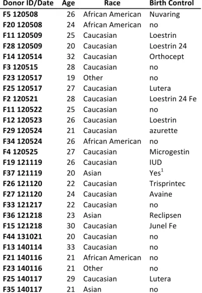
Table 2.2: Characterization of CVM samples: Age, Race, and birth control.
1Donor did not specify the brand of birth control, just expressed the fact that the birth control used were
oral contraceptives.
REFERENCES
1. Cone, R., Mucus, in Handbook of Mucosal Immunology, P.L. Ogra, J. Mestecky, M.E. Lamm, W. Strober, J. Bienenstock, and J.R. McGhee, Editors. 1999, Academic Press: San Diego. p. 43-64.
2. Lai, S.K., Y.Y. Wang, and J. Hanes, Mucus-penetrating nanoparticles for drug and gene delivery to mucosal tissues. Adv. Drug Deliv. Rev., 2009. 61(2): p. 158-71.
3. Lai, S.K., Y.Y. Wang, D. Wirtz, and J. Hanes, Micro- and macrorheology of mucus. Adv. Drug Deliv. Rev., 2009.
4. Kieweg, S.L., A.R. Geonnotti, and D.F. Katz, Gravity-induced coating flows of vaginal gel formulations: in vitro experimental analysis. J Pharm Sci, 2004. 93(12): p. 2941-52.
5. Kieweg, S.L. and D.F. Katz, Squeezing flows of vaginal gel formulations relevant to microbicide drug delivery. J Biomech Eng, 2006. 128(4): p. 540-53.
6. Boskey, E.R., R.A. Cone, K.J. Whaley, and T.R. Moench, Origins of vaginal acidity: high D/L lactate ratio is consistent with bacteria being the primary source. Hum. Reprod., 2001. 16(9): p. 1809-13.
7. Gipson, I.K., S.B. Ho, S.J. Spurr-Michaud, A.S. Tisdale, Q. Zhan, E. Torlakovic, J. Pudney, D.J. Anderson, N.W. Toribara, and J.A. Hill, 3rd, Mucin genes expressed by human female
reproductive tract epithelia. Biol. Reprod., 1997. 56(4): p. 999-1011.
8. Lai, S.K., Y.Y. Wang, K. Hida, R. Cone, and J. Hanes, Nanoparticles reveal that human
cervicovaginal mucus is riddled with pores larger than viruses. Proc Natl Acad Sci U S A, 2010. 107(2): p. 598-603.
9. Lai, S.K., K. Hida, S. Shukair, Y.Y. Wang, A. Figueiredo, R. Cone, T.J. Hope, and J. Hanes, Human immunodeficiency virus type 1 is trapped by acidic but not by neutralized human cervicovaginal mucus. J Virol, 2009. 83(21): p. 11196-200.
10. Lai, S.K., D.E. O'Hanlon, S. Harrold, S.T. Man, Y.Y. Wang, R. Cone, and J. Hanes, Rapid transport of large polymeric nanoparticles in fresh undiluted human mucus. Proc Natl Acad Sci U S A, 2007. 104(5): p. 1482-7.
12. Usala, S.J., F.O. Usala, R. Haciski, J.A. Holt, and G.F. Schumacher, IgG and IgA content of vaginal fluid during the menstrual cycle. J. Reprod. Med., 1989. 34(4): p. 292-4.
13. Wang, Y.Y., A. Kannan, K.L. Nunn, M.A. Murphy, D.B. Subramani, T. Moench, R. Cone, and S.K. Lai, IgG in cervicovaginal mucus traps HSV and prevents vaginal Herpes infections. Mucosal Immunol, 2014.
14. Ravel, J., P. Gajer, Z. Abdo, G.M. Schneider, S.S. Koenig, S.L. McCulle, S. Karlebach, R. Gorle, J. Russell, C.O. Tacket, R.M. Brotman, C.C. Davis, K. Ault, L. Peralta, and L.J. Forney, Vaginal microbiome of reproductive-age women. Proc Natl Acad Sci U S A, 2011. 108 Suppl 1: p. 4680-7.
15. Cohn, S.E. and R.A. Clark, Sexually transmitted diseases, HIV, and AIDS in women. Med Clin North Am, 2003. 87(5): p. 971-95.
16. Cottrell, B.H., An updated review of of evidence to discourage douching. MCN Am J Matern Child Nurs, 2010. 35(2): p. 102-7; quiz 108-9.
17. Helfgott, A., N. Eriksen, C.M. Bundrick, R. Lorimor, and B. Van Eckhout, Vaginal infections in human immunodeficiency virus-infected women. Am J Obstet Gynecol, 2000. 183(2): p. 347-55.
18. O'Hanlon, D.E., T.R. Moench, and R.A. Cone, In vaginal fluid, bacteria associated with bacterial vaginosis can be suppressed with lactic acid but not hydrogen peroxide. BMC Infect Dis, 2011. 11: p. 200.
19. O'Hanlon, D.E., T.R. Moench, S. Harrold, and R.A. Cone. Microbicide production by vaginal lactobailli: Vaginal Acidity and lactic acid are more potent than previously reported (Poster TA-237). in Microbicides 2008. 2008. New Delhi.
20. Boskey, E.R., R.A. Cone, K.J. Whaley, and T.R. Moench, Origins of vaginal acidity: high D/L lactate ratio is consistent with bacteria being the primary source. Hum Reprod, 2001. 16(9): p. 1809-13.
21. Aroutcheva, A., D. Gariti, M. Simon, S. Shott, J. Faro, J.A. Simoes, A. Gurguis, and S. Faro, Defense factors of vaginal lactobacilli. Am J Obstet Gynecol, 2001. 185(2): p. 375-9.
23. Redondo-Lopez, V., R.L. Cook, and J.D. Sobel, Emerging role of lactobacilli in the control and maintenance of the vaginal bacterial microflora. Rev Infect Dis, 1990. 12(5): p. 856-72.
24. Valore, E.V., C.H. Park, S.L. Igreti, and T. Ganz, Antimicrobial components of vaginal fluid. Am J Obstet Gynecol, 2002. 187(3): p. 561-8.
25. Aldunate, M., D. Tyssen, A. Johnson, T. Zakir, S. Sonza, T. Moench, R. Cone, and G. Tachedjian, Vaginal concentrations of lactic acid potently inactivate HIV. J Antimicrob Chemother, 2013.
26. Graver, M.A. and J.J. Wade, The role of acidification in the inhibition of Neisseria gonorrhoeae by vaginal lactobacilli during anaerobic growth. Ann Clin Microbiol Antimicrob, 2011. 10: p. 8.
27. Linhares, I.M., P.R. Summers, B. Larsen, P.C. Giraldo, and S.S. Witkin, Contemporary perspectives on vaginal pH and lactobacilli. Am J Obstet Gynecol, 2011. 204(2): p. 120 e1-5.
28. Witkin, S.S., S. Alvi, A.M. Bongiovanni, I.M. Linhares, and W.J. Ledger, Lactic acid stimulates interleukin-23 production by peripheral blood mononuclear cells exposed to bacterial
lipopolysaccharide. FEMS Immunol Med Microbiol, 2011. 61(2): p. 153-8.
29. Mossop, H., I.M. Linhares, A.M. Bongiovanni, W.J. Ledger, and S.S. Witkin, Influence of lactic acid on endogenous and viral RNA-induced immune mediator production by vaginal epithelial cells. Obstet Gynecol, 2011. 118(4): p. 840-6.
30. Mihm, S. and W. Droge, Regulation of cytotoxic T-lymphocyte activation by L-lactate and pyruvate. Cell Immunol, 1985. 96(1): p. 235-40.
31. Fichorova, R.N., H.S. Yamamoto, M.L. Delaney, A.B. Onderdonk, and G.F. Doncel, Novel vaginal microflora colonization model providing new insight into microbicide mechanism of action. MBio, 2011. 2(6): p. e00168-11.
32. Shukair, S.A., S.A. Allen, G.C. Cianci, D.J. Stieh, M.R. Anderson, S.M. Baig, C.J. Gioia, E.J. Spongberg, S.M. Kauffman, M.D. McRaven, H.Y. Lakougna, C. Hammond, P.F. Kiser, and T.J. Hope, Human cervicovaginal mucus contains an activity that hinders HIV-1 movement. Mucosal Immunol, 2013. 6(2): p. 427-34.
34. Yuan, S., D.B. Cohen, J. Ravel, Z. Abdo, and L.J. Forney, Evaluation of methods for the extraction and purification of DNA from the human microbiome. PLoS ONE, 2012. 7(3): p. e33865.
35. Srinivasan, S., N.G. Hoffman, M.T. Morgan, F.A. Matsen, T.L. Fiedler, R.W. Hall, F.J. Ross, C.O. McCoy, R. Bumgarner, J.M. Marrazzo, and D.N. Fredricks, Bacterial communities in women with bacterial vaginosis: high resolution phylogenetic analyses reveal relationships of microbiota to clinical criteria. PLoS ONE, 2012. 7(6): p. e37818.
36. Gevers, D., M. Pop, P.D. Schloss, and C. Huttenhower, Bioinformatics for the Human Microbiome Project. PLoS Comput Biol, 2012. 8(11): p. e1002779.
37. Peterson, J., S. Garges, M. Giovanni, P. McInnes, L. Wang, J.A. Schloss, V. Bonazzi, J.E. McEwen, K.A. Wetterstrand, C. Deal, C.C. Baker, V. Di Francesco, T.K. Howcroft, R.W. Karp, R.D. Lunsford, C.R. Wellington, T. Belachew, M. Wright, C. Giblin, H. David, M. Mills, R. Salomon, C. Mullins, B. Akolkar, L. Begg, C. Davis, L. Grandison, M. Humble, J. Khalsa, A.R. Little, H. Peavy, C. Pontzer, M. Portnoy, M.H. Sayre, P. Starke-Reed, S. Zakhari, J. Read, B. Watson, and M. Guyer, The NIH Human Microbiome Project. Genome Res, 2009. 19(12): p. 2317-23.
38. Edgar, R.C., B.J. Haas, J.C. Clemente, C. Quince, and R. Knight, UCHIME improves sensitivity and speed of chimera detection. Bioinformatics, 2011. 27(16): p. 2194-200.
39. Wang, Q., G.M. Garrity, J.M. Tiedje, and J.R. Cole, Naïve Bayesian Classifier for Rapid
Assignment of rRNA Sequences into the New Bacterial Taxonomy. Applied and Environmental Microbiology, 2007. 73(16): p. 5261-5267.
40. Gajer, P., R.M. Brotman, G. Bai, J. Sakamoto, U.M. Schutte, X. Zhong, S.S. Koenig, L. Fu, Z.S. Ma, X. Zhou, Z. Abdo, L.J. Forney, and J. Ravel, Temporal dynamics of the human vaginal microbiota. Science Translational Medicine, 2012. 4(132): p. 132ra52.
41. Nugent, R.P., M.A. Krohn, and S.L. Miller, Reliability of diagnosing bacterial vaginosis is improved by a standardized method of gram stain. J Clin Microbiol, 1991. 29(2): p. 297-301.
42. Whitkin, S.S., H. Mendes-Soares, L.M. Linhares, A. Jayaram, J.L. William and L.J. Forney, Influence of vaginal bacteria and D- and L-lactic acid isomers on vaginal extracellular matrix metalloproteinase inducer: Implications for protection against upper genital tract infections. MBio, 2013. 4(4): p. e00460-13.