Diabetic Microangiopathy*
J. M. B. BLOODWORTH, JR.
Department of Pathology, University of Wisconsin Medical School, Madison, and Madison Veterans Administration Hospital
Microangiopathy is the term ap-plied to the abnormal state of the capillaries, arterioles, and venules found in the diabetic patient. It is characterized principally by thick-ening of the basement membrane of these small vessels. It might be worth mentioning that while we are concerned here with the smaller blood vessels, diabetic patients also show thickening of the basement membrane beneath the endothelium of arteries of all sizes. Also, the basement membrane-like material that surrounds each smooth muscle fiber in the wall of arteries shows similar thickening in diabetic pa-tients. With this thickening, there is an increased glycoprotein con-tent of the arterial wall.
Diabetic microangiopathy is as-sociated with well known clinical disturbances in the retina and in the kidney, but involves as well capillaries throughout the body.
Let us consider the structure of the normal retinal capillary as re-vealed by the electron microscope (fig. 1). The lumen of the capillary is lined by endothelial cells. The nuclei of these endothelial cells protrude into the capillary lumen, but elsewhere the cytoplasm of these cells is spread out in a con-tinuous thin layer around the capil-lary to form its inner wall. In turn; a layer of basement membrane
*Supported in part by grant #AM-07118 from the National Institutes of Health, U.S.P.H.S.; the Wisconsin Heart Association; U.S.P.H.S. in-stitutional grant funds of the Univer-sity of Wisconsin; and Veterans Ad-ministration research funds.
completely surrounds each endo-thelial cell. External to the single layer of endothelial cells are cells whose cytoplasm extends like ten-tacles around the capillary. These outside cells are the pericytes, al-though Dr. Cogan in Boston ( 1961) prefers to call them mural cells. Whatever they are called, they give support and strength to the capil-lary wall. Projections of basement membrane extend out between the cytoplasmic projections of the peri-cytes, and around the body of the pericytes.
One difference between retinal capillaries and other capillaries is that the retinal capillaries are in-timately surrounded by the ele-ments of the retina, the nerve fibers, and the glial cells. These structures are right up against the retinal capillaries and perhaps support them to some extent. At times the basement membrane material of the capillary appears to trail off into the adjacent nerve fibers or glial cells.
If we examine that retinal capil-lary of an elderly man or an elderly dog, we can observe two changes. There is some thickening of the capillary basement membrane, and in the outer layers of the capillary wall it has developed vacuoliza-tion, giving a Swiss cheese appear-ance. These changes occur with age in man and in the dog, but not in the elderly rat. It is of some in-terest that retinopathy will develop in the diabetic man and the diabetic dog, but apparently not in the dia-betic rat.
electron microscope. The mildest change noted is the thickening of the basement membrane. The nor-mal basement membrane is 700 or 800 A in thickness, by our method; that of the elderly human a little thicker; but that of the diabetic much thicker, from 1100 to 3600 A. The diabetic shows the Swiss cheese type of vacuolization of the outer portion of the basement mem-brane, and in addition in the dia-betic the vacuolizations seem to be filled up with some kind of debris. Sometimes, the thickened base-ment membrane of the diabetic ap-pears slightly laminated with simi-lar osmiophillic debris between the layers. Another finding in the dia-betic is the occurrence of collagen fibrils within the thickened layer of basement membrane.
The internal structure of the endothelial cells and the pericytes appears entirely normal in the dia-betic. One sees normal nuclei and organdelles; that is, normal mito-chondria, normal pinocytic vesicles,
normal endoplasmic reticulum and ribosomes.
Let us now turn to the more classic light microscope findings in diabetic retinopathy; the micro-aneurysms, exudates, hemorrhages, and changes of retinitis proliferans. We really do not know whether these are related to the ultrastruc-tural abnormalities seen in the cap-illaries or not. For one thing, the ultrastructural capillary changes are generalized throughout the retina while the microaneurysms and ex-udates are spotty, tending to occur mostly around the nerve head.
In the diabetic, the degenerative changes in the capillaries begin shortly after birth and progress throughout life, similar to the changes accompanying ageing in the non-diabetic. We see collapsed, acellular capillaries composed of basement membrane material in which are interspersed the processes of glial cells. This is evidently the end stage of the capillary degenera-tion. Whether it has something to do with the development of
micro-DIABETIC MICROANGIOPATHY
aneurysms we do not know. Microaneurysms tend to occur in focal groups or clumps (fig. 3).
They will occur in one group of capillaries coming off an arteriole,
but not in another group of capil-laries coming off the same arteriole. There must be some focal factor that we do not understand.
Microaneurysms occur only in the inner half of the retina, which is the only part supplied by retinal capillaries. They occur in the gan-glion cell layer just beneath the nerve fibers or as deep as the outer molecular layer, but not in the deeper structures such as the rods and cones.
Microaneurysms can be divided into the thin-walled type and the thick-walled type. Actually all of them have thicker walls than the normal capillary. The thin-walled microaneurysm appears as a great ballooning out of the capillary into a globular shape with the capillary entering at one end and coming out the other. I think this is the first stage, and the thickening of the wall comes later.
There is no change in the struc-ture of the wall of the capillary at the point where it begins to balloon out to form a microaneurysm. The endothelial cells are intact and appear healthy (fig. 4). The base-ment membrane is thicker than usual and has debris in it, and may contain some collagen fibrils. These findings are diffuse and cannot ex-plain the microaneurysm. The base-ment membrane material of the aneurysm must have been produced in increased amount to allow this ballooning out, with still an in-creased thickness. Also, there must have been proliferation or hyper-trophy of the endothelial cells to allow them to continue to line the greatly enlarged surface of the aneurysm. This seems to suggest a vital, living process going on.
Cogan and his group in Boston (1961) found certain large capil-laries with hypercellularity of the endothelial cells, but with un-healthy appearing or dead pericytes
Fig. 1-Electron micrograph of a retinal capillary from a non-diabetic human patient for comparison with Figure 2. The capillary luman (CAP) contains a red blood cell (RBC). The lumen is surrounded by a thin layer of endothelial cytoplasm which, to the left of the lumen, contains a nucleus (EN). The endothelial cytoplasm is,
in turn, surrounded by an irregular basement membrane (BM) (4200X).
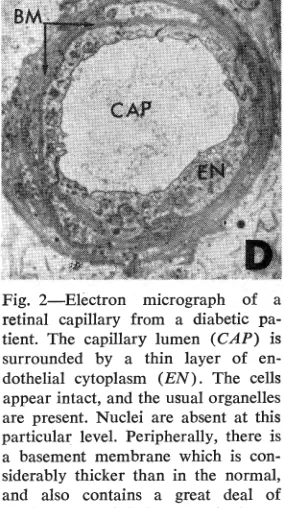
Fig. 2-Electron micrograph of a retinal capillary from a diabetic pa-tient. The capillary lumen (CAP) is surrounded by a thin layer of en-dothelial cytoplasm (EN). The cells appear intact, and the usual organelles are present. Nuclei are absent at this particular level. Peripherally, there is a basement membrane which is con-siderably thicker than in the normal,
(or mural cells), which he calls "ghost cells." He believes that it is the pericyte that gives strength to the capillary, and that when this is damaged or dies, the capillary dilates. He calls this type of dam-aged vessel a "shunt vessel," and it is from these that microaneurysms develop. Adjacent capillaries may show no blood and no cells, only a basement membrane, and occa-sionally atrophied remnants of cells and glial fibers. I cannot find enough of these large, hypercellu-lar "shunt vessels" to explain even a small portion of the total changes seen in diabetic retinopathy. I would be inclined to consider them as another phenomenon seen in diabetic retinopathy, but not as the basic etiology of the microaneu-rysms.
The thick-walled microaneurysms have a lot of glial cells around them and usually show accumulations of platelets. These collect along the endothelial lining, but some plate-lets may be seen within the vessel wall, along with red blood cells and fibrin. It seems evident that the thick-walled aneurysm is leaking,
and the thickness results from the blood elements trapped in the wall and the glial reaction about the vessel.
The second classic finding of diabetic retinopathy is the exudate. The smallest exudates are those that appear immediately around micro-aneurysms, and represent leaked plasma constituents. They can hardly be distinguished from the aneurysm itself on ophthalmoscopic examination. The larger, waxy exu-dates are large collections of PAS-positive material, usually in the plexiform layers. Under the elec-tron microscope, the exudate has a fine granular component and a fibrillar component. We believe this is a mixture of plasma proteins in-cluding fibrin that has leaked from the capillary. Apparently capillary leaks can occur in all eyes, but they
are vastly more frequent in diabetic
eyes. Around these exudates we may see glial cells attempting to
J. M. B. BLOODWORTH, JR.
Fig. 3- Flat preparation of the retina from a diabetic patient. Note the artery (A) and vein (V) between which there is a capillary plexus. Numerous micro-aneurysms (arrows) are present on the capillary network. At the right of the photograph, the dark gray areas represent exudates associated with the
micro-aneurysms in this area (90X).
phagocytize them. Apparently they can be phagocytized and removed eventually.
Hemorrhages are of two major types. The small, round hemor-rhages occur in the deeper layers, from rupture of or leakage from microaneurysms. These are reab-sorbed in a few days and probably cause no appreciable damage. Larger hemorrhages occur super-ficially, and may leak into the vitreous or may dissect beneath the retina and cause detachment. Strands of fibrin are laid down in the vitreous, and new blood vessels grow into these. These delicate new capillaries are easily disrupted, with further bleeding, and a vicious cycle is set up. This is the situation known as retinitis proliferans, the principle cause of blindness in dia-betic patients.
Along with these vascular changes, there seems to be some degeneration of ganglion cells. In the ganglion cell layer, there seems to be a dropping out of cells and some of the remaining cells appear atrophic. In some areas, special
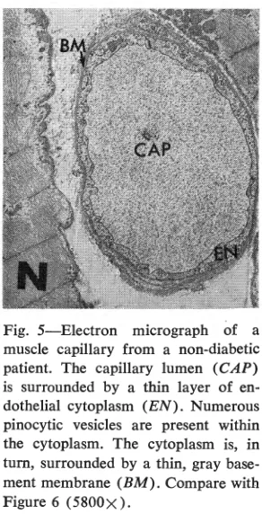
Fig. 5-Electron micrograph of a muscle capillary from a non-diabetic patient. The capillary lumen (CAP) is surrounded by a thin layer of en-dothelial cytoplasm (EN). Numerous pinocytic vesicles are present within the cytoplasm. The cytoplasm is, in turn, surrounded by a thin, gray base-ment membrane (BM). Compare with Figure 6 (5800x).
DIABETIC MICROANGIOPATHY
stains reveal a swelling and fibrillar fragmentation of the nerve proc-esses.
The cotton wool plaque looks like a mass of debris replacing a portion of the inner layers of the retina. It appears to be a micro-infarction. Attempts at revasculari-zation are seen. These structures are not specific for diabetes.
Evidence of microangiopathy may be seen in patients who have just developed diabetes, and in pre-diabetics with no demonstrable ab-normality of carbohydrate metab-olism. This had led some people to think that the changes of micro-angiopathy are due to an inherited defect in the capillary itself, in-herited in parallel with the defect in metabolism. I think our evidence is definitely against this view. There are a fair number of examples of microangiopathy in humans with non-hereditary diabetes. In dogs made diabetic with alloxan or with growth hormone, I have seen all forms of microangiopathy, includ-ing retinopathy, with all the features described in human beings. I think
the microangiopathy results from
some abnormality of metabolism
that we are not just yet able to measure. I think we will some day find out what this is.
I would like to point out that
most of the other capillaries in the body of the diabetic show the same thickening of the basement mem-brane. Muscle capillaries (compare figs. 5 and 6) show this, as do capil-laries from the skin, ear lobe, toe, uterus and mammary gland. The
one exception is the capillaries in
fat tissue which show no thickening of the basement membrane in dia-betes. This is true in the diabetic dog as well as in the diabetic hu-man being. This must have some real importance, but we do not know the explanation. It might be related to differences in the utiliza-tion of insulin by muscle and fat, but this remains to be determined.
I should mention the work of
Dr. Siperstein ( 1964 and 1965), who found thickening of the muscle capillary basement membrane in his diabetics and in prediabetics, but did not find any increase in thick-ness with age in the non-diabetic. I
think there is an increase in the
basement membrane thickness with age in non-diabetics, and am not
sure the increased thickness he
re-ports in his prediabetics is signifi-cant. He reports about 1,200 A for his normals, 1,500 A for his predia-betics, and around 2,500 A for the diabetics. The error inherent in the method of performing basement membrane measurements is rather large.
Blumenthal and his associates at St. Louis have reported the finding of proliferative lesions in the capil-laries and small vessels of the dia-betic. Three pathology laboratories, including my own, have studied this problem and have concluded that
these changes are not specific for
diabetes. They apparently occur in
areas of trauma and infection, as
around the edges of the gangrenous
area in a diabetic leg. The
im-portance of these lesions remains
speculative.
J. M. B. BLOODWORTH, JR.
REFERENCES
BLOODWORTH, J. M. B., JR. Diabetic retinopathy. Diabetes 11: 1, 1962.
BLOODWORTH, J. M. B., JR. Diabetic microangiopathy. Diabetes 12: 99, 1963.
SIPERSTEIN, M. D., A. R. COLWELL,
SR., AND K. MEGER (EDS.). Small
Blood Vessel Involvement in Dia-betes Mellitus. Washington, D. C.: Amer. Inst. Biol. Sci., 1964.
COGAN, D. G., TOUSSAINT, D., AND
KUWAHARA, T. Retinal vascular pat-terns. IV. Diabetic retinopathy. Arch. Ophthal. 66: 366, 1961. SrPERSTEIN, M. D. Diabetic vascular
changes in human muscle. Presented
to symposium on Microangiopathy
of the Eye in Diabetes, Univ. of California School of Medicine, San
Francisco Med. Center, Dec. 2-4,