R E V I E W A R T I C L E
Open Access
Myofascial trigger points in migraine and
tension-type headache
Thien Phu Do, Gerda Ferja Heldarskard, Lærke Tørring Kolding, Jeppe Hvedstrup and Henrik Winther Schytz
*Abstract
Background:A myofascial trigger point is defined as a hyperirritable spot in skeletal muscle that is associated with a hypersensitive palpable nodule in a taut band. It has been suggested that myofascial trigger points take part in chronic pain conditions including primary headache disorders. The aim of this narrative review is to present an overview of the current imaging modalities used for the detection of myofascial trigger points and to review studies of myofascial trigger points in migraine and tension-type headache.
Findings:Different modalities have been used to assess myofascial trigger points including ultrasound, microdialysis, electromyography, infrared thermography, and magnetic resonance imaging. Ultrasound is the most promising of these modalities and may be used to identify MTrPs if specific methods are used, but there is no precise description of a gold standard using these techniques, and they have yet to be evaluated in headache patients.
Active myofascial trigger points are prevalent in migraine patients. Manual palpation can trigger migraine attacks. All intervention studies aiming at trigger points are positive, but this needs to be further verified in placebo-controlled environments. These findings may imply a causal bottom-up association, but studies of migraine patients with comorbid fibromyalgia syndrome suggest otherwise. Whether myofascial trigger points contribute to an increased migraine burden in terms of frequency and intensity is unclear.
Active myofascial trigger points are prevalent in tension-type headache coherent with the hypothesis that peripheral mechanisms are involved in the pathophysiology of this headache disorder. Active myofascial trigger points in pericranial muscles in tension-type headache patients are correlated with generalized lower pain pressure thresholds indicating they may contribute to a central sensitization. However, the number of active myofascial trigger points is higher in adults compared with adolescents regardless of no significant association with headache parameters. This suggests myofascial trigger points are accumulated over time as a consequence of TTH rather than contributing to the pathophysiology.
Conclusions:Myofascial trigger points are prevalent in both migraine and tension-type headache, but the role they play in the pathophysiology of each disorder and to which degree is unclarified. In the future, ultrasound elastography may be an acceptable diagnostic test.
Keywords:Headache, Myofascial trigger point, Muscle, Treatment, Trigemino-cervical-complex, Migraine, Tension-type headache, Diagnostic test
Background
Migraine affects 16% of the population in Europe [1]
with high individual and socioeconomic costs [2,3].
Sev-eral mechanisms have been proposed to be involved in its pathophysiology including vascular, peripheral and
central mechanisms [4–9]. Jes Olesen systematically
de-scribed pericranial tenderness in migraine patients, both
during and outside of migraine attacks [10, 11], leading
to speculations that myofascial mechanisms may be
in-volved in migraine [12].
Tension-type headache (TTH) is the most prevalent
primary headache disorder worldwide [13]. Tenderness
in pericranial myofascial tissue is correlated with the
in-tensity and frequency of headache in TTH [14–16], and
studies show increased muscle stiffness in TTH patients
[17, 18]. Thus, myofascial structures may be associated
with TTH pathophysiology. * Correspondence:henrik.winther.schytz.01@regionh.dk
Headache Diagnostic Laboratory, Danish Headache Center and Department of Neurology, Rigshospitalet Glostrup, Faculty of Health Sciences, University of Copenhagen, Glostrup, Denmark
The term myofascial trigger point (MTrP) was popu-larized in the 1950s and is defined as a hyperirritable spot in skeletal muscle that is associated with a
hyper-sensitive palpable nodule in a taut band [19, 20]. The
spot is painful on compression and can cause referred pain, referred tenderness, motor dysfunction and auto-nomic phenomena. The interest in myofascial symptoms has been ongoing for centuries with similar descriptions of localized thickenings of muscles with regional pain
[21]. There have been inconsistencies and controversies
in the literature on the underlying pathology, and even
the existence of MTrPs [22]. While attempts have been
made to visualize MTrPs [22], the gold standard for
de-tection of MTrPs has been unchanged since the 1950s
[22] and remains to be by way of palpation of the
affected muscles. However, this technique proves to be poorly reproducible as practitioners disagree on the location of MTrPs when blindly examining different
pa-tient groups [23]. Nevertheless, MTrPs have come to
play a central role in the diagnosis and treatment of
myofascial pain syndrome [19]. Furthermore, MTrPs
have been proposed to take part in primary headache
disorders and other chronic pain conditions [12]. The
aim of this narrative review is to present an up-to-date overview on MTrPs in general and then in migraine and TTH, respectively.
Review
Myofascial trigger points
In the comprehensive trigger point manual by Travell and
Simons [19], MTrPs are subclassified into different types,
e.g., active and latent amongst others. An active MTrP produces a constant pain complaint while a latent only
produces pain during manual palpation [19]. It was
hypothesized that a sustained muscle contraction in MTrPs promotes hypoxia and ischemia with a following increase in concentrations of substances such as calcitonin
gene-related peptide (CGRP) and substance P (SP) [24].
Consequently, this would lead to increased peripheral
nociceptive transmission [24]. This hypothesis is only
sup-ported in active MTrPs, as they have been shown to be as-sociated with higher levels of these substances in the local
milieu compared to latent MTrPs [25,26]. Other
proper-ties such as the consistency of the tissue have also been
suggested to play a key role in MTrPs [27].
Investigations of myofascial trigger points
Ultrasound imaging
Different ultrasound modalities in ultrasound imaging
have visualized MTrPs. Lewis et al. [28] conducted a pilot
study to assess the use of ultrasound in determining soft tissue changes in the region of active MTrPs in 11 sub-jects. They found no correlation between clinical identified
active MTrPs and ultrasound. In contrast, Turo et al. [29]
were able to differentiate between symptomatic MTrPs and asymptomatic muscle tissue with texture-based ana-lysis. Sikdar et al. investigated the stiffness of active and la-tent MTrPs, using ultrasound elastography by Doppler
variance imaging in nine subjects while inducing
vibrations with an external handheld massage vibrator
[27]. MTrPs appeared as focal, hypoechoic regions on
two-dimensional ultrasound images and with reduced vi-bration amplitude, indicating increased stiffness. Further-more, they describe hypoechoic regions that were not identified during palpation prior to ultrasound. In another study by the same group, MTrPs showed reduced vibra-tion amplitude on elastography indicating increased
stiff-ness and distinct blood flow waveform patterns [30].
Ballyns et al. [31] used elastography to investigate MTrPs
in 44 subjects with acute cervical pain. They were able to measure the size and distinguish type (active, latent) of MTrPs with elastography. In addition, Doppler waveforms of blood flow showed different characteristics in active
sites compared to normal tissue. Takla et al. [32] compared
elastography with two-dimensional grayscale ultrasound in identifying MTrPs. They found that MTrPs had an accur-acy of 100% for both active and latent MTrPs while two-dimensional grayscale ultrasound could only identify 33 and 35%, respectively.
Microdialysis
Microdialysis has been used to measure endogenous and exogenous molecules in the local milieu of MTrPs. Shah
et al. [25] used microdialysis to investigate subjects with
active or latent MTrP, and controls without MTrP were detected by manual palpation by two experienced clini-cians. The authors measured selected substances (pH, bradykinin (BK), CGRP, SP, tumor necrosis factor alpha
(TNF-α), interleukin 1 beta (IL-1β), interleukin 6 (IL-6),
interleukin 8 (IL-8), serotonin, and norepinephrine (NE)) in standardized locations of the trapezius muscle and gastrocnemius muscle. Subjects with active MTrPs in the trapezius muscle showed increased concentrations of all substances compared to the other groups. Shah et al.
[26] found similar results in the trapezius muscle of
sub-jects with neck pain and active MTrP compared to a group with neck pain and no MTrP present and healthy controls. The results showed that the active MTrP group
had higher concentrations of BK, CGRP, SP, TNF-α,
IL-1β, serotonin, NE.
Electromyography
Electromyography (EMG) can be used to measure the electrical activity of skeletal muscles. Simons et al. com-pared the prevalence of motor endplate potentials in ac-tive MTrPs, endplate zones, and taut bands of skeletal
muscles in subjects with palpable MTrPs [33]. The
MTrPs than in sites outside of the trigger point, even within the same endplate zone. Ge et al. evaluated intra-muscular muscle activity in a synergistic muscle during isometric contraction in 15 subjects with latent MTrPs
[34]. The needle was inserted into a latent MTrP or a
non-MTrP in the upper trapezius at rest and during contraction. The EMG activities were recorded from the middle deltoid muscle and the upper, middle, and lower parts of the trapezius muscle. The intramuscular EMG activity in the upper trapezius muscle was significantly higher at rest and during contraction at latent MTrPs compared with non-MTrPs. Yu et al. measured max-imum voluntary isometric contraction, endurance, me-dian frequency, and muscle fatigue index in three groups of participants: an active MTrP group, a latent MTrP
group, and a control group [35]. The active MTrP group
had a higher median frequency and muscle fatigue index
than the control group. Wytrążek et al. compared the
EMG activity of muscle motor units at rest and maximal
contraction with surface EMG recordings [36]. The
re-sults showed MTrPs correlated with an increase in EMG amplitude at rest.
Infrared thermography
Infrared thermography can be used to measure the skin
temperature. Dibai-Filho et al. [37] have reviewed the
lit-erature on infrared thermography investigations of MTrPs. The authors included three comparative studies
[38–40] and one accuracy study [41]. The conclusion of
the review is that the included studies do not agree on skin temperature patterns in the presence of MTrPs. The included studies of the review are briefly presented
in the following. Merla et al. [38] found that individuals
with myofascial pain had a greater difference between the right and left side in skin temperature over the mas-seter and sternocleidomastoid muscles before and after maximal voluntary clenching compared to healthy vol-unteers. They also found that the myofascial pain group had a greater temperature change over the measured muscles after maximum voluntary clenching. Kimura et
al. [39] evaluated the vasoconstrictor response after
pro-voking pain in MTrPs with an intramuscular glutamate injection. Furthermore, they activated the sympathetic outflow by using a breath-holding maneuver. They found a decrease in skin temperature over time in latent
MTrPs. In contrast, Zhang et al. [40] did not find that
the skin temperature was affected following an intra-muscular glutamate injection into latent MTrPs. Haddad
et al. [41] compared infrared thermography and
alg-ometer measurements of MTrPs in the masticatory mus-cles. The authors found a positive correlation between skin surface temperature and pressure pain threshold. Regarding diagnosing MTrPs, infrared thermography
had an accuracy of 0.564 to 0.609 (area under the re-ceiver operating characteristic curve).
Magnetic resonance imaging
Chen et al. [42] examined 65 patients with myofascial
pain-associated taut bands using magnetic resonance elastography. They found that agreement between physi-cians and imaging raters were relatively poor (63%; 95% CI, 50%–75%), but that these bands could be assessed quantitatively using magnetic resonance elastography. The authors suggest that clinicians might overestimate while magnetic resonance elastography may underesti-mate MTrPs.
Migraine and myofascial trigger points
Pericranial tenderness in migraine patients was system-atically described by Jes Olesen in 1978, both during and
outside of attacks [10, 11] leading to speculations that
myofascial mechanisms may be involved in migraine
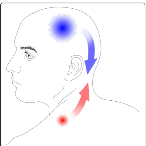
[12]. The bottom-up model states that increased
periph-eral nociceptive transmission sensitizes the central nervous system to lower the threshold for perceiving pain while the top-down model suggests these changes are already
present in the central nervous system [43]. While it can be
argued that pericranial tenderness in migraine may be caused by a top-down central sensitization, a bottom-up association was implied in 1981 when Tfelt-Hansen et al.
[44] demonstrated that injections of lidocaine and saline
into tender trigger points could relieve migraine attacks. They infiltrated the most tender spots of 26 cranial and neck muscles and tendon insertions in 50 migraine patients. The most frequent sites of tenderness were sternocleidomastoid, anterior temporal, neck and shoulder muscles, the coronoid process and occipital insertions. The tender points in the mentioned study do not necessarily
overlap with Travell and Simons’definition of MTrPs, but
the implication stands that peripheral myofascial mecha-nisms may be involved in migraine pathophysiology. Con-sequently, there has been an interest in exploring MTrPs
in migraine (Table1) [45–58].
The occurrence of myofascial trigger points in migraine Several studies have demonstrated a high occurrence of
active and latent MTrPs in migraine patients [45–49].
Studies show that there is a significantly higher preva-lence of active MTrPs in migraine patients compared to
healthy controls [45, 46, 58]. There are conflicting
re-sults in which muscles are the most affected [47, 48].
Fernández-de-Las-Peñas et al. [46] observed that active
MTrPs were most prevalent ipsilateral to the migraine headaches. More unclear is whether the amount of MTrPs is correlated with the frequency and intensity of
headache attacks. Calandre et al. [45] found a positive
frequency and duration of migraine attacks, whereas two
studies by Ferracini et al. [47, 48] found no such
correl-ation. Interestingly, Landgraf et al. [54] could visualize
MTrPs on MR imaging as focal signal alterations in a small pilot study.
Neck mobility and specific muscles
There appears to be an association between neck mobility
and MTrPs [46,48,49,58]. Ferracini et al. [48] found that
a higher number of active MTrPs was positively correlated with a reduction in cervical lordosis and head extension of the head on the neck. In addition, that lower cervical an-gles were correlated higher then the number of active
MTrPs. Florencio et al. [49] hypothesized that active
MTrPs in the cervical musculature alters the activity of the related muscles and that this would be reflected in EMG readings. They observed that the presence of active MTrPs in the cervical musculature had different activation in the neck flexor muscles compared to those without ac-tive MTrPs in the same muscles regardless of the presence
of pain. Palacios-Ceña et al. [55] found that the number of
active MTrPs in head, neck and shoulder muscles were as-sociated with widespread pressure hypersensitivity in a migraine population.
Provocation and intervention studies
Two unblinded studies show that manual palpation of
MTrPs can provoke a migraine attack [45,53]. Calandre
et al. provoked a migraine attack in one-third of a
mi-graine population by palpating MTrPs [45]. Landgraf et
al. provoked migraine headache by inducing pressure to MTrPs and could not replicate this by pressure to non-trigger points in the trapezius in an adolescent
mi-graine population [53].
Interventions targeted at MTrPs show promising
re-sults [50–52, 56, 57], but the quality of studies varies
greatly and lack placebo-control. Giambierardino et al. demonstrated that local anesthetic infiltration of MTrPs resulted in a reduction of migraine symptomatology in
terms of frequency and intensity [52]. Furthermore,
there was a reduction of hyperalgesia, not only at the in-jection site but also in referred areas overlapping with migraine pain sites. Similar, Ranoux et al. injected botu-linum toxin in MTrPs with similar results in terms of
re-duction in headache days [56]. Gandolfi et al. improved
the outcome of prophylactic botulinum toxin treatment in chronic migraine patients with manipulative
treat-ment of MTrPs [50]. The outcome was a lower
con-sumption of analgesics, improvement in pressure pain threshold and increased cervical range of motion. Like-wise, Ghanbari et al. reported that combined positional release therapy targeted at MTrPs with medical therapy is more effective than the sole pharmacological
treat-ment [51]. Interestingly, sessions of magnetic stimulation
of active MTrPs reduced headache frequency and
inten-sity in adolescent migraineurs [57]. Though these
find-ings need to be verified in a placebo-controlled study. There has not been any studies on the effect of systemic
musculoskeletal analgesics on MTrPs [59], which would
be of interest for future studies.
Tension-type headache and myofascial trigger points
Both peripheral and central mechanisms have been
sug-gested as important components of TTH [14–16,60].
Ten-derness in pericranial myofascial tissue is correlated with
the intensity and frequency of headache [14–16].
Further-more, there has been demonstrated increased muscle
stiff-ness in the trapezius muscle in TTH patients [17,18] not
differing between headache and non-headache days [18].
Although a recent study found no increased muscle stiff-ness in TTH patients, this may be due to the method used
[61]. Studies show that the referred pain elicited by active
MTrPs reproduce the headache pattern in TTH patients
[62–66]. Accordingly, there has been an interest in
investi-gating the occurrence of MTrPs in TTH (Table2) [62–80].
The occurrence of myofascial trigger points in tension-type headache
There is a high occurrence of active and latent MTrPs in
patients with TTH [63–67,69–72,79] Active MTrPs are
found almost only in TTH patients compared to
con-trols [63, 65, 69, 72, 80]. MTrPs are more prevalent on
the dominant side of the patients [66]. The number of
active MTrPs is higher in adults in comparison to ado-lescents regardless of no significant association between the number of active MTrPs and headache frequency,
duration and intensity [62]. Other studies have found
that active MTrPs are correlated with the severity of
TTH [65,67,78,80] with a greater occurrence of MTrPs
in chronic TTH in comparison to episodic TTH [80].
Furthermore, studies show that active MTrPs are corre-lated with the intensity, duration and frequency of
head-ache episodes in TTH [65,80]. In contrast, other studies
failed to show a correlation between MTrPs and chronic
and frequent episodic TTH [78] and showed no
correl-ation between MTrPs and headache parameters either in
episodic TTH patients [69].
Neck mobility and specific muscles
Episodic TTH patients had less neck mobility compared
to controls [69]. Patients with active MTrPs had a greater
forward head position than subjects only with latent
MTrPs [69]. However, neither forward head position or
neck mobility was correlated with headache parameters
[69]. In a different study, active MTrPs in the right upper
trapezius muscle and left sternocleidomastoid muscle was correlated with a greater headache intensity and duration
Table 2 Tension-type headache and myofascial trigger points
First author (year)
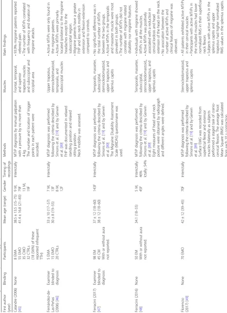
Blindi ng Participants Mean age (range) Gender Timi ng of recordings Method s Musc les Main fin dings Alons o-Blanco (2011) [ 62 ] None 20 CTTH adu lt patien ts 20 CTTH adolescent patien ts 41 (18 – 47) 8( 6 – 12) 10 M, 10F 10 M, 10F Interi ctally MTrP dia gnosis as de scribed by Simons et al. [ 19 ] Temp oralis, subo ccipital, sternoc leidom astoid, and upper trapezi us • Th en u m b e ro f ac ti veM Tr P s w e re hi gher in ad ul ts versus chil d ren. • Refe rred p ain el icite d from ac ti ve MT rPs shared simi lar pa in pa tterns as sp ont an e o us C TT H in bo th gr ou ps . No si gnifi cant associa tion b e twee n th en u m b e ro fa ct iv eM Tr P s an d head ache p ar ameter s. Couppé (2007) [ 67 ] Do uble-blin ded 20 CTTH patien ts 20 CTRLs 37.5 (33.3 – 41.6 ) Not reported Ictally MTrP dia gnosis as de scribed by Simons et al. [ 19 ] EMG exa minatio n at a MT rP and a control point in the same sub ject. Upp er trapezius • Th en u m b e ro f ac ti veM Tr P s w e re higher in pa tients ver sus controls • No differ ence in electromyogra phic act ivity between MTrPs versus cont rol points.
Fernández- de-las- Peñas (2011)
[ 63 ] Exa miner blin ded to dia gnosis 50 CTTH patien ts 50 CTRLs 8( 6 – 12) 14 M, 36F Interi ctally MTrP dia gnosis as de scribed by Simons et al. [ 19 ]. Temp oralis, super ior oblique, masse ter, subo ccipital, sternoc leidom astoid, levator scapu lae, an d upper trapezius • Acti ve MTrP s were only foun d in pat ients. • In the C TTH p ati ents, the num b er of active TrPs correl ated wi th the dura tion of a h eadache atta ck. • The local and referred pai ns el icited from activ e MTrP s share d simil ar pai n pattern as spont aneou s CT TH.
Fernández- de-las- Peñas (2009)
[ 68 ] None 40 CTTH 40 (20 – 57) 40F Interi ctally < 4 on a 11 NRS MTrP dia gnosis was performed followi ng the cri teria desc ribed by Simons et al. [ 19 ] and Gerwin et al. [ 89 ] PPT was assesse d using an algome ter. Temp oralis (9 landmar ks total, 3 eac h res pectively in the anterior, medial and poste rior part) • The analysis o f va ria nce d id not detect sign ificant dif fe rences in the referred p ain pattern b etwee n active MTrPs. • Th e to p og ra p h ic al pr es su re pa in sensitivity m aps showed the d istinc t dist ribution of the M TrPs in di cate d b y locations wit h low P P Ts.
Fernández- de-las- Peñas (2007)
[ 69 ] Exa miner blin ded to dia gnosis 15 ETTH 15 CTRLs 39 ± 17 (20 – 70) 37 ± 12 (21 – 70) 3 M, 12F 4 M, 11F Interi ctally MTrP dia gnosis as de scribed by Simons et al. [ 19 ] and Gerwin et al. [ 89 ] FHP was note d bot h seated an d standing. Tem p orali s, ste rnocleidomastoid, an d u p p er tr ap ezius • Acti ve MTrP s in the affecte d mu scles were onl y found within the ETT H group. • MTrP s we re not related to any clinic al variable conc ernin g the inten sity and the te mporal profile of headache.
Fernández- de-las- Peñas (2007)
Table 2 Tension-type headache and myofascial trigger points (Continued)
First author (year)
Blindi ng Participants Mean age (range) Gender Timi ng of recordings Method s Musc les Main fin dings
Fernández- de-las- Peñas (2007)
[ 66 ] Exa miner blin ded to dia gnosis 30 CTTH 30 CTRLs 39 ± 16 (18 – 65) 39 ± 12 (19 – 65) 9 M, 21F 9 M, 21F < 4 cm on a1 0c m VAS MTrP dia gnosis as de scribed by Simons et al. [ 19 ] and by Gerwin et al. [ 89 ] Temp oralis • Ref erred pain was evok ed in 87 and 54% o n the domin ant and n on -domi nant sides in CT TH patients, which was si gnificantly h igher than in controls (1 0% vs. 17% ,r espec tively ). • CTTH patien ts wit h activ e MTrP s in ei ther right or le ft temp oralis mu scle showe d longe r head ache dur ation than thos e with latent MTrP s. • CTTH patien ts sho wed significan tly lowe r pre ssure pai n threshol d whe n compared with cont rols.
Fernández- de-las- Peñas (2006)
[ 71 ] Exa miner blin ded to dia gnosis 10 ETTH 10 CTRLs 35 ± 15 (18 – 66) 34 ± 13 (18 – 66) 2M ,8 F 3M ,7 F Interi ctally MTrP dia gnosis as de scribed by Simons et al. [ 19 ] and by Gerwin et al. [ 89 ] Suboc cipital • In the ETTH group, 60% showe d activ e MTrP s; 40% showe d lat ent trigger point s. In the ETT H group, head ache intens ity, frequency and dur ation did not differ de pending on wh ether the MTrP s we re activ e or latent .
Fernández- de-las- Peñas (2006)
[ 72 ] Exa miner blin ded to dia gnosis 25 CTTH 25 CTRLs 40 ± 16 (18 – 72) 38 ± 9 (18 – 73) 8 M, 17F 9 M, 16F < 4 cm on a1 0c m VAS MTrP dia gnosis was performed followi ng the cri teria desc ribed by Simons et al. [ 19 ] and by Gerwin et al. [ 89 ] FHP was note d bot h seated an d standing. Tem p orali s, ster nocleidomastoid, and up per tra pe zius • Acti ve MTrP s were only foun d in CT TH patie nts. • There w as significant association b et w ee n th ep re se n ceo f ac tiv eM Tr P s and h eadache intensity and d u ration.
Fernández- de-las- Peñas (2006)
[ 65 ] Exa miner blin ded to dia gnosis 20 CTTH 20 CTRLs 38 ± 18 (18 – 70) 35 ± 10 (20 – 68) 9 M, 11F 12 M, 8F Pain intens ity < 4 on a 10 cm VAS MTrP dia gnosis was performed followi ng the cri teria desc ribed by Simons et al. [ 19 ] and by Gerwin et al. [ 89 ] FHP was note d bot h seated an d standing. Suboc cipital • Acti ve MTrP s were only foun d in CT TH patie nts. • CTTH patien ts wit h activ e MTrP s rep orted gre ater headache inten sity and frequ ency than thos e with latent . • A craniovertebra l small er angle was posi tively corr elated with increased head ache frequency and negatively corr elated with headache dur ation .
Fernández- de-las- Peñas (2005)
[ 64 ] Exa miner blin ded to dia gnosis 15 CCTH 15 ETTH 15 CTRLs 37 ± 16 38 ± 14 38 ± 14 Range not reported 5 M, 10F 4 M, 11F 5 M, 10F CTTH: Pai n intens ity < 4 cm on a1 0c m
VAS TTH: Interi
Table 2 Tension-type headache and myofascial trigger points (Continued)
First author (year)
Table
2
Tension-type
headache
and
myofascial
trigger
points
(Continued)
First author (year)
Blindi
ng
Participants
Mean
age
(range)
Gender
Timi
ng
of
recordings
Method
s
Musc
les
Main
fin
dings
Palacios- Ceña (2016)
[
78
]
Exa
miner
blin
ded
to dia
gnosis
77
CTTH
80
ETTH
46
(42
–
50)
47
(43
–
51)
46
M,
111F
Interi
ctally
MTrP
dia
gnosis
was
performed
followi
ng
the
cri
teria
desc
ribed
by
Simons
et
al.
[
19
]
PPT
was
assesse
d
over
the
trigemi
nal
area,
extra-trigemin
al
area
an
d
two
distant
pain
free
points
usi
ng
an
algome
ter.
Temp
oralis,
mas
seter,
subo
ccipital,
sternoc
leidom
astoid,
spleni
us
capitis
,
and
upper
trap
ezius
•
No
differenc
e
in
numbe
r
of
MTrPs
an
d
PPT
in
the
tw
o
groups.
•
There
was
a
significan
t
negati
ve
corr
elatio
n
betwee
n
the
numbe
r
of
trigger
point
s
(act
ive
or
lat
ent)
and
PP
T.
Romero- Morales (2017)
[
79
]
None
60
ETTH
60
CTRLs
38,30
±
10,05
34
±
8,20
Range
not
reported
24
M,
32F
27
M,
33F
Not repo
rted
MTrP
dia
gnosis
was
performed
followi
ng
the
cri
teria
desc
ribed
by
Simons
et
al.
[
19
]
PPT
was
assesse
d
using
an
algome
ter.
Temp
oralis
and
upp
er
trapezius
Min
imum
clinical
differ
ences
in
PP
T
betw
een
TTH
and
CTRLs
were
•
Right
upper
trapezius
;
0,85
kg/cm
2
•
Left
upper
trapezius;
0;76
kg/c
m
2
•
Right
temporalis;
0;16
kg/cm
2
•
Left
temp
orals;
0,17
kg/cm
2
Sohn (2012)
[
80
]
Exa
miner
blin
ded
to dia
gnosis
23
CTTH
36
ETTH
42
CTRLs
53.43
±
16.97
51.11
±
14.42
51.69
±
16.18
Range
not
reported
2
M,
21F
7
M,
29F
8
M,
34F
Hea
dache
intens
ity
<
3
on
a
10
cm
VAS
MTrP
dia
gnosis
was
performed
followi
ng
the
cri
teria
desc
ribed
by
Simons
et
al.
[
19
]
and
by
Gerwin
et
al.
[
89
]
FHP
was
used
to
eva
luate
posture
abnorm
alities.
Measurement
of
neck
mob
ility
was
used
to
evaluate
me
chanical
abnorm
alities.
Temp
oralis,
subo
ccipital,
sternoc
leidom
astoid,
and
upper
trapezius
•
The
num
ber
of
activ
e
MTrP
s
was
sign
ificantly
greater
in
CT
TH
subje
cts
than
in
ET
TH
subjects.
•
The
num
ber
of
activ
e
MTrP
s
we
re
corr
elated
with
the
frequency
and
dur
ation
of
head
ache.
•
No
correlations
were
obse
rved
for
FHP
or
neck
mob
ility.
CTTH
chronic
tension-type
headache,
ETTH
episodic
tension-type
headache,
CTRLs
healthy
controls,
F
female,
M
male,
MTrP
myofascial
trigger
point,
EMG
electromyography,
PPT
pressure
pain
threshold,
FHP
frontal
head
position,
VAS
visual
analog
scale,
NRS
numeric
rating
temporalis muscles correlated with longer headache
dur-ation and greater headache intensity, respectively [72].
Suboccipital active MTrPs correlated with increased
in-tensity and frequency of headache [65]. Chronic TTH
pa-tients with active MTrPs in the analyzed muscles had a greater forward head position than those subjects only
with latent MTrPs [65, 72]. Sohn et al. [80] identified a
greater occurrence of MTrPs in chronic TTH compared to episodic TTH and that the number of active MTrPs correlated with the frequency and duration of headache, although they did not find any correlations for forward head posture and neck mobility in contrast to
Fernández--de-las-Peñas et al. [65,72].
Pressure pain threshold
The number of active and latent MTrPs was signifi-cantly and negatively associated with pressure pain thresholds on the temporalis muscle, C5/C6 zygapo-physeal joint, second metacarpal, and tibialis anterior
muscle [78]. Thus, a higher number was associated
with a more generalized sensitization regardless of the frequency of headache. Another study observed that the location of active MTrPs in the temporalis muscle corresponded to areas with lower pain pressure thresholds which establishes a relationship between
the two [68]. The same group found that chronic
TTH patients with bilateral active MTrPs in the tra-pezius muscles have a significantly lower pain
pres-sure threshold compared to patients with only
unilateral active MTrPs [70]. Minimum clinical
differ-ences in pressure pain thresholds in TTH patients
may be used to evaluate treatment of MTrPs [79].
Therapeutic studies targeting myofascial trigger points
Karadas et al. [81] investigated pericranial lidocaine
injections in MTrPs in 108 patients with frequent epi-sodic TTH using a double-blind placebo-controlled randomized study design. Repeated local lidocaine injections into the MTrPs in the pericranial muscles reduced both the frequency and intensity of pain compared to placebo. Another placebo-controlled study found similar results with lidocaine injections in MTrPs in chronic TTH with a reduction in pain frequency, pain
in-tensity, and analgesic use [74]. In addition, there was a
sig-nificant effect on anxiety and depression of the subjects. A randomized, double-blind, placebo-controlled pilot study of botulinum toxin A injections in MTrPs included 23
pa-tients with chronic TTH [73]. The subjects were assessed
at 2 weeks, 1, 2 and 3 months after injection. The botu-linum toxin A group reported a reduction in headache fre-quency that disappeared by week 12. There was no difference in intensity between the groups. In a random-ized, placebo-controlled clinical trial, Moraska et al. ap-plied massage focused on MTrPs of patients with TTH
[77]. For both active and placebo groups, there was a
de-crease in headache frequency, but not for intensity or dur-ation. Thus, there was no difference between massage and
placebo [81].
Discussion
Ultrasound and EMG appear to be the most promis-ing modalities to be used as a diagnostic test for MTrPs. While the use of ultrasound in headache
dis-orders has primarily been focused on vascular
changes and not on myofascial structures [82],
ultra-sound may also be used to identify MTrPs if specific
analysis methods are applied [29] or with the use of
elastography [27, 30, 32]. However, there is no precise
description of a gold standard using these techniques, and they have yet to be evaluated in headache pa-tients. Active MTrPs affect the electrical activity at rest and during muscle contraction in EMG studies
[33–36]. Out of the two modalities, ultrasound is
pre-sumably the most viable candidate as a diagnostic test as it has an immediate availability at most treatment sites, it is time-efficient and is non-invasive. Although there are currently no studies investigating if it is possible to identify MTrPs with ultrasound without prior manual palpation. Future studies should investi-gate if ultrasound is comparable with manual palpa-tion in identifying MTrPs. The other modalities do not appear to be suitable as microdialysis show mixed results regarding whether the local milieu of MTrPs is changed and needs further exploration before a
conclusion can be made [25, 26, 83]. According to
the review by Dibai-Filho et al. [37], infrared
therm-ography appears to be a promising non-invasive method, but it should still only be used as an auxil-iary tool in the evaluation of MTrPs due to
conflict-ing results. Magnetic resonance elastography in
diagnosing MTrPs has only been investigated in a few studies, and the sensitivity may be too low for
suit-able use as a diagnostic test [42].
Studies show a high occurrence of active and latent
MTrPs [45–49] and correlation between neck mobility
and MTrPs in migraine patients [46, 48, 49, 58].
How-ever, there are conflicting results in which muscles are
the most affected [47, 48], and it is unclear whether
there is a positive correlation with the degree of head-ache frequency or intensity due to conflicting results. Palpation of MTrPs may provoke a migraine attack in
some patients [45, 53] but needs further confirmation
in placebo-controlled studies. Intervention studies
tar-geting MTrPs are mostly positive [50–52, 56, 57], but
they lack placebo-control. Thus, a bottom-up
associ-ation between MTrPS and migraine [44] cannot be fully
supported based on the evidence (Fig. 1). In addition,
has been shown that migraine attacks exacerbate fibro-myalgia symptoms, suggesting a top-down central
sensitization [84] as fibromyalgia symptoms include
specific tender points [85]. Although a study showed
migraine severity was similar in migraine patients with
and without fibromyalgia [86]. One would expect an
as-sociation between migraine severity and co-existing fibromyalgia if a top-down central is taking place in patients with this comorbidity. It is possible that MTrPs may have an important role in some subpopu-lations of migraine patients. This calls for therapeutic studies targeting patients with a high degree of MTrPs, but this is only speculative at this point.
The prevalence of active MTrPs in TTH [65–67,73,
74, 77, 78,80, 81] is coherent with the hypothesis that
peripheral mechanisms are involved in the
pathophysi-ology of TTH [14–16, 60]. It has been speculated that
an increased peripheral nociception increases the
sensitization of central mechanisms resulting in an
in-crease in the sensitivity to peripheral pain (Fig.1).
Ac-tive MTrPs may contribute to a central sensitization as they are correlated with lower pain pressure thresholds
[68, 70, 78]. This would also provide an explanation
for the efficacy of injections of lidocaine in MTrPs [74,
81] as these would reduce the transmission of
periph-eral nociception. However, these assumptions are in contrast with a study showing that the number of ac-tive MTrPs is higher in adults in comparison to adoles-cents, regardless of no significant association with
headache parameters [62]. This suggests that active
MTrPs are accumulated over time as a consequence of
TTH [62] instead of being an integrated part of the
pathophysiology of TTH. Previous studies of botu-linum toxin A injections in pericranial muscles have
been shown to have no effect in TTH [87]. The efficacy
of botulinum toxin A in MTrPs [73] might be
ex-plained by its possible action of modulating the release of nociceptive and inflammatory mediators e.g., CGRP
and SP [88]. These inflammatory mediators may be
in-creased in the local milieu of MTrPs [25, 26]. This
would also account for its poor efficacy in injection protocols targeting fixed landmarks in pericranial
mus-cles instead of MTrPs [87], as these substances appear
to be concentrated at MTrPs [25,26].
There are many overlapping findings in studies of MTrPs in migraine or TTH. In both disorders, MTrPs are prevalent and may be related to neck mobility. Pal-pation of MTrPs can, in some cases, provoke an attack in migraine patients, while palpation of MTrPs in TTH can provoke pain resembling the usual headache pat-tern of patients. Intervention studies are promising in both disorders. The quality of studies in both disorders varies greatly as many of the reviewed studies lacked
blinding (Table 3). Furthermore, true blinding is
diffi-cult to achieve as active MTrPs by definition cause re-ferred pain.
Conclusion
In conclusion, ultrasound elastography is the most
promising tool to assess MTrPs [27, 30, 32], but still
needs to be performed combined with palpation, which introduces risk of bias and interobserver vari-ation. MTrPs are very frequent in both migraine
pa-tients [45–49] and TTH patients [65–67, 73, 74, 77,
78, 80, 81] compared to healthy controls. Active
Table 3An overview on the use of blinding, control groups and placebo
Migraine Tension-type headache Total
Blinding 36% (5/14 relevant studies) 79% (15/19 relevant studies) 61% (19/33 relevant studies)
Control group 44% (4/9 relevant studies) 79% (11/14 relevant studies) 65% (15/23 relevant studies)
Placebo 40% (2/5 relevant studies) 80% (4/5 relevant studies) 60% (6/10 relevant studies) Fig. 1The bottom-up model states that increased peripheral
MTrPs are especially interesting as these are rarely found in control groups. However, their role in the pathophysiology of each disorder and to which degree is still unclear. The results of the provocation and
intervention studies support the hypothesis of a
trigemino-cervical-complex pathophysiology model in
both migraine [45,50–53, 56, 57] and TTH [73,74,81].
Whether MTrPs contribute to an increased disease
bur-den in migraine is uncertain [45,47,48] and needs further
exploration [50, 52]. Future research should aim to
in-crease the quality of studies before further speculations are made. To elucidate this, large-scale studies to stratify the headache populations into more homogenous sub-groups should be conducted.
Abbreviations
BK:Bradykinin; CGRP: Calcitonin gene-related peptide; CTRL: Healthy control;
EMG: Electromyography; F: Female; FHP: Forward head posture; IL-1β: Interleukin
1 beta; IL-6: Interleukin 6; IL-8: Interleukin 8; M: Male; MA: Migraine with aura; MO: Migraine without aura; MTrP: Myofascial trigger point; NE: Norepinephrine; NRS: Numeric rating scale; PPT: Pressure pain threshold; SP: Substance P;
TNF-α: Tumor necrosis factor alpha; TTH: Tension-type headache;
VAS: Visual analog scale
Funding
TPD and JH were funded by a grant from Candys Foundation. LTK was funded by a grant from the Lundbeck Foundation.
Availability of data and materials
Data sharing is not applicable to this article as no datasets were generated or analysed during the current study.
Authors’contributions
TPD contributed with data interpretation, drafting and revision of the manuscript for intellectual content. GFH, LTK and JH contributed with revision of the manuscript for intellectual content. HWS contributed with conceptualization, data interpretation and revision of the manuscript for intellectual content. All authors read and approved the final manuscript.
Ethics approval and consent to participate
Not applicable.
Consent for publication
Not applicable.
Competing interests
HWS has received travel grants or speaking fees from Pfizer, Autonomic Technologies and Novartis. TPD, GFH, LTK and JH declare that they have no competing interests.
Publisher’s Note
Springer Nature remains neutral with regard to jurisdictional claims in published maps and institutional affiliations.
Received: 21 August 2018 Accepted: 3 September 2018
References
1. Stovner LJ, Andree C (2010) Prevalence of headache in Europe: a review for
the Eurolight project. J Headache Pain. 11:289–299
2. Lyngberg AC, Rasmussen BK, Jørgensen T et al (2005) Secular changes in
health care utilization and work absence for migraine and tension-type
headache: a population based study. Eur J Epidemiol 20:1007–1014
3. Olesen J, Sobscki P, Truelsen T et al (2008) Cost of disorders of the brain in
Denmark. Nord J Psychiatry 62:114–120
4. Olesen J, Burstein R, Ashina M et al (2009) Origin of pain in migraine:
evidence for peripheral sensitisation. Lancet Neurol 8:679–690
5. Noseda R, Burstein R (2013) Migraine pathophysiology: anatomy of the
trigeminovascular pathway and associated neurological symptoms, CSD,
sensitization and modulation of pain. Pain 154:44–53
6. Shevel E (2011) The extracranial vascular theory of migraine--a great story
confirmed by the facts. Headache 51:409–417
7. Goadsby PJ (2009) The vascular theory of migraine--a great story wrecked
by the facts. Brain 132:6–7
8. Asghar MS, Hansen AE, Amin FM et al (2011) Evidence for a vascular factor
in migraine. Ann Neurol 69:635–645
9. Amin FM, Asghar MS, Hougaard A et al (2013) Magnetic resonance angiography
of intracranial and extracranial arteries in patients with spontaneous migraine
without aura: a cross-sectional study. Lancet Neurol 12:454–461
10. Hay KM (1979) Pain thresholds in migraine. Practitioner 222:827–833
11. Olesen J (1978) Some clinical features of the acute migraine attack. An
analysis of 750 patients. Headache 18:268–271
12. Olesen J (1991) Clinical and pathophysiological observations in migraine
and tension-type headache explained by integration of vascular, supraspinal
and myofascial inputs. Pain 46:125–132
13. GBD 2015 Neurological Disorders Collaborator Group (2017) Global,
regional, and national burden of neurological disorders during 1990–2015: a
systematic analysis for the Global Burden of Disease Study 2015. Lancet
Neurol 16:877–897
14. Lipchik GL, Holroyd KA, O’Donnell FJ et al (2000) Exteroceptive suppression
periods and pericranial muscle tenderness in chronic tension-type headache:
effects of psychopathology, chronicity and disability. Cephalalgia 20:638–646
15. Buchgreitz L, Lyngberg AC, Bendtsen L et al (2006) Frequency of headache
is related to sensitization: a population study. Pain 123:19–27
16. Fernández-de-Las-Peñas C, Cuadrado ML, Arendt-Nielsen L et al (2007)
Increased pericranial tenderness, decreased pressure pain threshold, and headache clinical parameters in chronic tension-type headache patients.
Clin J Pain 23:346–352
17. Sakai F, Ebihara S, Akiyama M et al (1995) Pericranial muscle hardness in
tension-type headache. A non-invasive measurement method and its
clinical application. Brain 118(Pt 2):523–531
18. Ashina M, Bendtsen L, Jensen R et al (1999) Muscle hardness in patients
with chronic tension-type headache: relation to actual headache state. Pain
79:201–205
19. Simons D, Travell J (1999) Travell & Simons’myofascial pain and dysfunction:
the trigger point manual. Williams & Wilkins, Baltimore
20. Travell J, Simons D (1952) The myofascial genesis of pain. Postgrad Med 11:
434–452
21. Stockman R (1904) The causes, pathology and treatment of chronic
rheumatism. Edinburgh Med J 15:107–116
22. Shah JP, Thaker N, Heimur J et al (2015) Myofascial trigger point then and
now: a historical and scientific prespective. PM R J 7:746–761
23. Wolfe F, Simons D, Fricton J et al (1992) The fibromyalgia and myofascial
pain syndromes: a preliminary study of tender points and trigger points in persons with fibromyalgia, myofascial pain syndrome and no disease.
J Rheumatol 19:944–951
24. Fernández-De-Las-Peñas C, Dommerholt J Myofascial trigger points:
peripheral or central phenomenon? Curr Rheumatol Rep 16. Epub ahead of
print 2014.https://doi.org/10.1007/s11926-013-0395-2
25. Shah JP, Danoff JV, Desai MJ et al (2008) Biochemicals associated with pain
and inflammation are elevated in sites near to and remote from active
myofascial trigger points. Arch Phys Med Rehabil 89:16–23
26. Shah JP, Phillips TM, Danoff JV et al (2005) An in vivo microanalytical
technique for measuring the local biochemical milieu of human skeletal
muscle. J Appl Physiol 99:1977–1984
27. Sikdar S, Shah JP, Gebreab T et al (2009) Novel applications of ultrasound
technology to visualize and characterize myofascial trigger points and
surrounding soft tissue. Arch Phys Med Rehabil 90:1829–1838
28. Lewis J, Tehan P (1999) A blinded pilot study investigating the use of diagnostic
ultrasound for detecting active myofascial trigger points. Pain 79:39–44
29. Turo D, Otto P, Shah JP et al (2012) Ultrasonic tissue characterization of the
upper trapezius muscle in patients with myofascial pain syndrome. Conf
Proc IEEE Eng Med Biol Soc 2012:4386–4389
30. Sikdar S, Shah JP, Gilliams E et al (2008) Assessment of myofascial trigger
points (MTrPs): a new application of ultrasound imaging and vibration
31. Ballyns JJ, Shah JP, Hammond J et al (2011) Objective sonographic measures for characterizing myofascial trigger points associated with
cervical pain. J Ultrasound Med 30:1331–1340
32. Takla MKN, Razek NMA, Kattabei O et al (2016) A comparison between
different modes of real-time sonoelastography in visualizing myofascial
trigger points in low back muscles. J Man Manip Ther 24:253–263
33. Simons DG, Hong C-Z, Simons LS (2002) Endplate potentials are common
to midfiber myofacial trigger points. Am J Phys Med Rehabil 81:212–222
34. Ge HY, Monterde S, Graven-Nielsen T et al (2014) Latent myofascial
trigger points are associated with an increased intramuscular electromyographic activity during synergistic muscle activation. J Pain
15:181–187
35. Yu SH, Kim HJ (2015) Electrophysiological characteristics according to
activity level of myofascial trigger points. J Phys Ther Sci 27:2841–2843
36. Wytrążek M, Huber J, Lipiec J et al (2015) Evaluation of palpation, pressure
algometry, and electromyography for monitoring trigger points in young
participants. J Manip Physiol Ther 38:232–243
37. Dibai-Filho AV, Guirro RR (2015) Evaluation of myofascial trigger points
using infrared thermography: a critical review of the literature. J Manip
Physiol Ther 38:86–92
38. Merla A, Ciuffolo F, D’Attilio M et al (2004) Functional infrared imaging in the
diagnosis of the myofascial pain. Conf Proc IEEE Eng Med Biol Soc 2:1188–1191
39. Kimura Y, Ge H-Y, Zhang Y et al (2009) Evaluation of sympathetic
vasoconstrictor response following nociceptive stimulation of latent
myofascial trigger points in humans. Acta Physiol (Oxf) 196:411–417
40. Zhang Y, Ge H-Y, Yue S-W et al (2009) Attenuated skin blood flow response
to nociceptive stimulation of latent myofascial trigger points. Arch Phys
Med Rehabil 90:325–332
41. Haddad DS, Brioschi ML, Arita ES (2012) Thermographic and clinical
correlation of myofascial trigger points in the masticatory muscles.
Dentomaxillofac Radiol 41:621–629
42. Chen Q, Wang H, Gay RE et al (2016) Quantification of myofascial taut
bands. Arch Phys Med Rehabil 97:67–73
43. Eller-Smith OC, Nicol AL, Christianson JA (2018) Potential mechanisms
underlying centralized pain and emerging therapeutic interventions. Front Cell Neurosci 12:35
44. Tfelt-Hansen P, Lous I, Olesen J (1981) Prevalence and significance of
muscle tenderness during common migraine attacks. Headache 21:49–54
45. Calandre EP, Hidalgo J, García-Leiva JM et al (2006) Trigger point evaluation
in migraine patients: an indication of peripheral sensitization linked to
migraine predisposition? Eur J Neurol 13:244–249
46. Fernández-de-Las-Peñas C, Cuadrado ML, Pareja JA (2006) Myofascial trigger
points, neck mobility and forward head posture in unilateral migraine.
Cephalalgia 26:1061–1070
47. Ferracini GN, Florencio LL, Dach F et al (2017) Myofascial trigger points and
migraine-related disability in women with episodic and chronic migraine.
Clin J Pain 33:109–115
48. Ferracini GN, Chaves TC, Dach F et al (2016) Relationship between active
trigger points and head/neck posture in patients with migraine. Am J Phys
Med Rehabil. 95:831–839
49. Florencio LL, Ferracini GN, Chaves TC et al (2017) Active trigger points
in the cervical musculature determine the altered activation of superficial neck and extensor muscles in women with migraine. Clin J
Pain 33:238–245
50. Gandolfi M, Geroin C, Valè N et al Does myofascial and trigger point
treatment reduce pain and analgesic intake in patients undergoing OnabotulinumtoxinA injection due to chronic intractable migraine? A pilot, single-blind randomized controlled trial. Eur J Phys Rehabil Med. Epub
ahead of print 27 July 2017.https://doi.org/10.23736/S1973-9087.17.04568-3
51. Ghanbari A, Askarzadeh S, Petramfar P et al (2015) Migraine responds better
to a combination of medical therapy and trigger point management than
routine medical therapy alone. NeuroRehabilitation 37:157–163
52. Giamberardino MA, Tafuri E, Savini A et al (2007) Contribution of myofascial
trigger points to migraine symptoms. J Pain 8:869–878
53. Landgraf MN, Biebl JT, Langhagen T et al Children with migraine:
provocation of headache via pressure to myofascial trigger points in the trapezius muscle? - a prospective controlled observational study. Eur J Pain.
Epub ahead of print 26 September 2017.https://doi.org/10.1002/ejp.1127
54. Landgraf MN, Ertl-Wagner B, Koerte IK et al (2015) Alterations in the
trapezius muscle in young patients with migraine--a pilot case series with
MRI. Eur J Paediatr Neurol 19:372–376
55. Palacios-Ceña M, Ferracini GN, Florencio LL et al (2017) The number of
active but not latent trigger points associated with widespread pressure pain hypersensitivity in women with episodic migraines. Pain Med 18:
2485–2491
56. Ranoux D, Martiné G, Espagne-Dubreuilh G et al (2017) OnabotulinumtoxinA
injections in chronic migraine, targeted to sites of pericranial myofascial pain: an observational, open label, real-life cohort study. J Headache Pain. 18:75
57. Sollmann N, Trepte-Freisleder F, Albers L et al (2016) Magnetic stimulation
of the upper trapezius muscles in patients with migraine - a pilot study. Eur
J Paediatr Neurol 20:888–897
58. Tali D, Menahem I, Vered E et al (2014) Upper cervical mobility, posture and
myofascial trigger points in subjects with episodic migraine: case-control
study. J Bodyw Mov Ther 18:569–575
59. Affaitati G, Martelletti P, Lopopolo M et al (2017) Use of nonsteroidal
anti-inflammatory drugs for symptomatic treatment of episodic headache. Pain
Pract 17:392–401
60. Jensen RH (2017) Tension-type headache - the normal and most prevalent
headache. Headache:1–7
61. Kolding LT, Do TP, Ewertsen C et al (2018) Muscle stiffness in tension-type
headache patients with pericranial tenderness. Cephalalgia Reports 1: 251581631876029
62. Alonso-Blanco C, Fernández-de-las-Peñas C, Fernández-Mayoralas DM et al
(2011) Prevalence and anatomical localization of muscle referred pain from active trigger points in head and neck musculature in adults and children
with chronic tension-type headache. Pain Med 12:1453–1463
63. Fernández-de-las-Peñas C, Fernández-Mayoralas DM, Ortega-Santiago R et al
(2011) Referred pain from myofascial trigger points in head and neck-shoulder muscles reproduces head pain features in children with chronic
tension type headache. J Headache Pain. 12:35–43
64. Fernández-de-las-Peñas C, Cuadrado ML, Gerwin RD et al (2005) Referred
pain from the trochlear region in tension-type headache: a myofascial
trigger point from the superior oblique muscle. Headache 45:731–737
65. Fernández-de-las-Peñas C, Alonso-Blanco C, Cuadrado ML et al (2006)
Trigger points in the suboccipital muscles and forward head posture in
tension-type headache. Headache 46:454–460
66. Fernández-de-Las-Peñas C, Ge H-Y, Arendt-Nielsen L et al (2007) The local and
referred pain from myofascial trigger points in the temporalis muscle contributes
to pain profile in chronic tension-type headache. Clin J Pain 23:786–792
67. Couppé C, Torelli P, Fuglsang-Frederiksen A et al (2007) Myofascial trigger
points are very prevalent in patients with chronic tension-type headache: a
double-blinded controlled study. Clin J Pain 23:23–27
68. Fernández-de-las-Peñas C, Caminero AB, Madeleine P et al (2009) Multiple
active myofascial trigger points and pressure pain sensitivity maps in the temporalis muscle are related in women with chronic tension type
headache. Clin J Pain 25:506–512
69. Fernández-de-Las-Peñas C, Cuadrado ML, Pareja JA (2007) Myofascial trigger
points, neck mobility, and forward head posture in episodic tension-type
headache. Headache 47:662–672
70. Fernández-de-Las-Peñas C, Ge H-Y, Arendt-Nielsen L et al (2007) Referred
pain from trapezius muscle trigger points shares similar characteristics with
chronic tension type headache. Eur J Pain 11:475–482
71. Fernández-de-Las-Peñas C, Alonso-Blanco C, Cuadrado ML et al (2006)
Myofascial trigger points in the suboccipital muscles in episodic
tension-type headache. Man Ther 11:225–230
72. Fernández-de-Las-Peñas C, Alonso-Blanco C, Cuadrado ML et al (2006)
Myofascial trigger points and their relationship to headache clinical
parameters in chronic tension-type headache. Headache 46:1264–1272
73. Harden RN, Cottrill J, Gagnon CM et al (2009) Botulinum toxin a in the
treatment of chronic tension-type headache with cervical myofascial trigger points: a randomized, double-blind, placebo-controlled pilot study.
Headache 49:732–743
74. KaradaşÖ, Inan LE, UlaşÜH et al (2013) Efficacy of local lidocaine
application on anxiety and depression and its curative effect on patients
with chronic tension-type headache. Eur Neurol 70:95–101
75. Lattes K, Venegas P, Lagos N et al (2009) Local infiltration of gonyautoxin is
safe and effective in treatment of chronic tension-type headache. Neurol
Res 31:228–233
76. Moraska AF, Schmiege SJ, Mann JD et al (2017) Responsiveness of
myofascial trigger points to single and multiple trigger point release massages: a randomized, placebo controlled trial. Am J Phys Med Rehabil.
77. Moraska AF, Stenerson L, Butryn N et al (2015) Myofascial trigger point-focused head and neck massage for recurrent tension-type headache: a
randomized, placebo-controlled clinical trial. Clin J Pain 31:159–168
78. Palacios-Ceña M, Wang K, Castaldo M, et al. Trigger points are associated
with widespread pressure pain sensitivity in people with tension-type
headache. Cephalalgia. Epub ahead of print 14 November 2016.https://doi.
org/10.1177/0333102416679965
79. Romero-Morales C, Jaén-Crespo G, Rodríguez-Sanz D et al (2017)
Comparison of pressure pain thresholds in upper trapezius and temporalis muscles trigger points between tension type headache and healthy
participants: a case-control study. J Manip Physiol Ther 40:609–614
80. Sohn J, Choi H, Lee S-M et al (2010) Differences in cervical musculoskeletal
impairment between episodic and chronic tension-type headache.
Cephalalgia 30:1514–1523
81. KaradaşÖ, Gül HL, Inan LE (2013) Lidocaine injection of pericranial
myofascial trigger points in the treatment of frequent episodic tension-type headache. J Headache Pain 14:44
82. Schytz HW, Amin FM, Selb J et al (2017) Non-invasive methods for
measuring vascular changes in neurovascular headaches. J Cereb Blood Flow Metab 271678X17724138
83. Ashina M, Stallknecht B, Bendtsen L et al (2003) Tender points are not sites
of ongoing inflammation - In vivo evidence in patients with chronic
tension-type headache. Cephalalgia 23:109–116
84. Giamberardino MA, Affaitati G, Martelletti P et al (2015) Impact of migraine
on fibromyalgia symptoms. J Headache Pain. 17:28
85. Wolfe F, Smythe HA, Yunus MB et al (1990) The American College of
Rheumatology 1990 criteria for the classification of fibromyalgia. Report of
the multicenter criteria committee. Arthritis Rheum 33:160–172
86. Ifergane G, Buskila D, Simiseshvely N et al (2006) Prevalence of fibromyalgia
syndrome in migraine patients. Cephalalgia 26:451–456
87. Jackson JL, Kuriyama A, Hayashino Y (2012) Botulinum toxin a for
prophylactic treatment of migraine and tension headaches in adults: a
meta-analysis. JAMA 307:1736–1745
88. Do TP, Hvedstrup J, Schytz HW (2018) Botulinum toxin: a review of the
mode of action in migraine. Acta Neurol Scand 137:442–451
89. Gerwin RD, Shannon S, Hong CZ et al (1997) Interrater reliability in myofascial