R E V I E W
Open Access
New frontiers for platelet CD154
Antoine Dewitte
1,4, Annabelle Tanga
1, Julien Villeneuve
2,3, Sébastien Lepreux
1, Alexandre Ouattara
4,
Alexis Desmoulière
5, Christian Combe
1,6and Jean Ripoche
1*Abstract
The role of platelets extends beyond hemostasis. The pivotal role of platelets in inflammation has shed new light
on the natural history of conditions associated with acute or chronic inflammation. Beyond the preservation of
vascular integrity, platelets are essential to tissue homeostasis and platelet-derived products are already used in the
clinics. Unanticipated was the role of platelets in the adaptative immune response, allowing a renewed conceptual
approach of auto-immune diseases. Platelets are also important players in cancer growth and dissemination.
Platelets fulfill most of their functions through the expression of still incompletely characterized membrane-bound
or soluble mediators. Among them, CD154 holds a peculiar position, as platelets represent a major source of CD154
and as CD154 contributes to most of these new platelet attributes. Here, we provide an overview of some of the
new frontiers that the study of platelet CD154 is opening, in inflammation, tissue homeostasis, immune response,
hematopoiesis and cancer.
Keywords:
Platelets, CD154
Introduction
Platelets are cytoplasmic fragments released in the
blood-stream during the fragmentation of polyploid
megaka-ryocytes (MK), a phenomenon critically dependent on
thrombopoietin [1-3]. The mammalian platelet is thought
to result from a phylogenic trend to ensure hemostasis
under high vascular shear forces; indeed, it can specifically
form arterial thrombi sustaining high shear stress [4]. It is
thought that the platelet coopted attributes of a
nucle-ated cell ancestor endowed with a multifunctional role
in coagulation, inflammation and defense against
infec-tions [5,6]. Platelets have a short lifespan, of around
7 days; mechanisms responsible for their clearance are
ill-understood; lectin-carbohydrate recognition of aged
and damaged platelets by splenic and liver macrophages
and hepatocytes is emphasized [7]. The best-defined
func-tion of platelets is hemostasis. Disrupfunc-tion of the
endothe-lial cell (EC) lining leads to platelet activation, platelet
adherence and aggregation which temporarily plug the
damaged vessel. In this process, platelets also drive and
confine coagulation at sites of tissue damage. Indeed,
defi-ciencies in platelet production or function are associated
to bleeding disorders, while increases in platelet number
or gain of function are associated to thrombosis. The
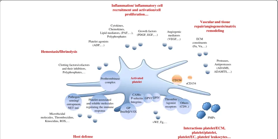
role of platelets in health and disease extends beyond
hemostasis; non-hemostatic platelet functions include
inflammation, innate and adaptative immune responses
and tissue homeostasis (Figure 1). Decisive advances in
understanding platelet function have been made through
the characterization of platelet receptors and their ligands
and platelet-derived mediators [8]. Among platelet
me-diators, CD154, the ligand of CD40, has attracted
spe-cific attention as it orchestrates many of these new
platelet attributes.
CD154
CD154, the CD40 ligand, a member of the Tumor
Necro-sis Factor (TNF) family, is central to the immune response
[9,10]. CD154 was discovered as mediating humoral
im-munity and was originally considered to be restricted to
activated helper T cells. The CD154/CD40 interaction
drives B cell proliferation, antibody production and
iso-type switching and is involved in thymic selection. This
interaction is required for B memory cell generation and
germinal center formation. Accordingly, CD154 deficiency
is associated with an impairment of the humoral
im-mune response to T-cell dependent antigens, including
defective immunoglobulin class switching; patients with
the X-linked hyper-IgM syndrome caused by mutations
* Correspondence:[email protected]1INSERM U1026, and Université de Bordeaux, F-33000 Bordeaux, France
Full list of author information is available at the end of the article
of the
CD154
gene, generally present low serum IgG
and IgA, but normal or increased serum IgM, and are
susceptible to opportunistic infections. Mice with a
dis-rupted
Cd154
gene fail to undergo isotype switching to
T-cell dependent antigens while normally responding to
T-cell independent antigens. In line with its regulatory
role on the adaptative immune response, the CD40/
CD154 interaction contributes to autoimmune disorders
in a number of animal models [11-15]. Manipulation of
the CD154/CD40 interaction has been used in efforts to
develop novel strategies in autoimmune diseases, results
in animal models being encouraging [13]. Clinical trials
have been launched with humanized anti-CD154
mono-clonal antibodies. Clinical interest of this strategy
re-mains mixed, and is strongly limited by thrombotic
complications [12-14].
Apart from B cells, CD40 is expressed by various cells,
including dendritic cells (DC), monocytes, T
lympho-cytes, EC, a variety of epithelial cells, smooth muscle
cells, fibroblasts; its expression is low in basal conditions
and is stimulated by inflammatory mediators [16-19].
CD40 expression is increased by CD154, however it is not
known whether this induction is direct or indirect [20,21].
CD40 is not the sole receptor for CD154; alternative
receptors have been described, such as integrins
α
5
β
1,
α
IIb
β
3 and
α
M
β
2; CD154 binding depends on their
activation states [22-25]. These additional receptors are of
significance in the pathophysiology of atherogenesis and
are important to consider when comparing CD40- and
CD154-deficient mouse phenotypes.
CD154 is a transmembrane protein and a proteolytic
soluble form, sCD154, which keeps the CD40-binding
do-main, is released by a partially understood mechanism.
The release of sCD154 was first documented in activated
T-lymphocytes [26]. CD154 has a trimeric configuration,
required for functional activity [27-30]. A complex
signal-ing cascade is triggered by CD40 ligation, involvsignal-ing TNF
receptor-associated factors (TRAF) as proximal
transdu-cing signal initiators [10,20]. Several signaling pathways,
including nuclear factor-
κ
B (NF-
κ
B), c-Jun N-terminal
kinase (JNK) and p38 mitogen-activated protein kinase
pathways, are activated by CD40 ligation; however, there
is a differential outcome depending upon which TRAF
member binds preferentially, and which cell/conditions
Hemostasis/fibrinolysis
Clotting factors/cofactors and their inhibitors, Polyphosphates,…
Vascular and tissue repair/angiogenesis/matrix
remodeling Inflammation/ inflammatory cell
recruitment and activation/cell proliferation…
Cytokines, Chemokines, Lipid mediators, (PAF,…),
Polyphosphates
Growth factors
(PDGF, EGF,…) Angiogenicmediators (VEGF,...)
Host defense
Pathogen sensing/ entrapment,
NET ind. Microbicidal molecules, Thrombocidins,
Kinocidins, ROS,…
Platelet-associated and soluble molecules regulating the immune
response
Interactions platelet/ECM, platelet/platelet, platelet/EC, platelet/ leukocytes…
Activated
platelet sCD154
ECM constituents (Fn, Vn,…)
Proteases, Antiproteases
(ADAMS, ADAMTS,…)
Others (CD9..)
PMPs CAMs:
P-selectin Integrins.. GP Ibα/Ibβ/V/IX
GPVI Platelet agonists
(ADP,…)
vWF, Fg,… Prothrombinase
complex CD154
GPIV Thrombin /agonist receptors
are involved [31]; the binding of TRAF-6 is critical in
vascular inflammation and metabolic complications
as-sociated with obesity [32,33].
CD154 expression is also observed in natural killer
cells, DC, cells of the monocyte/macrophage lineage,
endothelial, smooth muscle and epithelial cells [20]. Basal
CD154 expression is very low, or undetectable, as in EC
and epithelial cells for example [34], and is increased by a
variety of stimuli, most notably inflammatory cytokines
[20]. This suggests that CD154 expression may mostly
have relevance when induced, as in inflammation. CD154
is also expressed by blood platelets, being cryptic in
un-stimulated platelets and rapidly exposed at the platelet
surface following platelet activation [35].
CD154 expression by platelets
The distribution of CD154 in platelets is partly
under-stood. CD154 was found in
α
-granules, as shown by
immunoelectron microscopy or quantitative
immuno-fluorescence approaches [36,37]. Accordingly, patients
presenting a Gray-platelet syndrome, are characterized
by platelets that lack
α
-granules, and do not release
CD154 upon activation [37]. CD154 is highly coclustered
with insulin growth factor in
α
-granules, the signification
of which is unknown [36]. One question is whether
CD154 is also cytosolic, as found in resting platelets [38].
Pre-mRNAs and mature mRNAs are present in platelets
and a functional spliceosome and translational apparatus
allow platelets to process them, in response to
platelet-activating signals [39,40]. Detecting CD154 mRNA by
RT-PCR in platelets is challenging because of purity issues.
However, CD154 mRNA was evidenced in mouse
plate-lets, introducing other potential regulatory layers of
CD154 expression by platelets [34].
When activated, platelets express a membrane form and
release a soluble form of CD154
Platelets are activated by immobilized or soluble agonists.
The activation-driven secretion of granule content is a
pri-mary phenomenon [41-46]. Platelets also synthetize
media-tors, including interleukin-1
β
, tissue factor (TF), fibrinogen,
thrombospondin, von Willebrand Factor,
α
IIb
β
3, through a
translational-dependent pathway triggered by platelet
acti-vation [47,48].
Soluble CD154 is released by an activation-driven
pro-teolytic mechanism. Agonists, including thrombin,
throm-bin receptor-agonist peptide, ADP or collagen, stimulate
CD154 expression at the platelet membrane and the
re-lease of sCD154; long-term platelet activation leads to
complete conversion of CD154 to sCD154 [38,49-53]. A
matrix metalloproteinase (MMP)-dependent proteolytic
event is involved. The involvement of MMPs, MMP-2
and/or MMP-9, [51,54-57], differs from the release of
sCD154 by activated T-cells, which involves ADAM10
and 17 [58]. A role for
α
IIb/
β
3 has been put forward, as
α
IIb/
β
3 antagonists inhibit sCD154 release and as
Glanzmann platelets show reduced sCD154 release rate
[53,54,59]. An interaction between
α
IIb/
β
3 and MMP-2
is involved [57]. The roles of NADPH activation and
reactive oxygen species (ROS) generation as well as
CD154 binding to platelet CD40 have been underlined
[50,60]. The particularity of sCD154 release may explain
its specific response to agonists and secretion kinetics
[38,53]; however, how sCD154 is released remains be
fully understood, as shown for example by the effects of
in-hibitors added after platelet activation, suggesting complex,
intra-platelet mechanisms [53]. A debate remains about
the parallel biological activities of platelet-derived soluble
and membrane-associated CD154; recombinant soluble
forms, particularly trimeric forms, are active [50,61-63].
Fi-nally, sCD154 activates platelets by itself, suggesting
feed-back amplification of its secretion [64,65].
The megakaryocytic origin of platelet CD154
The assembly and loading of granules mainly occur in MK;
granules are distributed in proplatelets via a
microtubule-dependent mechanism [2,66,67]. The main origin of
plate-let CD154 is likely to be the MK that express CD154
mRNA, as shown in MK derived by differentiation of
human and mouse hematopoietic progenitor cells and
in MK of immune thrombocytopenic purpura (ITP)
pa-tients [68,69]. CD154 mRNA expression is increased
upon MK differentiation [69]. CD154 protein is also found
in MK cell lines and in MK from ITP patients [38,68,69].
As for T cells, the calcium-dependent activation of nuclear
factor of activated T cells-c2 and the early growth
re-sponse transcription factor EGR-1 contribute to
CD154
gene activation in MK [69,70].
Translation from endogenous mRNAs contributes to
platelet content. Its significance in quiescent platelets is
unclear. However, pre-mRNA processing and mRNA
translation are driven by platelet activation [40,48,71]. The
contribution of such mechanism in CD154 expression
during platelet lifespan is unknown.
Platelets also carry mediators present in plasma and
pos-sibly concentrated and/or modified within platelets [72,73].
Fibrinogen, albumin, immunoglobulins, amino acids,
in-flammatory and angiogenic mediators including vascular
endothelial growth factor (VEGF), histamine or serotonin,
are among them. Soluble CD154 is not detected in
plate-lets, making unlikely its uptake from plasma.
reduced [78], again suggesting relationship between the
platelet count and circulating sCD154. However, there are
contrasting studies, and a correlation between the platelet
count and sCD154 is not always found [79,80].
Importantly, platelet activation is associated to elevated
sCD154 and, indeed, platelet activation markers correlate
with sCD154 in blood [81-83]. For this reason, serum
seems inappropriate to evaluate circulating sCD154; in fact,
sCD154 levels are higher in serum than in plasma, clotting
resulting in increased sCD154 generation [52,79,80,84-88].
Hence the importance of a preanalytical standardization of
blood samples processing, conditions such as temperature,
length of storage, centrifugation, interfering with
meas-urement [84,89]. Further, plasma/serum sCD154 may
correspond to a pool of free soluble and
microparticle-bound CD154 [84] and ELISA may not discriminate
between sCD154 and platelet microparticles
(PMP)-associated CD154 [90]. Circulating sCD154 is linked to
platelet activation state; in patients with recent
throm-botic events, plasma sCD154 correlates with platelet
count, but this correlation is not found in patients with
non-thrombotic, non-inflammatory conditions [84].
Fi-nally, in patients with cardiovascular conditions,
com-monly used drugs such as statins, interfere with sCD154
releasing, a point that has also to be considered [91-93].
The baseline presence of sCD154 in the plasma of
healthy subjects may be secondary to basal platelet
acti-vation, as in high shear stress flow areas [94]. PMP are
released upon platelet activation [95]. A functional
CD154 is expressed by PMP [63,96]. The importance of
the contribution of PMP-bound CD154, in comparison
with the
“
true
”
soluble CD154, to plasma sCD154 has
been emphasized [90]. Questions also remain on the fate
and half-life of sCD154 in blood and how the CD154
information can be delivered at distance from platelet
activation sites.
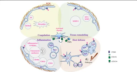
Platelet CD154: a critical mediator of the inflammatory
reaction
Platelets orchestrate a subtle balance between tissue injury
and repair; they are a key source of material for
reestab-lishing tissue homeostasis but they also contribute to
tis-sue injury. CD154 mediates several platelet functions in
tissue homeostasis (Figure 2).
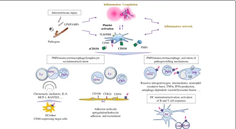
Platelet CD154 and inflammation
Regardless of its
cause, the inflammatory milieu is rich in platelet-activating
material, including chemokines [98]. The dialog between
EC and platelets in inflammation has been widely studied
as EC are primary platelet partners. Upon CD40 ligation,
EC switch to an activated phenotype, expressing molecules
that contribute to an inflammatory and thrombotic
scenario, including cytokines/chemokines, adhesion
mole-cules, and tissue factor [16,20,99]. Platelets/EC reciprocal
activation is critical in atherosclerosis and cardiovascular
conditions [100-103]. The pathogenic role of platelet
CD154 is a major theme in atherosclerosis and
cardio-vascular diseases [25,62,74,100-109].
The role of platelet CD154 in inflammation extends
beyond the dialog with EC, as activated platelets interact
with various CD40 expressing-cells. Platelets are brought
to inflammatory sites via vascular injury/permeability,
at-tachment to activated leukocytes, and also chemotactic
recruitment [110]. CD40 ligation on inflammatory cells
at sites of tissue injury is a potent stimulus for the
expres-sion of a variety of proinflammatory mediators including
cytokines, chemokines, eicosanoids, products of the
proteolytic cascades, ROS generation, and of adhesion
molecules [49,111], making platelet CD154 a versatile
fuel for inflammation. The platelet contribution in many
inflammation-associated disorders, including rheumatic,
lung, gastrointestinal, neuro-inflammatory and metabolic
diseases is actively studied [112-120] and the specific
pathogenic role played by platelet CD154 in these
disor-ders is a recently opened frontier. Soluble CD154 levels
were found to correlate with disease activity as in systemic
lupus erythematosus [121]; whether sCD154 could
repre-sent a potential useful marker in inflammation-associated
disorders is an interesting question. PMP also
contrib-ute to inflammatory disorders [122-128]; the specific
role of PMP-associated CD154 remains however to be
fully understood.
Platelet CD154 and tissue repair
The effectors of
The specific role of CD154 has been mainly studied in
EC. CD154 promotes EC survival, proliferation and
migration, capillary-like tube formation
in vitro
and
angiogenesis
in vivo
. Mechanisms include activation of
the phosphatidylinositol-3 kinase/Akt pathway,
induc-tion of angiogenic mediators and matrix remodeling
protein production [155-157]. CD40 signaling
contrib-utes to neointima repair, TRAF6 signaling intermediate
being critical [32,158,159]. However, platelet CD154 was
shown to inhibit the VEGF-induced EC migration via
in-creased ROS generation, and sCD154 to inhibit
VEGF-induced angiogenesis [160]. Soluble CD154 also promotes
oxidative stress in endothelial outgrowth cells (EOC),
redu-cing their viability and proliferation [161], while promoting
endothelial repair via increased production of MMP-9 by
EOC [162]. These findings may be context-dependent; they
emphasize the importance of platelet CD154 in vascular
homeostasis and the complexity of its biological interfaces.
Other tissues for which platelet CD154 is likely to show
importance for repair are skin and bone. CD40 ligation
stimulates keratinocyte differentiation, suggesting
contri-bution to skin wound repair [163]. Regulation of
osteo-clastogenesis by CD154 is suggested by the reduced bone
mineral density together with elevated urine markers of
osteoclast activity in patients with the X-linked hyper-IgM
syndrome, and the reduced bone mineral density in
CD154 deficient mice [164,165]. CD40 is expressed by
osteoblastic cells and CD154 is anti-apoptotic in these cells
[166]. Therefore, much remains to be found about the role
of platelet CD154 in tissue repair. As CD40 is largely
dis-tributed, platelet CD154 could be conjectured to be
gener-ally involved, to one degree or another, in tissue repair.
Platelet CD154 as a mediator of tissue injury
The
model of platelets promoting tissue repair is to be
com-pared to their deleterious role in acute and chronic
tis-sue injury. Difficult points are raised by this friend or foe
facet, implicating balanced therapeutic approaches [119].
Tissue
MNC
PMN
-9
Ischemia/reperfusion (I/R) underscores platelet
deleteri-ous role, and the importance to control platelet activation
in this context. In I/R, platelet activation in the
microcir-culation vascular bed leads to tissue injury, as shown in
lung, liver or kidney. Platelet depletion or antiplatelet
treatments are protective in several experimental I/R
models [167-169]; CD154 is contributing: mice deficient
in CD154 are protected from I/R-mediated injury in brain,
lung, liver or intestine; in lung I/R-mediated injury platelet
CD154 is specifically contributing [170-172].
Platelet CD154 and the immune response: unanticipated
new frontiers
Platelets participate to the control of infection via direct
and indirect mechanisms [6,173-178]. The significance
of platelet Toll-like receptors (TLR) has been
empha-sized; TLR ligation activates platelet secretion of
media-tors regulating the immune response, including sCD154
[6,179-184]. Platelets also regulate several steps of the
adaptative immune response [6,182-194]. Moreover,
plate-lets can present antigen [195]; they express MHC class I
molecules and T cell costimulatory molecules, including
CD86 and CD40 and harbor a functional proteasome
[196-199]. Among platelet mediators, CD154 proved to be
critical in linking platelet and immunity (Figure 3).
Although much remains to be understood, particularly
with reference to the innate immune response, the specific
role of platelet CD154 in immunity is strengthening.
Several pathogen-clearing mechanisms are stimulated by
CD154, including platelet aggregation [173], phagocytosis
and production of defense proteins, such as complement
proteins and interferon-
α
, by cells of the innate immune
system [6,20,201]. CD40 contributes to the regulation of
innate immune response, including induction of TLR
expression, cooperation in TLR-mediated B cell
activa-tion, engagement in the crosstalk between intracellular
MHC class II molecules and TLR signaling pathway
[202-204]. The specific role of platelet CD154 in these
mechanisms remains to be precised. However, it is now
appreciated that platelet CD154 controls many facets of
the interface between innate and adaptive immune
re-sponses [173,187,191,205]. Platelet CD154 induces DC
maturation, can activate B cells, antibody production and
isotype switching, contributes to germinal center formation,
and enhances CD8
+T cell responses [188,206-213]. Platelet
CD154 helps mounting a protective cytotoxic T cell
im-mune response to viral or bacterial challenge [206,214].
Platelet CD154 may promote the immune response in the
context of low antigen challenge by lowering the antigen
threshold, and improve B cell response in regulatory
T-cell limiting settings [210,215]. Further, sCD154
per se
induces cardiac allograft rejection [212]. Many questions
remain. How platelet CD154 enters the draining lymph
nodes to regulate the adaptive immune response
machin-ery is not known; PMP may convey this information, as
CD154 associated to PMP is functional: it enhances DC
activation, germinal center formation, B cell proliferation
and IgG production [63,216]. Several questions are also
raised with reference to platelet CD154 in autoimmunity;
this
“
dark side
”
[14,217] feature of platelet CD154 is a
recently opened frontier. Platelet CD154 is competent
to increase production of antiplatelet antibodies in
im-mune thrombocytopenic purpura [68] and, in systemic
lupus erythematosus, platelet CD154 activates antigen
presenting cells contributing to enhanced interferon-
α
production [218].
Platelet CD154: a new hematopoietic regulator?
Hematopoiesis can be adapted in response to
inflamma-tion/infection by signals generated at bone marrow distal
sites [219-224]. Platelets are activated at sites of
inflamma-tion/infection and are a major source of circulating
sCD154. Could platelets deliver a CD154 signal, through
sCD154, platelet- or PMP-associated CD154 that regulates
hematopoiesis? Platelet mediators enhance hematopoietic
stem cell proliferation and platelet-derived signals may
contribute to CD34+ cell mobilization [225,226].
Sev-eral studies have demonstrated CD154 involvement in
hematopoiesis. CD154 regulation of early B cell
lympho-poiesis is suggested by the sCD154-induced increased
number of B cell progenitors (BCP) in mice after bone
marrow transplantation (BMT) [227]. CD40 is expressed
on BCP, and a positive effect of CD40 ligation on BCP
proliferation can be observed on pre- and immature B
cells in human and pro-B cells in the mouse [228,229]. In
the mouse, there is clear experimental evidence for a
posi-tive role of CD154 in B cell hematopoiesis and,
particu-larly in stress conditions, as after BMT [229]. However,
normal numbers of circulating B cells in patients with
X-linked hyper-IgM syndrome would rule out an absolute
requirement for the CD154/CD40 signaling in early B cell
development. CD154 may therefore mostly play a
signifi-cant role in emergency B cell hematopoiesis [229]. More
is known about CD154 regulation of the lymphoid system
maturation, which has been fully reviewed [230]. A role
for platelet CD154 on myelopoiesis is suggested by the
sCD154-mediated increased granulocyte and platelet
re-covery after BMT in the mouse and by the neutropenia
and thrombocytopenia observed in patients with X-linked
hyper-IgM syndrome [227].
In vitro
, sCD154 promotes the
differentiation of CD34+ cells towards the granulocytic/
monocytic and megakaryocytic lineages in CD34+/stromal
cell cocultures. The mode of action of sCD154 appears
to be essentially indirect, through the induction of
hematopoietic cytokines by bone marrow stromal cells
[231,232]. Platelet CD154 may therefore play a role in
regulating emergency hematopoiesis. However, many
questions remain unsolved, particularly which and how
platelet CD154 signals could be delivered and interact
with bone marrow stem/progenitor cells.
Platelet CD154 and cancer: a rapidly expanding frontier
There is strong evidence for the involvement of platelets
in cancer progression; mechanisms are multiple [233-240].
Platelets are activated in the tumor environment and bind
tumor cells. Mediators released upon platelet activation
are key to tumor angiogenesis [241,242] and are likely to
contribute to the tumor-supporting inflammatory
environ-ment [243,244]. Platelets play a positive role in metastasis
[234,238,245-249]. However, this may not be true for all
organs [250]. In hematogenous dissemination, platelet/
cancer cell microthrombi provide protection, including
shielding from shear flow, or immune evasion; during
the arrest and extravasation phases, platelet mediators
facilitate tumor cell arrest on EC, extravasation, survival
and growth after seeding [251]. Platelet MPs are also
contributing [124,252,253].
the tumor cell environment, this study is made complex
as there are extra platelet sources of CD154.
Conclusion
There have been recent and rapid advances in our current
knowledge of the non-hemostatic functions of platelets,
placing them in the middle of the spectrum of
mecha-nisms that maintain homeostasis, and highlighting their
role in a variety of inflammatory and immune disorders.
However, platelets store and release such a wide diversity
of biologically active mediators that major gaps remain in
our understanding of which and how these mediators
collectively fulfill these functions. Platelet CD154 has
attracted considerable attention as it recapitulates several
of non-hemostatic platelet attributes. Considering the
large number of different cells expressing CD40, the
complex signaling cascade and the wide range of
effec-tors activated by the CD154/CD40 interaction, it can be
anticipated that future investigations will further extend
the contribution of platelet CD154 in health and
dis-ease. For example, recent publications on the CD154/
CD40 dyad have pointed to its role in obesity and
hep-atic steatosis [259-263], and it is tempting to speculate
that platelet CD154 contributes to metabolic
homeosta-sis. In the same direction, the number of physiological
or pathological conditions associated with platelet
acti-vation is enlarging. For example, platelet actiacti-vation has
been found associated to aging, to emotional or
environ-mental stresses
…
; platelet CD154 might represent a
sig-nificant link between these conditions and accompanying
pathologies, such as cardiovascular events [264]. However,
platelet CD154 is always acting in a multicytokine context,
including inhibitors and activators released at the same
time by platelets; understanding how this complexity is
tuned and evidencing the specific role of platelet CD154
remains a difficult challenge.
Competing interests
The authors declare that they have no competing interests.
Authors’contributions
All authors contributed to the writing of the manuscript. All authors read and approved the manuscript.
Acknowledgments
A.T. acknowledges support from the Amadeus LabEx, Université de Bordeaux. J.V. acknowledges support from a Marie Curie international outgoing fellowship within the 7thEuropean community framework program. The support of the Association pour la Recherche en Néphrologie is acknowledged.
Author details
1
INSERM U1026, and Université de Bordeaux, F-33000 Bordeaux, France.2Cell and Developmental Biology Programme, Centre for Genomic Regulation, 08003 Barcelona, Spain.3Department of Molecular and Cell Biology, Howard Hughes Medical Institute, University of California, Berkeley, CA 94720-3200, USA.4Service d’Anesthésie-Réanimation II, CHU de Bordeaux, F-33600 Pessac, France.5EA 6309, University of Limoges, F-87025 Limoges, France.6Service de Néphrologie Transplantation Dialyse, CHU de Bordeaux, F-33076 Bordeaux, France.
Received: 13 January 2015 Accepted: 3 February 2015
References
1. Kaushansky K. The molecular mechanisms that control thrombopoiesis. J Clin Invest. 2005;115(12):3339–47.
2. Thon JN, Italiano JE. Platelet formation. Semin Hematol. 2010;47(3):220–6. 3. Machlus KR, Italiano Jr JE. The incredible journey: From megakaryocyte
development to platelet formation. J Cell Biol. 2013;201(6):785–96. 4. Schmaier AA, Stalker TJ, Runge JJ, Lee D, Nagaswami C, Mericko P, et al.
Occlusive thrombi arise in mammals but not birds in response to arterial injury: evolutionary insight into human cardiovascular disease. Blood. 2011;118(13):3661–9.
5. Weyrich AS, Lindemann S, Zimmerman GA. The evolving role of platelets in inflammation. J Thromb Haemost. 2003;1(9):1897–905.
6. Semple JW, Italiano Jr JE, Freedman J. Platelets and the immune continuum. Nat Rev Immunol. 2011;11(4):264–74.
7. Grozovsky R, Hoffmeister KM, Falet H. Novel clearance mechanisms of platelets. Curr Opin Hematol. 2010;17(6):585–9.
8. Coller BS. Historical perspective and future directions in platelet research. J Thromb Haemost. 2011;9 Suppl 1:374–95.
9. Grewal IS, Flavell RA. CD40 and CD154 in cell-mediated immunity. Annu Rev Immunol. 1998;16:111–35.
10. van Kooten C, Banchereau J. CD40-CD40 ligand. J Leukoc Biol. 2000;67(1):2–17. 11. Howard LM, Miller SD. Immunotherapy targeting the CD40/CD154
costimulatory pathway for treatment of autoimmune disease. Autoimmunity. 2004;37(5):411–8.
12. Toubi E, Shoenfeld Y. The role of CD40-CD154 interactions in autoimmunity and the benefit of disrupting this pathway. Autoimmunity. 2004;37(6–7):457–64. 13. Law CL, Grewal IS. Therapeutic interventions targeting CD40L (CD154) and
CD40: the opportunities and challenges. Adv Exp Med Biol. 2009;647:8–36. 14. Peters AL, Stunz LL, Bishop GA. CD40 and autoimmunity: the dark side of a
great activator. Semin Immunol. 2009;21(5):293–300. 15. Alaaeddine N, Hassan GS, Yacoub D, Mourad W. CD154: an
immunoinflammatory mediator in systemic lupus erythematosus and rheumatoid arthritis. Clin Dev Immunol. 2012;2012:490148.
16. Hollenbaugh D, Mischel-Petty N, Edwards CP, Simon JC, Denfeld RW, Kiener PA, et al. Expression of functional CD40 by vascular endothelial cells. J Exp Med. 1995;182(1):33–40.
17. Karmann K, Hughes CC, Schechner J, Fanslow WC, Pober JS. CD40 on human endothelial cells: inducibility by cytokines and functional regulation of adhesion molecule expression. Proc Natl Acad Sci U S A. 1995;92(10):4342–6.
18. Yellin MJ, Brett J, Baum D, Matsushima A, Szabolcs M, Stern D, et al. Functional interactions of T cells with endothelial cells: the role of CD40L-CD40-mediated signals. J Exp Med. 1995;182(6):1857–64. 19. Schonbeck U, Libby P. CD40 signaling and plaque instability. Circ Res.
2001;89(12):1092–103.
20. Schonbeck U, Libby P. The CD40/CD154 receptor/ligand dyad. Cell Mol Life Sci. 2001;58(1):4–43.
21. Delmas Y, Viallard JF, Solanilla A, Villeneuve J, Pasquet JM, Belloc F, et al. Activation of mesangial cells by platelets in systemic lupus erythematosus via a CD154-dependent induction of CD40. Kidney Int. 2005;68(5):2068–78. 22. Andre P, Prasad KS, Denis CV, He M, Papalia JM, Hynes RO, et al. CD40L
stabilizes arterial thrombi by a beta3 integrin–dependent mechanism. Nat Med. 2002;8(3):247–52.
23. Leveille C, Bouillon M, Guo W, Bolduc J, Sharif-Askari E, El-Fakhry Y, et al. CD40 ligand binds to alpha5beta1 integrin and triggers cell signaling. J Biol Chem. 2007;282(8):5143–51.
24. Zirlik A, Maier C, Gerdes N, MacFarlane L, Soosairajah J, Bavendiek U, et al. CD40 ligand mediates inflammation independently of CD40 by interaction with Mac-1. Circulation. 2007;115(12):1571–80.
25. Hassan GS, Merhi Y, Mourad WM. CD154 and its receptors in inflammatory vascular pathologies. Trends Immunol. 2009;30(4):165–72.
26. Graf D, Muller S, Korthauer U, van Kooten C, Weise C, Kroczek RA. A soluble form of TRAP (CD40 ligand) is rapidly released after T cell activation. Eur J Immunol. 1995;25(6):1749–54.
28. Fanslow WC, Srinivasan S, Paxton R, Gibson MG, Spriggs MK, Armitage RJ. Structural characteristics of CD40 ligand that determine biological function. Semin Immunol. 1994;6(5):267–78.
29. Karpusas M, Hsu YM, Wang JH, Thompson J, Lederman S, Chess L, et al. 2 A crystal structure of an extracellular fragment of human CD40 ligand. Structure. 1995;3(10):1031–9.
30. Pietravalle F, Lecoanet-Henchoz S, Blasey H, Aubry JP, Elson G, Edgerton MD, et al. Human native soluble CD40L is a biologically active trimer, processed inside microsomes. J Biol Chem. 1996;271(11):5965–7. 31. Bishop GA, Moore CR, Xie P, Stunz LL, Kraus ZJ. TRAF proteins in CD40
signaling. Adv Exp Med Biol. 2007;597:131–51.
32. Donners MM, Beckers L, Lievens D, Munnix I, Heemskerk J, Janssen BJ, et al. The CD40-TRAF6 axis is the key regulator of the CD40/CD40L system in neointima formation and arterial remodeling. Blood. 2008;111(9):4596–604. 33. Chatzigeorgiou A, Seijkens T, Zarzycka B, Engel D, Poggi M, van den Berg S,
et al. Blocking CD40-TRAF6 signaling is a therapeutic target in obesity-associated insulin resistance. Proc Natl Acad Sci U S A. 2014;111(7):2686–91. 34. Horrillo A, Fontela T, Arias-Salgado EG, Llobat D, Porras G, Ayuso MS, et al.
Generation of mice with conditional ablation of the Cd40lg gene: new insights on the role of CD40L. Transgenic Res. 2014;23(1):53–66. 35. Henn V, Slupsky JR, Grafe M, Anagnostopoulos I, Forster R, Muller-Berghaus G,
et al. CD40 ligand on activated platelets triggers an inflammatory reaction of endothelial cells. Nature. 1998;391(6667):591–4.
36. Kamykowski J, Carlton P, Sehgal S, Storrie B. Quantitative
immunofluorescence mapping reveals little functional coclustering of proteins within platelet alpha-granules. Blood. 2011;118(5):1370–3. 37. Charafeddine AH, Kim EJ, Maynard DM, Yi H, Weaver TA, Gunay-Aygun M,
et al. Platelet-derived CD154: ultrastructural localization and clinical correlation in organ transplantation. Am J Transplant. 2012;12(11):3143–51.
38. Hermann A, Rauch BH, Braun M, Schror K, Weber AA. Platelet CD40 ligand (CD40L)–subcellular localization, regulation of expression, and inhibition by clopidogrel. Platelets. 2001;12(2):74–82.
39. Denis MM, Tolley ND, Bunting M, Schwertz H, Jiang H, Lindemann S, et al. Escaping the nuclear confines: signal-dependent pre-mRNA splicing in anucleate platelets. Cell. 2005;122(3):379–91.
40. Rowley JW, Schwertz H, Weyrich AS. Platelet mRNA: the meaning behind the message. Curr Opin Hematol. 2012;19(5):385–91.
41. Reed GL, Fitzgerald ML, Polgar J. Molecular mechanisms of platelet exocytosis: insights into the“secrete”life of thrombocytes. Blood. 2000;96(10):3334–42. 42. Jurk K, Kehrel BE. Platelets: physiology and biochemistry. Semin Thromb
Hemost. 2005;31(4):381–92.
43. Ren Q, Ye S, Whiteheart SW. The platelet release reaction: just when you thought platelet secretion was simple. Curr Opin Hematol. 2008;15(5):537–41. 44. Koseoglu S, Flaumenhaft R. Advances in platelet granule biology. Curr Opin
Hematol. 2013;20(5):464–71.
45. Wijten P, van Holten T, Woo LL, Bleijerveld OB, Roest M, Heck AJ, et al. High precision platelet releasate definition by quantitative reversed protein profiling–brief report. Arterioscler Thromb Vasc Biol. 2013;33(7):1635–8. 46. Golebiewska EM, Poole AW. Secrets of platelet exocytosis - what do we
really know about platelet secretion mechanisms? Br J Haematol. 2013;165(2):204–16.
47. Lindemann S, Gawaz M. The active platelet: translation and protein synthesis in an anucleate cell. Semin Thromb Hemost. 2007;33(2):144–50. 48. Weyrich AS, Schwertz H, Kraiss LW, Zimmerman GA. Protein synthesis by
platelets: historical and new perspectives. J Thromb Haemost. 2009;7(2):241–6. 49. Aukrust P, Muller F, Ueland T, Berget T, Aaser E, Brunsvig A, et al. Enhanced levels of soluble and membrane-bound CD40 ligand in patients with unstable angina. Possible reflection of T lymphocyte and platelet involvement in the pathogenesis of acute coronary syndromes. Circulation. 1999;100(6):614–20. 50. Henn V, Steinbach S, Buchner K, Presek P, Kroczek RA. The inflammatory
action of CD40 ligand (CD154) expressed on activated human platelets is temporally limited by coexpressed CD40. Blood. 2001;98(4):1047–54. 51. Jin Y, Nonoyama S, Morio T, Imai K, Ochs HD, Mizutani S. Characterization of
soluble CD40 ligand released from human activated platelets. J Med Dent Sci. 2001;48(1):23–7.
52. Nannizzi-Alaimo L, Rubenstein MH, Alves VL, Leong GY, Phillips DR, Gold HK. Cardiopulmonary bypass induces release of soluble CD40 ligand. Circulation. 2002;105(24):2849–54.
53. Otterdal K, Pedersen TM, Solum NO. Release of soluble CD40 ligand after platelet activation: studies on the solubilization phase. Thromb Res. 2004;114(3):167–77.
54. Furman MI, Krueger LA, Linden MD, Barnard MR, Frelinger 3rd AL, Michelson AD. Release of soluble CD40L from platelets is regulated by glycoprotein IIb/IIIa and actin polymerization. J Am Coll Cardiol. 2004;43(12):2319–25. 55. Menchen L, Marin-Jimenez I, Arias-Salgado EG, Fontela T,
Hernandez-Sampelayo P, Rodriguez MC, et al. Matrix metalloproteinase 9 is involved in Crohn’s disease-associated platelet hyperactivation through the release of soluble CD40 ligand. Gut. 2009;58(7):920–8.
56. Reinboldt S, Wenzel F, Rauch BH, Hohlfeld T, Grandoch M, Fischer JW, et al. Preliminary evidence for a matrix metalloproteinase-2 (MMP-2)-dependent shedding of soluble CD40 ligand (sCD40L) from activated platelets. Platelets. 2009;20(6):441–4.
57. Choi WS, Jeon OH, Kim DS. CD40 ligand shedding is regulated by interaction between matrix metalloproteinase-2 and platelet integrin alpha (IIb)beta(3). J Thromb Haemost. 2010;8(6):1364–71.
58. Yacoub D, Benslimane N, Al-Zoobi L, Hassan G, Nadiri A, Mourad W. CD154 Is Released from T-cells by a Disintegrin and Metalloproteinase Domain-containing Protein 10 (ADAM10) and ADAM17 in a CD40 Protein-dependent Manner. J Biol Chem. 2013;288(50):36083–93.
59. Nannizzi-Alaimo L, Alves VL, Phillips DR. Inhibitory effects of glycoprotein IIb/IIIa antagonists and aspirin on the release of soluble CD40 ligand during platelet stimulation. Circulation. 2003;107(8):1123–8.
60. Pignatelli P, Sanguigni V, Lenti L, Ferro D, Finocchi A, Rossi P, et al. gp91phox-dependent expression of platelet CD40 ligand. Circulation. 2004;110(10):1326–9.
61. Mazzei GJ, Edgerton MD, Losberger C, Lecoanet-Henchoz S, Graber P, Durandy A, et al. Recombinant soluble trimeric CD40 ligand is biologically active. J Biol Chem. 1995;270(13):7025–8.
62. Anand SX, Viles-Gonzalez JF, Badimon JJ, Cavusoglu E, Marmur JD. Membrane-associated CD40L and sCD40L in atherothrombotic disease. Thromb Haemost. 2003;90(3):377–84.
63. Sprague DL, Elzey BD, Crist SA, Waldschmidt TJ, Jensen RJ, Ratliff TL. Platelet-mediated modulation of adaptive immunity: unique delivery of CD154 signal by platelet-derived membrane vesicles. Blood. 2008;111(10):5028–36.
64. Inwald DP, McDowall A, Peters MJ, Callard RE, Klein NJ. CD40 is constitutively expressed on platelets and provides a novel mechanism for platelet activation. Circ Res. 2003;92(9):1041–8.
65. Prasad KS, Andre P, He M, Bao M, Manganello J, Phillips DR. Soluble CD40 ligand induces beta3 integrin tyrosine phosphorylation and triggers platelet activation by outside-in signaling. Proc Natl Acad Sci U S A. 2003;100(21):12367–71.
66. King SM, Reed GL. Development of platelet secretory granules. Semin Cell Dev Biol. 2002;13(4):293–302.
67. Schulze H, Shivdasani RA. Mechanisms of thrombopoiesis. J Thromb Haemost. 2005;3(8):1717–24.
68. Solanilla A, Pasquet JM, Viallard JF, Contin C, Grosset C, Dechanet-Merville J, et al. Platelet-associated CD154 in immune thrombocytopenic purpura. Blood. 2005;105(1):215–8.
69. Crist SA, Sprague DL, Ratliff TL. Nuclear factor of activated T cells (NFAT) mediates CD154 expression in megakaryocytes. Blood. 2008;111(7):3553–61. 70. Crist SA, Elzey BD, Ahmann MT, Ratliff TL. Early growth response-1 (EGR-1)
and nuclear factor of activated T cells (NFAT) cooperate to mediate CD40L expression in megakaryocytes and platelets. J Biol Chem. 2013;288(47):33985–96.
71. Weyrich AS, Dixon DA, Pabla R, Elstad MR, McIntyre TM, Prescott SM, et al. Signal-dependent translation of a regulatory protein, Bcl-3, in activated human platelets. Proc Natl Acad Sci U S A. 1998;95(10):5556–61. 72. Maguire PB, Fitzgerald DJ. Platelet proteomics. J Thromb Haemost.
2003;1(7):1593–601.
73. Gnatenko DV, Perrotta PL, Bahou WF. Proteomic approaches to dissect platelet function: Half the story. Blood. 2006;108(13):3983–91. 74. Andre P, Nannizzi-Alaimo L, Prasad SK, Phillips DR. Platelet-derived
CD40L: the switch-hitting player of cardiovascular disease. Circulation. 2002;106(8):896–9.
75. Viallard JF, Solanilla A, Gauthier B, Contin C, Dechanet J, Grosset C, et al. Increased soluble and platelet-associated CD40 ligand in essential thrombocythemia and reactive thrombocytosis. Blood. 2002;99(7):2612–4. 76. Nagasawa M, Zhu Y, Isoda T, Tomizawa D, Itoh S, Kajiwara M, et al. Analysis
77. Feng X, Scheinberg P, Wu CO, Samsel L, Nunez O, Prince C, et al. Cytokine signature profiles in acquired aplastic anemia and myelodysplastic syndromes. Haematologica. 2011;96(4):602–6.
78. Feng X, Scheinberg P, Samsel L, Rios O, Chen J, McCoy Jr JP, et al. Decreased plasma cytokines are associated with low platelet counts in aplastic anemia and immune thrombocytopenic purpura. J Thromb Haemost. 2012;10(8):1616–23.
79. Fan Y, Ge Y, Zhu H, Wang Y, Yang B, Zhuang Y, et al. Characterization and application of two novel monoclonal antibodies against CD40L: epitope and functional studies on cell membrane CD40L and studies on the origin of soluble serum CD40L. Tissue Antigens. 2004;64(3):257–63.
80. Mason PJ, Chakrabarti S, Albers AA, Rex S, Vitseva O, Varghese S, et al. Plasma, serum, and platelet expression of CD40 ligand in adults with cardiovascular disease. Am J Cardiol. 2005;96(10):1365–9.
81. Cipollone F, Mezzetti A, Porreca E, Di Febbo C, Nutini M, Fazia M, et al. Association between enhanced soluble CD40L and prothrombotic state in hypercholesterolemia: effects of statin therapy. Circulation. 2002;106(4):399–402. 82. Riondino S, Martini F, La Farina F, Spila A, Guadagni F, Ferroni P. Increased
plasma levels of soluble CD40 ligand correlate with platelet activation markers and underline the need for standardized pre-analytical conditions. Clin Biochem. 2010;43(7–8):666–70.
83. Burdess A, Michelsen AE, Brosstad F, Fox KA, Newby DE, Nimmo AF. Platelet activation in patients with peripheral vascular disease: reproducibility and comparability of platelet markers. Thromb Res. 2012;129(1):50–5.
84. Ahn ER, Lander G, Jy W, Bidot CJ, Jimenez JJ, Horstman LL, et al. Differences of soluble CD40L in sera and plasma: implications on CD40L assay as a marker of thrombotic risk. Thromb Res. 2004;114(2):143–8.
85. Thom J, Gilmore G, Yi Q, Hankey GJ, Eikelboom JW. Measurement of soluble P-selectin and soluble CD40 ligand in serum and plasma. J Thromb Haemost. 2004;2(11):2067–9.
86. Varo N, Nuzzo R, Natal C, Libby P, Schonbeck U. Influence of pre-analytical and analytical factors on soluble CD40L measurements. Clin Sci (Lond). 2006;111(5):341–7.
87. Weber M, Rabenau B, Stanisch M, Elsaesser A, Mitrovic V, Heeschen C, et al. Influence of sample type and storage conditions on soluble CD40 ligand assessment. Clin Chem. 2006;52(5):888–91.
88. Weber M, Rabenau B, Stanisch M, Nef HM, Mollmann H, Elsasser A, et al. Influence of sample type on soluble CD40 ligand assessment in patients with acute coronary syndromes. Thromb Res. 2007;120(6):811–4. 89. Ivandic BT, Spanuth E, Haase D, Lestin HG, Katus HA. Increased plasma
concentrations of soluble CD40 ligand in acute coronary syndrome depend on in vitro platelet activation. Clin Chem. 2007;53(7):1231–4.
90. Mobarrez F, Sjovik C, Soop A, Hallstrom L, Frostell C, Pisetsky DS et al. CD40L expression in plasma of volunteers following LPS administration: A comparison between assay of CD40L on platelet microvesicles and soluble CD40L. Platelets. 2014:1–5. [Epub ahead of print]
91. Schonbeck U, Gerdes N, Varo N, Reynolds RS, Horton DB, Bavendiek U, et al. Oxidized low-density lipoprotein augments and 3-hydroxy-3-methylglutaryl coenzyme A reductase inhibitors limit CD40 and CD40L expression in human vascular cells. Circulation. 2002;106(23):2888–93.
92. Semb AG, van Wissen S, Ueland T, Smilde T, Waehre T, Tripp MD, et al. Raised serum levels of soluble CD40 ligand in patients with familial hypercholesterolemia: downregulatory effect of statin therapy. J Am Coll Cardiol. 2003;41(2):275–9.
93. Li J, Zhao SP, Peng DQ, Xu ZM, Zhou HN. Early effect of pravastatin on serum soluble CD40L, matrix metalloproteinase-9, and C-reactive protein in patients with acute myocardial infarction. Clin Chem. 2004;50(9):1696–9. 94. Tamura N, Yoshida M, Ichikawa N, Handa M, Ikeda Y, Tanabe T, et al.
Shear-induced von Willebrand factor-mediated platelet surface translocation of the CD40 ligand. Thromb Res. 2002;108(5–6):311–5.
95. Heijnen HF, Schiel AE, Fijnheer R, Geuze HJ, Sixma JJ. Activated platelets release two types of membrane vesicles: microvesicles by surface shedding and exosomes derived from exocytosis of multivesicular bodies and alpha-granules. Blood. 1999;94(11):3791–9.
96. Baj-Krzyworzeka M, Majka M, Pratico D, Ratajczak J, Vilaire G, Kijowski J, et al. Platelet-derived microparticles stimulate proliferation, survival, adhesion, and chemotaxis of hematopoietic cells. Exp Hematol. 2002;30(5):450–9. 97. May AE, Kälsch T, Massberg S, Herouy Y, Schmidt R, Gawaz M. Engagement
of glycoprotein IIb/IIIa (aIIbb3) on platelets upregulates CD40L and triggers CD40L-dependent matrix degradation by endothelial cells. Circulation. 2002;106(16):2111–7.
98. Gear AR, Camerini D. Platelet chemokines and chemokine receptors: linking hemostasis, inflammation, and host defense. Microcirculation. 2003;10(3–4):335–50.
99. Dechanet J, Grosset C, Taupin JL, Merville P, Banchereau J, Ripoche J, et al. CD40 ligand stimulates proinflammatory cytokine production by human endothelial cells. J Immunol. 1997;159(11):5640–7.
100. Gawaz M, Langer H, May AE. Platelets in inflammation and atherogenesis. J Clin Invest. 2005;115(12):3378–84.
101. Davi G, Patrono C. Platelet activation and atherothrombosis. N Engl J Med. 2007;357(24):2482–94.
102. Projahn D, Koenen RR. Platelets: key players in vascular inflammation. J Leukoc Biol. 2012;92(6):1167–75.
103. Rondina MT, Weyrich AS, Zimmerman GA. Platelets as cellular effectors of inflammation in vascular diseases. Circ Res. 2013;112(11):1506–19. 104. Mach F, Schonbeck U, Libby P. CD40 signaling in vascular cells: a key role in
atherosclerosis? Atherosclerosis. 1998;137(Suppl):S89–95.
105. Mach F, Schonbeck U, Sukhova GK, Atkinson E, Libby P. Reduction of atherosclerosis in mice by inhibition of CD40 signalling. Nature. 1998;394(6689):200–3.
106. Danese S, Fiocchi C. Platelet activation and the CD40/CD40 ligand pathway: mechanisms and implications for human disease. Crit Rev Immunol. 2005;25 (2):103–21.
107. Antoniades C, Bakogiannis C, Tousoulis D, Antonopoulos AS, Stefanadis C. The CD40/CD40 ligand system: linking inflammation with atherothrombosis. J Am Coll Cardiol. 2009;54(8):669–77.
108. Lievens D, Eijgelaar WJ, Biessen EA, Daemen MJ, Lutgens E. The multi-functionality of CD40L and its receptor CD40 in atherosclerosis. Thromb Haemost. 2009;102(2):206–14.
109. Lievens D, Zernecke A, Seijkens T, Soehnlein O, Beckers L, Munnix IC, et al. Platelet CD40L mediates thrombotic and inflammatory processes in atherosclerosis. Blood. 2010;116(20):4317–27.
110. Czapiga M, Gao JL, Kirk A, Lekstrom-Himes J. Human platelets exhibit chemotaxis using functional N-formyl peptide receptors. Exp Hematol. 2005;33(1):73–84.
111. Kiener PA, Moran-Davis P, Rankin BM, Wahl AF, Aruffo A, Hollenbaugh D. Stimulation of CD40 with purified soluble gp39 induces proinflammatory responses in human monocytes. J Immunol. 1995;155(10):4917–25. 112. Danese S, de la Motte C, Sturm A, Vogel JD, West GA, Strong SA, et al.
Platelets trigger a CD40-dependent inflammatory response in the microvasculature of inflammatory bowel disease patients. Gastroenterology. 2003;124(5):1249–64.
113. Kornerup KN, Page CP. The role of platelets in the pathophysiology of asthma. Platelets. 2007;18(5):319–28.
114. Tabuchi A, Kuebler WM. Endothelium-platelet interactions in inflammatory lung disease. Vascul Pharmacol. 2008;49(4–6):141–50.
115. Yoshida H, Granger DN. Inflammatory bowel disease: a paradigm for the link between coagulation and inflammation. Inflamm Bowel Dis. 2009;15(8):1245–55.
116. Ripoche J. Blood platelets and inflammation: their relationship with liver and digestive diseases. Clin Res Hepatol Gastroenterol. 2011;35(5):353–7. 117. Boilard E, Blanco P, Nigrovic PA. Platelets: active players in the pathogenesis
of arthritis and SLE. Nat Rev Rheumatol. 2012;8(9):534–42.
118. Santilli F, Vazzana N, Liani R, Guagnano MT, Davi G. Platelet activation in obesity and metabolic syndrome. Obes Rev. 2012;13(1):27–42. 119. Gasparyan AY, Ayvazyan L, Pretorius E, Kitas GD. Platelets in Rheumatic
Diseases: Friend or Foe? Curr Pharm Des. 2014;20(4):552–66.
120. Langer HF, Chavakis T. Platelets and neurovascular inflammation. Thromb Haemost. 2013;110(5):888–93.
121. Kato K, Santana-Sahagùn E, Rassenti LZ, Weisman MH, Tamura N, Kobayashi S, et al. The soluble CD40 ligand sCD154 in systemic lupus erythematosus. J Clin Invest. 1999;104(7):947–55.
122. Diamant M, Tushuizen ME, Sturk A, Nieuwland R. Cellular microparticles: new players in the field of vascular disease? Eur J Clin Invest. 2004;34(6):392–401.
123. Tan KT, Lip GY. The potential role of platelet microparticles in atherosclerosis. Thromb Haemost. 2005;94(3):488–92.
124. Varon D, Shai E. Role of platelet-derived microparticles in angiogenesis and tumor progression. Discov Med. 2009;8(43):237–41.
126. Shantsila E, Kamphuisen PW, Lip GY. Circulating microparticles in
cardiovascular disease: implications for atherogenesis and atherothrombosis. J Thromb Haemost. 2010;8(11):2358–68.
127. Burger D, Schock S, Thompson CS, Montezano AC, Hakim AM, Touyz RM. Microparticles: biomarkers and beyond. Clin Sci (Lond). 2013;124(7):423–41. 128. Burnouf T, Goubran HA, Chou ML, Devos D, Radosevic M. Platelet
microparticles: detection and assessment of their paradoxical functional roles in disease and regenerative medicine. Blood Rev. 2014;28(4):155–66. 129. Nathan C. Points of control in inflammation. Nature. 2002;420(6917):846–52. 130. Barton GM. A calculated response: control of inflammation by the innate
immune system. J Clin Invest. 2008;118(2):413–20.
131. Medzhitov R. Origin and physiological roles of inflammation. Nature. 2008;454(7203):428–35.
132. Serhan CN, Savill J. Resolution of inflammation: the beginning programs the end. Nat Immunol. 2005;6(12):1191–7.
133. Gurtner GC, Werner S, Barrandon Y, Longaker MT. Wound repair and regeneration. Nature. 2008;453(7193):314–21.
134. Nurden AT. Platelets, inflammation and tissue regeneration. Thromb Haemost. 2011;105 Suppl 1:S13–33.
135. Gawaz M, Vogel S. Platelets in tissue repair: control of apoptosis and interactions with regenerative cells. Blood. 2013;122(15):2550–4. 136. Ho-Tin-Noe B, Demers M, Wagner DD. How platelets safeguard vascular
integrity. J Thromb Haemost. 2011;9 Suppl 1:56–65.
137. Verheul HM, Jorna AS, Hoekman K, Broxterman HJ, Gebbink MF, Pinedo HM. Vascular endothelial growth factor-stimulated endothelial cells promote adhesion and activation of platelets. Blood. 2000;96(13):4216–21. 138. Brill A, Elinav H, Varon D. Differential role of platelet granular mediators in
angiogenesis. Cardiovasc Res. 2004;63(2):226–35.
139. Klement GL, Yip TT, Cassiola F, Kikuchi L, Cervi D, Podust V, et al. Platelets actively sequester angiogenesis regulators. Blood. 2009;113(12):2835–42. 140. Lesurtel M, Graf R, Aleil B, Walther DJ, Tian Y, Jochum W, et al. Platelet-derived
serotonin mediates liver regeneration. Science. 2006;312(5770):104–7. 141. Markiewski MM, DeAngelis RA, Lambris JD. Liver inflammation and
regeneration: two distinct biological phenomena or parallel pathophysiologic processes? Mol Immunol. 2006;43(1–2):45–56. 142. Nocito A, Georgiev P, Dahm F, Jochum W, Bader M, Graf R, et al. Platelets
and platelet-derived serotonin promote tissue repair after normothermic hepatic ischemia in mice. Hepatology. 2007;45(2):369–76.
143. Doukas J, Blease K, Craig D, Ma C, Chandler LA, Sosnowski BA, et al. Delivery of FGF genes to wound repair cells enhances arteriogenesis and myogenesis in skeletal muscle. Mol Ther. 2002;5(5 Pt 1):517–27. 144. Norazit A, Nguyen MN, Dickson CG, Tuxworth G, Goss B, Mackay-Sim A,
et al. Vascular endothelial growth factor and platelet derived growth factor modulates the glial response to a cortical stab injury. Neuroscience. 2011;192:652–60.
145. Kim HK, Song KS, Chung JH, Lee KR, Lee SN. Platelet microparticles induce angiogenesis in vitro. Br J Haematol. 2004;124(3):376–84.
146. Brill A, Dashevsky O, Rivo J, Gozal Y, Varon D. Platelet-derived microparticles induce angiogenesis and stimulate post-ischemic revascularization. Cardiovasc Res. 2005;67(1):30–8.
147. Italiano Jr JE, Mairuhu AT, Flaumenhaft R. Clinical relevance of microparticles from platelets and megakaryocytes. Curr Opin Hematol. 2010;17(6):578–84. 148. Mause SF, Ritzel E, Liehn EA, Hristov M, Bidzhekov K, Muller-Newen G, et al.
Platelet microparticles enhance the vasoregenerative potential of angiogenic early outgrowth cells after vascular injury. Circulation. 2010;122(5):495–506.
149. Hayon Y, Shai E, Varon D, Leker RR. The role of platelets and their microparticles in rehabilitation of ischemic brain tissue. CNS Neurol Disord Drug Targets. 2012;11(7):921–5.
150. Anitua E, Andia I, Ardanza B, Nurden P, Nurden AT. Autologous platelets as a source of proteins for healing and tissue regeneration. Thromb Haemost. 2004;91(1):4–15.
151. Langer HF, Gawaz M. Platelets in regenerative medicine. Basic Res Cardiol. 2008;103(4):299–307.
152. Nurden AT, Nurden P, Sanchez M, Andia I, Anitua E. Platelets and wound healing. Front Biosci. 2008;13:3532–48.
153. Burnouf T, Goubran HA, Chen TM, Ou KL, El-Ekiaby M, Radosevic M. Blood-derived biomaterials and platelet growth factors in regenerative medicine. Blood Rev. 2013;27(2):77–89.
154. Textor J. Platelet-Rich Plasma (PRP) as a Therapeutic Agent: Platelet Biology, Growth Factors and a Review of the Literature. In: Andrade Santana MH,
Dias Belangero W, Malheiros Luzo AC, editors. Lana JFSD. Springer Berlin Heidelberg: Platelet-Rich Plasma. Lecture Notes in Bioengineering; 2014. p. 61–94. 155. Mach F, Schonbeck U, Fabunmi RP, Murphy C, Atkinson E, Bonnefoy JY,
et al. T lymphocytes induce endothelial cell matrix metalloproteinase expression by a CD40L-dependent mechanism: implications for tubule formation. Am J Pathol. 1999;154(1):229–38.
156. Melter M, Reinders ME, Sho M, Pal S, Geehan C, Denton MD, et al. Ligation of CD40 induces the expression of vascular endothelial growth factor by endothelial cells and monocytes and promotes angiogenesis in vivo. Blood. 2000;96(12):3801–8.
157. Deregibus MC, Buttiglieri S, Russo S, Bussolati B, Camussi G. CD40-dependent activation of phosphatidylinositol 3-kinase/Akt pathway mediates endothelial cell survival and in vitro angiogenesis. J Biol Chem. 2003;278(20):18008–14.
158. Li G, Sanders JM, Bevard MH, Sun Z, Chumley JW, Galkina EV, et al. CD40 ligand promotes Mac-1 expression, leukocyte recruitment, and neointima formation after vascular injury. Am J Pathol. 2008;172(4):1141–52. 159. Song Z, Jin R, Yu S, Nanda A, Granger DN, Li G. Crucial role of CD40
signaling in vascular wall cells in neointimal formation and vascular remodeling after vascular interventions. Arterioscler Thromb Vasc Biol. 2012;32(1):50–64.
160. Urbich C, Dernbach E, Aicher A, Zeiher AM, Dimmeler S. CD40 ligand inhibits endothelial cell migration by increasing production of endothelial reactive oxygen species. Circulation. 2002;106(8):981–6.
161. Hristov M, Gumbel D, Lutgens E, Zernecke A, Weber C. Soluble CD40 ligand impairs the function of peripheral blood angiogenic outgrowth cells and increases neointimal formation after arterial injury. Circulation. 2010;121(2):315–24.
162. Bou Khzam L, Boulahya R, Abou-Saleh H, Hachem A, Zaid Y, Merhi Y. Soluble CD40 ligand stimulates the pro-angiogenic function of peripheral blood angiogenic outgrowth cells via increased release of matrix
metalloproteinase-9. PLoS One. 2013;8(12):e84289.
163. Peguet-Navarro J, Dalbiez-Gauthier C, Moulon C, Berthier O, Reano A, Gaucherand M, et al. CD40 ligation of human keratinocytes inhibits their proliferation and induces their differentiation. J Immunol. 1997;158(1):144–52. 164. Lopez-Granados E, Temmerman ST, Wu L, Reynolds JC, Follmann D, Liu S,
et al. Osteopenia in X-linked hyper-IgM syndrome reveals a regulatory role for CD40 ligand in osteoclastogenesis. Proc Natl Acad Sci U S A. 2007;104(12):5056–61.
165. Li Y, Toraldo G, Li A, Yang X, Zhang H, Qian W, et al. B cells and T cells are critical for the preservation of bone homeostasis and attainment of peak bone mass in vivo. Blood. 2007;109(9):3839–48.
166. Ahuja SS, Zhao S, Bellido T, Plotkin LI, Jimenez F, Bonewald LF. CD40 ligand blocks apoptosis induced by tumor necrosis factor alpha, glucocorticoids, and etoposide in osteoblasts and the osteocyte-like cell line murine long bone osteocyte-Y4. Endocrinology. 2003;144(5):1761–9.
167. Bozza FA, Shah AM, Weyrich AS, Zimmerman GA. Amicus or adversary: platelets in lung biology, acute injury, and inflammation. Am J Respir Cell Mol Biol. 2009;40(2):123–34.
168. Hu H, Batteux F, Chereau C, Kavian N, Marut W, Gobeaux C, et al. Clopidogrel protects from cell apoptosis and oxidative damage in a mouse model of renal ischaemia-reperfusion injury. J Pathol. 2011;225(2):265–75. 169. Dixon JT, Gozal E, Roberts AM. Platelet-mediated vascular dysfunction
during acute lung injury. Arch Physiol Biochem. 2012;118(2):72–82. 170. Ishikawa M, Vowinkel T, Stokes KY, Arumugam TV, Yilmaz G, Nanda A, et al.
CD40/CD40 ligand signaling in mouse cerebral microvasculature after focal ischemia/reperfusion. Circulation. 2005;111(13):1690–6.
171. Ke B, Shen XD, Gao F, Tsuchihashi S, Farmer DG, Briscoe D, et al. The CD154-CD40 T-cell co-stimulation pathway in liver ischemia and reperfusion inflammatory responses. Transplantation. 2005;79(9):1078–83.
172. Lapchak PH, Ioannou A, Kannan L, Rani P, Dalle Lucca JJ, Tsokos GC. Platelet-associated CD40/CD154 mediates remote tissue damage after mesenteric ischemia/reperfusion injury. PLoS One. 2012;7(2):e32260. 173. Weyrich AS, Zimmerman GA. Platelets: signaling cells in the immune
continuum. Trends Immunol. 2004;25(9):489–95.
174. Fitzgerald JR, Foster TJ, Cox D. The interaction of bacterial pathogens with platelets. Nat Rev Microbiol. 2006;4(6):445–57.
175. Flaujac C, Boukour S, Cramer-Borde E. Platelets and viruses: an ambivalent relationship. Cell Mol Life Sci. 2010;67(4):545–56.
177. Herter JM, Rossaint J, Zarbock A. Platelets in inflammation and immunity. J Thromb Haemost. 2014;12(11):1764–75.
178. Yeaman MR. Platelets: at the nexus of antimicrobial defence. Nat Rev Microbiol. 2014;12(6):426–37.
179. Klinger MH, Jelkmann W. Role of blood platelets in infection and inflammation. J Interferon Cytokine Res. 2002;22(9):913–22. 180. Shiraki R, Inoue N, Kawasaki S, Takei A, Kadotani M, Ohnishi Y, et al.
Expression of Toll-like receptors on human platelets. Thromb Res. 2004;113(6):379–85.
181. Cognasse F, Hamzeh-Cognasse H, Lafarge S, Delezay O, Pozzetto B, McNicol A, et al. Toll-like receptor 4 ligand can differentially modulate the release of cytokines by human platelets. Br J Haematol. 2008;141(1):84–91. 182. Semple JW, Freedman J. Platelets and innate immunity. Cell Mol Life Sci.
2010;67(4):499–511.
183. Vieira-de-Abreu A, Campbell RA, Weyrich AS, Zimmerman GA. Platelets: versatile effector cells in hemostasis, inflammation, and the immune continuum. Semin Immunopathol. 2012;34(1):5–30.
184. Jenne CN, Urrutia R, Kubes P. Platelets: bridging hemostasis, inflammation, and immunity. Int J Lab Hematol. 2013;35(3):254–61.
185. Diacovo TG, Puri KD, Warnock RA, Springer TA, von Andrian UH. Platelet-mediated lymphocyte delivery to high endothelial venules. Science. 1996;273(5272):252–5.
186. Diacovo TG, Catalina MD, Siegelman MH, von Andrian UH. Circulating activated platelets reconstitute lymphocyte homing and immunity in L-selectin-deficient mice. J Exp Med. 1998;187(2):197–204.
187. Elzey BD, Sprague DL, Ratliff TL. The emerging role of platelets in adaptive immunity. Cell Immunol. 2005;238(1):1–9.
188. Li N. Platelet-lymphocyte cross-talk. J Leukoc Biol. 2008;83(5):1069–78. 189. McNicol A, Israels SJ. Beyond hemostasis: the role of platelets in
inflammation, malignancy and infection. Cardiovasc Hematol Disord Drug Targets. 2008;8(2):99–117.
190. Smyth SS, McEver RP, Weyrich AS, Morrell CN, Hoffman MR, Arepally GM, et al. Platelet functions beyond hemostasis. J Thromb Haemost. 2009;7(11):1759–66.
191. Sowa JM, Crist SA, Ratliff TL, Elzey BD. Platelet influence on T- and B-cell responses. Arch Immunol Ther Exp (Warsz). 2009;57(4):235–41. 192. Qu Z, Chaikof EL. Interface between hemostasis and adaptive immunity.
Curr Opin Immunol. 2010;22(5):634–42.
193. Li C, Li J, Li Y, Lang S, Yougbare I, Zhu G, et al. Crosstalk between Platelets and the Immune System: Old Systems with New Discoveries. Adv Hematol. 2012;2012:384685.
194. Garraud O, Hamzeh-Cognasse H, Pozzetto B, Cavaillon JM, Cognasse F. Bench-to-bedside review: Platelets and active immune functions - new clues for immunopathology? Crit Care. 2013;17(4):236.
195. Chapman LM, Aggrey AA, Field DJ, Srivastava K, Ture S, Yui K, et al. Platelets present antigen in the context of MHC class I. J Immunol. 2012;189(2):916–23. 196. Kao KJ, Cook DJ, Scornik JC. Quantitative analysis of platelet surface HLA by
W6/32 anti-HLA monoclonal antibody. Blood. 1986;68(3):627–32. 197. Yukawa M, Sakon M, Kambayashi J, Shiba E, Kawasaki T, Ariyoshi H, et al.
Proteasome and its novel endogeneous activator in human platelets. Biochem Biophys Res Commun. 1991;178(1):256–62.
198. Gupta N, Li W, Willard B, Silverstein RL, McIntyre TM. Proteasome proteolysis supports stimulated platelet function and thrombosis. Arterioscler Thromb Vasc Biol. 2014;34(1):160–8.
199. Zufferey A, Schvartz D, Nolli S, Reny JL, Sanchez JC, Fontana P.
Characterization of the platelet granule proteome: Evidence of the presence of MHC1 in alpha-granules. J Proteomics. 2014;101:130–40.
200. Jin R, Yu S, Song Z, Zhu X, Wang C, Yan J, et al. Soluble CD40 ligand stimulates CD40-dependent activation of the beta2 integrin Mac-1 and protein kinase C zeda (PKCzeta) in neutrophils: implications for neutrophil-platelet interactions and neutrophil oxidative burst. PLoS One. 2013;8(6):e64631.
201. Suttles J, Stout RD. Macrophage CD40 signaling: a pivotal regulator of disease protection and pathogenesis. Semin Immunol. 2009;21(5):257–64. 202. Hassan GS, Mourad W. An unexpected role for MHC class II. Nat Immunol.
2011;12(5):375–6.
203. Jain S, Chodisetti SB, Agrewala JN. CD40 signaling synergizes with TLR-2 in the BCR independent activation of resting B cells. PLoS One. 2011;6(6):e20651. 204. Liu X, Zhan Z, Li D, Xu L, Ma F, Zhang P, et al. Intracellular MHC class II
molecules promote TLR-triggered innate immune responses by maintaining activation of the kinase Btk. Nat Immunol. 2011;12(5):416–24.
205. von Hundelshausen P, Weber C. Platelets as immune cells: bridging inflammation and cardiovascular disease. Circ Res. 2007;100(1):27–40. 206. Elzey BD, Tian J, Jensen RJ, Swanson AK, Lees JR, Lentz SR, et al.
Platelet-mediated modulation of adaptive immunity. A communication link between innate and adaptive immune compartments. Immunity. 2003;19(1):9–19. 207. Kaneider NC, Kaser A, Tilg H, Ricevuti G, Wiedermann CJ. CD40 ligand-dependent maturation of human monocyte-derived dendritic cells by activated platelets. Int J Immunopathol Pharmacol. 2003;16(3):225–31. 208. Czapiga M, Kirk AD, Lekstrom-Himes J. Platelets deliver costimulatory signals
to antigen-presenting cells: a potential bridge between injury and immune activation. Exp Hematol. 2004;32(2):135–9.
209. Martinson J, Bae J, Klingemann HG, Tam Y. Activated platelets rapidly up-regulate CD40L expression and can effectively mature and activate autologous ex vivo differentiated DC. Cytotherapy. 2004;6(5):487–97. 210. Elzey BD, Grant JF, Sinn HW, Nieswandt B, Waldschmidt TJ, Ratliff TL. Cooperation between platelet-derived CD154 and CD4+ T cells for enhanced germinal center formation. J Leukoc Biol. 2005;78(1):80–4. 211. Solpov A, Shenkman B, Vitkovsky Y, Brill G, Koltakov A, Farzam N, et al.
Platelets enhance CD4+ lymphocyte adhesion to extracellular matrix under flow conditions: role of platelet aggregation, integrins, and non-integrin receptors. Thromb Haemost. 2006;95(5):815–21.
212. Xu H, Zhang X, Mannon RB, Kirk AD. Platelet-derived or soluble CD154 induces vascularized allograft rejection independent of cell-bound CD154. J Clin Invest. 2006;116(3):769–74.
213. Cognasse F, Hamzeh-Cognasse H, Lafarge S, Chavarin P, Cogne M, Richard Y, et al. Human platelets can activate peripheral blood B cells and increase production of immunoglobulins. Exp Hematol. 2007;35(9):1376–87. 214. Iannacone M, Sitia G, Isogawa M, Whitmire JK, Marchese P, Chisari FV, et al.
Platelets prevent IFN-alpha/beta-induced lethal hemorrhage promoting CTL-dependent clearance of lymphocytic choriomeningitis virus. Proc Natl Acad Sci U S A. 2008;105(2):629–34.
215. Elzey BD, Schmidt NW, Crist SA, Kresowik TP, Harty JT, Nieswandt B, et al. Platelet-derived CD154 enables T-cell priming and protection against Listeria monocytogenes challenge. Blood. 2008;111(7):3684–91.
216. Nomura S, Fujita S, Nakanishi T, Yokoi T, Shimamoto K, Miyamoto R, et al. Platelet-derived microparticles cause CD154-dependent activation of dendritic cells. Platelets. 2012;23(1):81–2.
217. Elzey BD, Ratliff TL, Sowa JM, Crist SA. Platelet CD40L at the interface of adaptive immunity. Thromb Res. 2011;127(3):180–3.
218. Duffau P, Seneschal J, Nicco C, Richez C, Lazaro E, Douchet I, et al. Platelet CD154 potentiates interferon-alpha secretion by plasmacytoid dendritic cells in systemic lupus erythematosus. Sci Transl Med. 2010;2(47):47ra63. 219. Metcalf D. Hematopoietic cytokines. Blood. 2008;111(2):485–91. 220. Baldridge MT, King KY, Goodell MA. Inflammatory signals regulate
hematopoietic stem cells. Trends Immunol. 2011;32(2):57–65. 221. Takizawa H, Boettcher S, Manz MG. Demand-adapted regulation of
early hematopoiesis in infection and inflammation. Blood. 2012;119(13):2991–3002.
222. Schuettpelz LG, Link DC. Regulation of hematopoietic stem cell activity by inflammation. Front Immunol. 2013;4:204.
223. Libregts SF, Nolte MA. Parallels between immune driven-hematopoiesis and T cell activation: 3 signals that relay inflammatory stress to the bone marrow. Exp Cell Res. 2014;329(2):239–47.
224. Manz MG, Boettcher S. Emergency granulopoiesis. Nat Rev Immunol. 2014;14(5):302–14.
225. Foss B, Bruserud O, Hervig T. Platelet-released supernatants enhance hematopoietic stem cell proliferation in vitro. Platelets. 2008;19(2):155–9. 226. de Boer HC, van Oeveren-Rietdijk AM, Rotmans JI, Dekkers OM, Rabelink TJ,
van Zonneveld AJ. Activated platelets correlate with mobilization of naive CD34(+) cells and generation of CD34(+) /KDR(+) cells in the circulation. A meta-regression analysis. J Thromb Haemost. 2013;11(8):1583–92. 227. Funakoshi S, Taub DD, Anver MR, Raziuddin A, Asai O, Reddy V, et al.
Immunologic and hematopoietic effects of CD40 stimulation after syngeneic bone marrow transplantation in mice. J Clin Invest. 1997;99(3):484–91.
228. Larson AW, LeBien TW. Cross-linking CD40 on human B cell precursors inhibits or enhances growth depending on the stage of development and the IL costimulus. J Immunol. 1994;153(2):584–94.