Abstract
Introduction: Bronchial challenge tests by inhalation of aerosolized methacholine (MCH) are commonly used in the clinical diagnosis of airway hyperrespon-siveness (AHR). While the detection of airway nar-rowing relies on the patient’s cooperation performing forced spirometry, body plethysmographic measure-ments of airway resistance are less depending on the patient’s cooperation and do not alter the respiratory tract by maximal maneuvers. Hence we compared both methods concerning their clinical value and cor-relation during MCH challenges in patients with asth-ma.
Material and methods: Cumulative MCH challenges test, consisting of up to 5 steps, evaluated with body plethysmography on each step were performed in 155 patients with bronchial asthma. Airway responses were recorded at each step of MCH application (Master-Screen Body, Cardinal Health, Höchberg). At the base-line test and after crossing the provocation dose (PD) threshold in body plethysmography (PD+100 sReff), forced expirations were performed and FEV1, FVC,
and FEV1 %FVC were measured. Using regression
analysis of the airway parameters and taking the MCH dose as the covariate, we could extrapolate to missing spirometric values and interpolate the estimated MCH dose when crossing the PD threshold (PD-20 FEV1)
between two consecutive measurements. The adminis-tered PD+100 MCH doses for specific airway resist-ance, sRtot, and sReff were compared with resistance parameters Rtot and Reff, and to PD-20 of FEV1and
FEV1 %FVC.
Results: Regarding sReff we found a mild, moderate, or severe AHR in 114 patients (75%), but only 50 (32%) according to FEV1. A statistical analysis showed
strongly linear correlated parameters of airway resist-ance, but no significant correlation between the results of body plethysmography and forced spirometry Conclusions: Using MCH challenges, we found specif-ic airway resistance to be the most sensitive parameter to detect AHR. Raw is largely independent of height and gender facilitating the interpretation of measure-ments carried out longitudinally.
Key words: bronchial challenge, methacholine, forced spirometry, asthma, airway hyperresponsiveness
I
NTRODUCTIONAirway hyperresponsiveness (AHR) is defined as an excessive response to an aerosolized drug provocation that elicits little or no response in a normal person. This feature, at some point in time, appears to distin-guish most patients with asthma, and underlies the ra-tionale for bronchoprovocation testing [1-3]. Bronchial challenge tests by inhalation of aerosolized methacholine (MCH) are recommended by both the American Thoracic Society (ATS) [4] and the Euro-pean Respiratory Society (ERS) [5]. Differences in the reaction on provocative concentrations or provocative doses of methacholine causing a fall in FEV1by 20%
and/or an increase in sRaw by 100% derived by the dosimeter method or the tidal breathing method are still in intensive discussion [6-8]. Currently, a 1-con-centration 4-step dosimeter protocol for MCH testing was developed [9]. The procedure was compared to the ATS standard protocol of five different concentra-tions [4] modified from the standard procedures of Chai et al [10] and Ryan et al [11].
Detection of airway narrowing in Anglo-American countries commonly relies on forced spirometry (FEV1). A fall in FEV1≥20% following MCH
inhala-tion is widely used to determine the degree of AHR. FEV1, however, is largely depending on the subject’s
cooperation, his compliance, and motivation. In addi-tion inspiraaddi-tion to TLC and forced expiratory maneu-vers widen the airway caliber and thus lower respirato-ry airflow flow resistance [12]. This effect was already described by Nadel and Tierney in 1961 [13]. Slas et al [14] showed that airways inflammation plays an essen-tial role in responses to deep inhalations in healthy subjects and patients with bronchial asthma causing bronchodilatation. Bronchidilatation by deep inhala-tion may however be of only minor importance in pa-tients with chronic obstructive pulmonary disease (COPD) and increased airway resistance [14].
In contrast, for body plethysmographic measure-ments of specific airway resistance (sRaw), or specific conductance (sGaw), which have lately been standard-ized and recommended by the ‘Deutsche Atemwegsli-ga’, only quiet breathing is required with a minimum of cooperation. The major part of airways resistance is localized in the first 8 to 12 bronchial generations Eur J Med Res (2009) 14(Suppl. IV): 170-176 © I. Holzapfel Publishers 2009
A
SSESSMENT OF
A
IRWAY
H
YPERREACTIVITY
: C
OMPARISON OF
F
ORCED
S
PIROMETRY AND
B
ODY
P
LETHYSMOGRAPHY FOR
M
ETHACHOLINE
C
HALLENGE
T
ESTS
F. Nensa1, W. Marek1, E. Marek1, H-J. Smith2, M. Kohlhäufl3
1Institute of Occupational Physiology, Augusta Kranken Anstalt, Bochum, Germany; 2Cardinal Health Inc, Würzburg, Germany; 3Klinik Schillerhöhe, Center for Pulmonolgy and Thoracic Surgery, Academic Teaching Hospital of the University of Tübingen,
and less in peripheral lungs. The peripheral lung, how-ever, is incorporated in sRaw or sGaw by their volume related component. An increase of sRaw of ≥200% from baseline value and its absolute value larger than 2.0 kPa . s as well as a fall in sGaw ≥40% from base-line with an absolute value below 0.5 1/(kPa . s) are recommended by both international societies, ATS and ERS, to determine AHR. ATS and ERS consider both spirometry ( FEV1) and body plethysmography
(sReff and sGeff) for bronchoprovocation testing; comparative studies are, however, lacking.
In the present study, we compared both methods in order to evaluate the concordance of forced spirome-try and body plethysmography, concerning their clini-cal value during MCH challenge testing in patients with confirmed bronchial asthma.
M
ATERIAL ANDE
THODSSTUDYPOPULATION
Cumulative MCH challenges test were performed in 155 patients (82 males, 73 females) with a mean age of 50.4 ±15.9 years (range 18–84 years), 170 ±10 cm height (range 154–194 cm) and a mean BMI of 28 ± 6.2 kg/m2 (range 16.8–48.9) during a clinical
assess-ment of patients with chronic cough or suspected mild asthma referred to a 205-bed-center for pul-monology and thoracic surgery prospectively. None of the patients had any cardiac or pulmonary disease oth-er than suspected asthma and none woth-ere receiving medication for upper or lower airway disease, other than inhaled short acting bronchodilators. Inclusion criteria: >18 yr of age, non-smoker, no respiratory in-fection or exacerbation of asthma within the preced-ing 6 weeks; FEV1≥70% pred and ≥1.5 L, no
previ-ous treatment with oral or inhaled steroids. Subjects were interviewed by a pulmonologist, and asked to re-frain from using caffeine-containing beverages, and gave informed written consent to the study.
METHACHOLINECHALLENGES
Methacholine chloride was dissolved in isotonic saline to a final concentration of 16 mg/ml. This standard MCH concentration was used at all provocation steps. The patients were advised to breathe tidal and slow without exceeding a maximal flow of 0.5 L/s. At in-halation after a time of 0.2 s, the nebulizer generated an aerosol bolus for defined periods of time (0.29 s, 0.59 s and 0.94 s). The MCH challenge procedure had the following design: after measuring baseline values and an initial inhalation of aerosolized isotonic saline, maximal 5 provocation steps (0.025, 0.050, 0.100, 0.201, and 0.401 mg) were performed. The challenge test was terminated by administration of the short act-ing bronchodilator. The APS provocation system (Car-dinal Health, Höchberg, Germany) uses the effective and reliable nebulizer from MedicAid with an aerody-namic mass median diameter of particles of MMAD 3.2 µm, which is optimal size for effective sedimenta-tion of the aerosol in the lung. The cumulative inhaled doses after each step of inhalation (0.025, 0.075, 0.175, 0.376, and 0.777 mg MCH) were obtained by
taking two breaths with MCH boluses at step one and two, 4 breaths at step 3, 8 breaths at step 4, and 10 breaths at step 5. Classification of AHR was per-formed in four steps: severe (MCH dose < 0.05 mg), moderate (0.05 mg < MCH dose < 0.20 mg) mild (0.20 < MCH dose < 0.65 mg), and normal responsive (MCH dose > 0.65 mg).
LUNGFUNCTIONMEASUREMENTS
Specific airway resistance (sRaw), specific conductance (sGaw), airway resistance (Raw), conductance (Gaw) and intrathoraxic gas volume (ITGV) were recorded by body plethysmography (MasterScreen Body, Cardi-nal Health, Höchberg, Germany). In the combined measurements, body plethysmography was performed first followed by forced spirometry. In both test the patient was sitting in upright position. To allow opti-mal instruction and motivation in the forced spiromet-ric maneuver, the door of the plethysmograph was opened for this test. According to the recommenda-tions of the manufactures the body plethysmograph was calibrated every morning.
The spirometric test results were compared to the reference equations of the ECCS [15]. Classification of body plethysmography based on the current recom-mendations of Criee at al [16]. At rest, after saline in-halation and after each step of MCH inin-halation body plethysmography was measured 2 minutes after MCH inhalation, with determination of Reff, sReff, Rtot, sR-tot, sGeff and sGsR-tot, and ITGV [17]. An increase of sRaw of ≥200% from 100% baseline value and its ab-solute value larger than 2.0 kPa . s or a fall in sGaw ≥40% and its absolute value lower than 0.5 1/(kPa . s) are recommended as thresholds to determine airways hyperresponsiveness. At baseline and after crossing the PD threshold in body plethysmography (PD+100 Raw, sRaw,) forced spirometric maneuvers were performed and FEV1, FVC, and FEV1 %FVC were determined
and the PD-20 FEV1extrapolated.
DATAANALYSIS
Because the challenge needed to be stopped as soon as patients showed significant symptoms based on airway narrowing and assessed via PD+100 sRaw, most spiro-metric datasets were left incomplete with some param-eters not reaching the diagnostic threshold PD-20 FEV1. Utilizing the regression analysis of the recorded
airway resistance parameters taking the metacholine dose as the covariate, we could extrapolate to missing spirometric values and interpolate the estimated meta-choline dose when crossing the threshold between two consecutive measurements.
Regression was done in a simple 3-way process of transforming data into linear space, doing linear re-gression and back transformation, where linear regres-sion was performed by built-in statistical functions of an Oracle© 10g XE database. Quality checking was
MCH dose when crossing the threshold of -20% for FEV1, between two consecutive measurements. The
regression analysis resulted in high values for the ‘goodness of fit’ (0.83 > r2 < 0.95). The required
PD+100 MCH doses for airway, specific airway resist-ance (Rtot, Reff, sRtot, and sReff), and specific con-ductance (sGaw) were compared to PD-20 of FEV1.
R
ESULTSLUNGFUNCTIONPARAMETERS OFPATIENTSINCLUDED IN THESTUDY
Under baseline conditions, the mean values of respira-tory parameters of males with mild asthma were in the normal range for sReff (mean 0.77 ±0.036 kPa . s, range 0.26-1.6), FEV1(3.51 ±0.90 L, range 0.95-5.55,
or 95% according ECCS reference values) and sGeff (mean 1.53 ±0.63 1/kPa . s) (Table 1). The mean base-line values for females corresponded to ECCS refer-ence values for FEV1 (2.45 ± 0.60 L, range 1.23-4.16
L, or 95.5% predicted) and to plethysmographically measured sReff (0.835 ±0.306 kPa . s, range 0.12-1.65) and sGeff (mean 1.37 ±0.63 1/kPa . s).
CLASSIFICATION OFAIRWAYHYPERRESPONSIVENESS
According to PD+100 sReff, 25 patients suffered from severe AHR, in 47 a moderate AHR was found,
in 41 patients a mild airway AHR, and in another 41 patients normal responses with an increase in sReff < 2.0 kPa/L . s or < 100%. So a total of 113 patients (75%) showed positive MCH challenges based on sR-eff (Fig.1).
Regarding PD-40 sGeff with a decrease of 40% as the threshold value, 28 patients had severe, 58 patients moderate, and 45 mild AHR, whereas only 19 patients had normal responses, so 88% of the patients had positive test results by sGeff.
Taking a fall in FEV1 >20% of baseline value (PD-20 FEV1) as a criterion, only 6 patients had severe AHR, 23 had moderate responses, 21 had mild responses, and the majority of 104 patients had no response. So, 50 from the 154 patients (32%) had a positive test result with FEV1 (Fig. 2).
The mean MCH-dose for diagnosis of severe AHR was 0.026 ±0.012 mg MCH. This dose was inhaled after the first or second MCH-inhalation step. Moder-ate AHR was diagnosed after step 3 or 4, at a mean cumulative dose of 0.122 ±0.046 mg MCH. The mean dose for a mild AHR was 0.356 ± 0.122 mg MCH.
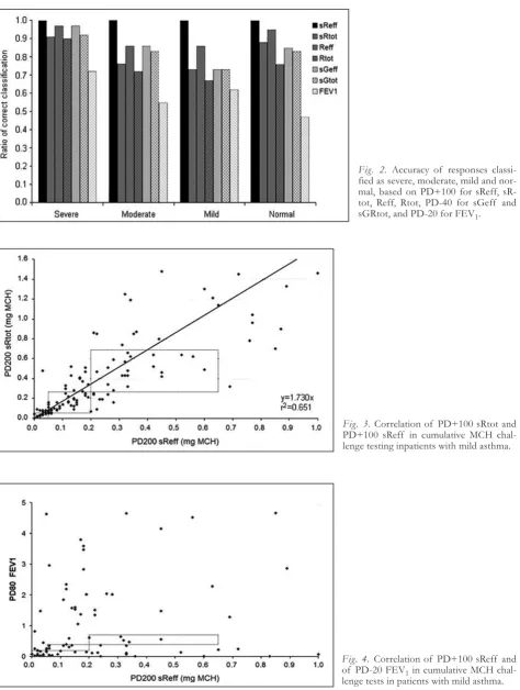
Statistical analysis revealed strongly linearly corre-lated parameters of airway resistance, but no signifi-cant relationship between the test results of body plethysmography and forced spirometry. PD+100 sR-eff and PD+100 sRtot significantly correlated in
pa-Fig. 1. Number of patients with MCH re-sponses, classified as severe, moderate, mild, and normal, based on PD+100 for sReff, sRtot, Reff, Rtot , PD-40 for sGeff and sGRtot and PD-20 for FEV1.
Table 1. Baseline respiratory parameters of patients with mild asthma included in the study.
FEV1 FEV1% Reff sReff Rtot sRtot sGeff sGtot
(L/s) FVC (%) (kPa/L . s) (kPa . s) (kPa/L . s) (kPa . s) (1/kPa . s) (1/kPa . s)
Males, n = 82
MW 3.51 82.2 0.19 0.77 0.22 0.87 1.53 1.29
SD ±0.91 ±7.60 ±0.07 ±0.36 ±0.07 ±0.34 ±0.62 ±0.41 Med 3.47 83.2 0.19 0.71 0.22 0.80 1.41 1.26
Females, n = 73
MW 2.45 85.0 0.26 0.84 0.29 0.92 1.40 1.19
tients with severe, moderate and mild degree of AHR (Fig. 3). For PD-20 FEV1and PD+100 sReff no sig-nificant correlation could be found (Fig. 4). Signifi-cant accuracy of PD+100 sReff and PD-40 sGeff could be stated.
Coinciding positive or negative results in Reff and sReff were found in 95% of the MCH tests, sReff and sRtot matched in 88% of the patients, 85% matched in sReff and sGeff, but only 47% coinciding responses were obtained in sReff and FEV1.
Fig. 2. Accuracy of responses classi-fied as severe, moderate, mild and nor-mal, based on PD+100 for sReff, sR-tot, Reff, RsR-tot, PD-40 for sGeff and sGRtot, and PD-20 for FEV1.
Fig. 3.Correlation of PD+100 sRtot and PD+100 sReff in cumulative MCH chal-lenge testing inpatients with mild asthma.
Fig. 4. Correlation of PD+100 sReff and of PD-20 FEV1in cumulative MCH
D
ISCUSSIONThe concordance of forced spirometry and tidal breathing recording, studied by body plethysmogra-phy, was investigated by bronchial challenge tests in patients with chronic cough or suspected mild asthma. In cumulative MCH challenges, we found specific airway resistance (sReff) to be the most useful parameter in the detection of AHR. The measure-ment of specific airway resistance (sRaw) by body plethysmography is largely independent of patient’s cooperation, facilitating the interpretation of meas-urements. Furthermore, higher MCH doses were necessary before FEV1, as a key parameter of forced spirometry, showed a significant decrease above 20%. Although most practitioners only meas-ure FEV1, reliance upon this measurement alone can lead to both false negative and false positive test re-sults.
Goldstein et al [18] reported that sensitivity of a MCH challenge determined by FEV1 alone was only
60%, but increased to 97% after the assessments of FVC, sGaw, and thoracic gas volume (TGV) were added to the analysis. This result is in accordance with the known axial heterogeneity of the response of air-ways of different caliber to bronchoactive agents. Fur-thermore, if patients not fully inhale to total lung ca-pacity (TLC), a fall in FEV1 can occur and cause a
false positive result. Generally, the sensitivity of a pos-itive MCH challenge test for the diagnosis of asthma is 85%. A positive predictive value is more limited, as false positive results may be seen in patients with aller-gic rhinitis, cystic fibrosis, congestive heart failure, COPD, and bronchitis. [1, 19]. However, patients with allergic rhinitis, cystic fibrosis, heart failure, or COPD were not included in this study.
IMPACT OFDEEPINSPIRATIONS DURINGFORCED
EXPIRATORYMANEUVERS ONMCH-TESTING
Deep breaths effectively modulate airway caliber. In healthy subjects, deep inhalations result in bronchodi-latation and reduce pharmacologically induced airways obstruction [20]. On the other hand, avoidance of maximal maneuvers enhances reactions of bron-choconstrictor agents [21]. Taking deep breaths before bronchial challenge tests diminshes obstructive re-sponses as a consequence [22, 23]. Therefore, deep in-spirations during forced expiratory maneuvers provide an effective protection against airways obstruction. However, the beneficial effects of deep inspirations are impaired in asthma [24, 25] and during acute exac-erbations of asthma. In these clinical settings deep in-spirations may even enhance obstructive responses [26]. In chronic obstructive pulmonary disease (COPD), bronchodilatory effects of deep inspirations are effectively reduced, which may depend on the de-gree of parenchymal damage [27]. Airway inflamma-tion is a typical feature of asthma and COPD, al-though the composition of inflammatory cells is dif-ferent [28, 29]. Inflammatory processes induce airway remodelling and thus increasing thickness of airway walls and reducing the elasticity of the airways [30]. The result would be a reduced strain transmission
from lung parenchyma to the airway walls and dimin-ished effects of stretching the airways [31].
Slats et al [14] demonstrated that the bronchodilato-ry effects of deep inspirations in asthma are related to inflammatory cell counts in bronchial walls and smooth muscle layers, whereas in moderate COPD this relationship could not be found. They conclude that the physiologic protection against narrowing of the airways by deep inspirations is impaired in asthma and COPD, but depends on different mechanisms.
CHANGES INAIRWAYRESISTANCE DURINGQUIET
BREATHING
Using body plethysmography for the assessment of airway resistance responses, measurements are per-formed during quiet breathing and are not masked by the bronchodilatory responses of deep inhalations or positive intraluminal pressure in the airways. In addi-tion, the results are largely independent of patients’ cooperation. There was a close correlation between Reff and Rtot, the two parameters for airway resist-ance, which are calculated from different approaches, and the PD+100 of both parameters. In addition, spe-cific airway resistance sReff and sRtot, including lung volume and changes in lung volume during airway ob-struction, closely correlate. Since Raw and intratho-racic gas volume (ITGV) both increase during acute airway obstruction, the largest response rates can be recorded in sReff and sRtot. A close correlation was also found between specific airway resistance and cific conductance, sGaw, the reciprocal value of spe-cific resistance. In further investigations, PD sGaw should be adjusted to PD+100 sReff.
CLINICALRELEVANCE OFNON-SPECIFICCHALLENGE
TESTING
AHR is an important defining feature of asthma and is a manifestation of reversible airflow obstruction due to smooth muscle contraction. AHR represents an exaggerated constrictor response to a variety of physi-cal, chemiphysi-cal, or environmental stimuli. AHR can be quantified by the dose response to pharmacologic agents such as methacholine or histamine, causing a 20% decrease in FEV1 from baseline. While AHR is
failure to show AHR argues against the diagnosis of asthma, AHR may be the sole objective evidence of airway dysfunction, AHR is quantitatively associated with the presence and severity of disease, the occur-rence of AHR in an asymptomatic person may help predict the future development of asthma, the degree of AHR in a symptomatic person can have prognostic and potentially therapeutic implications, the periodici-ty of asthma exists in parallel with changes in the de-gree of AHR [32, 33]. A false negative FEV1 in
pa-tients with asthma may have serious consequences for the patient due to undertreatment. Thus, recording re-sponses of airway resistance by measurements per-formed during quiet breathing, which are not masked by the bronchodilatory responses of deep inspirations may increase the diagnostic impact of bronchoprovo-cation.
S
UMMARY ANDC
ONCLUSIONThe authors compared forced spirometry and body plethysmography in order to evaluate the concordance of the forced respiratory maneuvers and measure-ments of airway resistance during normal breathing in MCH challenges. PD+100 Raw and PD-40 Gaw for the following plethysmographically measured parame-ters Rtot, Reff, sRtot, sReff, sGtot, and sGeff, are closely correlated with cumulative MCH-challenge tests. Our results did not show a correlation of PD+100 sReff and PD-20 FEV1. The reason for the
different responses might be found in the location of the plethymographically measured airway resistance and the flow reduction measured in forced spirometric maneuvers. From our results, we would recommend sReff and sGaw as the reliable parameters for classifi-cation of AHR. Additional investigations on healthy subjects and patients with asthma and COPD should be performed to compare the sensitivity and specifici-ty of body plethysmography and forced spirometry for MCH- challenge tests.
Conflicts of interest:No conflicts of interests were declared by the authors in relation to this article.
R
EFERENCES[1] Crapo RO, Casaburi R, Coates AL, et al. Guidelines for methacholine and exercise challenge testing - 1999. The official statement of the American Thoracic Society adopted by the ATS Board of Directors. Am J Respir Crit Care Med 2000; 161: 309-329.
[2] Juniper EF, Frith PA, Hargreave FE. Airway responsive-ness to histamine and methacholine: Relationship to mini-mum treatment to control symptoms of asthma. Thorax 1981; 36: 575-579.
[3] Cockroft DW, Killian DN, Mellon JA, et al. Bronchial re-activity to inhaled histamine: A method and clinical sur-vey. Clin Allergy 1977; 7; 235.
[4] American Thoracic Society. Guidelines for methacholine and exercise challenge testing. Am J Respir Crit Care Med. 1999; 161: 309-329.
[5] Sterk PJ, Fabbri LM, Quanjer PH. Airway responsiveness: standardized challenge testing with pharmacological, physical and sensitizing stimuli in adults. Eur Respir J 1993; 6: 53-83.
[6] Smith L, McFadden ER Jr. Bronchial hyperresponsiveness revisited. Ann Allergy Asthma Immunol 1995; 74: 454-469.
[7] Cockroft DW, Davis BE, Todd AC, et al. Methacholine challenge: comparison of two methods. Chest 2005; 127: 839-844.
[8] Prieto L, Lopez V, Llusar R, Rocio Rojas RN, Marin J. Dif-ferences in the response to methacholine between the tidal breathing and dosimeter methods. Influence of the dose of bronchoconstrictor agent delivered to the mouth. Chest 2008; 134: 699-703.
[9] Merget R, Jörres RA, Heinze E, Haufs MG, Taeger D, Brüning Th. Development of a 1-concentration-4-step dosimeter protocol for methacholine testing. Respir Med 2009; 103[4]: 607-613.
[10] Chai H, Farr RS, Froehlich LA, Mathison DA, McLean JA, Rosenthal RR. Standardization of bronchial inhala-tion challenge procedures. J Allergy Clin Immunol 1975; 56: 323-327.
[11] Ryan G, Dolovich MB, Obminski G, Cockroft DW, Ju-niper E, Hargreave FE. Standardization of inhalation provocation tests: influence of nebulizer output, particle size, and method of inspiration. J Appl Physiol 1981; 67: 156-161.
[12] Cockroft DW, Davis BE. The bronchoprotective effect of inhaling methacholine by using total lung capacity in-spirations has a marked influence on the interpretation of test results. J Allergy Clin Immunol 2006; 117: 1244-1248. [13] Nadel JA, Tierney DF. Effect of a previous deep
inspira-tion on airway resistance. J Appl Physiol 1961; 16: 717-719.
[14] Slats AM, Janssen K, van Schadewijk A, van der Plas DT, Schot R, van der Aardweg JG, de Jongste JC, Hiemstra PS, Mauad T, Rabe KF, Sterk PJ. Bronchial inflammation and airway responses to deep inspiration in asthma and chronic obstructive pulmonary disease. Am J Respir Crit Care Med 2007; 176: 121-128.
[15] Quanjer P, Tammeling GJ, Cotes JE, Pederson OF, Peslin R, Yernault JC. Lung volumes and forced ventilatory flows. Report of the working party standardization of lung functions tests. European Community for Steel and Coal. Eur Respir J 1993; 6 Suppl 16.
[16] Camarri B, Eastwood PR, Cecins NM, Thompson PJ, Jenkins S. Six minutes walk distance in healthy subjects aged 55-75 years. Respir Med 2006; 100: 658-665. [17] Criée CP, Berdel D, Heise D, Jörres RA, Kardos P,
Köh-ler D, Leupold W, Magnussen H, Marek W, Merget R, Mitfessel H, Rasche K, Rolke M, Smith HJ, Sorichter S, Worth H. Empfehlungen zur Ganzkörperplethysmogra-phie. München, Orlando: Dustri-Verlag Dr. Karl Feistle, 2009.
[18] Goldstein MF, Pacana SM, Dvorin DJ, Dunsky EH. Ret-rospective analysis of methacholine inhalation challenges. Chest 1994; 105: 1082-1088.
[19] Goldstein MF, Veza BA, Dunsky EH, Belecanech GA, Haralabatos JC. Comparisons of peak diurnal expiratory flow variation, postdilatator FEV1responses, and
metha-choline inhalation challenges in the evaluation of suspect-ed asthma. Chest 2001; 119: 1001-1010.
[20] Scichilone N, Permutt S, Togias A. The lack of the bron-choprotective and not the bronchodilatatory ability is as-sociated with airway hyperresponsiveness. Am J Respir Crit Care Med 2001; 163: 413-419.
[21] Skloot G, Permutt S, Togias A. Airway hyperresponsive-ness in asthma: a problem of limited smooth muscle re-laxation with inspiration. J Clin Invest 1995; 96: 2393-2403.
[23] Scichilone N, Kapsali T, Permutt S, Togias A. Deep in-spiration-induced bronchoprotection is stronger than bronchodilatation. Am J Respir Crit Care Med 2009; 162: 910-916.
[24] Jensen A, Atileh H, Suki B, Ingenito EP, Lutchen KR. Selected contribution: airway caliber in healthy and asth-matic subjects: effects of bronchial challenge and deep inspiration. J Appl Physiol 2001; 91: 506-515.
[25] Salome CM, Thorpe CW, Dipa C, Brown NJ, Berend N, King GG. Airway re-narrowing following deep inspira-tion in asthmatic and non asthmatic subjects. Eur Respir J 2003; 22: 62-68.
[26] Lim TK, Ang SM, Rossing TH, Ingenito EP, Ingram RH Jr. The effects of deep inhalation on maximal expiratory flow during intensive treatment of spontaneous asthmatic episodes. Am Rev Respir Dis 1989; 140: 340-343. [27] Fraishter RD. Airway hysteresis in normal subjects and
individuals with chronic airflow obstruction. J Appl Phys-iol 1985; 58: 1505-1510.
[28] Jeffrey PK. Remodelling and inflammation of the bronchi in asthma and chronic obstructive pulmonary disease. Proceedings American Thoracic Society 2004; 1: 176-183.
[29] Fabbri LM, Romagnoli M, Corbetta L, Casoni G, Busljet-ic K, Turato G, Ligabue G, Ciaccia A, Saetta M, Papi A. Differences in airway inflammation in patients with fixed airflow obstruction due to asthma and or chronic ob-structive pulmonary desease. Am J Respir Crit Care Med 2003; 167: 418-424.
[30] Elias JA, Zhu Z, Chupp G, Homer RJ. Airway remodel-ling in asthma. J Clin Invest 1999; 104:1001-1006. [31] Macklem PT. A theoretical analysis of the effects of
air-way smooth muscle load on airair-way narrowing. Am J Respir Crit Care Med 1996; 153: 83-89.
[32] Laprise C, Laviolette M, Boutet M, Boulet LP. Asympto-matic airway responsiveness: relationship with airway in-flammation and remodelling. Eur Respir J 1999; 14: 63-73.
[33] Weiss ST, van Natta ML, Zeiger RS. Relationship be-tween increased airway responsiveness and asthma severi-ty in the childhood asthma management program. Am J Respir Crit Care Med 2000; 162: 50-56.
Author’s address:
Dr. Wolfgang Marek,
Institute for Occupational Physiology Augusta Kranken Anstalt
Bergstr. 26 44791 Bochum Germany
Phone: +49 234 5172474,