R E S E A R C H A R T I C L E
Open Access
Use of Mitomycin C to reduce the incidence of
encapsulated cysts following ahmed glaucoma
valve implantation in refractory glaucoma
patients: a new technique
Minwen Zhou
2,3, Wei Wang
1, Wenbin Huang
1and Xiulan Zhang
1*Abstract
Background:To evaluate the surgical outcome of Ahmed glaucoma valve (AGV) implantation with a new
technique of mitomycin C (MMC) application.
Methods:This is a retrospective study. All patients with refractory glaucoma underwent FP-7 AGV implantation. Two methods of MMC application were used. In the traditional technique, 6 × 4 mm cotton soaked with MMC (0.25–0.33 mg/ml) was placed in the implantation area for 2–5mins; in the new technique, the valve plate first was encompassed with a thin layer of cotton soaked with MMC, then inserted into the same area. A 200 ml balanced salt solution was applied for irrigation of MMC. The surgical success rate, intraocular pressure (IOP), number of anti-glaucoma medications used, and postoperative complications were analyzed between the groups.
Results:The surgical outcomes of two MMC applied techniques were compared. The new technique group had
only one case (2.6%) of encapsulated cyst formation out of 38 eyes, while there were eight (19.5%) cases out of 41 eyes the in traditional group. The difference was statistically significant (P= 0.030). According to the definition of success rate, there was 89.5% in the new technique group and 70.7% in the traditional group at the follow-up end point. There was a significant difference between the two groups (P= 0.035). Mean IOP in the new technique group were significantly lower than those of the traditional group at 3 and 6 months (P< 0.05).
Conclusions:By using a thin layer of cotton soaked with MMC to encompass the valve plate, the new MMC
application technique could greatly decrease the incidence of encapsulated cyst and increase the success rate following AGV implantation.
Keywords:Refractory glaucoma, Ahmed glaucoma valve implantation, Encapsulated cyst, Mitomycin C
Background
Ahmed glaucoma valve (AGV) implantation has been widely used and has been proved to be an effective method for treating refractory glaucoma [1-4]. Several studies in the literature have reported success rates of AGV implantation ranging from 49% to 83.6% [1,3,5-7]. Encapsulated cyst formation is one of the main reasons for failure [8]. The proliferation of fibrous tissue around the implant plates blocks the diffusion of aqueous humor
and elevates intraocular pressure (IOP) [9]. Adjunctive use of antimetabolites can greatly inhibit fibrosis [10,11], and mitomycin C (MMC) has been used extensively in filter-ing and glaucoma drainage device implant surgery [12,13]. However, how to use MMC more effective has remained to be explored further. Heuer et al. [14]. Found that double-plate Molteno implantation more frequently af-fords IOP control than single-plate Molteno. Assuming that the expanded surface area of the implant plate allows reduced occurrence of encapsulated cyst, it is also sup-posed that expanding the MMC function area in the scleral bed where the AGV is placed may decrease encap-sulated cyst formation. Unfortunately, the cotton soaked * Correspondence:zhangxl2@mail.sysu.edu.cn
1
Zhongshan Ophthalmic Center, State Key Laboratory of Ophthalmology, Sun Yat-Sen University, 54S.Xianlie Road, Guangzhou 510060, China Full list of author information is available at the end of the article
with MMC and inserted into the implantation area often rolls into a mass, without a guarantee of enough size. Therefore, we improved the method by introducing a novel way for MMC to be used: the valve plate was first encompassed with a thin layer of cotton soaked with MMC, then insert into the implanted area. In this study, we evaluated its surgical outcomes to see whether the new method could produce better surgical results.
Methods
Patients and inclusion criteria
This was a retrospective study of patients diagnosed with refractory glaucomas (including failed filtration, uveitic glaucoma, pseudophakia, and traumatic glaucoma) who underwent AGV implantation at the Glaucoma Depart-ment of Zhongshan Ophthalmic Center. Consecutive pa-tients followed up at the Zhongshan Ophthalmic Center from October 2008 and January 2013 were included in this study. From October 2008 to January 2010, we employed the traditional method in our hospital, and we converted to the new technique from January 2010. It was approved by the Ethical Review Committee of Zhongshan Ophthalmic Center and adhered to the pro-visions of the Declaration of Helsinki for research in-volving human subjects.
Best corrected visual acuity (BCVA), IOP, number of antiglaucoma medications, and systemic diseases were examined by chart review. Demographic data, such as age, sex, prior surgery history, and subtypes of glaucoma were collected. All patients received MMC application during the surgery. Age less than 18 years, previous aqueous shunt surgery in the same eye, prior scleral buckling procedures, and without MMC application, were all factors for excluding patients from the study.
Surgical techniques and MMC application
One glaucoma specialist (XZ) performed all the FP-7 AGV implantation surgeries, using the same techniques. A fornix-based flap of the conjunctiva and Tenon cap-sule was created in the superior temporal quadrant. However, in the patients who had undergone previous eye surgery, such as trabeculectomy, causing scarring of the conjunctiva of the superior temporal quadrant, we used an inferior temporal quadrant incision. The tube of the AGV was flushed with a balanced salt solution through a scleral track to ensure patency before inser-tion. In order to decrease the possibility of overfiltering following AGV implantation, the tube was ligated tightly to restrict aqueous flow, using 8–0 polyglactin absorb-able sutures, in all patients. The AGV was positioned in the middle of the quadrant, with the anterior edge of the plate 10 mm or more posterior to the superior temporal corneoscleral limbus. Before the AGV was placed, MMC (0.25–0.33 mg/ml, 2–5 min) was applied in all patients
(Figure 1). The concentration and time of MMC depended on the judgment of the risk of failure of the surgery by the surgeon. In the traditional manner, a piece of cotton (6 × 4 mm) soaked with MMC was placed in the appointed area. However, the wet cotton often rolled into a mass, without a guarantee of enough size. In the improved manner, the valve plate was first encompassed with a thin layer of cotton soaked with MMC, then inserted into the same area. After 2–5 mi-nutes, the cotton pieces and the encompassed AGVs were removed and irrigated with 200 ml of balanced salt solution. Then, the valve plate was sutured to the sclera with 6–0 nylon sutures through the anterior positional holes of the body of the valve plate. A half-thickness, rectangular, 4 × 6 mm, limbal-based scleral flap was cre-ated. The tip of the drainage tube was then cut and bev-eled upwards, in order to extend it by 2 mm into the anterior chamber. Paracentesis in the inferior temporal peripheral cornea was performed, and viscoelastic was injected to maintain the anterior chamber before tube insertion. A 23-gauge needle punctured the anterior chamber under the scleral flap, and the drainage tube was inserted. The tube was sutured to the episcleral sur-face with 8–0 polyglactin sutures. The scleral flap over the drainage tube was reattached to the sclera and su-tured with 10–0 nylon sutures. The conjunctiva and Tenon capsule were reapproximated to the limbus with 8–0 polyglactin sutures. Topical prednisolone acetate 1% (prednisolone acetate ophthalmic suspension, USP; PA) was administered four times daily for four weeks, and was then replaced with non-steroid anti-inflammatory drug eye drops (pranoprofen 0.1% [Senju, Japan; PF]) for two weeks. Glaucoma medications were prescribed when the postoperative IOP was greater than 21 mm Hg, and the medications were added or removed according to the IOP level. Topical β-blockers were the first line of therapy. Topical carbonic anhydrase inhibitor and top-ical α2-adrenergic agonists were added as a second line of therapy. Systemic medications to decrease IOP were applied if necessary. When the IOP was reduced after
treatment, antiglaucoma agents were gradually with-drawn during the follow-up.
Postoperative follow-up
Postoperative IOP and VA were recorded at each visit after AGV implantation surgery. The number of postop-erative glaucoma medications and postoppostop-erative compli-cations were recorded. The postoperative visits were performed after 1 day, 1 week, 1 month, 3 months, and every 6 months thereafter.
Evaluation criteria
Postoperative survival was defined as IOP <21 mm Hg, with or without glaucoma medications, and without sig-nificant visually threatening complications (endophthal-mitis, retinal detachment, suprachoroidal hemorrhage, preseptal cellulitis, or persistent hypotony [IOP < 5 mm Hg]). Failure was defined as IOP not less than 21 mm Hg, IOP < 5 mm Hg on two consecutive follow-up visits after three months, or loss of light perception, or the need for further surgery or laser to control IOP [15].
Statistical analysis
The data were processed and statistically analyzed using SPSS for Windows XP (Version 13.0; SPSS, Chicago, IL). The Mann–WhitneyUtest was used for variables with a skewed distribution, and the chi-square or Fisher’s exact test was used for categorical variables. An independent
samplet test was used to compare normally distributed continuous variables data between the two groups. To compare the IOPs and glaucoma medications at various time points before and after operation, the Wilcoxon signed-rank test was used. Success rates in both groups were compared using Kaplan–Meier survival curves and the log rank test.P values of <0.05 were considered sta-tistically significant.
Results
Seventy-nine eyes of 79 patients who fulfilled the inclu-sion criteria were included in the study. MMC applied with the traditional technique was performed in 41 eyes (traditional group), while the new technique was per-formed in the remaining 38 eyes (new technique group). The minimum required follow-up period after surgery was six months. Mean follow-up times were 19.89 ± 8.29 months for the new technique group and 18.10 ± 8.71 months for the traditional group (P= 0.938). The demographic and preoperative data of the two groups are presented in Table 1. There were no significant dif-ferences in sex, mean age, mean IOP, mean BCVA, mean glaucoma medication, or number of previous glaucoma surgeries between the two groups.
Compared with preoperative IOP, the two groups showed a statistically significant IOP decrease at all follow-up intervals (P< 0.05, Wilcoxon signed-rank test). IOP was lower in the traditional group than in the new
Table 1 Demographic and preoperative data of different group patients
New technique (n = 38) Traditional (n = 41) P
Age (y), mean ± SD 42.34 ± 13.69 38.29 ± 15.32 0.220a
Sex 0.150b
Male, n (%) 23 (60.5) 31 (75.6)
Female, n (%) 15 (39.5) 10 (24.4)
Mean IOP (mm Hg), mean ± SD 41.97 ± 10.58 43.15 ± 9.63 0.608a
Mean glaucoma medication,mean ± SD 3.13 ± 0.58 3.24 ± 0.92 0.460c
Mean follow-up time (month), mean ± SD 19.89 ± 8.29 18.10 ± 8.71 0.938a
Mean best corrected visual acuity (logMAR) 1.82 ± 1.25 2.24 ± 1.11 0.107c
Mean MMC concentration (mg/ml) 0.29 ± 0.04 0.28 ± 0.04 0.155a
Mean MMC duration (min) 3.24 ± 0.97 3.00 ± 1.02 0.296a
Previous glaucoma surgeries history, n (%) 17 (44.7) 19 (46.3) 0.886b
Diagnosis 0.992b
Uveitic glaucoma, n (%) 10 (26.3) 11 (26.8)
NVG, n (%) 13 (34.2) 15 (36.6)
Traumatic glaucoma, n (%) 4 (10.5) 3 (7.3)
ICE syndrome, n (%) 2 (5.3) 2 (4.9)
Failed trabeculectomy, n (%) 9 (23.7) 10 (24.4)
a
independent samplettest. b
chi-square test. c
Mann–WhitneyUtest.
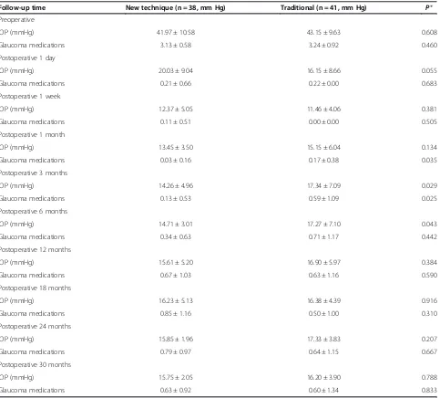
technique group in postoperative day 1 and week 1. Nevertheless, the new technique group showed lower IOPs thereafter up to the end of the study in postopera-tive month 30. The new technique group, compared with traditional group, showed significantly lower IOP at month 3 (P= 0.029) and month 6 (P= 0.043). Table 2 display the mean IOPs at all time intervals in both groups.
Table 2 compares the mean numbers of antiglaucoma medications required in both groups at all time intervals. Medication use for both groups after surgery was signifi-cantly decreased at all follow-up time points when co-mpared with preoperative values (P< 0.05, Wilcoxon signed-rank test). There were no statistically significant
differences between the groups at most of time point in terms of the mean number of medications. However, the new technique group had a significantly lower mean number of medications than the traditional group at the month 1 (P= 0.035) and month 3 (P= 0.025) postopera-tive follow-up visit.
Kaplan–Meier survival analysis showed that the suc-cess rates for the new technique and traditional groups were 97.4% and 87.8% at 12 months, respectively, and 89.5% and 73.2% at 24 months, respectively. The success rate of the new technique group was significantly higher than that of the traditional group (P= 0.035, log rank test) (Figure 2). After the endpoint of follow-up, failure had occurred in 4 patients (10.5%) in the new technique
Table 2 Mean IOP and mean glaucoma medications required in both groups at all follow-up time intervals (mean ± SD)
Follow-up time New technique (n = 38, mm Hg) Traditional (n = 41, mm Hg) P* Preoperative
IOP (mmHg) 41.97 ± 10.58 43.15 ± 9.63 0.608
Glaucoma medications 3.13 ± 0.58 3.24 ± 0.92 0.460
Postoperative 1 day
IOP (mmHg) 20.03 ± 9.04 16.15 ± 8.66 0.055
Glaucoma medications 0.21 ± 0.66 0.22 ± 0.00 0.683
Postoperative 1 week
IOP (mmHg) 12.37 ± 5.05 11.46 ± 4.06 0.381
Glaucoma medications 0.11 ± 0.51 0.00 ± 0.00 0.505
Postoperative 1 month
IOP (mmHg) 13.45 ± 3.50 15.15 ± 6.04 0.134
Glaucoma medications 0.03 ± 0.16 0.17 ± 0.38 0.035
Postoperative 3 months
IOP (mmHg) 14.26 ± 4.96 17.34 ± 7.09 0.029
Glaucoma medications 0.13 ± 0.53 0.59 ± 1.09 0.025
Postoperative 6 months
IOP (mmHg) 14.71 ± 3.01 17.27 ± 7.10 0.043
Glaucoma medications 0.34 ± 0.63 0.71 ± 1.17 0.442
Postoperative 12 months
IOP (mmHg) 15.61 ± 5.20 16.90 ± 5.97 0.384
Glaucoma medications 0.67 ± 1.03 0.63 ± 1.16 0.590
Postoperative 18 months
IOP (mmHg) 16.23 ± 5.13 16.38 ± 4.39 0.916
Glaucoma medications 0.85 ± 1.16 0.50 ± 1.00 0.310
Postoperative 24 months
IOP (mmHg) 15.85 ± 1.96 17.33 ± 3.83 0.207
Glaucoma medications 0.79 ± 0.97 0.64 ± 1.15 0.667
Postoperative 30 months
IOP (mmHg) 15.75 ± 2.05 16.20 ± 3.90 0.788
Glaucoma medications 0.63 ± 0.92 0.60 ± 1.34 0.833
group and 11 patients (26.8%) in the traditional group. Table 3 showed the reasons for failure in both groups.
During the follow-up period, visual acuity remained unchanged relative to pre-operative values. There were no significant differences in visual acuity between the 2 groups at all time points (Table 4).
As shown in Table 5, postoperative complications in-cluded encapsulated cyst formation, choroidal effusion, flat anterior chamber, hypotony maculopathy, and hyphema. The most common complication in the eyes of the trad-itional group was encapsulated cyst formation, with inci-dences in eight eyes (19.5%), while there was only an incidence in one eye (2.6%) in the new technique group. Statistically significant differences were detected between the two groups when comparing encapsulated cyst forma-tion complicaforma-tions (P= 0.030). Flat anterior chamber oc-curred in five eyes (13.2%) in the new technique group. There were no statistically significant differences in inci-dences of other postoperative complications between the groups.
Discussion
AGV implantation allows aqueous drainage via a tube inserted into the anterior chamber to a posterior plate sutured to the episclera. The aqueous humor crosses the surrounding bleb wall by passive diffusion, and it is
removed from the periocular space by venous capillaries or lymphatics [16,17]. However, when proliferation of fi-brous tissue around the plate forms, it restricts aqueous humor diffusion through the capsule, followed by a gradual elevation of IOP, and then, encapsulated cyst formation [8]. Encapsulated cyst formation is the most frequent reason for glaucoma drainage device implant surgery failure [8]. Adjunctive use of MMC is still con-troversial; while most studies have concluded that ad-junctive use of MMC is beneficial for improving success rates [11,18,19], other studies have found that MMC did not increase the short- or intermediate-term success rates of AGV implantation [20,21]. Thus, further study is expected to reveal whether adjunctive use of MMC is beneficial, as well as how to use it more effectively in AGV implantation.
In the process of AGV implantation, the traditional method for placing MMC is to take a piece of cotton or sponge soaked with MMC into the middle of the quad-rant where the valve was to be implanted. In fact, the cotton or sponge often folds or rolls into a mass at the scleral bed, limiting the anti-fibrotic function of MMC. In this study, the new technique, which overcame this shortcoming, was able to guarantee enough fixed space for MMC to function. Therefore, the novel technique Figure 2Cumulative survival curves showed new technique
group had a greater survival than traditional group after AVG implantation.There was significant difference between the 2 groups (P= 0.035).
Table 3 Reasons for failure in both groups
New technique (n = 38)
Traditional (n = 41)
High IOP* (>21 mmHg) 2 (5.3%) 9 (22.0%)
Low IOP* (<5 mmHg) 1 (2.6%) 0
Progression to NLP 1 (2.6%) 1(2.4%)
Additional glaucoma surgery 0 1(2.4%)
*IOP-related failures require 2 consecutive visits at or after 3 months in which the criterion is not met.
Table 4 Mean best corrected visual acuity (logMAR) in both groups at all follow-up time intervals (mean ± SD)
Follow-up time New technique (n = 38, mm Hg)
Traditional (n = 41, mm Hg) P
*
Preoperative 1.82 ± 1.25 2.24 ± 1.11 0.107
Postoperative 1 day 1.89 ± 1.22 2.30 ± 1.09 0.124
Postoperative 1 week 1.87 ± 1.21 2.18 ± 1.18 0.225
Postoperative 1 month 1.79 ± 1.25 2.12 ± 1.17 0.177
Postoperative 3 months 1.79 ± 1.25 2.10 ± 1.19 0.228
Postoperative 6 months 1.91 ± 1.35 2.09 ± 1.20 0.386
Postoperative 12 months 2.04 ± 1.21 2.11 ± 1.26 0.745
Postoperative 18 months 2.06 ± 1.24 2.02 ± 1.26 0.991
Postoperative 24 months 1.86 ± 0.91 2.28 ± 1.25 0.398
Postoperative 30 months 1.77 ± 0.92 2.62 ± 0.86 0.141
*Mann–WhitneyUtest.
Abbreviations: IOPintrocular pressure,SDstandard deviation.
Table 5 Postoperative complications in both groups
Complications New technique (n = 38)
Traditional (n = 41)
P
Encapsulated cyst formation, n (%) 1 (2.6%) 8 (19.5%) 0.030*
Choroidal effusion, n (%) 3 (7.9%) 1 (2.4%) 0.612
Flat anterior chamber, n (%) 5 (13.2%) 1 (2.4%) 0.072
Hypotony maculopathy, n (%) 1 (2.6%) 0 (0%) 0.481
Hyphema, n (%) 2 (5.3%) 4 (9.8%) 0.676
Fisher’s exact test.
could greatly decrease encapsulated cyst incidences and significantly increase surgical outcomes.
This is the first study to compare the surgical outcome and complication rates associated with the use of MMC in AGV implantation, using the traditional and new methods. Both methods showed efficacy and safety dur-ing AGV implantation, and they showed a similar trend in postoperative IOP control and the use of glaucoma medication. Kaplan–Meier survival curves showed statis-tically significant differences between the groups, which might be a result of the lower incidences of encapsulated cyst formation in the new technique group (one eye, 2.6% vs. eight eyes, 19.5% in the traditional group).
Comparing our findings with other reported series is problematic, as some authors do not consider the forma-tion of an encapsulated cyst as a complicaforma-tion and, therefore, do not report it [22-24]. However, other stud-ies have reported differences in incidences of encapsu-lated cyst. Lai [25], in a series of 65 eyes undergoing AGV implantation, reported that 16 eyes (24.6%) devel-oped encapsulated cyst as a postoperative complication. Similarly, a prospective, comparative study showed that five eyes (14.7%) had incidences of encapsulated cyst after AGV implantation [6]. In short, encapsulated cyst formation is often referred to as a late complication after glaucoma implant insertion in adults, with an appear-ance varying from 5% to 30%, depending on study de-sign, follow-up time, and patient selection. In our study, encapsulated cyst occurred in only 2.6% of the new tech-nique group. Therefore, by using a thin layer of cotton soaked with MMC to encompass the valve plate, this novel MMC application technique could greatly de-crease the incidence of encapsulated cyst.
On the other hand, the incidence of flat anterior chamber using the new technique method was higher than using the traditional method. In fact, to avoid post-operative hypotony, the tube was ligatured tightly, using 8–0 polyglactin suture, to restrict aqueous flow during the surgery. Therefore, the use of adjunctive MMC may be another cause of hypotony, besides leakage around the tube, a decrease in aqueous production, and overfil-tration [26]. Whether the new technique method of MMC application allows more range for MMC func-tioning to cause more flat chamber incidence needs to be investigated further.
The main limitation of this study is the nonrandomized design. We took every possible step to reduce potential bias, and the final data were subjected to careful statistical analysis. The crucial criterion for any“randomization” is to have groups at the baseline comparable in demographic and clinical characteristics. In this study, that was the case. The second limitation is that different MMC concentra-tions and times in different patient might affect result of study.
Conclusions
In conclusion, this study indicates that the new technique for MMC application may provide a better chance for pa-tients to decrease the incidence of encapsulated cyst, when compared with the traditional method. In addition, there was a tendency for lower IOP and higher complete suc-cess rate in the new technique group.
Competing interests
The authors declare that they have no competing interests.
Authors’contributions
All authors conceived of and designed the experimental protocol. MZ and WW collected the data. All authors were involved in the analysis. MZ wrote the first draft of the manuscript. MZ and XZ reviewed and revised the manuscript and produced the final version. All authors read and approved the final manuscript.
Acknowledgements
Supports: This research was supported by the National Natural Science Foundation of China (81371008).
Author details
1
Zhongshan Ophthalmic Center, State Key Laboratory of Ophthalmology, Sun Yat-Sen University, 54S.Xianlie Road, Guangzhou 510060, China. 2Department of Ophthalmology, Shanghai First People’s Hospital, School of
Medicine, Shanghai JiaoTong University, Shanghai, China.3Shanghai Key Laboratory of Fundus Disease, Shanghai, China.
Received: 7 December 2013 Accepted: 7 August 2014 Published: 6 September 2014
References
1. Souza C, Tran DH, Loman J, Law SK, Coleman AL, Caprioli J:Long-term outcomes of Ahmed glaucoma valve implantation in refractory glaucomas.Am J Ophthalmol2007,144:893–900.
2. Teixeira SH, Doi LM, Freitas SA, Silva KD, Paes AT, Higa FS, Mendonca M, Prata JA, Paranhos A:Silicone ahmed glaucoma valve with and without intravitreal triamcinolone acetonide for neovascular glaucoma: randomized clinical trial.J Glaucoma2012,21:342–348.
3. Budenz DL, Barton K, Feuer WJ, Schiffman J, Costa VP, Godfrey DG, Buys YM: Treatment outcomes in the Ahmed Baerveldt Comparison Study after 1 year of follow-up.Ophthalmology2011,118:443–452.
4. Shen CC, Salim S, Du H, Netland PA:Trabeculectomy versus Ahmed Glaucoma Valve implantation in neovascular glaucoma.Clin Ophthalmol 2011,5:281–286.
5. Papadaki TG, Zacharopoulos IP, Pasquale LR, Christen WB, Netland PA, Foster CS:Long-term results of Ahmed glaucoma valve implantation for uveitic glaucoma.Am J Ophthalmol2007,144:62–69.
6. Lima FE, Magacho L, Carvalho DM, Susanna RJ, Avila MP:A prospective, comparative study between endoscopic cyclophotocoagulation and the Ahmed drainage implant in refractory glaucoma.J Glaucoma2004, 13:233–237.
7. Christakis PG, Kalenak JW, Zurakowski D, Tsai JC, Kammer JA, Harasymowycz PJ, Ahmed II:The Ahmed Versus Baerveldt study: one-year treatment out-comes.Ophthalmology2011,118:2180–2189.
8. Eibschitz-Tsimhoni M, Schertzer RM, Musch DC, Moroi SE:Incidence and management of encapsulated cysts following Ahmed glaucoma valve insertion.J Glaucoma2005,14:276–279.
9. Classen L, Kivela T, Tarkkanen A:Histopathologic and
immunohistochemical analysis of the filtration bleb after unsuccessful glaucoma seton implantation.Am J Ophthalmol1996,122:205–212. 10. Lee D, Shin DH, Birt CM, Kim C, Kupin TH, Olivier MM, Khatana AK, Reed SY:
The effect of adjunctive mitomycin C in Molteno implant surgery.
Ophthalmology1997,104:2126–2135.
11. Alvarado JA, Hollander DA, Juster RP, Lee LC:Ahmed valve implantation with adjunctive mitomycin C and 5-fluorouracil: long-term outcomes.
12. Lusthaus JA, Kubay O, Karim R, Wechsler D, Booth F:Primary trabeculectomy with mitomycin C: safety and efficacy at 2 years.
Clin Experiment Ophthalmol2010,38:831–838.
13. Al-Mobarak F, Khan AO:Two-year survival of Ahmed valve implantation in the first 2 years of life with and without intraoperative mitomycin-C.
Ophthalmology2009,116:1862–1865.
14. Heuer DK, Lloyd MA, Abrams DA, Baerveldt G, Minckler DS, Lee MB, Martone JF:Which is better? One or two? A randomized clinical trial of single-plate versus double-plate Molteno implantation for glaucomas in aphakia and pseudophakia.Ophthalmology1992,99:1512–1519. 15. Gedde SJ, Schiffman JC, Feuer WJ, Herndon LW, Brandt JD, Budenz DL:
Treatment outcomes in the Tube Versus Trabeculectomy (TVT) study after five years of follow-up.Am J Ophthalmol2012,153:789–803. 16. Schocket SS:Investigations of the reasons for success and failure in
the anterior shunt-to-the-encircling-band procedure in the treatment of refractory glaucoma.Trans Am Ophthalmol Soc1986,84:743–798. 17. Prata JJ, Mermoud A, LaBree L, Minckler DS:In vitro and in vivo flow characteristics of glaucoma drainage implants.Ophthalmology1995, 102:894–904.
18. Perkins TW, Cardakli UF, Eisele JR, Kaufman PL, Heatley GA:Adjunctive mitomycin C in Molteno implant surgery.Ophthalmology1995,102:91–97. 19. Susanna RJ, Nicolela MT, Takahashi WY:Mitomycin C as adjunctive therapy
with glaucoma implant surgery.Ophthalmic Surg1994,25:458–462. 20. Costa VP, Azuara-Blanco A, Netland PA, Lesk MR, Arcieri ES:Efficacy and
safety of adjunctive mitomycin C during Ahmed Glaucoma Valve implantation: a prospective randomized clinical trial.Ophthalmology2004, 111:1071–1076.
21. Kurnaz E, Kubaloglu A, Yilmaz Y, Koytak A, Ozerturk Y:The effect of adjunctive Mitomycin C in Ahmed glaucoma valve implantation.
Eur J Ophthalmol2005,15:27–31.
22. Valimaki J, Tuulonen A, Airaksinen PJ:Capsule excision after failed Molteno surgery.Ophthalmic Surg Lasers1997,28:382–386.
23. Chen PP, Palmberg PF:Needling revision of glaucoma drainage device filtering blebs.Ophthalmology1997,104:1004–1010.
24. Knape RM, Szymarek TN, Tuli SS, Driebe WT, Sherwood MB, Smith MF: Five-year Outcomes of Eyes With Glaucoma Drainage Device and Penetrating Keratoplasty.J Glaucoma2012,21:608–614.
25. Lai JS, Poon AS, Chua JK, Tham CC, Leung AT, Lam DS:Efficacy and safety of the Ahmed glaucoma valve implant in Chinese eyes with complicated glaucoma.Br J Ophthalmol2000,84:718–721.
26. Krupin T, Ritch R, Camras CB, Brucker AJ, Muldoon TO, Serle J, Podos SM, Sinclair SH:A long Krupin-Denver valve implant attached to a 180 degrees scleral explant for glaucoma surgery.Ophthalmology1988, 95:1174–1180.
doi:10.1186/1471-2415-14-107
Cite this article as:Zhouet al.:Use of Mitomycin C to reduce the incidence of encapsulated cysts following ahmed glaucoma valve implantation in refractory glaucoma patients: a new technique.BMC
Ophthalmology201414:107.
Submit your next manuscript to BioMed Central and take full advantage of:
• Convenient online submission
• Thorough peer review
• No space constraints or color figure charges
• Immediate publication on acceptance
• Inclusion in PubMed, CAS, Scopus and Google Scholar
• Research which is freely available for redistribution