
R E S E A R C H
Open Access
Preclinical effects of
APOE
ε
4 on
cerebrospinal fluid A
β
42 concentrations
Ronald Lautner
1,2*, Philip S. Insel
3,4,5, Tobias Skillbäck
1,2, Bob Olsson
1,2, Mikael Landén
1,6, Giovanni B. Frisoni
7,
Sanna-Kaisa Herukka
8, Harald Hampel
9, Anders Wallin
1, Lennart Minthon
3,10, Oskar Hansson
3,10, Kaj Blennow
1,2,
Niklas Mattsson
3,11and Henrik Zetterberg
1,2,12Abstract
Background:From earlier studies it is known that theAPOEε2/ε3/ε4 polymorphism modulates the concentrations of cerebrospinal fluid (CSF) beta-amyloid1–42(Aβ42) in patients with cognitive decline due to Alzheimer’s disease
(AD), as well as in cognitively healthy controls. Here, in a large cohort consisting solely of cognitively healthy individuals, we aimed to evaluate how the effect ofAPOEon CSF Aβ42 varies by age, to understand the association betweenAPOEand the onset of preclinical AD.
Methods:APOEgenotype and CSF Aβ42 concentration were determined in a cohort comprising 716 cognitively healthy individuals aged 17–99 from nine different clinical research centers.
Results:CSF concentrations of Aβ42 were lower inAPOEε4 carriers than in noncarriers in a gene dose-dependent manner. The effect ofAPOEε4 on CSF Aβ42 was age dependent. The age at which CSF Aβ42 concentrations started to decrease was estimated at 50 years inAPOEε4-negative individuals and 43 years in heterozygousAPOE
ε4 carriers. HomozygousAPOEε4 carriers showed a steady decline in CSF Aβ42 concentrations with increasing age throughout the examined age span.
Conclusions:People possessing theAPOEε4 allele start to show a decrease in CSF Aβ42 concentration almost a decade beforeAPOEε4 noncarriers already in early middle age. HomozygousAPOEε4 carriers might deposit Aβ42 throughout the examined age span. These results suggest that there is anAPOEε4-dependent period of early alterations in amyloid homeostasis, when amyloid slowly accumulates, that several years later, together with other downstream pathological events such as tau pathology, translates into cognitive decline.
Keywords:Alzheimer’s disease,APOE, Cerebrospinal fluid, Beta amyloid
Background
Alzheimer’s disease (AD) is a neurodegenerative disease characterized by the accumulation of extracellular beta-amyloid (Aβ) plaques and intracellular tau tangles [1]. AD pathology is multifactorial with both genetic and environ-mental risk factors, with the most prominent susceptibility gene being apolipoprotein E (APOE) [2]. TheAPOEgene is polymorphic, with three different alleles, of which the ε4 allele is associated with an increased risk, as well as a
lower age at onset, of AD. HeterozygousAPOEε4 carriers have an approximately 3-fold increase of risk compared with individuals lacking theε4 allele, whereas the increase of risk is up to 12-fold in homozygousAPOEε4 carriers [3]. The underlying pathophysiological mechanisms for this strong genetic association are still unknown, but may involve direct or indirect effects on Aβ aggregation or clearance [4, 5].
Measurement of the 42 amino acid isoform of Aβ(Aβ42) in the cerebrospinal fluid (CSF) is used alongside CSF total tau (T-tau) and CSF phosphorylated tau (P-tau) as a diag-nostic tool for AD [6]. Decreased concentrations of CSF Aβ42 are indicative of cerebral amyloid pathology during the entire course of the disease, from preclinical asymptom-atic disease to mild cognitive impairment (MCI) and
* Correspondence:[email protected]
1Department of Psychiatry and Neurochemistry, Institute of Neuroscience
and Physiology, Sahlgrenska Academy at the University of Gothenburg, Gothenburg, Sweden
2Clinical Neurochemistry Laboratory, Sahlgrenska University Hospital, Mölndal,
Sweden
Full list of author information is available at the end of the article
dementia, and may even indicate disturbed amyloid metab-olism before amyloid deposition may be visualized by amyl-oid PET imaging [7–9]. An association between theAPOE genotype and CSF concentrations of Aβ42 has been de-scribed previously among patients with AD and MCI, as well as in healthy controls, with theAPOE ε4 allele being associated with lower CSF Aβ42 concentrations in a gene dose-dependent manner [10–15]. However, because most studies have only analyzed older people, it is not clear whether this effect is present in all age groups irrespective of preexisting amyloid pathology, especially since an earlier study showed the effect to be absent in a small cohort of younger cognitively healthy individuals [14].
Consequently, the question that arises is at what age the potential effects of APOE ε4 on CSF Aβ42 can be de-tected. To tackle this question, we analyzed CSF Aβ42 as well as theAPOEε4 genotype in a large cohort consisting of 716 cognitively healthy individuals from 17 to 99 years of age. Specifically, we tested how CSF Aβ42 concentra-tions differ by age in the differentAPOEε4 carrier groups.
Methods Cohorts
The total cohort consisted of 716 cognitively healthy in-dividuals from nine different centers in Sweden, Finland, Germany, and Italy with age ranging from 17 to 99 years. All centers were specialized memory clinics, except one, which is a psychiatry clinic specialized in affective disor-ders. All subjects underwent neurological examination as well as cognitive testing to exclude cognitive impair-ment. One of the subcohorts contained 138 patients with bipolar disorder, whereas the rest of the partici-pants (n= 578) were healthy volunteers. Most study par-ticipants (except the bipolar disorder patients, who were recruited among patients at the specialized affective dis-orders clinic) were recruited by advertisement or among relatives or friends of patients who were evaluated on suspicion of cognitive dysfunction.
Lumbar puncture
CSF samples were obtained by lumbar puncture in the L3/4 or L4/5 interspace, collected in polypropylene tubes, centrifuged, and stored frozen at–80 °C until analysis ac-cording to standard operating procedures [6]. The time frame during which samples were collected in each center in relation to the time of sample analysis was less than 5 years in all cohorts. Long-term stability of CSF Aβ42 at –80 °C has been evaluated in several studies [16–18], all of which show that CSF Aβ42 is stable at–80 °C. The ma-jority of the biomarker analyses were performed at the Clinical Neurochemistry Laboratory at the Sahlgrenska University Hospital, Gothenburg, Sweden, but samples from Kuopio, Finland and Munich, Germany as well as from Italy were analyzed in local laboratories.
CSF analyses
CSF Aβ42 concentrations were measured using a sandwich enzyme-linked immunosorbent assay (INNOTEST β -amyloid[1–42]; Fujirebio, Ghent, Belgium) designed to detect the 1st and 42nd amino acids in the Aβprotein as described previously [19]. A subset of the samples was ana-lyzed using a multiplex semiautomated assay platform (xMAP Luminex AlzBio3; Fujirebio) as described previously [20]. All analyses were performed by experienced laboratory technicians who were blinded to all clinical information.
To adjust for variation in biomarker concentrations be-tween the different laboratories, data were normalized by defining the largest center cohort as the reference group and then calculating factors between theAPOEε4-negative individuals from each participating center and the APOE ε4-negative individuals in the reference group. These fac-tors were then applied to all data, hence relating biomarker concentrations in all of the different center cohorts to those in the reference group. There were no significant correla-tions between age and CSF Aβ42 concentrations in all but one of the subcohorts (in which the effect was minor,r2=– 0.036, P= 0.037), which points toward a lack of a primary relation between these two parameters. Note that since the center cohort that was defined as the reference group used the xMAP Luminex AlzBio3 assay, the normalized concen-trations of Aβ42 in this material were lower than the corresponding concentrations when using the INNOTEST β-amyloid[1–42] assay.
APOEalleles
Genotyping for APOE (gene map locus 19q13.2) was performed using allelic discrimination technology (Taq-Man; Applied Biosystems) or equivalent techniques. Ge-notypes were obtained for the two single nucleotide polymorphisms that define theε2,ε3, andε4 alleles.
Statistical analysis
Comparisons of biomarker concentrations between
APOE ε4 carrier groups were performed by one-way analysis of variance (ANOVA) for several independent samples. Comparisons of genotype frequencies between patients with bipolar disorder and healthy volunteers were performed using Pearson’s chi-squared test. Statis-tical significance was defined at P< 0.05 and all statis-tical calculations were performed using SPSS version 19 (SPSS Inc., Chicago, IL, USA).
for age andAPOE ε4 group. Age at the initial decline of CSF Aβ42 concentrations was taken to be the maximum Aβ42 concentration prior to a monotone descent with increasing age. Confidence intervals for age at initial de-cline were estimated using the margins (2.5 and 97.5 percentiles) of 500 bootstrap samples.
Results
Demographic, genetic, and biochemical data
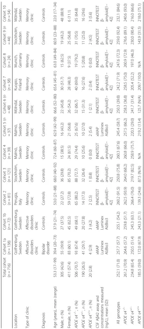
The majority of individuals in the total cohort (70.7%) lacked the APOEε4 allele, with 26.5% being heterozygous and 2.8% being homozygous APOE ε4 carriers (Table 1). The subcohort consisting of patients with bipolar disorder had similarAPOEε4 genotype frequency (P= 0.633) as well as similar concentrations of CSF Aβ42 (P= 0.302) com-pared to the healthy volunteers and was therefore pooled with the rest of the total cohort (data not shown). There were neither any gender differences with regards toAPOE ε4 genotype frequency (P= 0.586) or CSF Aβ42 concentra-tions (P= 0.534). Table 2 presents detailed demographic and biochemical data for all of the subcohorts included in the analysis.
CSF Aβ42 concentrations in relation toAPOEgenotype
In the total cohort, CSF Aβ42 concentrations were lower in APOEε4 carriers than in noncarriers in a gene dose-dependent manner (P< 0.001, Table 1), which is in keep-ing with earlier findkeep-ings [14]. However, when dividkeep-ing the total cohort into tertiles according to age, the effect was present in the middle and upper tertiles among individuals aged 46 or older (P< 0.001, Table 1), whereas in the lower tertile, containing individuals aged 45 or younger, the dif-ference was nonsignificant (P= 0.203, Table 1).
CSF Aβ42 concentrations across different age groups
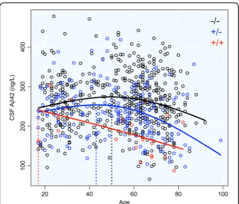
The estimated curves showed an initial upslope of CSF Aβ42 concentrations inAPOEε4-negative individuals and heterozygous APOE ε4 carriers followed by a steep des-cent (Fig. 1). Aβ42 concentrations in homozygousAPOE ε4 carriers, however, descended from an early age lacking the initial upslope. The age of initial descent, defined as the age at which CSF Aβ42 reaches its maximum, was es-timated at 50 (95% confidence interval (CI) 42–54) years forAPOEε4-negative individuals and 43 (95% CI 17–48) years for heterozygous APOE ε4 carriers. This number could not be estimated in homozygousAPOEε4 carriers, as they lacked the initial upslope.
Discussion
We conducted a large multicenter study to assess how ef-fects of the APOE ε2/ε3/ε4 polymorphism on CSF Aβ42 concentrations vary by age in cognitively healthy individuals. The main findings were that: theAPOEε4 allele was associ-ated with lower CSF Aβ42 concentrations overall in cogni-tively healthy people; the effects ofAPOEε4 were present in older people but not in young people; and CSF Aβ42 started to decline at age 50 in people without theAPOEε4 allele, at age 43 in people carrying oneAPOEε4 allele, and even earl-ier in people carrying twoAPOEε4 alleles. Taken together, these findings show that APOE ε4 strongly modulates the effect of age on CSF Aβ42 in cognitively healthy people, and points to important age-dependent effects ofAPOE ε4 on the development of preclinical AD.
Comparisons of CSF Aβ42 and amyloid PET imaging in cognitively healthy people suggest that the first decline in CSF Aβ42 does not always translate to widespread cerebral amyloid deposition [7, 8, 21]. The age at which CSF Aβ42
Table 1Demographic, genetic, and biochemical data in the total cohort as well as divided into three age tertiles
Total cohort (n= 716) ≤45 years (n= 237) 46–64 years (n= 242) ≥65 years (n= 237)
Demographic
Age (years), mean (range) 53.3 (17–99) 29.9 (17–45) 57.3 (46–64) 72.6 (65–99)
Male,n(%) 305 (42.6) 114 (48.1) 104 (43.0) 87 (36.7)
Female,n(%) 411 (57.4) 123 (51.9) 138 (57.0) 150 (63.3)
Genetic
APOEε4–/–,n(%) 506 (70.7) 162 (68.4) 172 (71.1) 172 (72.6)
APOEε4+/–,n(%) 190 (26.5) 69 (29.1) 64 (26.4) 57 (24.1)
APOEε4+/+,n(%) 20 (2.8) 6 (2.5) 6 (2.5) 8 (3.4)
Biochemical: CSF Aβ42 (ng/L), mean (SD)
All genotypes 252.1 (71.0) 251.9 (67.8) 264.9 (67.7) 239.2 (75.2)
APOEε4–/– 261.2 (70.9) 257.2 (69.5) 274.5 (68.1) 251.8 (73.4)
APOEε4+/– 234.8 (65.4) 241.4 (64.0) 248.1 (58.3) 211.9 (69.7)
APOEε4+/+ 185.5 (59.0) 231.3 (51.8) 167.8 (39.1) 164.4 (62.1)
Pvalue* <0.001 0.203 <0.001 <0.001
*Pvalues indicate comparisons of CSF Aβ42 concentrations between theAPOEε4 carrier groups (for the total cohort as well as for each of the tertiles)
Table 2 Demographic, genetic, and biochemical data in the individual subcohorts Total cohort ( n =7 1 6 ) Cohort 1a ( n = 138) Cohort 1b ( n = 72) Coho rt 2 ( n = 81) Coh ort 3 ( n =1 2 1 ) Coh ort 4 ( n = 39) Cohort 5 ( n = 37) Cohort 6 ( n = 48) Coho rt 7 ( n = 58) Coho rt 8 ( n =2 4 ) Coh ort 9 ( n = 44) Co hort 10 ( n = 54) Loca tion Gothenb urg, Sweden Gothenb urg, Swed en Perug ia, Italy Stoc kholm, Sw eden Mal mö, Sw eden Malmö, Sweden Mölndal, Sweden Kuopio , Finland Mun ich, Germ any Sto ckholm, Sw eden Möl ndal, Sw eden Type of clinic
Affective disorders clinic
Affecti ve disorde rs clinic Mem ory clinic Mem ory clinic Mem ory clinic Memo ry clinic Mem ory clinic Mem ory clinic Mem ory clinic Mem ory clinic Me mory cl inic Diag nosis Bipolar disorder Contr ols Contr ols Con trols Con trols Contro ls Contr ols Contr ols Contr ols Con trols Co ntrols Age (years), mean (range) 53.3 (17 – 99) 39.4 (20 – 73) 37.9 (21 – 74) 53.0 (21 – 88) 68.0 (40 – 92 ) 72 .4 (60 – 87) 62.9 (42 – 99) 66.6 (52 – 80) 65.6 (45 – 81) 63.0 (49 – 84 ) 60.9 (23 – 88) 22 .0 (17 – 34) Male, n (%) 305 (42. 6) 55 (39. 9) 27 (37.5) 22 (27.2) 36 (29.8 ) 15 (38.5 ) 16 (43. 2) 22 (45. 8) 30 (51.7) 15 (62.5 ) 19 (43.2 ) 48 (88.9 ) Fema le, n (%) 411 (57. 4) 83 (60. 1) 45 (62.5) 59 (72.8) 85 (70.2 ) 24 (61.5 ) 21 (56. 8) 26 (54. 2) 28 (48.3) 9 (37.5 ) 25 (56.8 ) 6 (11.1) APOE ε 4 – /
–,n
(%) 506 (70. 7) 93 (67. 4) 49 (68.1) 65 (80.2) 88 (72.7 ) 29 (74.4 ) 25 (67. 6) 32 (66. 7) 40 (69.0) 19 (79.2 ) 31 (70.5 ) 35 (64.8 ) APOE ε 4 +/
–,n
concentrations start to decrease may therefore be the start-ing point for preclinical pathological disturbances in amyl-oid homeostasis, which ultimately results in amylamyl-oid accumulation that later becomes detectable on amyloid PET imaging. The data from this study suggest that the dis-turbed amyloid homeostasis occurs on average from age 50 inAPOEε4-negative individuals, and almost a decade earl-ier inAPOEε4 heterozygous people. InAPOEε4 homozy-gous people we estimated a descent in CSF Aβ42 already from age 17, but the sparsity of data among young homozy-gous APOEε4 carriers makes this estimate uncertain, and we can only conclude that the decline in CSF Aβ42 starts considerably earlier in homozygousAPOEε4 carriers com-pared with heterozygous APOE ε4 carriers or noncarriers. Importantly, previous studies provide convergent evidence that emerging amyloid pathology, defined as decreased CSF Aβ42 concentrations, or CSF Aβ42 concentrations slightly above conventional thresholds for amyloid positivity, may have deleterious effects on brain structure, brain function, and cognition [22–25]. This highlights the importance of detecting the earliest effects of APOEε4 on CSF Aβ42 in order to provide very early diagnostics and potentially initi-ate prevention of AD.
The fact that APOE ε4 affected CSF Aβ42 concen-trations already from 43 years of age is interesting since a previous study found that APOE ε4 was asso-ciated with cognitive decline only after 50 years of age [26]. We therefore suggest that there is an intermediate period of early alterations in amyloid homeostasis before cognitive decline becomes detect-able [23], when amyloid accumulation slowly builds up together with downstream pathological events
(including spread of tau tangles), which ultimately translate to cognitive decline several years later.
This study has several limitations. First, we used cross-sectional data from several cohorts and several assays to measure CSF Aβ42. Although we employed normalization measures to bridge all results, the variability in cohorts and assays increases the variance of our models and estimates. Future studies are needed to verify these results in a mono-center setting, obviating the need for data normalization across cohorts. Second, the low number ofAPOEε4 homo-zygous people, along with the sparsity of data in the age span between 85 and 100 years, limits our ability to model effects of APOE ε4 homozygosity and effects in the final part of the natural life span. Also, the lack of APOE ε4 homozygous people between age 35 and 50 makes it im-possible to define whether there is a plateau in Aβ42 con-centrations before decline or whether the concon-centrations drop directly from age 17 in homozygousAPOEε4 carriers.
Conclusions
To sum up, the results of this study suggest that the process of preclinical Aβ pathology might start in early middle age in APOEε4 carriers. Hence, we hypothesize that theAPOEε4 allele affects CSF Aβ42 concentrations by speeding up the process of preclinical Aβ accumula-tion and deposiaccumula-tion in the brain. Studies addressing the molecular mechanisms behind the association between ApoE and cerebral Aβbuild-up are needed to verify this.
Abbreviations
AD:Alzheimer’s disease; ANOVA: Analysis of variance;APOE: Apolipoprotein E; Aβ42: Beta-amyloid1–42; CI: Confidence interval; CSF: Cerebrospinal fluid;
MCI: Mild cognitive impairment; PET: Positron emission tomography; P-tau: Phosphorylated tau; T-P-tau: Total tau
Acknowledgements Not applicable.
Funding
This study was supported by grants from the Swedish Research Council, the European Research Council, Frimurarestiftelsen, Stiftelsen Gamla Tjänarinnor, the Swedish Alzheimer Foundation, the Swedish Brain Foundation, the Knut and Alice Wallenberg Foundation, the Marianne and Marcus Wallenberg Foundation, and the Torsten Söderberg Foundation. HH is supported by the AXA Research Fund, the Fondation Université Pierre et Marie Curie, and the Fondation pour la Recherche sur Alzheimer, Paris, France. The research leading to these results has received funding from the program
“Investissements d’avenir”ANR-10-IAIHU-06 (to HH). Availability of data and materials
The datasets used and/or analyzed during the current study are available from the corresponding author on reasonable request.
Authors’contributions
RL, KB, NM, and HZ created the concept and design for the study. RL, BO, ML, GBF, S-KH, HH, AW, OH, KB, NM, and HZ acquired, analyzed, and interpreted data. RL, PSI, NM, and HZ drafted the manuscript. RL, PSI, and NM performed the statistical analyses. The study was supervised by NM and HZ. All authors contributed critical revision for important intellectual content and read and approved the final manuscript.
Fig. 1CSF Aβ42 concentrations plotted against age. Mean curves
Ethics approval and consent to participate
The study received approval from regional ethical committees at the Universities of Gothenburg and Lund (Sweden), Brescia (Italy), Kuopio (Finland), and Munich (Germany) and followed the tenets of the Helsinki declaration. Written informed consent was obtained from all participants.
Consent for publication Not applicable.
Competing interests
ML declares that, over the past 36 months, he has received lecture honoraria from Lundbeck and AstraZeneca Sweden, and served as scientific consultant for EPID Research Oy; he has no other equity ownership, profit-sharing agreements, royalties, or patents. HH declares no competing financial interests related to the present article; he serves as Senior Associate Editor for the journalAlzheimer's &
Dementia; he has been a scientific consultant and/or speaker and/or attended scientific advisory boards of Axovant, Anavex, Eli Lilly and company, GE Healthcare, Cytox, Qynapse, Roche, Biogen Idec, Takeda-Zinfandel, Oryzon Genomics; he receives research support from the Fondation for Alzheimer Research (Paris), COLAM Initiatives (Paris), IHU-A-ICM (Paris), Pierre and Marie Curie University (Paris), Pfizer & Avid (paid to institution); and he has patents as inventor, but received no royalties. OH has acquired research support (for the institution) from Roche, GE Healthcare, Biogen, AVID Radiopharmaceuticals, Fujirebio, and Euroimmun; in the past 2 years, he has received consultancy/speaker fees (paid to the institution) from Lilly, Roche, and Fujirebio. KB has served as a consultant or on advisory boards for Alzheon, BioArctic, Biogen, Eli Lilly, Fujirebio Europe, IBL International, Pfizer, and Roche Diagnostics. HZ has served on the scientific advisory board for Roche Diagnostics, Eli Lilly and Pharmasum Therapeutics. KB and HZ are cofounders of Brain Biomarker Solutions in Gothenburg AB, a GU Ventures-based platform company at the University of Gothenburg. RL, PSI, TS, BO, GBF, S-KH, AW, LM, and NM declare that they have no competing interests.
Publisher’s Note
Springer Nature remains neutral with regard to jurisdictional claims in published maps and institutional affiliations.
Author details
1
Department of Psychiatry and Neurochemistry, Institute of Neuroscience and Physiology, Sahlgrenska Academy at the University of Gothenburg,
Gothenburg, Sweden.2Clinical Neurochemistry Laboratory, Sahlgrenska
University Hospital, Mölndal, Sweden.3Clinical Memory Research Unit,
Department of Clinical Sciences Malmö, Lund University, Lund, Sweden.
4Department of Veterans Affairs Medical Center, Center for Imaging of
Neurodegenerative Diseases, San Francisco, CA, USA.5Department of
Radiology and Biomedical Imaging, University of California, San Francisco,
CA, USA.6Department of Medical Epidemiology and Biostatistics, Karolinska
Institutet, Stockholm, Sweden.7Istituto di Ricovero e Cura a Carattere
Scientifico Centro San Giovanni di Dio Fatebenefratelli, Brescia, Italy.
8Department of Neurology, University of Eastern Finland, Kuopio University
Hospital, Kuopio, Finland.9AXA Research Fund and UPMC Chair, Sorbonne
Universités, Université Pierre et Marie Curie (UPMC) Paris 06, Inserm, CNRS, Institut du cerveau et de la moelle (ICM), Département de Neurologie,
Institut de la Mémoire et de la Maladie d’Alzheimer (IM2A), Hôpital
Pitié-Salpêtrière, Boulevard de l’hôpital, F-75013 Paris, France.10Memory
Clinic, Skåne University Hospital, Malmö, Sweden.11Department of
Neurology, Skåne University Hospital, Lund, Sweden.12Department of
Molecular Neuroscience, UCL Institute of Neurology, Queen Square, London WC1N 3BG, UK.
Received: 12 June 2017 Accepted: 3 October 2017
References
1. Blennow K, de Leon MJ, Zetterberg H. Alzheimer's disease. Lancet. 2006; 368:387–403.
2. Corder EH, Saunders AM, Strittmatter WJ, Schmechel DE, Gaskell PC, Small GW, Roses AD, Haines JL, Pericak-Vance MA. Gene dose of apolipoprotein E type 4 allele and the risk of Alzheimer's disease in late onset families. Science. 1993;261:921–3.
3. Holtzman DM, Herz J, Bu G. Apolipoprotein E and apolipoprotein E receptors: normal biology and roles in Alzheimer disease. Cold Spring Harb Perspect Med. 2012;2:a006312.
4. Castellano JM, Kim J, Stewart FR, Jiang H, DeMattos RB, Patterson BW, Fagan AM, Morris JC, Mawuenyega KG, Cruchaga C, et al. Human apoE isoforms differentially regulate brain amyloid-βpeptide clearance. Sci Transl Med. 2011;3:89ra57.
5. Verghese PB, Castellano JM, Garai K, Wang Y, Jiang H, Shah A, Bu G, Frieden C, Holtzman DM. ApoE influences amyloid-β(Aβ) clearance despite minimal apoE/Aβassociation in physiological conditions. Proc Natl Acad Sci U S A. 2013;110:E1807–16.
6. Blennow K, Hampel H, Weiner M, Zetterberg H. Cerebrospinal fluid and plasma biomarkers in Alzheimer disease. Nat Rev Neurol. 2010;6:131–44.
7. Mattsson N, Insel PS, Donohue M, Landau S, Jagust WJ, Shaw LM, Trojanowski JQ, Zetterberg H, Blennow K, Weiner MW. Independent information from cerebrospinal fluid amyloid-βand florbetapir imaging in Alzheimer's disease. Brain. 2015;138:772–83.
8. Palmqvist S, Mattsson N, Hansson O. Cerebrospinal fluid analysis detects cerebral amyloid-βaccumulation earlier than positron emission tomography. Brain. 2016;139:1226–36.
9. Blennow K, Mattsson N, Scholl M, Hansson O, Zetterberg H. Amyloid biomarkers in Alzheimer's disease. Trends Pharmacol Sci. 2015;36: 297–309.
10. Galasko D, Chang L, Motter R, Clark CM, Kaye J, Knopman D, Thomas R, Kholodenko D, Schenk D, Lieberburg I, et al. High cerebrospinal fluid tau and low amyloidβ42 levels in the clinical diagnosis of Alzheimer disease and relation to apolipoprotein E genotype. Arch Neurol. 1998;55:937–45. 11. Sunderland T, Mirza N, Putnam KT, Linker G, Bhupali D, Durham R, Soares H,
Kimmel L, Friedman D, Bergeson J, et al. Cerebrospinal fluidβ-amyloid1-42
and tau in control subjects at risk for Alzheimer's disease: the effect of APOE ε4 allele. Biol Psychiatry. 2004;56:670–6.
12. Shaw LM, Vanderstichele H, Knapik-Czajka M, Clark CM, Aisen PS, Petersen RC, Blennow K, Soares H, Simon A, Lewczuk P, et al. Cerebrospinal fluid biomarker signature in Alzheimer's disease neuroimaging initiative subjects. Ann Neurol. 2009;65:403–13.
13. Vemuri P, Wiste HJ, Weigand SD, Knopman DS, Shaw LM, Trojanowski JQ, Aisen PS, Weiner M, Petersen RC, Jack Jr CR. Effect of apolipoprotein E on biomarkers of amyloid load and neuronal pathology in Alzheimer disease. Ann Neurol. 2010;67:308–16.
14. Lautner R, Palmqvist S, Mattsson N, Andreasson U, Wallin A, Palsson E, Jakobsson J, Herukka SK, Owenius R, Olsson B, et al. Apolipoprotein E genotype and the diagnostic accuracy of cerebrospinal fluid biomarkers for Alzheimer disease. JAMA Psychiat. 2014;71:1183–91.
15. Prince JA, Zetterberg H, Andreasen N, Marcusson J, Blennow K. APOEε4 allele is associated with reduced cerebrospinal fluid levels of Aβ42. Neurology. 2004;62:2116–8.
16. Schipke CG, Jessen F, Teipel S, Luckhaus C, Wiltfang J, Esselmann H, Frolich L, Maier W, Ruther E, Heppner FL, et al. Long-term stability of Alzheimer's disease biomarker proteins in cerebrospinal fluid. J Alzheimers Dis. 2011;26:255–62.
17. Kuhlmann J, Andreasson U, Pannee J, Bjerke M, Portelius E, Leinenbach A, Bittner T, Korecka M, Jenkins RG, Vanderstichele H, et al. CSF Aβ1-42—an
excellent but complicated Alzheimer's biomarker—a route to standardisation. Clin Chim Acta. 2017;467:27–33.
18. Bjerke M, Portelius E, Minthon L, Wallin A, Anckarsater H, Anckarsater R, Andreasen N, Zetterberg H, Andreasson U, Blennow K. Confounding factors influencing amyloid beta concentration in cerebrospinal fluid. Int J Alzheimers Dis. 2010;2010:986310.
19. Andreasen N, Hesse C, Davidsson P, Minthon L, Wallin A, Winblad B, Vanderstichele H, Vanmechelen E, Blennow K. Cerebrospinal fluidβ -amyloid(1-42)in Alzheimer disease: differences between early- and
late-onset Alzheimer disease and stability during the course of disease. Arch Neurol. 1999;56:673–80.
20. Olsson A, Vanderstichele H, Andreasen N, De Meyer G, Wallin A, Holmberg B, Rosengren L, Vanmechelen E, Blennow K. Simultaneous measurement of β-amyloid(1-42), total tau, and phosphorylated tau (Thr181) in cerebrospinal
fluid by the xMAP technology. Clin Chem. 2005;51:336–45.
22. Insel PS, Mattsson N, Donohue MC, Mackin RS, Aisen PS, Jack Jr CR, Shaw LM, Trojanowski JQ, Weiner MW. The transitional association betweenβ-amyloid pathology and regional brain atrophy. Alzheimers Dement. 2015;11:1171–9. 23. Insel PS, Mattsson N, Mackin RS, Scholl M, Nosheny RL, Tosun D, Donohue
MC, Aisen PS, Jagust WJ, Weiner MW. Accelerating rates of cognitive decline and imaging markers associated withβ-amyloid pathology. Neurology. 2016;86:1887–96.
24. Mattsson N, Insel PS, Nosheny R, Tosun D, Trojanowski JQ, Shaw LM, Jack Jr CR, Donohue MC, Weiner MW. Emergingβ-amyloid pathology and accelerated cortical atrophy. JAMA Neurol. 2014;71:725–34.
25. Mattsson N, Insel PS, Donohue M, Jagust W, Sperling R, Aisen P, Weiner MW. Predicting reduction of cerebrospinal fluidβ-amyloid 42 in cognitively healthy controls. JAMA Neurol. 2015;72:554–60.
26. Caselli RJ, Dueck AC, Osborne D, Sabbagh MN, Connor DJ, Ahern GL, Baxter LC, Rapcsak SZ, Shi J, Woodruff BK, et al. Longitudinal modeling of age-related memory decline and the APOEε4 effect. N Engl J Med. 2009;361:255–63.
• We accept pre-submission inquiries
• Our selector tool helps you to find the most relevant journal
• We provide round the clock customer support
• Convenient online submission
• Thorough peer review
• Inclusion in PubMed and all major indexing services
• Maximum visibility for your research
Submit your manuscript at www.biomedcentral.com/submit

![catena Poly[2,2′,2′′ nitrilotris(ethanaminium) [tri μ oxido tris[dioxidovanadate(V)]] monohydrate]](data:image/gif;base64,R0lGODlhAQABAIAAAP///wAAACH5BAEAAAAALAAAAAABAAEAAAICRAEAOw==)