R E V I E W
Open Access
microRNA Regulation in Estrogen
Receptor-Positive Breast Cancer and Endocrine
Therapy
Erin W. Howard and Xiaohe Yang
*Abstract
As de novo and acquired resistance to standard first line endocrine therapies is a growing clinical challenge for estrogen receptor-positive (ER+) breast cancer patients, understanding the mechanisms of resistance is critical to develop novel therapeutic strategies to prevent therapeutic resistance and improve patient outcomes. The widespread post-transcriptional regulatory role that microRNAs (miRNAs) can have on various oncogenic pathways has been well-documented. In particular, several miRNAs are reported to suppress ERαexpression via direct binding with the 3’UTR ofESR1mRNA, which can confer resistance to estrogen/ERα-targeted therapies. In turn, estrogen/ ERαactivation can modulate miRNA expression, which may contribute to ER+breast carcinogenesis. Given the reported oncogenic and tumor suppressor functions of miRNAs in ER+breast cancer, the targeted regulation of specific miRNAs is emerging as a promising strategy to treat ER+breast cancer and significantly improve patient responsiveness to endocrine therapies. In this review, we highlight the major miRNA-ER regulatory mechanisms in context with ER+breast carcinogenesis, as well as the critical miRNAs that contribute to endocrine therapy resistance or sensitivity. Collectively, this comprehensive review of the current literature sheds light on the clinical applications and challenges associated with miRNA regulatory mechanisms and novel miRNA targets that may have translational value as potential therapeutics for the treatment of ER+breast cancer.
Keywords:Estrogen receptor alpha (ERα), ER+breast cancer, microRNA (miRNA), miRNA biogenesis, Endocrine therapy resistance, ER-receptor tyrosine kinase (RTK) crosstalk
Background
Estrogen receptor-positive (ER+) breast cancer accounts for nearly 65% of breast cancers. In particular, due to the asso-ciation of hormone levels and ER activation/regulation, the ER+breast cancer subtype is particularly prevalent in post-menopausal women taking hormone replacement therapy. The activation of ERα, a major transcription factor, pro-motes the transcription of a number of target genes that regulate oncogenic processes, including cell cycle progres-sion, cell survival, and epithelial/luminal cell differentiation. Although anti-estrogens, selective ER modulators (SERMs), and selective ER downregulators (SERDs), such as tamoxi-fen and fulvestrant, are leading treatment strategies that
can block mitogenic estrogen activity and have significantly improved ER+breast cancer patient outcomes, therapeutic resistance remains a significant clinical challenge. There-fore, understanding the mechanisms that regulate estrogen/ ER-mediated oncogenic activity will shed light on novel therapeutic targets to more effectively treat ER+ breast cancer.
Within the past 15 years, substantial research has highlighted the role of microRNA (miRNA) regulation that can contribute to ER+ breast cancer risk or preven-tion. In particular, studies have not only demonstrated that miRNAs are targets of ERα/hormonal signaling, but have also shown that ERαis a regulatory target of mul-tiple miRNAs. In this review, we highlight miRNA signa-tures that are associated with ER+and ER−breast cancer subtypes, as well as key tumor suppressor miRNAs and oncomiRs that are deregulated in ER+breast cancer and modulate therapeutic resistance. Importantly, we also * Correspondence:xyang@nccu.edu
Julius L. Chambers Biomedical/Biotechnology Research Institute, Department of Biological and Biomedical Sciences, North Carolina Central University, North Carolina Research Campus, 500 Laureate Way, NRI 4301, Kannapolis, North Carolina 28081, USA
discuss the potential clinical applications of novel thera-peutic strategies targeting miRNAs, and their role in the treatment of therapeutic-resistant ER+breast cancers.
miRNAs Associated with ER+Versus ER−Breast Cancer Subtypes
The identification miRNA profiles that are associated with specific cancers or predict clinical outcomes has been a major research focus in the past decade or more. In particular, miRNA expression profiling of human breast cancer subtypes, as classified by ER, progesterone receptor, and ErbB2/Her2 receptor statuses, has revealed miRNA signatures that not only correlate with the mo-lecular subtype, but can also serve as potential prognos-tic markers and indicators of patient responses to facilitate personalized treatment approaches. As summa-rized in Table 1, numerous reports have identified miR-NAs that are differentially expressed in ER+ versus ER− breast cancer samples. Many of the miRNAs that are downregulated in ER+ breast cancer are tumor suppres-sor miRNAs, such as miR-206 [1, 2], that function to suppress cell proliferation and survival. The inhibition of tumor suppressor miRNAs consequently contributes to the oncogenic phenotypes associated with ER+ breast cancer, such as a high proliferative index in the intrinsic Luminal B subtype of ER+breast cancers [3]. Indeed, the development of targeted therapeutics to upregulate these particular tumor suppressor miRNAs may serve as a promising strategy to treat ER+ breast cancer. In con-trast, several oncomiRs have consistently been reported to be upregulated in ER+breast cancer, such as miR-10b and miR-21 [4–6]. In particular, the tumor suppressor
PTEN, which dephosphorylates PIP3and in turn blocks PI3K/Akt signaling, is a direct target of many oncomiRs that are overexpressed in ER+ breast cancer (Table 1). Thus, the attenuation ofPTEN-mediated blockage of the PI3K/Akt signaling by the aberrant overexpression of oncomiRs in ER+ breast cancer contributes to down-stream Akt signaling, which promotes oncogenic cell growth, survival, and migration. The targeted inhibition of these oncomiRs could also provide alternative treat-ment options for ER+ breast cancer patients. As dis-cussed in the following sections, miRNAs play an integral role in the regulation of estrogen/ERα signaling and many miRNAs can also be modulated by ERα acti-vation. Yet, miRNA-mediated regulation of ERα expres-sion/activity and other oncogenic signaling pathways are linked to resistance to standard first-line endocrine therapies.
miRNAs and Estrogen/ERαRegulation
miRNAs that Regulate ERα
Many miRNAs that are associated with ER+/ER− breast cancer subtypes also directly target or indirectly regulate
ESR1/ERα. To this end, manyESR1-targeting miRNAs are downregulated in ER+breast cancer or are upregulated in endocrine therapy-resistant breast cancers (Table 2). In this section, the role of theseESR1-targeting miRNAs in ER signaling regulation will be discussed in context with ER+breast carcinogenesis and prevention.
miR-22
miR-22 is a classical example of the negative correlation betweenESR1-targeting miRNA expression and ER status in breast cancer. As such, miR-22 is downregulated in ER+ breast cancer cell lines, as well as clinical samples [7]. Studies have also consistently reported that miR-22 can post-transcriptionally inhibit ERα expression/function by directly binding to the 3’UTR ofESR1[7–9]. Indeed, the overexpression of miR-22 blocks ERα-dependent cell pro-liferation and ER-mediated transcriptional activity in vitro [9]. miR-22 was additionally found to indirectly inhibit ERα activity through the direct suppression of the ERα transcriptional coactivator, Sp1 [10]. The induction of cel-lular senescence by miR-22 overexpression also appeared to correlate with a less invasive cellular phenotype in hu-man breast and cervical cancer cell lines [10]. These in vitro data were consistent with results demonstrating that miR-22 overexpression also promotes senescence and re-duces the proliferative index of mammary tumor cells in a murine fat pad orthotopic mammary tumor model. In turn, these miR-22-induced cellular responses suppressed overall mammary tumor growth and reduced the appear-ance of metastatic tumors in vivo [10]. Together, the dir-ect and indirdir-ect mechanisms of ESR1/ERα inhibition utilized by miR-22 suppress the oncogenic phenotypes as-sociated with downstream ERα signaling activity, which makes miR-22 a promising candidate miRNA for thera-peutic strategies to treat ER+breast cancer.
miR-206
Table 1Differentially expressed miRNAs in ER+vs. ER−breast cancers and their major direct mRNA targets
miRNAs Expression levels in ER+(vs. ER−) breast cancer
Direct miRNA targets [117]
let-7a-c/f High [4] CDK6, KRAS, NRAS, HRAS, ITGB3,NF2, HMGA1/2, EWSR1, DICER, AGO1, LIN28A/B, CASP3/8/9, PARP1, IL6/10, E2F2,CCND1/2, CDC34, CDC25A, EZH2, WNT1, MAPK4K4, IRS2,IGF1R, TGFBR1, AKT2,
MYC, NUMB
miR-9 Low [118] ITGB1, RCOR1, FOXO1/3, CDH1, MAPK1/3, MMP9, TAZ, NOTCH2, HES1, CBX7, E2F1, RAB34, NFKB1, SIRT1, CCNG1, SOCS5, CREB1, DICER1,NF1, CXCR4, TGFBR2, BECN1
miR-10a/b High [4,5] MAP3K7,EPHA4, HOXA1, HOXB3, ACTG1,PTEN, USF2, PIK3CA/G,SERPINE1, MMP14,NCOR2, CKDN1A/2A, KLF4, PPARA,NF1, PLK1, CCNA2, ZEB1
miR-18a/b High [57]
Low [2,4,11] ESR1
, KRAS,PTEN, CTGF, NR3C1, HIF1A, TGFBR2, SMAD2/4, HSF2, ATM, DICER,BCL2, IRF2, RUNX1, MEF2D, CBX7, TNFAIP3, FOXN1
miR-19a/b Low [83] ESR1, HOXA5,PTEN,CCND1, ERBB4, ATXN1, KAT2B, SOCS1, TGFBR2, BMPR2, KIT, TLR2, TNF, TNFAIP3, FOXP1, BTG1, PIK3CA, MAP3K5, CUL5, AKT1, FGFR2, HIPK1
miR-21 High [6] RASGRP1, CDC25A,BCL2, JAF1, SMARCA4, SPRY2, DUSP10, TIMP3, SOX2/5, MTAP, DOCK5/7, RECK, TGFBR2/3,PTEN, E2F1, TGFBI,SP1, APAF1, BTG2,PDCD4, RHOB, BMPR2,NCOA3, TP63, MSH2/6, TIAM1, EGFR, ERBB2, ICAM1, PPARA, NTF3,COL4A1, SMAD7, MAP2K3, MAT2A/2B, STAT3, LRP6, FZD6, BMI1, SOCS1, FOXO1, CASP8, VEGFA
miR-22 Low [7] PTMS, ERBB3, ARPC5,BMP7, PPARA,ESR1,NCOA1, HDAC4/6, HMGB1,SP1, RAB5B, TET2,CDKN1A, WNT1, SIRT1, NET1, TIAM1, MMP14, SNAI1, CXCR2, AKT1, CDK6
miR-25 Low [4] BCL2L11, CCL26, KLF4, CDKN1C, KAT2B,TP53, CDH1,MDM2,PTEN, EZH2, SMAD7, ERBB2
miR-26a/b High [36] HMGA1/2, CCND2, CCNE1/2,ESR1, CDK6, CDC6,PTEN, EZH2, SERBP1, SMAD1/4, REB1, MAP3K2, GSK3B, NOS2, CHD1, FGF9, ATM, HGF, IGF1, LIN28B, DNMT3B, WEE1, ADAM17, CHEK1, ITGA5, DUSP4/5, NRAS, E2F2, PTGS2, JAG1,MYC, GATA4, TAB1, RB1, COX2
miR-29b High [36] TGFB1/3, HDAC4, COL1A1, COL3A1,COL4A1/2, COL5A2,SP1, CDK6, PPP1R13B,PTEN, DNMT1, DNMT3A/B, MCL1,BCL2, VEGFA, TET1/2, CDC42, MMP2/9/15/24, ADAM12, BMP1, HMGA2, GSK3B, INFG, PIK3R1, CCND2, SNAI3, AKT2/3, ITGA6,GATA3, PDGFRA, PDGRA-C, STAT3,
miR-30a-d High [4,36] DTL, SMAD1, CDK6, NOTCH1, BECN1, SNAI1, PIK3CD, PRDM1, ABL1,VIM,ESR2, RUNX2, BCL9, SOX4, CBX3,TP53, NCAM1, DNMT1, ITGB3, ATF1, CCND2, CCNE1/2, RPA1, TET1, NOTCH1, KLF9, CAT, ATG12,
SERPINE1, DLL4, PDGFRB,BCL2, EIF5A2, ADAM12, HOXA1, SOCS1/3, RUNX2, CASP3, PAK1, MCL1, FOXO3, EZH2
miR-93 Low [4,119] CDKN1A, TP53INP1, E2F1, VEGFA, ITGB8, KAT2B, TUSC2,PTEN, LATS2, TGFBR2, DAB2, SMAD7, ZBTB4, CXCL8,PDCD4, ANG, PTENP1, NEDD4L, MMP3, ZNRF3,FOXA1
miR-106b Low [4] ITCH, APP,CDKN1A, E2F1, KAT2B, RB1,PTEN, APC, CASP7,JAK1, SETD2, SMAD7, STAT3, ZBTB4, TWIST1, HIF1A, TRIM8, RUNX3, DAB2, PTENP1, CYBB, TNFSF10A/11
miR-130a High [4] HOXA5/10, ATXN1, KLF4, PPARG,ESR1, DICER1, RUNX3, RAB5A, SMAD4, TNF, TGFBR2, TGFB1, PPARA,
MYC,PTEN, DLL4, MAP3K12, MECP2, MET
miR-135b Low [120] APC, KLF4, CASR, PPP2R5C, LATS2, SMAD5, MID1, MTCH2, BMPR2, TGFBR1, FOXO1
miR-142 Low [4] RAC1, ARNTL, TGFBR1/2, BOD1, PROM1, ROCK2, CCNT2, TAB2, HMGA1/B1, PTPN23, TP53INP1, IL1A, HSPA1B, SMAD3, ABCG2, LGR5, ZEB1, SIRT1, HIF1A, SOCS1,PTEN, DOCK6
miR-145 High [121] BNIP3, KLF4/5, SOX2/9, MUC1,CDKN1A, ITGB8, STAT1, YES1, CLINT1, IRS1/2, VEGFA, HOXA9,MYC, FLI1, IFNB1,IGF1R, FZD7, CDK4/6,SERPINE1,ESR1, JAG1, NEDD9, PAK4, ERG, NRAS, ADAM17, CDH2, EPAS1, ETS1, CD44, BRAF, SMAD2/3, TGFBR3, CTNND1,SP1/7, TNFSF13, DDX6, ARF6, ADD3, HMGA2, ROCK1, HDAC11, SENP1, NAIP, TUG1, TGFB2, EGFR, ACTB
miR-148 Low [4] DNMT1/3B, TGIF2, MCL1, IRS1,BCL2, ITGA5/B8, ROCK1, PIK3CA, NRAS, CSF1, CDC25B, MAP3K4/9, MMP7, WNT1/10B, CDKN1B,SERPINE1, SMAD2, MET, USP4, STAT3, ALCAM, TGFB2, AKT2, BAX, CYBB, NRP1,
miR-150 Low [4] MYB, MUC4, EGR2, ZEB1, EP300,TP53, CBL,SP1, CREB1, STAT1/5B, CTNNB1, NANOG, MMP14, ZNF350,
IGF1R, BIRC5
miR-155 Low [4] TAB2, SOCS1/6, MSH2/6, MLH1, DET1, SMAD1/2/5, ZNF652, ZIC3, BACH1, APC, TRIM32, RHOA, TP53INP1, SPI1, FOXO3, RUNX2,JUN, ETS1, ICAM1, IRAK3, MYB, SKI, SOX6, FADD, BCL6, NOS3,CCND1, NFKB1, E2F2,MYC, DOCK1, RAD51,PTEN, ERBB2, RPTOR, TFAM, STAT1, SIRT1,TP53
miR-185 Low [36] RHOA, CDC42, SIX1, DNMT1, EPAS1, ARC, NFATC3, VEGFA, MZB1, HMGA1/2,IGF1R, DUSP4, CASP14, EPHB2, SMAD7, TRIM29, TGFB1, AKT1, CCNE1, CDK6, ATR, EZH2
miR-187 Low [4] TNF, BCL6, CRMP1,CYP1B1, FOXA2, ALDH1A3
miR-206 also directly targets the 3’UTR ofMET, which is associated with aggressive breast cancer phenotypes [12, 17]. Together, these reports further validate the miR-206-induced effects on ERα expression/activation and ER-mediated breast carcinogenesis [14]. The cell cycle and growth-promoting targets of miR-206 also highlight the potential application of miR-206 expression restoration as a potential therapeutic strategy for ER+breast cancer, par-ticularly the luminal B subtype that is typically character-ized by a high proliferative index.
miR-221/222
Various studies have demonstrated that miR-221/222 target oncogenes, as well as tumor suppressor genes. Of particular importance in ER+breast cancer, miR-221/222 targets ESR1 [8, 11]. The negative regulation of ESR1/ ERα has significant clinical implications related to ac-quired resistance to endocrine therapies, as discussed later in this review. On the other hand, miR-221/222 can indirectly promote ERα/estrogen signaling through dir-ect inhibition of BECN1/Beclin1, which is a key
regulator of autophagy. Beclin1 can induce autophagy and suppress the tumorigenic properties of MCF-7 breast cancer xenografts in vivo [18]. Although cytokine
mda-7/IL-24-induced suppression of miR-221 has been found to result in Beclin1-associated autophagic cell death [19], Beclin1 acts as a negative regulator of ERα/ estrogen signaling, which may interfere with the efficacy of anti-estrogen therapies [20].
In addition to the regulation of estrogen/ERα signal-ing, miR-221/222 have a number of tumor suppressor targets, which suggest the oncogenic functional mecha-nisms of miR-221/222. As a potential mechanism of miR-221/222-associated tumorigenesis, key cell cycle in-hibitors have been identified as targets of miR-221/222, which contributes to oncogenic cell cycle progression. In particular, miR-221/222 are negative post-transcriptional regulators ofCDKN1B/p27Kip1, a cell cycle inhibitor [21, 22]. As such, miR-221/222 overexpression suppresses p27Kip1 expression and correspondingly promotes G1/S transition in MCF-7 and MDA-MB-231 breast cancer cell lines [23,24].In vitro studies using breast and other
Table 1Differentially expressed miRNAs in ER+vs. ER−breast cancers and their major direct mRNA targets(Continued)
miRNAs Expression levels in ER+(vs. ER−) breast cancer
Direct miRNA targets [117]
miR-191 High [36] MDM4, TMC7, NDST1, SOX4, CDK6/9, SATB1, CEBPB, BASP1, NOTCH2, EGR1, CCND2
miR-193b High [2] ESR1,CCND1, PLAU, PRAP1, MCL1, ETS1, MAX, KRAS, RAD51, MYB,NF1, SMAD3, STMN1
miR-199a/b High [4] MET, HIF1A, SMARCA2, CD44, EZH2, IKBKB, MAPK1, JUNB, DDR1, MAP3K11, CAV1/2, ERBB2, ERBB3, MTOR, SIRT1, PTGS2, HSPA5, ATF6, ERN1, CDH1/2, ZHX1, HGF, BECN1, IGF1, NFKB1, VEGFA, SNAI1, GSK3B, WNT2, FZD4/6, JAG1, HK2, TFAM, CCR7, TGFB2, PIK3CD, PAK4, CDK7, ITGA3, YAP1, MAP4K3m TGFBR1, FLT1, KDR, FOXA2, SLC27A1, AKT1, MAPK8/9/14, HES1, SET, DDR1, PAK4,
miR-206 Low [1,2,36] MET,NOTCH3,ESR1, PAX3, CCND2, CDK4, NR1H3, KRAS, SFRP1,CCND1, ANXA2, NR4A2, SMAD2, TWF1, CCL2, VEGFA, SOD1, AKT1
miR-212 Low [36] RB1, MECP2, TJP1, PEA15, PTCH1, KCNJ2, RBP2,MYC, ACHE, PXN, SOX4, SGK3, SMAD2, MAF, CCNA2/B1, AGO1
miR-214 High [4] EZH2, XBP1,PTEN, MAP2K3, MAPK1/8, ING4, TWIST1, GALNT7,TP53, CTNNB1, JAG1, FGFR1, NRAS, BIRC5, CDK3/6, RAB15, E2F2, ITCH, SUFU, CPD, PIM1
miR-217 High [120] SIRT1, ROBO1,PTEN, EZH2, E2F3, DACH1, FOXO3, PTPN14, DMNT1,IGF1R
miR-218 High [120] LAMB3, RICTOR, BIRC5/6, LASP1, IKBKB,SP1, VOPP1, ACTN1, ROBO1, SFRP2, HOXB3, DKK2, TOB1, CDK6, BMI1, HMGB1, LEF1, SLIT3, PDGFRA, GLI2, RUNX2, CDH2,TFF1, EGFR, LGR4, SMO, E2F2, CDC27, KIT, BCL9
miR-221 High [2] CDKN1B/C, BMF, FOXO3, DICER1, KIT, TMED7, ETS1, BBC3, DKK2,DDIT4, TIMP3, ICAM1, FOS,ESR1, TICAM1,PTEN, TRPS1, WEE1, ZEB2, RB1, APAF1, RECK, SIRT1,MDM2, MGMT, SOCS1/3, ARF4,CXCL12,
IL6R, BECN1, RUNX1/2, DVL2, PIK3R1, MMP2, RAD51
miR-224 High [4] KLK10, CXCR4, CDC42, SMAD4, DIO1,NCOA6, FOSB, API5, CDH1, CASP3/7, ATG5, MTOR, FUT4, KRAS,
BCL2, PAK2
miR-299 Low [120] CDKN1A,SPP1, IGF1
miR-342 High [4,120,122,123] GEMIN4,BMP7, DNMT1, ID4, SREBF1/2, CTBP2, BIRC6, E2F1
miR-375 High [118] TIMM8A, PDK1, JAK2, YAP1, MTDH, RASD1,SP1, MAP3K8, LDHB, CIP2A,TP53, ERBB2,IGF1R, PIK3CA, DEPTOR, RUNX3, MALAT1, MYCN
miR-519a Low [124] CDKN1A,PTEN, YES1, DICER1, TIMP1, RB1, FOXF2, STAT3
miR-520g Low [122] VEGFA, SMAD7, MMP2
cancer models have further corroborated the oncogenic role of miR-221/222 by demonstrating TIMP3, PTEN, and PUMA, as putative targets of miR-221/222 [25–29]. The inhibition of miR-221/222 targets induces effects on multiple signaling pathways, which results in a range of cellular responses involved in cell proliferation, apoptosis, and invasion/migration. TIMP3 exhibits anti-cancer ac-tions via the inhibition of metalloproteinases, including several ADAMs, which has been shown to block cell mi-gration and invasion associated with cancer cell metasta-sis. Moreover, PTEN is an inhibitor of the PI3K/Akt pathway and has been well-established as a tumor suppressor. In MCF-7 ER+breast cancer cells, PTEN sup-pression via miR-221/222 overexsup-pression was associated with PI3K/Akt-mediated promotion of cell proliferation, migration/invasion, and stemness/self-renewal [28]. Fur-thermore, PUMA is a pro-apoptotic protein that is regu-lated by p53. Zhang et al. (2010) found that miR-221/222 knockdown induced PUMA-dependent apoptosis in MCF-7 breast cancer and A549 lung cancer cells [29]. The pro-apoptotic function PUMA was further demon-strated by the concurrent induction of Bax and suppres-sion of Bcl2.
In context with miR-221/222 expression and breast cancer in patients, recent reports indicate that miR-221/ 222 expression does not significantly correlate with over-all and disease-free survival in breast cancer patients not grouped by subtype [30, 31]. However, according to a study by Han et al. (2017), high expression levels of miR-222 in patients with ER+breast cancer were signifi-cantly (P= 0.021) associated with decreased disease-free survival as compared to ER+breast cancer patients exhi-biting low levels of miR-222 [31]. In contrast, the effect of miR-222 expression was not correlated with signifi-cant differences in disease-free survival of ER− breast cancer patients. Thus, these reports provide supportive Table 2miRNAs that regulateESR1/ERα
miRNA Effects onESR1/ERαand known mechanisms of ER regulation
References
miR-1 SuppressesESR1expression [7]
miR-9 SuppressesESR1expression ➢Directly targetsESR1
[7,11,125]
miR-18a/b SuppressesESR1expression ➢Directly targetsESR1
Inhibits ERαtranscriptional activity
[7,11,57,82]
miR-19a/b SuppressesESR1expression ➢Directly targetsESR1
Inhibits ERαtranscriptional activity
[57]
miR-20a/b SuppressesESR1expression ➢Directly targetsESR1
➢Directly targetsNCOA3
Inhibits ERαtranscriptional activity
[57]
miR-22 SuppressesESR1expression ➢Directly targetsESR1
➢Directly targetsSP1
[7–9,11]
miR-26a/b SuppressesESR1expression ➢Directly targetsESR1
[126,127]
miR-93 SuppressesESR1expression [11]
miR-103 SuppressesESR1expression [7]
miR-107 SuppressesESR1expression [7]
miR-122 SuppressesESR1expression [7]
miR-129 SuppressesESR1expression [7]
miR-130a/b SuppressesESR1expression [7,11]
miR-145 SuppressesESR1expression, but does not affectESR1or ERα stability
➢Directly targetsESR1
[128]
miR-146b SuppressesESR1expression [7]
miR-181a-d SuppressesESR1expression [11]
miR-192 SuppressesESR1expression ➢Directly targetsESR1
[129]
miR-193b SuppressesESR1expression ➢Directly targetsNCOA3
[11,125]
miR-206 SuppressesESR1expression and ER target genes ➢Directly targetsESR1
➢Directly targetsNCOA1
andNCOA3
➢Directly targetsGATA3
➢Directly targetsMET
[1,7,11–14]
miR-219 SuppressesESR1expression [11]
miR-221 SuppressesESR1expression ➢Directly targetsESR1
[8,11]
miR-222 SuppressesESR1expression ➢Directly targetsESR1
[7,8,11]
miR-301a/b SuppressesESR1expression [11]
miR-302a-e SuppressesESR1expression [11]
miR-335 SuppressesESR1expression ➢Directly targetsESR1
[130]
miR-372 SuppressesESR1expression [11]
miR-373 SuppressesESR1expression [11]
Table 2miRNAs that regulateESR1/ERα(Continued)
miRNA Effects onESR1/ERαand known mechanisms of ER regulation
References
miR-517a/c SuppressesESR1expression [11]
miR-520a-e SuppressesESR1expression [11]
miR-548p SuppressesESR1expression [7]
miR-583 SuppressesESR1expression [7]
miR-590 SuppressesESR1expression [7]
miR-643 SuppressesESR1expression [7]
miR-874 SuppressesESR1expression [7]
miR-885 SuppressesESR1expression [7]
miR-934 SuppressesESR1expression [7]
evidence that miR-221/222 are key regulators of ER+ breast carcinogenesis.
miRNAs Regulated by Estrogen/ERα
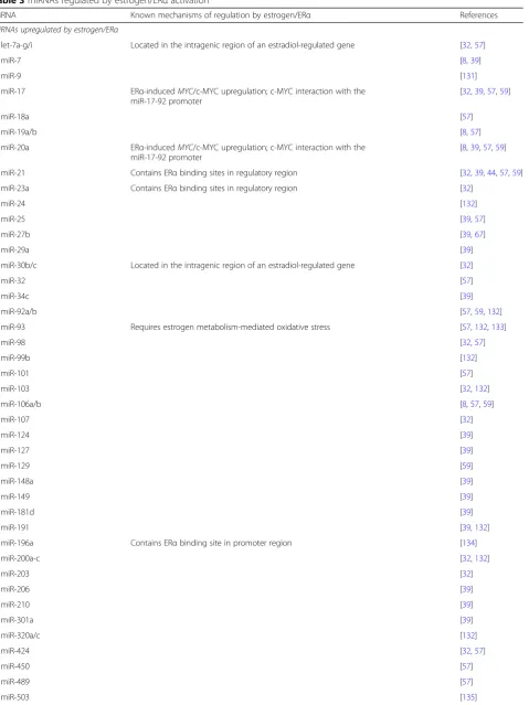
Estradiol/estrogen signaling has been shown to regulate hundreds of genes through direct ERαbinding site inter-action, transcription factor activation, and miRNA regu-lation. miRNA microarray analyses have demonstrated that estradiol can upregulate the expression of numerous individual miRNAs and miRNA families, including the let-7, miR-17-92, and miR-200 family members (Table3) [32]. In this section, several estradiol-regulated miRNAs will be highlighted and the functional implications of their regulation will be discussed in context with ER+ breast carcinogenesis.
miR-21
miR-21 is a well-studied oncomiR with anti-cancer tar-gets, like PDCD4 [33], TIMP3 [34], and PTEN [35], and is overexpressed in human breast cancer tissues as com-pared to healthy breast tissues [34, 36]. Indeed, miR-21 expression levels increase with tumor grade and inva-siveness of the cancer [37]. Thus, given the strong asso-ciation between miR-21 expression and breast cancer, how miR-21 is regulated is an important question. A re-cent review by Petrović(2016) postulated that the onco-genic role of miR-21 in breast cancer may lie predominately in the regulation of cancer cell growth and invasion rather than the events that contribute to cancer initiation [38]. Moreover, miR-21 expression was reported to exhibit a 2-fold increase in ER+breast cancer cells exposed to estradiol [32, 39]. However, in contrast to reports demonstrating that miR-21 is upregulated in response to estradiol, several studies have reported that estradiol suppresses miR-21 expression in vitro [40–43]. In context with the conflicting effects of estradiol on miR-21 expression, ERα binding sites found in the miR-21 regulatory region are key to determining the up-regulation or downup-regulation of miR-21. In particular, Bhat-Nakshatri et al. (2009) found that unliganded ERα suppresses miR-21 expression, whereas ligand-bound/ estradiol-activated ERα promotes miR-21 upregulation [32]. Additionally, overexpression of a truncated form of ERα (ERα46) may trigger estradiol-induced miR-21 up-regulation [44]. ERα activator, AF-1, was also found to be an integral factor in estradiol-induced miR-21 sup-pression. Although in vivo data are still missing to fully understand and confirm the effects of estradiol on miR-21 expression, these contrasting reports indicate a complex regulatory mechanism between estradiol and miR-21 that may be differentially regulated at different breast cancer stages.
In regards to the potential of miR-21 as a target for breast cancer inhibition, studies using cell models of ER+ breast
cancer have consistently reported that miR-21 inhibition suppresses ER+breast carcinogenesis [45,46]. Interestingly, several reports have indicated that non-pharmaceutical in-terventions can modulate miR-21 expression. For instance, exercise training can reduce the expression of miR-21, which was postulated to contribute to the decreased mam-mary tumorigenesis observed in in vivo ER+ breast cancer models [47, 48]. These studies found that exercise down-regulated miR-21 and rescued the expression of miR-21 tar-gets, including PDCD4. Moreover, Khori et al. (2015) found that exercise training was equally effective as tamoxifen in reducing estradiol levels and ERα expression, and both tamoxifen and exercise training significantly reduced tumor burden in a BALB/c mouse model of ER+ breast cancer [47]. Since PDCD4 downregulation is a major marker of poor prognosis and endocrine therapy resistance in ER+ breast cancer patients [49], the pharmacological, as well as non-pharmacological, suppression of miR-21 represents a promising treatment strategy for ER+ breast cancer pa-tients; however, questions remain to be addressed regarding the regulatory mechanisms that control miR-21 expression during breast carcinogenesis, as well as the mechanism by which exercise training alters miR-21 expression. Uncover-ing these key mechanisms would significantly advance our understanding of how physiological responses can regulate miRNAs that are associated with oncogenic processes.
miR-7
miR-7 expression is elevated in ER+ breast cancer cell lines and is associated with tumor aggressiveness, as indicated by the positive correlation between miR-7 expression and increased tumor size, grade, and metasta-sis in ER+ breast cancer patients [8,50,51]. In contrast, miR-7 expression is inversely correlated with the invasiveness of breast cancer cell lines in vitro [52]. Moreover, miR-7 is reported to influence the G2/M cell cycle checkpoint and DNA repair via inhibition of key regulators, such as WEE1, GADD45A,TP53, and ATM
Table 3miRNAs regulated by estrogen/ERαactivation
miRNA Known mechanisms of regulation by estrogen/ERα References
miRNAs upregulated by estrogen/ERα
let-7a-g/i Located in the intragenic region of an estradiol-regulated gene [32,57]
miR-7 [8,39]
miR-9 [131]
miR-17 ERα-inducedMYC/c-MYC upregulation; c-MYC interaction with the miR-17-92 promoter
[32,39,57,59]
miR-18a [57]
miR-19a/b [8,57]
miR-20a ERα-inducedMYC/c-MYC upregulation; c-MYC interaction with the miR-17-92 promoter
[8,39,57,59]
miR-21 Contains ERαbinding sites in regulatory region [32,39,44,57,59]
miR-23a Contains ERαbinding sites in regulatory region [32]
miR-24 [132]
miR-25 [39,57]
miR-27b [39,67]
miR-29a [39]
miR-30b/c Located in the intragenic region of an estradiol-regulated gene [32]
miR-32 [57]
miR-34c [39]
miR-92a/b [57,59,132]
miR-93 Requires estrogen metabolism-mediated oxidative stress [57,132,133]
miR-98 [32,57]
miR-99b [132]
miR-101 [57]
miR-103 [32,132]
miR-106a/b [8,57,59]
miR-107 [32]
miR-124 [39]
miR-127 [39]
miR-129 [59]
miR-148a [39]
miR-149 [39]
miR-181d [39]
miR-191 [39,132]
miR-196a Contains ERαbinding site in promoter region [134]
miR-200a-c [32,132]
miR-203 [32]
miR-206 [39]
miR-210 [39]
miR-301a [39]
miR-320a/c [132]
miR-424 [32,57]
miR-450 [57]
miR-489 [57]
in ER− MDA-MB-231 breast cancer cells, but not ER+ MCF-7 breast cancer cells [52]. Although estradiol medi-ates miR-7 upregulation to confer ER-dependent cell growth, miR-7 downregulation may contribute to endo-crine therapy resistance due to the lack of RTK inhibition, which consequently activates compensatory oncogenic
signaling pathways. Alternatively, in ER− breast cancers, miR-7 overexpression has been considered as a potential therapeutic strategy to exploit miR-7-mediated suppression of EGFR and IGF1R, which are often overexpressed in triple-negative breast cancer (TNBC). Indeed, miR-7 over-expression can reduce the metastatic potential of TNBC
Table 3miRNAs regulated by estrogen/ERαactivation(Continued)
miRNA Known mechanisms of regulation by estrogen/ERα References
miR-542 [57]
miR-638 [132]
miR-1275 [132]
miR-1915 [132]
miRNAs downregulated by estrogen/ERα
let-7a/c/f/g Transcriptionally repressed by estradiol-induced ER [40,132]
miR-9 Located in the intragenic region of an estradiol-regulated gene [32]
miR-15b [132]
miR-21 Mediated by AF-1; Transcriptionally repressed by estradiol-induced ER; Contains ERα
binding sites in promoter/regulatory region; Mediated by unliganded ERα
[40–44,132]
miR-22 [57,59]
miR-23b Transcriptionally repressed by estradiol-induced ER [40]
miR-26a/b [40,132]
miR-27a/b Contains ERαbinding sites in regulatory region; Transcriptionally repressed by
estradiol-induced ER
[32,40,57]
miR-127 [59]
miR-143 [32]
miR-148a [136]
miR-181a/b/d Contains ERαbinding sites in regulatory region; Transcriptionally repressed by estradiol-induced ER; Mediated by unliganded ERα
[40,57,137]
miR-198 [57]
miR-200c [40]
miR-206 [13]
miR-221/222 Contains ERαbinding sites in promoter region [12]
miR-302b [32]
miR-487b [57]
miR-494 [57]
miR-500 [57]
miR-506 [32]
miR-519e [8]
miR-524 [32]
miR-584 [57]
miR-663 [57]
miR-671 [57]
miR-1228 [8]
cells in vitro [52]. Given the potential breast cancer subtype-specific effects of miR-7 expression, understanding the role of ER-associated regulation of miR-7 and its down-stream targets will underscore the clinical applications of targeting miR-7 as novel therapeutic strategies for ER+ breast cancer patients, as well as TNBC patients.
miR-17 and miR-17-92 Cluster
miR-17 is a member of the miR-17-92 cluster, which in-cludes the miR-17, miR-19, and miR-92 families. The miR-17 family includes miR-17, miR-18a, and miR-20a; the miR-19 family includes miR-19a and miR-19b; and miR-92 belongs to its own family. Based on several stud-ies, estradiol appears to be a major stimulator of many miRNAs in the miR-17-92 cluster. Specifically, miR-17 and miR-20a have been shown to be upregulated by 1.7– 3.36 fold in estradiol-treated ER+breast cancer cell lines via ERα-induced MYC/c-MYC upregulation [32, 39, 57, 58]. Consistent with these in vitro microarray data, Kovalchuk et al. (2007) reported that estradiol exposure upregulated miR-17, miR-20a, and miR-92 in a rat model of estradiol-induced breast carcinogenesis, which was accompanied by oncogenic changes in mammary gland morphology, including lobular hyperplasia with increased alveoli and duct numbers [59]. As well, c-MYC was also found to be significantly upregulated in estradiol-exposed rats, further supporting estradiol/ ERα-induced c-MYC upregulation as a major regulatory mechanism of the miR-17-92 cluster. Estradiol-induced modulation of gene and histone methylation may also contribute to these associated changes in miRNA ex-pression. Nevertheless, additional studies are necessary as there is a major gap in our understanding of the mechanisms that mediate estradiol/ERα-associated regu-lation of miRNAs.
miR-17 is often characterized as an oncomiR given its inhibition of putative targets involved in tumor suppres-sion, including p21 [58,60]. However, several genes that promote cell growth and survival, such as CCND1,
STAT3, andE2F1, are also suppressed by miR-17, as well as miR-20a [58, 61–63]. Thus, miR-17 may have onco-genic or tumor suppressor functions that are dependent on specific cellular conditions that determine the need for cell survival. To note, AIB1 (also known as NCOA3 or SRC3) is a putative target of miR-17 that is implicated in breast carcinogenesis and endocrine therapy resist-ance, indicating that miR-17 may also function as a regulator of ERα signaling [64–66]. As well, it has re-cently been reported that induced overexpression of miR-17 can sensitize cells to tamoxifen-mediated apop-tosis in vitro [63]. Given the evidence that miR-17 can exhibit functions as both an oncomiR and a tumor sup-pressor, additional studies are warranted to elucidate the microenvironmental/cellular context that may influence
the cancer-promoting or anti-cancer properties of miR-17 and its related family of miRNAs.
miR-27a/b
Reports examining the effects of estradiol on miR-27a/b expression have produced conflicting data. For instance, microarray data have indicated that estradiol can elicit a 0.6 to 2.65 fold change in miR-27a/b expression in MCF-7 cells [32,39,40, 67]. Despite conflicting in vitro reports, Tang et al. (2012) confirmed that high miR-27a expression and low ZBTB10 expression, a putative target of miR-27a, were associated with poor patient survival [68]. In context with the regulatory role of miR-27a/b in ER+ breast cancer, ZBTB10 is a major inhibitor of Sps, including Sp1 [69,70]. Sp1 interactions with ERαare in-tegral for ERα transcriptional activity; therefore, the in-direct regulation of Sp1 via miR-27a may in-direct the activation or inhibition of ERα activity. miR-27b is also shown to target CYP1B1, a gene that encodes for an en-zyme that is important for the hydroxylation of estradiol, which can produce free radicals and lead to DNA damage [71]. Since miR-27b expression is reported at higher levels in normal breast tissues as compared to breast cancer tis-sues [71], the suppression of miR-27b by estradiol [32] may be a cancer-promoting mechanism that results in the in-creased expression of CYP1B1 and subsequent DNA dam-age. Moreover, miR-27b levels are also reduced in tamoxifen-resistant breast cancer cell lines as well as pa-tient samples, which are associated with the increased ex-pression of miR-27b targetsNR5A2/LRH1andCREB1[67]. The upregulation of NR5A2 has a particularly important oncogenic function as it directly binds to the ERαpromoter to stimulate transcription [72] and promote MYC expres-sion [73], which has previously been reported to upregulate the expression of other oncomiRs (i.e. miR-17 and miR-20a) [58]. Thus, further studies are needed to dissect the factors that regulate the balance between the physio-logical and oncogenic functions of miR-27a/b.
Feedback Mechanisms that Regulate Estrogen/ER Signaling
be repressed downstream of ERα, indicating a potential role in ductal growth regulation [74]. Other estrogen/ ER-associated miRNAs may also participate in regulatory loops to modulate estrogen/ER signaling. miR-221/222, which directly target ESR1, are other key examples as ERα knockdown in MCF-7 cells revealed that miR-221/ 222 can be regulated by ERα activity in vitro (Fig. 1b) [12]. Specifically, estrogen-mediated ERα activation re-cruited corepressors NCoR and SMRT to the transcrip-tion start site of miR-221/222. Garofalo et al. (2009) also proposed a mechanism of miR-221/222 upregulation in non-small cell lung cancer (NSCLC) and hepatocellular carcinoma (HCC) models involving c-MET/MET-me-diated upregulation of the transcription factor AP-1 [25]. This mechanism may be potentially translated to miR-221/222 upregulation associated with ER+ breast cancer models; however, further in vitro and in vivo evi-dence is warranted.
As previously discussed, miR-17 and miR-27a/b may also be involved in estrogen/ERα pathway regulation. The expression of these miRNAs has been found to be upregulated by estradiol/ERα in numerous reports [32, 39, 58, 59]; however, the attenuation of their putative targets involved in ERα regulation, such as AIB1 by miR-17 andZBTB10by miR-27a, results in indirect ERα signaling inhibition and activation, respectively (Fig. 1c) [64,67,69]. Additionally, miR-375 is reportedly involved in a feedback mechanism that regulates ERα expression and signaling. Simonini et al. (2010) found that hypome-thylation of the miR-375 promoter epigenetically stimu-lated miR-375 expression, which was positively correstimu-lated with ERαexpression and activity [75]. The authors further identifiedRASD1, a negative regulator of ERα, as a direct target of miR-375 (Fig. 1d). In contrast, others have reported that miR-375 can directly target Sp1, an ERα co-activator, in HCC, colorectal, and cervical cancer models [76–78]. Although miR-375-mediated Sp1 regulation has not been verified in breast cancer models, this potential mechanism highlights a facet of miR-375-associated estro-gen/ERα signaling regulation. Collectively, miRNA in-volvement in positive/negative feedback loops that regulate ERαactivity can, at times, produce contradictory cellular responses, which further underscores the com-plexity of these regulatory networks that need to be unrav-eled to advance the potential development of miRNA-targeting therapeutics for ER+
breast cancer.
miRNAs and Drug Resistance
In ER+ breast cancer, several therapeutic strategies are employed to selectively inhibit estrogen/ERα signaling, including ER antagonists that directly bind to ERα and enzyme inhibitors that block estrogen synthesis. How-ever, de novo and acquired resistance to conventional endocrine therapies can occur in more than 30% of patients
[79,80]. Due to general targeting of estrogen/ER signaling, cross-resistance to multiple endocrine therapies is also a major concern. Therefore, elucidating the mechanisms of therapeutic resistance is integral to preventing and over-coming this significant clinical challenge. Several mecha-nisms of endocrine therapy resistance have been identified, including decreased ER expression, ER-independent signal transduction, and even miRNA regulation. Notably, miR-NAs are promising targets for the novel therapeutic strat-egies due to the broad range of mRNA targets that ultimately regulate many of the ER-independent mecha-nisms associated with endocrine therapy resistance. In this section, resistance to tamoxifen, fulvestrant, and aromatase inhibitors will be discussed in context with miRNAs that may confer the resistant phenotype and/or may be promis-ing therapeutic targets to increase patient responsiveness to the first-line endocrine therapies.
Tamoxifen Resistance
As discussed in the previous section, although miR-221/ 222 are shown to target and inhibit the translation of oncogenic proteins associated with ER+ breast cancer, several studies have linked miR-221/222 expression to tam-oxifen resistance. Miller et al. (2008) determined that miR-221/222 expression was positively correlated with resist-ance to tamoxifen-mediated cell growth inhibition, apoptosis, and cell cycle inhibition in MCF-7 breast cancer cells [23]. In context with the 3’UTR of CDKN1B mRNA as a known tar-get of miR-221/222 [21, 22, 24],CDKN1B mRNA/p27Kip1 protein regulation appears to be an essential mediator of miR-221/222-induced tamoxifen resistance in MCF-7 cells. Indeed, ectopic overexpression of p27Kip1rescues tamoxifen sensitivity in MCF-7 cells [23]. A report by Wei et al. (2014) suggested that exosomal secretion of miR-221/222 may be a mechanism that confers tamoxifen resistance [81]. Interest-ingly, secreted exosomes from tamoxifen-resistant MCF-7 cells contained miR-221/222 and could be transferred into tamoxifen-sensitive cells, which rendered the cells resistant to tamoxifen-induced cell growth inhibition and apoptosis. Consistent with miR-221/222 upregulation, ERαand p27Kip1 protein levels were also decreased in the MCF-7 cells exposed to exosomes from tamoxifen-resistant cells. As an-other potential mediator of tamoxifen resistance, miR-21 was found to be activated by 4-hydroxytamoxifen (4-OHT)-and fulvestrant-mediated blockage of estrogen/estradiol [41]. This in vitro study presented evidence of miR-21 expression as a resistance factor in ER+breast cancer; nevertheless, add-itional studies are warranted to fully understand the onco-genic mechanisms of miR-21 and miR-221/222 associated with therapeutic resistance.
miRNAs, such as miR-221/222 [8], miR-18a [82], miR-19a/b [83], and miR-22 [9], may predict the clinical responsiveness of ER+ breast cancer patients to tamoxi-fen. Moreover, Beclin1-overexpressing MCF-7 cells exhib-ited estradiol-induced colocalization of Beclin1 and ERα, which contributed to decreased sensitivity to tamoxifen derivatives, raloxifene and 4-OHT [20].
Tamoxifen Sensitization
In recent years, several strategies have been proposed to increase breast cancer cell sensitivity to tamoxifen. First, targeted inhibition of miRNAs associated with tamoxifen resistance has provided in vitro evidence of increased tamoxifen sensitivity in ER+breast cancer cells. As such, miR-221/222 knockdown upregulated TIMP3 mRNA and protein expression, which corresponded with en-hanced tamoxifen-induced inhibition of cell viability in MCF-7 ER+ breast cancer cells [26]. To note, miR-221/ 222 inhibition did not affect the responsiveness of MDA-MB-231 triple-negative breast cancer cells to tam-oxifen, which is consistent with the link between miR-221/222 expression, estrogen/ERα signaling, and tamoxifen activity. Furthermore, miR-221/222 silencing in tamoxifen-resistant MCF-7 cells protected wild-type cells from exosomal miR-221/222-induced tamoxifen re-sistance [81].
The inhibition of known oncomiRs, such as miR-21, has also provided a promising strategy to suppress ER+breast carcinogenesis and enhance the effects of tamoxifen and other anti-estrogen treatments. Recently, Yu et al. (2016) reported that ectopic suppression of miR-21 via transfec-tion with a miR-21 inhibitor results in Beclin1-mediated autophagic cell death, as well as increased sensitivity to both tamoxifen and fulvestrant, as demonstrated by an in-creased induction of apoptosis in vitro [46]. Moreover, nanoparticles co-loaded with anti-miR-21 and 4-OHT have shown promising anti-proliferative effects on MCF-7 ER+ breast cancer cells in vitro [84]. As compared to the 4-OHT treatment alone, the anti-miR-21 + 4-OHT nano-particles induced a significant decrease in cell prolifera-tion, which was accompanied by cell cycle arrest in G2/M phase. Furthermore, miR-34a expression has been re-ported to suppress miR-21 via the post-transcriptional in-hibition of CD24 and Src, which were found to induce miR-21 expression, in in vitro breast and colon cancer models [85]. Importantly, CD24 expression has been posi-tively correlated with ERαand ErbB2 status in breast can-cer [86], and Src is involved in the phosphorylation/ activation and intracellular localization of ERαand subse-quent DNA synthesis [87]. Together, these data provide supportive evidence for testing miR-34a mimics and/or miR-21 inhibitors to treat ER+breast cancer and to over-come tamoxifen resistance.
Fig. 1Feedback mechanisms that regulate estrogen/ERαsignaling. miR-206 (a), miR-221/222 (b), miR-17 and miR-27a (c), and miR-375 (d) are
Alternatively, the induced upregulation of miRNAs that are reportedly downregulated in tamoxifen-resistant cells has been shown to significantly improve ER+breast cancer cell sensitivity to tamoxifen. For instance, miR-378 is downregulated in tamoxifen-resistant MCF-7 cells and is found at lower levels in human breast cancer tissues as compared to normal adjacent tissues [88]. Low miR-378 expression was further associated with decreased recurrence-free survival in ER+breast cancer patients tak-ing tamoxifen as an adjuvant therapy [88]. Importantly, key mechanistic data suggest that miR-378-mediated in-hibition of GOLT1A, which is involved in the fusion of transport vesicles with the Golgi membrane, plays a crit-ical role in tamoxifen resensitization in vitro, which is fur-ther corroborated by clinical data demonstrating increased relapse-free survival in breast cancer patients with low GOLT1A expression. miR-27b is also reportedly downregulated in tamoxifen-resistant ER+ breast cancer cells and patient tumor tissue samples [67,89]. Zhu et al. (2016) showed that miR-27b-3p is downregulated in tamoxifen-resistant ER+ breast cancer cells [67]. Interest-ingly, studies have reported conflicting miR-27b regulation by estrogen/estradiol. As previously mentioned, miR-27b was identified as a miRNA that is repressed by estradiol by 0.6 fold in MCF-7 cells [32]. In contrast, a recent report found that estradiol upregulated miR-27b-3p expression, and tamoxifen dose- and time-dependently suppressed miR-27b-3p expression in MCF-7 and T47D cells [67]. Moreover, overexpression via miR-27b mimics signifi-cantly enhanced ER+ breast cancer cell sensitivity to tamoxifen-induced cell growth inhibition and apoptosis. miR-27b overexpression also rendered tamoxifen-resistant breast cancer cells sensitive to tamoxifen in both in vitro cell line and in vivo xenograft tumor models [67]. The up-regulation of miR-27b sensitization to tamoxifen in ER+ breast cancer cells was further associated with the sup-pression of direct targets, NR5A2/LRH1 and CREB1, which are implicated in tamoxifen resistance via the in-duction of ERαand aromatase, respectively [72,90]. The negative correlation between miR-27b and NR5A2 and CREB1 expression has further been corroborated in hu-man breast cancer tissues from tamoxifen-untreated and tamoxifen-resistant patients, suggesting that miR-27b may be a predictive marker for tamoxifen responsiveness, as well as a potential therapeutic target to overcome tamoxi-fen resistance [67].
Modulated miR-342 expression is also correlated with tamoxifen resistance/sensitivity in ER+ breast cancer. Numerous studies have consistently reported that miR-342 is downregulated in tamoxifen/4-OHT-resistant breast cancer cell lines and clinical samples [23, 91,92]. In particular, miR-342 expression was reduced by 55% in 4-OHT-resistant MCF-7 cells as compared to parental cells [23]. Likewise, miR-342 downregulation is associat
ed with tamoxifen treatment failure and decreased over-all survival in ER+breast cancer patients [30,91]. In pre-clinical models of tamoxifen resistance, restoring the expression of miR-342 markedly sensitized ER+ breast cancer cells to tamoxifen-mediated cell growth inhib-ition and apoptosis [91]. Interestingly, He et al. (2013) reported that miR-342 expression is positively correlated with ERα expression in clinical breast cancer samples and breast cancer cell lines, which is proposed as a po-tential mechanism of enhanced tamoxifen sensitivity in breast cancer cell lines with ectopic miR-342 overexpres-sion [92]. Together, these reports indicate that miR-342 is not only a potential biomarker for ER+ breast cancer patient survival and tamoxifen efficacy, but also may serve as a therapeutic target to enhance patient re-sponses to tamoxifen.
The overexpression of EGFR, ErbB2, and other RTKs is linked to tamoxifen resistance as compensatory oncogenic signaling pathways [93,94]. Therefore, targeted inhibition of these pathways has been presented as a potential strategy to overcome tamoxifen resistance. For example, miR-26a/b are reported to target the 3’UTR ofERBB2and suppress ErbB2 protein expression in tamoxifen-resistant ER+ breast cancer cells. In particular, miR-26 is suggested to be suppressed by estrogen/estradiol [95] and is found to be downregulated in tamoxifen-resistant breast cancer cells. However, high miR-26a expression is associated with decreased expression of known cell cycle regulatory targets, including Cyclin E1, EZH2, and Cdk1, and positive clinical outcomes in patients with metastatic breast cancer taking tamoxifen [96]. Overex-pression of the tumor suppressor miR-34a has also been re-ported to suppress ErbB2 expression and breast cancer cell growth and invasion [97]. Given the oncogenic function of ErbB2 in acquired tamoxifen resistance, miR-34a expression may be a prognostic target to predict tamoxifen responsive-ness in ER+ breast cancer patients. In addition, the knock-down of Beclin1, a target of miR-221 [19], has been reported to suppress ErbB2 expression and restore tamoxifen sensitiv-ity in resistant MCF-7 cells in vitro [98]. Consistently, clinical data from ER+breast cancer patients taking tamoxifen have demonstrated that high Beclin1 expression is correlated with decreased patient survival [98]. Based on reports highlighting the oncogenic role of Beclin1 in ER+ breast cancer, miR-221-induced inhibition of Beclin1 would theoretically inhibit ER+ breast carcinogenesis and promote tamoxifen sensitivity in ER+ breast cancers. Nevertheless, the anti-cancer mechanism involving Beclin1 knockdown, in context with reports indicating the cancer-promoting activ-ities of miR-221/222, further adds to the complex role of miR-221/222 in ER+breast cancer promotion and inhibition.
Fulvestrant Resistance
Zhou et al. (2018) recently reported that miRNA profiles generally differ between fulvestrant- and tamoxifen-resistant MCF-7 breast cancer cells [99]. Nevertheless, there are some miRNAs that are associated with resistance to both thera-peutic agents because of the broad range of mRNA targets. As such, miR-21 overexpression has also been found to con-fer tamoxifen and fulvestrant resistance through the inhib-ition of its direct targets, including PTEN [46]. Interestingly, treatment with fulvestrant can upregulate miR-21 expression [49], which indicates that miR-21 may have a critical role in the development of fulvestrant resistance.
Other examples of miRNAs that are involved in both tamoxifen and fulvestrant resistance are miR-221/222. miR-221/222 are upregulated in both tamoxifen- and fulvestrant-resistant MCF-7 cells, but to a greater extent in the fulvestrant-resistant cell line, and confer the resistant phenotype [100]. Similar to the report indicating that exosomes can transfer miR-221/222 to promote tam-oxifen resistance in nearby cells, microvesicles containing miR-221 produced from cancer-associated fibroblasts can transfer fulvestrant resistance to non-tumor cells in an IL6-dependent manner [101]. Fulvestrant, but not tamoxi-fen, also significantly upregulates miR-221/222 expression in parental MCF-7 cells, which may be an indicator of the development of fulvestrant-resistance. As well, miR-221/ 222 upregulation promotes fulvestrant resistance via mul-tiple pathways, including the targeted inhibition of ERα and p27Kip1. The inhibition of ERα upon miR-221/222 overexpression consequently induces ERα-independent cell growth, which is a mechanism that confers fulvestrant resistance. RTK and Wnt/β-catenin pathways have been implicated in ERα-independent cell growth in fulvestrant-resistant cells [100, 102]. Particularly, the expression of critical genes involved in the Wnt/β-catenin pathway, in-cluding CTNNB1, LRP6, and WNT11, was previously found to be upregulated in fulvestrant-resistant MCF-7 cells, which coincided with hypomethylation of their gene promoters [102]. Moreover,β-catenin overexpression was found to confer fulvestrant resistance, but to a lesser ex-tent than miR-221/222 overexpression, suggesting that miR-221/222 regulation of other targets is necessary to in-duce fulvestrant resistance. Together, these reports indi-cate that perhaps miR-221/222 may functionally regulate Wnt/β-catenin signaling through an epigenetic mechan-ism that modulates promoter methylation.
More recently, the activation of other oncogenic signaling pathways has been connected to miR-221/222 expression and acquired fulvestrant resistance, including the upregula-tion of key markers in the TGFβ, Notch, Jak-STAT, MAPK, and p53 signaling pathways [100]. Furthermore, miR-221/ 222 overexpression promoted TGFβ-induced cell survival in parental MCF-7 cells, whereas miR-221/222 knockdown en-hanced the tumor suppressor activities of TGFβwith signifi-cantly decreased cell survival in fulvestrant-resistant MCF-7
cells. Thus, Rao et al. (2011) postulated that miR-221/222 may have an additional regulatory role that preferably stimu-lates the oncogenic function of TGFβ to promote fulvestrant-resistant breast cancer cell growth [100].
Fulvestrant Sensitization
Integrative computational analyses of miRNA-gene ex-pression in endocrine therapy-resistant breast cancer cell lines have revealed a miR-222-CDKN1B/p27Kip1 expression network that is conserved in both tamoxi-fen-resistant and fulvestrant-resistant MCF-7 cells [103]. Similar strategies to enhance ER+ breast cancer cell sensitivities to tamoxifen and fulvestrant are be-ing tested. The knockdown of oncogenic miR-221/222 in fulvestrant-resistant MCF-7 cells resulted in the con-current inhibition of cell growth and colony formation, as well as the upregulation of p27Kip1upon fulvestrant treat-ment [100]. In addition to the well-studied regulatory function of miR-221/222 on p27Kip1and ERαexpression, mRNA expression profiling revealed that 428 gene probes were upregulated and 224 gene probes were downregu-lated in response to miR-221/222 knockdown in fulves-trant-resistant MCF-7 cells. These data highlight the widespread and complex functional role of miR-221/222 in fulvestrant resistance, and uncover novel pathways or key mediators, such as Wnt/β-catenin, TGFβ, EGFR, and ErbB2, that may be valuable targets for future therapeutic development or combined treatment strategies. Import-antly, the targeted inhibition of EGFR, ErbB2, and β-catenin in tamoxifen-resistant and fulvestrant-resistant cells has demonstrated sensitization to the growth inhibi-tory effects proportional to the degree of phosphorylation/ activation of these targets in the parental and resistant cells [102]. Given the correlation between these oncogenic compensatory pathways that promote ERα-independent cell growth and tamoxifen/fulvestrant resistance, the tar-geted inhibition of miR-221/222 expression is a promising strategy to prevent the acquisition of fulvestrant resistance and even treat fulvestrant-resistant breast cancer patients.
cell death via activation of its direct target PTEN and subse-quent inhibition of the PI3K/Akt/mTOR pathway to en-hance sensitivity to tamoxifen and fulvestrant [46]. To note, the effects of miR-21 knockdown on autophagy in resistant breast cancer cells has not been investigated. Moreover, in-creased autophagy has been reported in cells treated with 4-OHT and fulvestrant, which may be a potential mechan-ism that leads to the acquisition of therapeutic resistance [105]. miR-214 overexpression was shown to suppress 4-OHT- and fulvestrant-induced autophagy in MCF-7 cells through the inhibition of its direct target UCP2. Import-antly, miR-214-mediated suppression of UCP2 led to the inhibition of the PI3K/Akt/mTOR pathway [105]. Based on these findings, the regulation of autophagy may have different consequences in context with the therapeutic sensitivity/resistance of the cells, which warrants further investigation. Advancing our under-standing of the role of autophagy in fulvestrant/tam-oxifen resistance will support the development of novel strategies that modulate autophagy to prevent or overcome endocrine therapy resistance.
miRNA-Regulated Pathways Associated with Aromatase Inhibitor (AI) Resistance
AIs, such as letrozole and anastrozole, are typically pre-scribed to postmenopausal ER+ breast cancer patients. However, the prolonged estrogen deprivation in these pa-tients can lead to acquired AI resistance, which is a critical clinical challenge. Emerging studies have determined that, like tamoxifen and fulvestrant resistance, AI resistance can also be mediated by miRNAs in ER+breast cancer. Al-though the number of studies examining the connection between AI resistance and miRNA regulation in ER+ breast cancer is generally limited, several specific pathways appear to be the main targets associated with miRNA-me-diated regulation of AI resistance.
According to a miRNA microarray using letrozole- and anastrozole-resistant MCF-7 cells, Vilquin et al. (2015) re-ported that both miRNA signatures in the resistant cells were associated with the regulation of the various signaling pathways, including MAPK signaling, focal adhesion, insu-lin signainsu-ling, ErbB signainsu-ling, and mTOR signainsu-ling pathways that converged on Akt regulation [106]. The microarray analyses determined that miR-125b, miR-205, and miR-30a were all significantly upregulated, while miR-424 was sig-nificantly downregulated, in both AI-resistant cell lines as compared to the sensitive cells. Interestingly, miR-125b and miR-205 overexpression and miR-424 suppression were able to confer resistance to both letrozole and anastrozole, as well as promote mammosphere/stem cell self-renewal, which may contribute to the aggressive nature of the resist-ant cells [106]. The activation of the PI3K/Akt/mTOR pathway appeared to be a critical mechanism of miR-125b/ miR-205/miR-424-mediated AI resistance. In contrast, the
targeted suppression of miR-125b/miR-205 and overexpres-sion of miR-424 may rescue the sensitivity of the cells to AIs. Clinical data demonstrating that miR-125b overexpres-sion is correlated with decreased relapse-free survival in primary ER+breast cancer samples further support the role of miRNAs, particularly miR-125b, miR-205, and miR-424, in AI-resistance [106]. Compensatory activation of ErbB2 and downstream MAPK and Akt signaling in AI-resistant ER+ breast cancer cells resulted in the upregulation of miR-21 and downregulation of its target PDCD4 [49]. In clinical cohorts of ER+breast cancer patients, PDCD4 ex-pression is strongly linked to patient outcomes, as demon-strated by patients with low PDCD4 expression levels having poor disease-free survival and increased tumor grade. As PDCD4 is a direct target of miR-21, targeted in-hibition of miR-21 significantly reduced cell proliferation in AI/letrozole-resistant MCF-7 cells in vitro [49], demon-strating that miR-21 may also be a potential target to treat AI resistance. Collectively, these preclinical and clinical data provide promising evidence regarding the potential clinical application of the targeted regulation of these miRNAs to overcome AI resistance and underscore the functional con-tribution of PI3K/Akt/mTOR pathway activation in AI resistance.
The TGFβsignaling pathway is another major pathway that can be modulated by miRNAs to promote AI resist-ance. Particularly, TGFβRI is identified as a direct target of miR-128a, which is overexpressed in letrozole-resistant MCF-7 cells [107]. TGFβinduces a growth inhibitory ef-fect in letrozole-sensitive cells, but does not significantly alter cell growth in the resistant cells, suggesting that the miR-128-mediated regulation of TGFβRI may contribute to the differential responses in the letrozole-sensitive and -resistant cell lines. In turn, suppression of miR-128a was found to inhibit TGFβRI in letrozole-resistant cells, which promoted the growth inhibitory effects of TGFβ. As the correlation between miR-128a expression and letrozole re-sistance has only been reported in in vitro cell models, fur-ther examination in additional preclinical models and verification in clinical datasets are needed to substantiate the clinical value of miR-128a as a potential therapeutic target or marker for AI resistance.
glycolysis. Indeed, targeting the miR-155/miR-143-HK2 axis may be a promising strategy to overcome letrozole resistance. As well, miR-155 expression may serve as a prognostic biomarker to predict the clinical responsive-ness to AIs in women with ER+breast cancer.
Mechanisms that Regulate miRNA Biogenesis in ER+Breast Cancer
As discussed previously, estrogen/ERα signaling can modulate the expression of various oncogenic and tumor suppressor miRNAs. However, the specific regulatory mechanisms have not been fully uncovered. In particular, the regulation of miRNA biogenesis and its functional ma-chinery is a key mechanism that controls miRNA expres-sion. In this section, the mechanisms of regulation and consequences of deregulated miRNA biogenesis will be discussed in context with ER+breast carcinogenesis.
The process of miRNA biogenesis and the components that form essential complexes, such as Drosha, DGCR8, Dicer1, and Ago2, have been extensively studied. Particu-larly, the oncogenic consequences of miRNA biogenesis deregulation have been well-documented [110–112]. Es-trogen/ERα can modulate key machinery involved in miRNA biogenesis, which may have key functional conse-quences in ER+breast cancer. For instance, Dicer, a cyto-plasmic endonuclease required for cleavage of the hairpin loop from the pre-miRNA, can be induced by estradiol as Dicer is reported to associate with an ERα binding site, which promotesDicermRNA transcription [32]. Accord-ingly, Dicer expression is upregulated in ER+breast cancer [4]. To note,Dicerexpression can be modulated by miR-NAs, including miR-221/222, miR-29a, and miR-200c [8]. Conversely, Ago1/2 were found to be downregulated in ER+ breast cancers versus ER−breast cancers despite up-regulation of other components of the RISC (i.e. Dicer and TRBP) [4, 113]. Drosha expression was also signifi-cantly lower in ER+ breast cancer samples. As well, it has been proposed that activated ERαmay interfere with the Drosha-DGCR8 microprocessor complex that is respon-sible for pri-miRNA cleavage [114]. Nevertheless, as a po-tential functional consequence of estradiol-mediated Dicer induction and the activation of other cell signaling path-ways that stimulate miRNA biogenesis, the overall expres-sion level of miRNAs is significantly increased in ER+ breast cancer cell lines and patient samples [4, 8]. Given the differential expression/regulation of miRNA process-ing machinery in ER+breast cancer, it is critical to under-stand the factors that regulate these key components and their specific role in cancer promotion or suppression. Overall, defective miRNA biogenesis and machinery ap-pears to be an important mechanism that ultimately con-tributes to ER-associated breast carcinogenesis. Thus, further exploration of these regulatory mechanisms is ne-cessary to advance our understanding of how miRNA
gene regulation can contribute to breast cancer, and may also reveal novel therapeutic strategies/targets for treating ER+breast cancer.
Role of miRNAs in ER-RTK Crosstalk Regulation in Breast Cancer
Studies unraveling the complex oncogenic signal transduc-tion networks associated with breast cancer initiatransduc-tion and progression have revealed that crosstalk between key signal-ing pathways is an important factor contributsignal-ing to breast carcinogenesis and resistance to therapeutic agents. In par-ticular, crosstalk between the ER and RTK pathways has emerged as a critical mechanism of compensatory pathway activation, particularly in endocrine therapy-resistant ER+ breast cancers [115]. The RTK family includes a number of growth factor-mediated receptors, such as IGF1R, EGFR, and ErbB2/Her2, that are reported to promote several hall-marks of breast cancer. Arpino et al. (2005) found that high ErbB2/Her2 and EGFR expression was independently asso-ciated with a significant decrease in disease-free survival in ER+breast cancer patients taking tamoxifen [116]. Import-antly, studies have found that several miRNAs can regulate targets involved in both the ER and RTK pathways, thus repressing ER-RTK crosstalk. Of particular importance, miRNAs involved in the regulation of ER-RTK crosstalk may serve as leading candidates for the development of novel miRNA-based breast cancer therapies targeting mul-tiple oncogenic pathways.
Summary and Future Perspectives
Abbreviations
4-OHT:4-hydroxytamoxifen; AI: Aromatase inhibitor; ER+: Estrogen
receptor-positive; ERα: Estrogen receptor alpha; HCC: Hepatocellular carcinoma; HK2: Hexokinase 2; LTED: Long-term estrogen deprivation; miRNA: microRNA; NSCLC: Non-small cell lung cancer; RTK: Receptor tyrosine kinase;
SERD: Selective estrogen receptor downregulator; SERM: Selective estrogen receptor modulator; TNBC: Triple-negative breast cancer
Funding
This work was supported in part by an R21 grant from the NIEHS (5R21 ES025337–02), a U54 grant from the NIAAA (5 U54 AA019765–08), and a U54 grant from the NIMHD (1 U54 MD012392–01).
Authors’Contributions
EWH wrote the manuscript. XY contributed to conceptualization and supervision. All authors read and approved the final manuscript.
Ethics Approval and Consent to Participate Not applicable.
Consent for Publication Not applicable.
Competing Interests
The authors declare that they have no competing interests.
Publisher’s Note
Springer Nature remains neutral with regard to jurisdictional claims in published maps and institutional affiliations.
Received: 8 August 2018 Accepted: 29 August 2018
References
1. Kondo N, Toyama T, Sugiura H, Fujii Y, Yamashita H. MiR-206 expression is down-regulated in estrogen receptorα–positive human breast cancer. Cancer Res. 2008;68:5004–8.
2. Yoshimoto N, Toyama T, Takahashi S, Sugiura H, Endo Y, Iwasa M, Fujii Y, Yamashita H. Distinct expressions of microRNAs that directly target estrogen receptorαin human breast cancer. Breast Cancer Res Treat. 2011;130:331–9. 3. Cheang MC, Chia SK, Voduc D, Gao D, Leung S, Snider J, Watson M, Davies
S, Bernard PS, Parker JS. Ki67 index, HER2 status, and prognosis of patients with luminal B breast cancer. J Natl Cancer Inst. 2009;101:736–50. 4. Blenkiron C, Goldstein LD, Thorne NP, Spiteri I, Chin S-F, Dunning MJ,
Barbosa-Morais NL, Teschendorff AE, Green AR, Ellis IO, et al. MicroRNA expression profiling of human breast cancer identifies new markers of tumor subtype. Genome Biol. 2007;8:R214.
5. Nassar FJ, El Sabban M, Zgheib NK, Tfayli A, Boulos F, Jabbour M, Talhouk R, Bazarbachi A, Calin GA, Nasr R. miRNA as potential biomarkers of breast cancer in the Lebanese population and in young women: a pilot study. PLoS One. 2014;9:e107566.
6. PetrovićN, MandušićV, DimitrijevićB, RoganovićJ, LukićS, TodorovićL, StanojevićB. Higher miR-21 expression in invasive breast carcinomas is associated with positive estrogen and progesterone receptor status in patients from Serbia. Med Oncol. 2014;31:977.
7. Xiong J, Yu D, Wei N, Fu H, Cai T, Huang Y, Wu C, Zheng X, Du Q, Lin D, Liang Z. An estrogen receptorαsuppressor, microRNA-22, is downregulated in estrogen receptorα-positive human breast cancer cell lines and clinical samples. FEBS J. 2010;277:1684–94.
8. Cochrane DR, Cittelly DM, Howe EN, Spoelstra NS, McKinsey EL, LaPara K, Elias A, Yee D, Richer JK. MicroRNAs link estrogen receptor alpha status and Dicer levels in breast cancer. Hormones and Cancer. 2010;1:306–19. 9. Pandey DP, Picard D. miR-22 inhibits estrogen signaling by directly
targeting the estrogen receptorαmRNA. Mol Cell Biol. 2009;29:3783–90. 10. Xu D, Takeshita F, Hino Y, Fukunaga S, Kudo Y, Tamaki A, Matsunaga J,
Takahashi R-u, Takata T, Shimamoto A, et al. miR-22 represses cancer progression by inducing cellular senescence. J Cell Biol. 2011;193:409–24. 11. Leivonen S-K, Mäkelä R, Östling P, Kohonen P, Haapa-Paananen S, Kleivi K,
Enerly E, Aakula A, Hellström K, Sahlberg N, et al. Protein lysate microarray analysis to identify microRNAs regulating estrogen receptor signaling in breast cancer cell lines. Oncogene. 2009;28:3926–36.
12. Di Leva G, Gasparini P, Piovan C, Ngankeu A, Garofalo M, Taccioli C, Iorio MV, Li M, Volinia S, Alder H, et al. MicroRNA cluster 221-222 and estrogen receptorαinteractions in breast cancer. J Natl Cancer Inst. 2010;102:706–21. 13. Adams BD, Furneaux H, White BA. The micro-ribonucleic acid (miRNA)
miR-206 targets the human estrogen receptor-α(ERα) and represses ERα messenger RNA and protein expression in breast cancer cell lines. Mol Endocrinol. 2007;21:1132–47.
14. Adams BD, Cowee DM, White BA. The role of miR-206 in the epidermal growth factor (EGF) induced repression of estrogen receptor-α(ERα) signaling and a luminal phenotype in MCF-7 breast cancer cells. Mol Endocrinol. 2009;23:1215–30.
15. Eeckhoute J, Keeton EK, Lupien M, Krum SA, Carroll JS, Brown M. Positive cross-regulatory loop ties GATA-3 to estrogen receptorαexpression in breast cancer. Cancer Res. 2007;67:6477–83.
16. Y-q R, H-j W, Y-q Z, Y-b L. WBP2 modulates G1/S transition in ER+ breast cancer cells and is a direct target of miR-206. Cancer Chemother Pharmacol. 2017;79:1003–11.
17. Graveel CR, DeGroot JD, Su Y, Koeman J, Dykema K, Leung S, Snider J, Davies SR, Swiatek PJ, Cottingham S, et al. Met induces diverse mammary carcinomas in mice and is associated with human basal breast cancer. Proc Natl Acad Sci. 2009;106:12909–14.
18. Liang XH, Yu J, Brown K, Levine B. Beclin 1 contains a leucine-rich nuclear export signal that is required for its autophagy and tumor suppressor function. Cancer Res. 2001;61:3443–9.
19. Pradhan AK, Talukdar S, Bhoopathi P, Shen X-N, Emdad L, Das SK, Sarkar D, Fisher PB.mda-7/IL-24mediates Cancer cell–specific death via regulation of miR-221 and the Beclin-1 Axis. Cancer Res. 2017;77:949–59.
20. John S, Nayvelt I, Hsu H-C, Yang P, Liu W, Das GM, Thomas T, Thomas T. Regulation of estrogenic effects by beclin 1 in breast cancer cells. Cancer Res. 2008;68:7855–63.
21. le Sage C, Nagel R, Egan DA, Schrier M, Mesman E, Mangiola A, Anile C, Maira G, Mercatelli N, Ciafrè SA, et al. Regulation of the p27Kip1 tumor suppressor by miR-221 and miR-222 promotes cancer cell proliferation. EMBO J. 2007;26:3699–708.
22. Visone R, Russo L, Pallante P, De Martino I, Ferraro A, Leone V, Borbone E, Petrocca F, Alder H, Croce CM, Fusco A. MicroRNAs (miR)-221 and miR-222, both overexpressed in human thyroid papillary carcinomas, regulate p27Kip1 protein levels and cell cycle. Endocr Relat Cancer. 2007;14:791–8. 23. Miller TE, Ghoshal K, Ramaswamy B, Roy S, Datta J, Shapiro CL, Jacob S,
Majumder S. MicroRNA-221/222 confers tamoxifen resistance in breast cancer by targeting p27Kip1. J Biol Chem. 2008;283:29897–903. 24. Li Y, Liang C, Ma H, Zhao Q, Lu Y, Xiang Z, Li L, Qin J, Chen Y, Cho WC,
et al. miR-221/222 promotes S-phase entry and cellular migration in control of basal-like breast cancer. Molecules. 2014;19:7122–37.
25. Garofalo M, Di Leva G, Romano G, Nuovo G, Suh S-S, Ngankeu A, Taccioli C, Pichiorri F, Alder H, Secchiero P, et al. miR-221&222 regulate TRAIL resistance and enhance tumorigenicity through PTEN and TIMP3 downregulation. Cancer Cell. 2009;16:498–509.
26. Gan R, Yang Y, Yang X, Zhao L, Lu J, Meng Q. Downregulation of miR-221/ 222 enhances sensitivity of breast cancer cells to tamoxifen through upregulation of TIMP 3. Cancer Gene Ther. 2014;21:290–6. 27. Petrovic N, Davidovic R, Jovanovic-Cupic S, Krajnovic M, Lukic S,
Petrovic M, Roganovic J. Changes in miR-221/222 levels in invasive and in situ carcinomas of the breast: differences in association with estrogen receptor and TIMP3 expression levels. Molecular Diagnosis & Therapy. 2016;20:603–15.
28. Li B, Lu Y, Wang H, Han X, Mao J, Li J, Yu L, Wang B, Fan S, Yu X, Song B. miR-221/222 enhance the tumorigenicity of human breast cancer stem cells via modulation of PTEN/Akt pathway. Biomed Pharmacother. 2016;79:93–101.
29. Zhang C, Zhang J, Zhang A, Wang Y, Han L, You Y, Pu P, Kang C. PUMA is a novel target of miR-221/222 in human epithelial cancers. Int J Oncol. 2010; 37:1621–6.
30. Young J, Kawaguchi T, Yan L, Qi Q, Liu S, Takabe K. Tamoxifen sensitivity-related microRNA-342 is a useful biomarker for breast cancer survival. Oncotarget. 2017;8:99978–89.
31. Han S-H, Kim HJ, Gwak JM, Kim M, Chung YR, Park SY. MicroRNA-222 expression as a predictive marker for tumor progression in hormone receptor-positive breast cancer. J Breast Cancer. 2017;20:35–44.