Opinion
Although we can instinctively tell left from right, these two terms prove strangely difficult to define. The develop ing embryo, however, reproducibly performs this task; the mechanisms underlying this process have intrigued embryologists for decades [1]. The human body shows a clear leftright (LR) asymmetry in the placement and patterning of the internal organs and associated vasculature: the heart apex, stomach and spleen lie to the left and the liver to the right. The normal pattern of sidedness (situs), is called ‘situs solitus’, whereas a mirror symmetric inversion of sidedness is called ‘situs inversus’. Situs solitus is seen in almost everyone, so it is clear that the determination of situs is controlled by a robust developmental mechanism. Moreover, organ asymmetry is strongly conserved throughout the vertebrate lineage, arguing that it is of ancient origin and has been evolu tionarily conserved. Defects in situs determination are rare, but when they do occur they are particularly asso ciated with congenital heart disease (CHD) [2]. Indeed, it has been argued that very minor situs defects may mani fest solely as cardiac defects, suggesting that the heart is more sensitive to situs defects than the other organs [3,4]. Strong associations are also seen between LR defects and many ciliopathies (diseases resulting from defective cilia); this association is due to a requirement for both motile and immotile cilia in LR determination [5,6]. LR
patterning defects also occur at a significant frequency in patients with extrahepatic biliary atresia [7]. The resultant defects in bileduct function mean that most of these patients require a liver transplant during infancy; the mechanism underlying this association remains un known, although simple geometric considerations may come into play.
The role of the node in establishing L-R asymmetry in early mammalian development
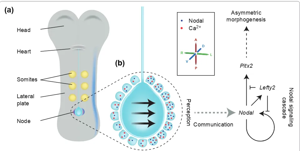
In the mouse embryo, the LR axis is established at approxi mately 8.25 days of development. The 8.25day embryo is relatively simple; an obvious head and heart lie at the anterior end and a midline containing the noto chord runs down the middle, with the pit shaped struc ture known as the node at its posterior end (Figure 1). Immediately to either side of the midline sits the paraxial mesoderm, containing the somites (from which trunk muscle and skeletal tissue develop). This is flanked by the lateral plate mesoderm (LPM), a lineage that will in later development contribute to asymmetric organ structure. Overlying the mesodermal tissue is a thin layer of endoderm that gives rise to the gut. As revealed in the papers to be discussed here, the endoderm also plays a previously unrecognized part in LR patterning.
Over the past 15 years, a general model of the establishment of LR asymmetry has emerged (Figure 1). The first indication that bilateral symmetry of the embryo has been broken is the LR asymmetric expression of certain genes in regions flanking the node as well as more laterally, in the LPM. Upstream of asymmetric gene expres sion, the rotation of motile cilia within the node (or the equivalent structure in other vertebrates) causes a leftward flow of fluid, called ‘nodal flow’ [811]. In the mouse, cilia project from the ventral surface of the node; these cilia are polarized with respect to the anterior posterior axis and by rotating in a clockwise direction, drive nodal flow leftwards [12]. Nodal flow has been shown to be both necessary and sufficient to define the left side of the mouse embryo [13,14]. The high incidence of LR patterning defects in humans with immotile cilia suggests that the same is true in humans [15]; approxi mately 50% of patients with immotile or abnormally
Abstract
The clockwise rotation of cilia in the developing mammalian embryo drives a leftward flow of liquid; this genetically regulated biophysical force specifies left-right asymmetry of the mammalian body. How leftward flow is interpreted and information propagated to other tissues is the subject of debate. Four recent papers have shed fresh light on the possible mechanisms.
Cilia, calcium and the basis of left-right asymmetry
Dominic P Norris*
OpiniOn
Open Access
*Correspondence: d.norris@har.mrc.ac.uk
Mammalian Genetics Unit, MRC Harwell, Harwell Science and Innovation Campus, Oxfordshire, OX11 0RD, UK
motile cilia exhibit situs inversus [16]. Downstream of nodal fl ow, asymmetric Ca2+ signaling is seen at the edges of the node, with stronger signaling on the left side than on the right [17]. In the interests of simplicity, this article will focus on our understanding of the determination of LR asymmetry in the mouse, and will address two ques tions: how is LR asymmetry established at the mouse node; and how is that asymmetry subsequently trans ferred over several cell diameters to the LPM.
Establishing and maintaining asymmetry: the nodal signaling cascade
Downstream of the initial breaking of symmetry at the node, the Nodal signaling cascade is activated in the left, but not the right, LPM (Figure 1). Nodal, a member of the transforming growth factorbeta (TGFbeta) signal ing family of intercellular signaling proteins, functions as a dimer. Importantly, only those cells in the left and right LPM are competent to respond to Nodal signaling. In the left LPM, Nodal signaling induces expression of the No dal gene itself, the Lefty2 gene,which encodes an antagonist of Nodal signaling, and the Pitx2 gene, which encodes a transcription factor that acts downstream of Nodal. Lefty2 is also a TGFbeta family member, but unlike
Nodal it functions as a monomer, and diff uses faster and further than Nodal [1820]. Once Nodal is expressed in the left LPM, the resultant production of Lefty2 sup presses the Nodal cascade in the right LPM, helping to lock in asymmetry [21]. Nodal and Lefty2 are expressed for only 6 to 8 hours. In contrast, once activated, Pitx2 remains asymmetrically expressed in the left LPM for the next two days, so that Pitx2 protein is present in the left LPM during organogenesis [2224]. Th is has led to the proposition that Pitx2is the ultimate eff ector of leftness [25]. While this is not absolutely the case, asymmetry of Pitx2 expression does underlie asymmetry of many organs [26,27].
Detecting fl ow in the node: the three hypotheses
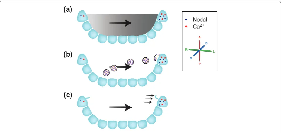
Th e mechanisms by which nodal fl ow is ‘perceived’ by the embryo remain the subject of debate, with three main hypotheses currently in contention (Figure 2). Th e ‘morphogen hypothesis’ argues that a shortlived mole cule becomes enriched on the left side of the node in response to nodal fl ow and that this higher concentration on the left is detected, leading to a LR asymmetric signal [8]. Both computational and experimental investigation argue that such asymmetric enrichment is plausible for Figure 1. The left-right asymmetry pathway.(a) An 8.25-day mouse embryo showing asymmetric Nodal expression (in blue) at the node as well as in the left, but not the right, lateral plate, and asymmetric Cerl2 expression in purple. (b) A higher magnifi cation of the node. The central pit of the node contains motile cilia (not shown) that drive a leftwards nodal fl ow (indicated by arrows). Surrounding the node are the node crown cells; these express Nodal (represented by blue dots) in an asymmetric fashion, with expression stronger on the left than on the right. An asymmetric calcium signal (represented by red stars) is also stronger on the left than the right. Downstream of nodal fl ow and asymmetric gene expression at the node, a ‘leftness’ signal is communicated several cell diameters to the left lateral plate mesoderm (LPM). Here it activates the Nodal signaling cascade, ultimately resulting in left-side-specifi c Pitx2 expression and asymmetric morphogenesis. The primary axes are shown: anterior-posterior (A-P); dorsal-ventral (D-V); left-right (L-R).
Communication
Perception
Nodal
Ca2+
Nodal
Lefty2 Pitx2
Asymmetric morphogenesis
Nodal signaling
cascade
Head
Heart
Somites
Node Lateral plate
(a)
(b)
A
D
L
P
V
molecules between 15 and 50 kDa in size, although the nature of the morphogen and the receptors are unknown [28,29]. A second hypothesis, the ‘nodal vesicular parcel (NVP) hypothesis’ posits the presence of membrane bounded vesicles that are carried leftwards, breaking in a ciliadependent fashion on the left side of the node where they deliver a cargo of morphogens [30]. Although very appealing, elements of this hypothesis (such as the mecha nism of NVP breaking) clearly need to be modifi ed [31]. Finally, the ‘twocilia hypothesis’ argues that immotile sensory cilia detect fl ow directly on the left but not the righthand side of the node [17]. Th is model was pre dicated on the known function of the polycystic kidney disease 1 (Pkd1) and polycystic kidney disease 2 (Pkd2) genes in the kidney the proteins encoded by these genes form a complex that detects urine fl ow and gives rise to a Ca2+ signal in response [32]. Although Pkd1 is not required for LR determination [33], both Pkd2 and the Pkd1 homologue Pkd1like 1 (Pkd1l1) are involved in LR patterning, being needed for the embryo to respond to nodal fl ow [34,35]. However, whether nodal fl ow pushing outwards on the lefthand side of the node can truly be diff erentiated from the pull exerted by nodal fl ow on the righthand side of the node, a requirement of the two cilia model, has been questioned [36]. For all these
models, the outcome is an asymmetric (L greater than R) Ca2+ signal at the node. Currently, only the twocilia hypo thesis (and the known function of Pkd1l1/Pkd2) provides a mechanism to explain how this signal might be generated [17].
investigating ciliary function
A recent study from Hamada and colleagues (Shinohara et al. [37]) utilizes a mixture of genetics, biophysics and imaging to examine the establishment of LR asymmetry at the node. Th e authors make the striking fi nding that just two rotating cilia are suffi cient to break LR sym metry. In previous studies, nodal fl ow has been examined by following the movement of small numbers of particles across the node, allowing overall direction a lity and speed of fl ow to be assessed. For this study Shinohara et al. used an approach called particle image velocimetry (PIV), which they have customized for nodalfl ow analysis [38]. Utilizing a high density of fl uorescent beads within a living node and highspeed confocal imaging of a single optical plane, they followed small variations in particle position over many frames, building up a vector map of fl ow and forces across the entire node. At a local level this provides far more information than the particletracking approaches used up to now, and it seems likely that PIV Figure 2. Three models of how fl ow breaks symmetry at the node. The node is represented in section, with the axes rotated by 90° from Figure 1; the axes are marked. (a) The morphogen hypothesis posits that a morphogen produced within the node becomes asymmetrically localized between left and right in response to fl ow (represented by the gray gradient). The resulting stronger left-sided signal is detected, thereby breaking symmetry. (b) The nodal vesicular parcel (NVP) hypothesis contends that morphogen-containing vesicles are carried leftwards by nodal fl ow, breaking in contact with cilia on the left side of the node. This delivers the morphogens within the NVPs asymmetrically, resulting in enriched morphogen signaling on the left-hand side of the node, thereby breaking symmetry. (c) The two-cilia hypothesis argues that fl ow itself is detected on the left side of the node by cilia-localized polycystic kidney disease (polycystin or PKD) family molecules, releasing a left-sided Ca2+
signal, thereby breaking symmetry. Single cilia of crown cells on the left and right sides of the node are represented, the one on the left becoming deformed in response to nodal fl ow.
(a)
(b)
(c)
Nodal Ca2+
A D
L
analysis Shinohara et al. confirm that the early node (8.0 day embryo) has a weak leftward flow, which they demon strate to be present at the time when the first asymmetric gene expression becomes apparent at the node: asymmetry of Cerberuslike 2 (Cerl2; also known as Dand5), an antagonist of Nodal signaling expressed more strongly on the right than the left side of the node. However, asymmetry of the Nodal cascade in the LPM occurs slightly later, once a stronger, more robust, leftwards nodal flow is acting at around the two to three somite stage (8.25day embryo). Using a nontoxic viscous solution (methylcellulose), Shinohara et al. slowed, and even stopped, nodal flow. This allowed them to demon strate that only a weak flow and/or a small temporal window of flow is required to break LR symmetry and drive asymmetric gene expression at the node (Cerl2) and in the left LPM (the Nodal cascade).
In order to investigate the role of altered nodal flow in greater detail, Shinohara et al. sought genetic approaches to perturb it. The Rfx3 locus encodes a transcription factor required for normal ciliogenesis in the node; muta tion of this gene leads to a massive reduction in nodal cilia number and to embryos demonstrating overt LR patterning defects [39]. Loss of the Dpcd (deleted in primary ciliary dyskinesia) locus similarly results in LR patterning defects [40]. Both loci show incomplete penetrance, which led Shinohara et al. to examine Rfx3-mutant embryos in greater detail [37]. Analysis of nodal cilia motility by light microscopy revealed that a few rotating cilia were present in these nodes. By maintaining the embryos in culture, the authors were able to image nodal cilia motility and correlate it with subsequent asym metric gene expression. They found mildly affected embryos to have four or five rotating cilia, accompanied by normal LR asymmetric gene expression. In contrast, severely affected embryos had at most a single rotating cilium and showed symmetrical Cerl2 expression at the node and complete absence of LPM Nodal expression. Further analysis revealed that only two rotating cilia were required to establish normal sidedness. In embryos with three or more rotating cilia, the addition of methyl cellulose to reduce flow resulted in loss of sidedness, underlining the role of the remaining level of flow in situs determination. Finally, the position of the rotating cilia within the node (whether they were near the left or right side) was addressed, and strikingly, it emerged that their position within the node bore no relationship to their ability to determine situs: something that might have implications for all three models. Intriguingly, there also seems to be a reduction in the number of immotile cilia at the periphery of the node in these embryos, although this clearly does not affect their ability to detect flow.
argue for only a few motile cilia being required to estab lish LR asymmetry, when perhaps 200 such cilia are present within a wildtype node. This raises the question of whether this apparent excess of cilia is truly required, or whether it is an evolutionary aberration or hangover. Unused function tends to be lost when evolutionary selection pressure is removed, as in the case of eye and pigment loss in cavedwelling animals [41]. Three obvious possible explanations present themselves. First, the pre sence of higher numbers of motile cilia and prolonged flow could have subtle effects on LR determination that are not being assessed in these studies, possibly influ encing the precise timing or extent of asymmetric gene expression, or some other unknown event downstream of nodal flow. The final outcome of these events on the anatomy and physiology of the adult mouse is what is being selected. Second, the presence of many motile cilia might add robustness to the symmetrybreaking event such that deleterious outcomes (affecting cardiac pattern ing, for example) become extremely rare. Third, the system may not currently be under selection. In this case we might expect a loss of function to be occurring, and perhaps for variation to be evident between different strains and species of mice. Studies in other types of organism and the production of adult mice that have developed from embryos with small numbers of motile cilia may be able to shed some light on this.
Shinohara et al. do not directly address the question of what mechanism might underlie flow sensing in the node. The weak flow produced by two rotating cilia would primarily change morphogen concentrations only very locally, and the authors surmise that this would slow down, but not necessarily destroy, a morphogenbased mecha nism. The impact of weak flow on mechanosensa tion (in the twocilia model) would also be noticeable, although the authors argue how it might be possible for forces created by these cilia to be directly transduced (almost instantaneously) across the node. On the basis of developmental timing, they argue that the twocilia model is more likely to be true.
normally broadly expressed in the early embryo) is required solely in the node crown cells for normal LR patterning to occur; expression driven specifi cally in the remainder of the node did not rescue LR patterning. Th ey then showed that Pkd2 protein must be localized to cilia for it to function in LR patterning. Finally, utilizing the Kif3anull mutant (Kif3a encodes a motor protein required for cilia formation), Yoshiba et al. created embryos in which cilia were present only in the node crown cells. By then applying an artifi cial fl ow across the node, they were able to activate the normal downstream LR pathway in these mice. Th is led them to propose a model in which fl ow is detected through cilialocalized Pkd2 protein in node crown cells, which in turn leads to repression of Cerl2 on the left side of the node. While these data fi t well with the twocilia hypothesis, the mechanism by which fl ow or a morphogen is detected remains unaddressed. Clearly, Pkd1l1 must be a prime candidate for performing this function, although it remains to be established whether Pkd1l1 responds to a morphogen or to fl ow [34,43].
Transferring asymmetry from the node: intra- or extracellular communication?
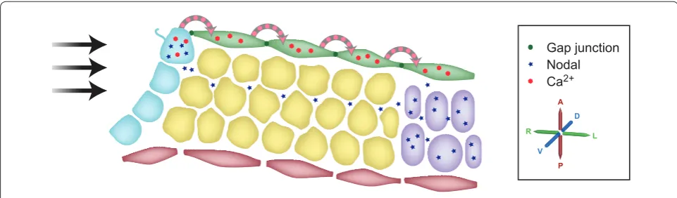
How asymmetric information moves from the node out to the LPM is also the subject of competing hypotheses (Figure 3). When Brueckner and colleagues [17] originally described the generation of asymmetric Ca2+ at the node, they commented that (at least on occasions) the asym metric signal spread as far as the lateral plate. Th is clearly provides one possible mechanism by which asymmetric information might travel out from the node by Ca2+ moving intracellularly from cell to cell. In contrast, Hamada and colleagues built on two other facts: that Nodal expression at the node is required for Nodal
activation in the LPM [44,45]; and the recognized ability of Nodal to activate its own expression [46,47]. Th ey proposed that asymmetrically distributed extracellular Nodal protein at the node is transported more readily leftwards, through the extracellular matrix [48]. Two recent papers provide signifi cant advances in our under standing of the mechanisms of these processes.
In one paper, Hadjantonakis and colleagues (Viotti et al. [49]) report that Sox17null embryos exhibit defective LR patterning; Sox17 encodes an Srybox containing protein that is required for normal defi nitive endoderm formation [50]. Th ese embryos do not express the Nodal cascade in either the left or the right LPM but, signifi cantly, retain asymmetric gene expression at the node. Th is clearly suggests that communication between the node and the LPM requires the defi nitive endoderm, a result reminiscent of the observations of McGrath et al. [17]. Together, these data suggested that calciuminduced calcium release was signaling between cells in the defi ni tive endoderm; such signals are known to travel via gap junctions. By surveying gapjunction protein expression in normal embryos, Viotti et al. found that the core gap junction protein connexin 43 (also known as Gja1) was expressed in the defi nitive endoderm. However, it proved to be absent from the endoderm of Sox17 mutant embryos. Loss of connexin 43 does not, of course, directly prove a loss of gapjunction function. Viotti et al. therefore assessed this by injecting dye into defi nitive endoderm cells, revealing that in wildtype embryos small, but not large, dye molecules moved between cells through gap junctions. Th e dyes did not cross into other cell lineages and, signifi cantly, never moved into or crossed the midline, demonstrating that the left and right sides of the embryo are distinct and not linked by gap junctions. In the absence of such a barrier, both sides of the embryo Figure 3. Two models for communicating signals from the node to the left lateral plate. A cartoon representation (not to scale) of a section through the left side of the embryo, including the left side of the node (as represented in Figure 2) and tissues lateral to the node. The cells of the node are shown in blue, the endoderm in green, the ectoderm in red, paraxial mesoderm in yellow and lateral plate mesoderm in purple. Viotti and colleagues’ [49] and Saund and colleagues’ work [51] argues that calcium signaling, via gap junctions, carries signals from the node leftwards through the endoderm. Oki and colleagues’ analyses [48] argue that Nodal protein itself travels leftwards through an extracellular, but intraembryonic, route and directly activates the Nodal locus in the lateral plate.
Gap junction Nodal Ca2+
A D
L
endoderm. When Sox17-null embryos were examined, the dyes did not migrate between cells, demonstrating a loss of gapjunction connections. The role of gap junctions was firmly established when pharmacological agents were used to block gapjunction function in wild type embryos, and this reproduced the LR patterning defects seen in the Sox17-null embryos. This work shows that the definitive endoderm and gap junctions are required for the transfer of LR asymmetry signals from the node to the LPM.
While Viotti et al. have shown that a gapjunction dependent Ca2+ signal can travel from the node to the left LPM, they stop short of providing a mechanism by which Ca2+ might activate Nodal expression in the LPM. In contrast, a model proposed by Oki et al. [48] does provide an explanation for Nodal activation in the LPM Nodal moving out from the node. However, the Oki model provides no explanation of the role of asymmetric Ca2+ signaling in the endoderm. It is of course tempting to combine these two models: a simple combined Viotti Oki model might propose that calcium signaling in the endoderm influences the underlying cell matrix, which in turn affects the ability of Nodal protein to propagate from the node to the left LPM.In a contemporaneous study, Saijoh and colleagues (Saund et al. [51]) have independently identified the role of Sox17 and the definitive endoderm in LR patterning [51]. These authors specifically investigated the link between loss of Sox17 function and the proposed intraembryonic, extra cellular route for Nodal. They examined extracellular matrix proteins previously implicated in the translocation of Nodal protein, revealing a defect in a proportion of Sox17 mutant embryos. However, a smaller proportion of embryos exhibited such defects than exhibited abnormal LR patterning. Saund et al. therefore argue that this change is not the primary cause of the LR defects in the Sox17 mutants. Of course, this does not fully exclude the possibility that a combination of similar defects, includ ing those changes that they detected, are influencing intraembryonic extracellular transport of Nodal to the left LPM.
prospects and questions
Although significant advances in the understanding of LR determination have been made, gaps still remain. Even following these most recent studies, it is evident that we do not fully understand how nodal flow leads to LR asymmetry. Both the twocilia and morphogen hypo theses remain entirely plausible, and both have their champions within the field. Discriminating between them is not simple, and short of the identification of the putative ligand central to the morphogen hypothesis, may remain so, as if the twocilia hypothesis is correct,
the realm of the biophysicist, and will increasingly require an understanding of equations and modeling. An appreciation of the node as a (low Reynolds number) microfluidic environment, in which inertia effectively disappears, is required. In such an environment, our ‘realworld’ experiences can lead us to expect outcomes that are in fact incorrect, taking us far from reality; a very accessible discussion of such environments, and life at low Reynolds number, is available in the excellent article by Purcell [52].
Is it possible that both the twocilia and morphogen mechanisms are acting in concert, providing two signals of ‘leftness’ in the node? Clearly, in such a scenario these might both have an impact on Nodal expression, but it is also possible to envisage that they might have different targets. Indeed, Pitx2, the most downstream gene of the Nodal signaling cascade, does not affect gross cardiac situs [26], implying that there must be additional asym metrically expressed loci controlling this process. The simple explanation is that such loci are directly controlled by asymmetric Nodal expression in the LPM; in other words, that there are additional unidentified Nodal target genes at the end of the Nodal cascade. However, arguments also exist for Nodalindependent asymmetric gene expression in the LPM: analysis of the Ablim1 locus reveals it to be asymmetrically expressed in the left but not right LPM in the absence of Nodal expression [53]. Moreover, both galanin (Gal), a neuropeptide with a role in neuronal inhibition, and Pitx2 retain LR asymmetric expression in early cardiac tissue (at the anterior end of the LPM) in the absence of the Nodal coreceptor Cryptic, which is required for Nodal expression in the LPM [54]. Whether these loci are affected by one rather than another of the putative mechanisms remains uninvestigated. Further uncertainty remains downstream of Pitx2,where the target genes that facilitate asymmetric morphogenesis remain to be identified.
The presence of very early LR asymmetry, established by the initial cleavage of the embryo, has been strongly argued in Xenopus [55]. However, few such suggestions have been made for the mouse. The one exception is a purely embryological study by Gardner [56], which revealed that manipulation of the early blastomeres can affect the direction of embryonic axial rotation, but not other aspects of situs. While at present there is no explanation of how this might function at a molecular level, it implies that another system of LR determination may be acting in addition to that driven by nodal flow.
but not right side), but no such association has been detected in the mouse or suggested in humans. The human LMO4 locus shows asymmetric expression in the brain of 12weekold human embryos, with stronger rightsided than leftsided expression [57]. This is, however, at a much later stage of development than the establishment of visceral asymmetry, suggesting that it may be a downstream event and/or that neural asym metry is entirely independent of visceral asymmetry. Intriguingly, expression of the mouse Lmo4 locus, while also asymmetrical, appears to be random, with individual embryos demonstrating a left or a rightsided preference in their expression [57]. Whether this reflects innate differences between mouse and human brains (and neural asymmetry) remains to be determined.
Continued study, in the mouse as well as in other organisms, will be required to unravel the nature of LR determination and its evolution. Understanding the differences between the mechanisms by which various organisms pattern their LR axes should reveal which elements of the process have remained constant and which have varied. Ultimately, this knowledge will provide insight into the evolution of LR asymmetry, how processes such as nodal flow have been gained and lost in different organisms, and perhaps to our understanding the evolutionary driving forces involved.
Acknowledgements
DPN is supported by the UK Medical Research Council. I thank Daniel Grimes and other members of my group for discussion and for reading drafts, and Steve Thomas for extensive work on the figures.
Published: 19 December 2012
References
1. Neville AC: Animal asymmetry.Studies Biol 1976, 67.
2. Ramsdell AF: Left-right asymmetry and congenital cardiac defects: Getting to the heart of the matter in vertebrate left-right axis determination.Dev Biol 2005, 288:1-20.
3. Franco D, Campione M: The role of Pitx2 during cardiac development. Linking left-right signaling and congenital heart diseases.Trends Cardiovasc Med 2003, 13:157-163.
4. Kathiriya IS, Srivastava D: Left-right asymmetry and cardiac looping: implications for cardiac development and congenital heart disease.Am J Med Genet 2000, 97:271-279.
5. Drummond IA: Cilia functions in development.Curr Opin Cell Biol 2012, 24:24-30. 6. Nakamura T, Hamada H: Left-right patterning: conserved and divergent
mechanisms.Development 2012, 139:3257-3262.
7. Carmi R, Magee CA, Neill CA, Karrer FM: Extrahepatic biliary atresia and associated anomalies: etiologic heterogeneity suggested by distinctive patterns of associations.Am J Med Genet 1993, 45:683-693.
8. Nonaka S, Tanaka Y, Okada Y, Takeda S, Harada A, Kanai Y, Kido M, Hirokawa N:
Randomization of left-right asymmetry due to loss of nodal cilia generating leftward flow of extraembryonic fluid in mice lacking KIF3B motor protein.Cell 1998, 95:829-837.
9. Feistel K, Blum M: Three types of cilia including a novel 9+4 axoneme on the notochordal plate of the rabbit embryo.Dev Dyn 2006, 235:3348-3358. 10. Essner JJ, Vogan KJ, Wagner MK, Tabin CJ, Yost HJ, Brueckner M: Conserved
function for embryonic nodal cilia.Nature 2002, 418:37-38.
11. Kramer-Zucker AG, Olale F, Haycraft CJ, Yoder BK, Schier AF, Drummond IA:
Cilia-driven fluid flow in the zebrafish pronephros, brain and Kupffer’s vesicle is required for normal organogenesis.Development 2005,
132:1907-1921.
12. Hirokawa N, Tanaka Y, Okada Y: Cilia, KIF3 molecular motor and nodal flow. Curr Opin Cell Biol 2012, 24:31-39.
13. Okada Y, Nonaka S, Tanaka Y, Saijoh Y, Hamada H, Hirokawa N: Abnormal nodal flow precedes situs inversus in iv and inv mice.Mol Cell 1999,
4:459-468.
14. Nonaka S, Shiratori H, Saijoh Y, Hamada H: Determination of left-right patterning of the mouse embryo by artificial nodal flow.Nature 2002,
418:96-99.
15. Brueckner M: Heterotaxia, congenital heart disease, and primary ciliary dyskinesia.Circulation 2007, 115:2793-2795.
16. Kartagener M, Horlacher A: Bronchiektasen bei Situs viscerum inversus. Scweiz Med Wochenschr 1935, 16:782-784.
17. McGrath J, Somlo S, Makova S, Tian X, Brueckner M: Two populations of node monocilia initiate left-right asymmetry in the mouse.Cell 2003,
114:61-73.
18. Sakuma R, Ohnishi Yi Y, Meno C, Fujii H, Juan H, Takeuchi J, Ogura T, Li E, Miyazono K, Hamada H: Inhibition of Nodal signaling by Lefty mediated through interaction with common receptors and efficient diffusion.Genes Cells 2002, 7:401-412.
19. Marjoram L, Wright C: Rapid differential transport of Nodal and Lefty on sulfated proteoglycan-rich extracellular matrix regulates left-right asymmetry in Xenopus.Development 2011, 138:475-485.
20. Muller P, Rogers KW, Jordan BM, Lee JS, Robson D, Ramanathan S, Schier AF:
Differential diffusivity of Nodal and Lefty underlies a reaction-diffusion patterning system.Science 2012, 336:721-724.
21. Meno C, Takeuchi J, Sakuma R, Koshiba-Takeuchi K, Ohishi S, Saijoh Y, Miyazaki J, ten Dijke P, Ogura T, Hamada H: Diffusion of nodal signaling activity in the absence of the feedback inhibitor Lefty2.Dev Cell 2001, 1:127-138. 22. Piedra ME, Icardo JM, Albajar M, Rodriguez-Rey JC, Ros MA: Pitx2 participates
in the late phase of the pathway controlling left-right asymmetry.Cell
1998, 94:319-324.
23. Ryan AK, Blumberg B, Rodriguez-Esteban C, Yonei-Tamura S, Tamura K, Tsukui T, de la Peña J, Sabbagh W, Greenwald J, Choe S, Norris DP, Robertson EJ, Evans RM, Rosenfeld MG, Izpisúa Belmonte JC: Pitx2 determines left-right asymmetry of internal organs in vertebrates.Nature 1998, 394:545-551. 24. Yoshioka H, Meno C, Koshiba K, Sugihara M, Itoh H, Ishimaru Y, Inoue T,
Ohuchi H, Semina EV, Murray JC, Hamada H, Noji S: Pitx2, a bicoid-type homeobox gene, is involved in a lefty-signaling pathway in determination of left-right asymmetry.Cell 1998, 94:299-305.
25. Logan M, Pagan-Westphal SM, Smith DM, Paganessi L, Tabin CJ: The transcription factor Pitx2 mediates situs-specific morphogenesis in response to left-right asymmetric signals.Cell 1998, 94:307-317.
26. Liu C, Liu W, Lu MF, Brown NA, Martin JF: Regulation of left-right asymmetry by thresholds of Pitx2c activity.Development 2001, 128:2039-2048. 27. Liu C, Liu W, Palie J, Lu MF, Brown NA, Martin JF: Pitx2c patterns anterior
myocardium and aortic arch vessels and is required for local cell movement into atrioventricular cushions.Development 2002, 129:5081-5091.
28. Cartwright JH, Piro O, Tuval I: Fluid-dynamical basis of the embryonic development of left-right asymmetry in vertebrates.Proc Natl Acad Sci U S A
2004, 101:7234-7239.
29. Okada Y, Takeda S, Tanaka Y, Belmonte JC, Hirokawa N: Mechanism of nodal flow: a conserved symmetry breaking event in left-right axis
determination.Cell 2005, 121:633-644.
30. Tanaka Y, Okada Y, Hirokawa N: FGF-induced vesicular release of Sonic hedgehog and retinoic acid in leftward nodal flow is critical for left-right determination.Nature 2005, 435:172-177.
31. Cartwright JH, Piro N, Piro O, Tuval I: Fluid dynamics of nodal flow and left-right patterning in development.Dev Dyn 2008, 237:3477-3490. 32. Harris PC, Torres VE: Polycystic kidney disease.Annu Rev Med 2009,
60:321-337.
33. Karcher C, Fischer A, Schweickert A, Bitzer E, Horie S, Witzgall R, Blum M: Lack of a laterality phenotype in Pkd1 knock-out embryos correlates with absence of polycystin-1 in nodal cilia.Differentiation 2005, 73:425-432. 34. Field S, Riley KL, Grimes DT, Hilton H, Simon M, Powles-Glover N, Siggers P,
Bogani D, Greenfield A, Norris DP: Pkd1l1 establishes left-right asymmetry and physically interacts with Pkd2.Development 2011, 138:1131-1142. 35. Pennekamp P, Karcher C, Fischer A, Schweickert A, Skryabin B, Horst J, Blum
M, Dworniczak B: The ion channel polycystin-2 is required for left-right axis determination in mice.Curr Biol 2002, 12:938-943.
cavity are sufficient to break left-right symmetry in the mouse embryo. Nat Commun 2012, 3:622.
38. Hashimoto M, Shinohara K, Wang J, Ikeuchi S, Yoshiba S, Meno C, Nonaka S, Takada S, Hatta K, Wynshaw-Boris A, Hamada H: Planar polarization of node cells determines the rotational axis of node cilia.Nat Cell Biol 2010,
12:170-176.
39. Bonnafe E, Touka M, AitLounis A, Baas D, Barras E, Ucla C, Moreau A, Flamant F, Dubruille R, Couble P, Collignon J, Durand B, Reith W: The transcription factor RFX3 directs nodal cilium development and left-right asymmetry specification.Mol Cell Biol 2004, 24:4417-4427.
40. Vogel P, Read R, Hansen GM, Freay LC, Zambrowicz BP, Sands AT: Situs inversus in Dpcd/Poll-/-, Nme7-/- , and Pkd1l1-/- mice. Vet Pathol 2010,
47:120-131.
41. Jeffery WR: Regressive evolution in Astyanax cavefish.Annu Rev Genet 2009,
43:25-47.
42. Yoshiba S, Shiratori H, Kuo IY, Kawasumi A, Shinohara K, Nonaka S, Asai Y, Sasaki G, Belo JA, Sasaki H, Nakai J, Dworniczak B, Ehrlich BE, Pennekamp P, Hamada H: Cilia at the node of mouse embryos sense fluid flow for left-right determination via Pkd2.Science 2012, 338:226-231.
43. Kamura K, Kobayashi D, Uehara Y, Koshida S, Iijima N, Kudo A, Yokoyama T, Takeda H: Pkd1l1 complexes with Pkd2 on motile cilia and functions to establish the left-right axis.Development 2011, 138:1121-1129.
44. Brennan J, Norris DP, Robertson EJ: Nodal activity in the node governs left-right asymmetry.Genes Dev 2002, 16:2339-2344.
45. Saijoh Y, Oki S, Ohishi S, Hamada H: Left-right patterning of the mouse lateral plate requires nodal produced in the node.Dev Biol 2003,
256:160-172.
46. Adachi H, Saijoh Y, Mochida K, Ohishi S, Hashiguchi H, Hirao A, Hamada H:
Determination of left/right asymmetric expression of nodal by a left side-specific enhancer with sequence similarity to a lefty-2 enhancer.Genes Dev
1999, 13:1589-1600.
47. Norris DP, Robertson EJ: Asymmetric and node-specific nodal expression patterns are controlled by two distinct cis-acting regulatory elements. Genes Dev 1999, 13:1575-1588.
48. Oki S, Hashimoto R, Okui Y, Shen MM, Mekada E, Otani H, Saijoh Y, Hamada H:
Sulfated glycosaminoglycans are necessary for Nodal signal transmission
49. Viotti M, Niu L, Shi SH, Hadjantonakis AK: Role of the gut endoderm in relaying left-right patterning in mice.PLoS Biol 2012, 10:e1001276. 50. Kanai-Azuma M, Kanai Y, Gad JM, Tajima Y, Taya C, Kurohmaru M, Sanai Y,
Yonekawa H, Yazaki K, Tam PP, Hayashi Y: Depletion of definitive gut endoderm in Sox17-null mutant mice.Development 2002, 129:2367-2379. 51. Saund RS, Kanai-Azuma M, Kanai Y, Kim I, Lucero MT, Saijoh Y: Gut endoderm
is involved in the transfer of left-right asymmetry from the node to the lateral plate mesoderm in the mouse embryo.Development 2012,
139:2426-2435.
52. Purcell EM: Life at low Reynolds number.Am J Physics 1977, 45:3-11. 53. Stevens J, Ermakov A, Braganca J, Hilton H, Underhill P, Bhattacharya S, Brown
NA, Norris DP: Analysis of the asymmetrically expressed Ablim1 locus reveals existence of a lateral plate Nodal-independent left sided signal and an early, left-right independent role for nodal flow.BMC Dev Biol 2010,
10:54.
54. Schweickert A, Deissler K, Britsch S, Albrecht M, Ehmann H, Mauch V, Gaio U, Blum M: Left-asymmetric expression of Galanin in the linear heart tube of the mouse embryo is independent of the nodal co-receptor gene cryptic. Dev Dyn 2008, 237:3557-3564.
55. Levin M: Left-right asymmetry in embryonic development: a comprehensive review.Mech Dev 2005, 122:3-25.
56. Gardner RL: Normal bias in the direction of fetal rotation depends on blastomere composition during early cleavage in the mouse.PLoS ONE
2010, 5:e9610.
57. Sun T, Patoine C, Abu-Khalil A, Visvader J, Sum E, Cherry TJ, Orkin SH, Geschwind DH, Walsh CA: Early asymmetry of gene transcription between embryonic human left and right cerebral cortex.Science 2005,
308:1794-1798.
doi:10.1186/1741-7007-10-102