ABSTRACT
Pathologic fractures of the jaw are usually associated with aggressive, destructive lesions or malignant lesions of the jaws. Aneurysmal bone cysts (ABC) of the jaws are benign lesions which normally do not cause destruction to such an extent. This article reports a relatively aggressive
ABC
of the mandible in a19 year-old male which destroyed the buccal and lingual cortices
as well as the inferior border of the mandible resulting in a pathologic fracture. Although pathologic fractures associated with ABC's of the skeleton are common, however, to our knowledge this is the frrst report of an AB C associated with a pathologic fracture of the jaw.MJIRI, Vol. 12, No.2, 185-189, 1998.
Keywords: Cyst, bone, aneurysmal; mandible.
INTRODUCTION
The aneurysmal bone cyst (ABC) is an enigmatic and misnamed lesion displaying variable etiopathogenic, histologic and radiographic characteristics.'·6 In essence, it is a giant cell lesion with numerous caverns or blood clefts and no epitheliallining.4.8.,o.'4 Jaffe and Lichtenstein2 fIrst recognizeditin 1942; however,Bernier andBhaskarreported the fIrst case occurring in the jaws in
1958.3
Since then, many reports of ABC's have been published with varying clinicopathologic characteristics. The majority of ABC's are seen in adolescents, predominantly under the age of 20, but they can occur earlier or later in life.4.'2,'7.'8 ABC's are found more frequently in the mandible than the maxilla (3: 1), andmore commomym the molar regIOns, but have alSO invol ved other bones of the face such as the zygoma and the zygomatic arch.4.'2.'9,2o A history of trauma preceding it's* Assistant Professor of Oral and Maxillofacial Surgery, Attending Surgeon, Clinic of Oral and Maxillofacial Surgery, Baqiyatallah Medical Center, Baqiyatallah and Azad Universities of Medical Sciences, Tehran, I.R. Iran.
development can often be obtained. 4·11 They have been found to occur in association with numerous pathologic entities such as the dentigerous cyst, osteoclastoma, central giant cell tumor, fibrous dysplasia, osteosarcoma, nonosteogenic fibroma, cementifying fibroma, ameloblastoma, and hemangioepithelioma.9.'3"sThe clinical course of the ABC known today may range from a self-limited lesion' to a progressive, aggressive and rapidly destructive lesion which may mimic a malignancy.7,8 Treatment of the aneurysmal bone cyst is generally directed towards removal of the lesion and has ranged from no treatment, simple curettage and cryotherapy, to excision and bone grafting. 1.7·9.21·23 Recurrence of the jaw lesions has been attributed to incomplete removal of the lesion.7.9.'2
Case report
A
19
year-old male was referred to the Oral and Maxillofacial Surgery Clinic o n November1, 1994
complaining of swelling over the right side of the mandibular body and malocclusion. He stated that he had rolled off of his bed several days ago but that trauma was trivial. On clinical examination the right side of the face wasFig.
1. Photograph of the patient upon admission: note swelling of the right side of the mandible (A). Fracture of the rightmandibular body with displacement evident on the,.right lateral oblique radiograph (B).asymmetrical, swollen, but not tender, and a step could be palpated at the inferior border of the mandible near the parasymphyseal region on the ipsilateral side (Fig.
IA).
There was a slight degree of paresthesia in the area innervated by the right mental nerve, which the patient noted had developed subsequent to the trauma There were no palpable lymph nodes in the neck, supraclavicular or axillary regions, and no prior history of noticeable weight loss or cachexia.Fig.
2. Panoramic radiograph of the patient demonstrating a unicystic multilocular radiolucency with scalloped borders and a fracture at the inferior border of the right mandibular body. Note telescoping of the fracture and open bite in occlusion on the contralateral side necessitating intraoral open reduction.examination revealed a malocclusion due to premature contact of the lower right posterior teeth, and resultant anterior and contralateral open bite.
A
fracture was clinically evident. Mobility and crepitation were noticed on manual manipulation of the right anterior and posterior segments. Swelling was also noticed intraorally in the right premolar region, which was compressible. There were no carious teeth or restorations. Physical examination was otherwise non-contributory and the patient was in good general health. Radiographic exami.nation revealed a displaced fracture in the area of the right body of the mandible in the premolar area, with a step at the inferior border evident on the right lateral oblique view (Fig.IB).
The fracture had occurred within a solitary radiolucent lesion with ragged, poorly defined, scalloped borders extending from below the right firstmolar to the right lateral incisor (Fig. 2). The roots of the right canine and premolars were apparently within the confines of the lesion; however, there was no evident root resorption or displacementAn
aspiration was performed using a large bore needle but no aspirate could be obtained. The CBC, differential WBC, ESR and other routine laboratory tests including measurement of serum calcium, phosphorus, and alkaline phosphatase levels were within normal limits.The patient was admitted to the hospital and antibiotics -were iJ)itiated. In view of the clinical, laboratory and radiographic findings, and the age of the patient, it was decided to operate the patient and perform an excisional biopsy and reduce the fracture simultaneously.
Under general anesthesia via nasoendotracheal intubation, a marginal sulcular incision was made from the right lower central incisor to thefrrst molar, with a releasing incision anterior to the right central incisor, and a mucoperiosteal flap was reflected.
A
round, soft, brown,perforated the buccal cortex. The lesion was completely curretted from the bone and meticulously dissected from the mental neurovascular bundle, which it encompassed. Bleeding was moderate during the procedure. The jaw was then placed in occlusion and open reduction of the fracture and intermaxillary fixation was performed (Fig. 3A).
The patient's postoperative course was uneventful and healing was nonnal. The pathology report gave a diagnosis of an aneurysmal bone cyst of the mandible (Fig. 3B). Union was complete after
8
weeks and the patient was followed for approximately two years. Normal sensation had returned in the area innervated by the right mental nerve and the lesion had resolved clinically and radiographically and there was no evidence of recurrence (Fig.4).
Facial symmetry was also restored (Fig.5).
Fig.
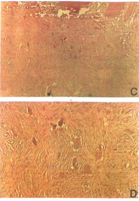
3. Immediate postoperative view of the patient (A). Low power photo micro graph of AB C (H & E x60) revealing fibro blastic connective tissue stroma, with numerous fibroblasts and scattered giant cells (B). Osteoid seen in approximation to a blood cleft (C). High power magnification (H&E x4(0) revealing numerous multinucleated giant cells dispersed in the fibroblastic stroma (D).DISCUSSION
The aneurysmal bone cyst (ABC) generally affects young persons, predominantly under age
20.
There is usually no sex predilection,4.!5 although some6 report itto occur more in females while others7,8 report a greater occurrence in males. It is more common in the mandible than the maxilla (3:1); the site predilection in the jaws is the molar area; however, it can also occur in other sites.4-11 The ABC comprises5 %
of all lesions of the cranial and maxillofacial bones.6 ABCs may displace but usually do not resorb the dentition, although this finding has been reported.24 Paresthesiais
usually not a feature of this disorder.4.!5.7,8Clinical presentations of ABCs are extremely variable, ranging from a small, indolent, asymptomatic lesion being first noticed as a radiolucency on a routine radiographic
Fig. 4. Two-year postoperative radiog raph of the patient Jemonstrating complete bone formation (A,B).
examination, to a rapidly growing, expansile, destructive lesion that may clinicall y mimic a malignancy
Y
Aspiration fmdings may also vary, yielding no blood (solid type) or syringes full of bright red, partially Qxygenated blood (vascular type) or, due to subsequent infection or thrombosis of the lesion, nothing at all.3.5,7,8,IS.17The radiographic features are not pathognomonic. The lesion may appear as a unicystic, multilocular soap bubble or moth-eaten radiolucency with demarcated or irregular margins causing pain or destruction of the bony corticies.s, 7,8,10·17
Histologically, the ABC consists of a fibrous connective tissue stroma with numerous blood-filled caverns or sinusoids, which may undergo thrombosis. Young fibroblasts and giant cells are abundant, making it si.nilar to the giant cell granuloma, but the latter
is
devoid of vascular caverns. Hemosiderin, osteoid, and bone formation are variable.4•17 Although it may appear cyst-like on radiography, it does not have an epithelial lining and thusis
not a cyst 4.5,10·17CONCLUSION
Fig. 5.
Full face photograph demonstrating restoration of facial symmetry,cyst (ABC) remains obscure and it is not known whether the lesion arises
de
novo or represents the result of some form of vascular accident in a pre-existing lesion,22 However, many of the cases of ABC present with a history of previous trauma; thus it may be a possible predisposing factor.4.5,9.11 Although aneurysmal bone cysts are more commonin
the shafts of long bones orin the vertebral column,lesions of the jaws are relatively rare.22 Waldron22 found fewer than50
cases reported in the literature up to1987.
In our review of the international literature, we have found78
reported cases of maxillofacialAB
C's up to January1997.
The treatment of choice for ABC is complete curettage and although recurrence rates of up to50%
have been reported, the studies of Kalantar Motamedi, Khodayari, and Yazdi have shown thatif
the lesion is completely removed, recurrence is rare.S•8,10.I8,2I,23 Thus, en bloc excision or resection is usually reserved for recurrent cases or for cases in association with other lesions that necessitate such treatment.S•8,10.18 To our knowledge, there have been no published reports of pathologic fractures associated with mandibular ABCs.REFERENCES
I, Hernandez GA. Castro A, Castro G, Amador E: Aneurysmal bone cyst versus hemangioma of the mandible, Report of a long-term follow-up of a self-limiting case. Oral Surg O ral
Med Oral Pathol 76 (12): 790-796,1993.
2. Jaffe HL, Lichtenstein L: Solitary unicameral bone cyst with emphasis on the roentgen picture, thepathological appearance
of the jaws: analysis of 11 cases. J Oral Maxillofac Surg 52: 20. Eveson IW, Moos KF, MacDonald DG: Aneurysmal bone
471-47.5,1994. cyst of the zygomatic arch. Br J Oral Surg 15: 259-264, 1978.
9. Eldeeb M, Sedano MO, Waite DE: Aneurysmal bone cyst 21. Trent C, Byl FM: Aneurysmal bone cyst of the mandible. Ann ofthe jaws: report of a case associated with fibrous dysplasia. Otol Rhinol Laryngol 102: 917-24, 1993.
Int J Oral Surg 9: 301, 1980. 22. Waldron CA: Bone pathology, In: Neville BW, Damm �O, 10. Steidler N: Aneurysmal bone cysts of the jaws. Br J Oral Surg Allen CM, Bouquot JE (eds.), Oral and Maxillofacial
16: 254, 1975. Pathology. Philadelphia, PA: WB Saunders, pp. 459-461,
11. Struthers P, Shear M: Aneurysmal bone cyst of the jaws. 1995.
Pathogenesis.Int J Oral Surg 13: 92,1984. 23. Wiatrak BJ, Myer CM, Andrews TM: Alternatives in the 12. Struthers P, Shear M: Aneurysmal bone cyst of the jaws. management of aneurysmal bone cysts of the mandible. Int J
Clinicopathological features. Int J Oral Surg 13: 85, 1984. Pediatr Otorhinolaryngol 31: 247-257, 1995.
13. Levy W: Aneurysmal bone cyst secondary to other osseous 24. Hardee PS, Whear NM, Morgan PR: Aneurysmal bone cyst
lesions: report of 57 cases. Am J Clin Pathol 63: 1, 1975. of the maxilla-an association with tooth resorption. J
14. Tillman B, Dahlin 0: Aneurysmal bone cyst: an analysis of Craniomaxillofac Surg 20: 266-269, 1992.