R E V I E W A R T I C L E
Automation, consolidation, and integration in autoimmune
diagnostics
Renato Tozzoli1•Federica D’Aurizio1•Danilo Villalta2•Nicola Bizzaro3
Received: 15 April 2015 / Accepted: 1 June 2015 / Published online: 3 July 2015 The Author(s) 2015
Abstract Over the past two decades, we have witnessed an extraordinary change in autoimmune diagnostics, char-acterized by the progressive evolution of analytical tech-nologies, the availability of new tests, and the explosive growth of molecular biology and proteomics. Aside from these huge improvements, organizational changes have also occurred which brought about a more modern vision of the autoimmune laboratory. The introduction of automation (for harmonization of testing, reduction of human error, reduction of handling steps, increase of pro-ductivity, decrease of turnaround time, improvement of safety), consolidation (combining different analytical technologies or strategies on one instrument or on one group of connected instruments) and integration (linking analytical instruments or group of instruments with pre-and post-analytical devices) opened a new era in immun-odiagnostics. In this article, we review the most important changes that have occurred in autoimmune diagnostics and present some models related to the introduction of automation in the autoimmunology laboratory, such as automated indirect immunofluorescence and changes in the two-step strategy for detection of autoantibodies; auto-mated monoplex immunoassays and reduction of
turnaround time; and automated multiplex immunoassays for autoantibody profiling.
Keywords Automated immunofluorescence Automated monoplex immunoassay
Automated multiplex immunoassay
Two-step diagnostic strategyTurnaround time Autoantibody profiling
Introduction
Over the past 25 years, autoimmune diagnostics has gone through an evolutionary period, which seems not yet ended, as clinical and basic knowledge in the field of autoimmunity is still growing. From year to year, important achievements in pathophysiology, development of new diagnostic tech-nologies and advances in therapy of autoimmune diseases (AIDs) have been steadily added. The engine of this evo-lutionary period is the improvement in AIDs diagnostics, which has allowed an increasing number of recognized cases of individuals (3–8 % of the population; 80 % women) suffering from one or more AIDs [1,2]. The main recent cornerstones in this field have been: (a) The discovery of new autoantibody-autoantigen systems (both systemic and organ-specific); (b) Recognition that autoantibodies may have predictive, pathogenic, or protective roles; (c) The availability of new diagnostic technologies (monoplex and multiplex immunoassays-IMA), and (d) Changes in orga-nizational processes (integration and automation) [3–6].
We could now wonder if such events could have been foreseen at the beginning of the era of modern autoimmune diagnostics. The answer to this question lies in the expected changes in clinical immunology laboratories as dictated in the early 1990s. Twenty years ago, Nakamura
& Renato Tozzoli
1 Department of Laboratory Medicine, Laboratory of Clinical
Pathology, S. Maria degli Angeli Hospital, Via Montereale, 24, 33170 Pordenone, Italy
2 Allergology-Clinical Immunology, Department of Laboratory
Medicine, S. Maria degli Angeli Hospital, Pordenone, Italy
3 Laboratory of Clinical Pathology, San Antonio Hospital,
Tolmezzo, Italy
and Bylund [7], during the 17th AO Beckman conference, identified the primary focus of the laboratory of clinical immunology and defined the main influencing factors of future changes: governmental regulations, healthcare eco-nomics, advances in technology and advances in develop-ment and use of therapeutic agents. At the same time, Hamilton [8], looking at the clinical immunology labora-tory of the future, emphasized the importance of stan-dardization of methods and development of ‘universal’ automated immunoanalyzers. Two years later, Normansell [9], at the 8th annual meeting of the American Association of Medical Laboratory Immunologists, foresaw the pro-gressive unification of academic specialty laboratories (chemistry, hematology, immunology, etc.) in a unique general laboratory (consolidation).
Most of these predictions have been achieved, and especially automation and consolidation paved the way to the modern laboratory [10,11]. During the past decade, we and others [12–16] have described the progressive evolu-tion of analytical methods and technologies, signaling the extraordinary dynamic development of autoimmune diag-nostics resulting from the availability of new tests, diffu-sion of improved immunoassay methods and instruments, and the explosive growth of molecular biology and pro-teomic medicine.
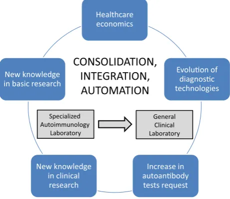
To meet both clinical need and growing demand for autoantibody testing, automation of autoimmune diagnostics has invaded the laboratory as an extension of the general technological improvement already achieved in almost all other diagnostic areas of the clinical laboratory [17, 18]. Currently, all stages of the analytical procedure for detection and quantification of autoantibodies are automated. The third generation of laboratory systems now encompasses most of the analytical steps of the laboratory workflow, enabling the clinical pathologists (autoimmunologists in this case) to focus on ‘value-added’ work, such as result validation and production of narrative reports for clinical interpretation [4]. The two key concepts of third generation systems are ‘consolidation’ (i.e., combining different analytical tech-nologies or strategies on one instrument or on one group of connected instruments) and ‘integration’ (linking analytical instruments or group of instruments with pre- and post-ana-lytical devices).
As predicted, in many cases this approach has brought about the reunification of diagnostics in the central general laboratory. It is now customary to manage the flow of highly demanded autoantibody tests (thyroperoxidase, thyroglobulin, tissue transglutaminase, citrullinated peptide autoantibodies) in ‘human-less’ robotic platforms, charac-terized by total laboratory automation (TLA) which includes pre-analytical, analytical, and post-analytical operations [19]. The rationale in favor of this approach lies in the advantage of placing as many assays on a single
analyzer, rather than maintaining two or more analyzers, because each instrument requires separate quality control (QC), preventive maintenance, record keeping, etc. (Fig-ure1). TLA combines a large variety of processes, including accessioning and sorting specimens, decapping tubes, centrifugation, aliquoting, delivery to analyzers, recapping tubes, and storage and archiving of samples. The prototype of instruments used in the autoimmunology laboratories is IMA platforms [4–6].
The advantages of TLA include harmonization of testing to improve patient care [20], reduction of human error, reduction of handling steps, increase of productivity, decrease and standardization of turnaround time (TAT), improvement of safety, use of state of the art informatics (i.e., interface with laboratory information systems-LIS and health information systems; middleware to bridge the analyzers and LIS). Most, if not all, the obstacles to automation as defined by Tomar as much as 15 years ago [10] are now completely removed.
In the next sections of this article, we present some models related to the introduction of automation in the autoimmunology laboratory: a. automated indirect immunofluorescence (IIF) and the changes in the two-step strategy for detection of autoantibodies, b. automated monoplex IMAs and the reduction of TAT, and c. auto-mated multiplex IMAs and autoantibody profiling.
Automated indirect immunofluorescence and changes in the two-step strategy for autoantibody detection
Recently, indirect immunofluorescence (IIF) automation for reading and interpretation of ANA and other autoanti-bodies was developed, supported by the American College
Healthcare economics
Evoluon of diagnosc technologies
Increase in autoanbody
tests request New knowledge
in clinical research New knowledge in basic research
Specialized Autoimmunology
Laboratory
General Clinical Laboratory
CONSOLIDATION,
INTEGRATION,
AUTOMATION
of Rheumatology recommendation that IIF is the reference method for anti-nuclear-cytoplasmic antibodies (NCA or ANA) [21, 22], anti-neutrophil cytoplasmic antibodies (ANCA) [23], and anti-dsDNA antibodies (DNAAb). At present, at least 6 automatic systems for the detection of these autoantibodies are available [4, 24, 25] (Table1). These systems allow for automated classification of sam-ples, with a high efficiency in discriminating between positive and negative ANA and an acceptable correlation with manual microscope reading [4, 26, 27], automated quantification of autoantibodies [28, 29], and implemen-tation of a quantitative internal QC system [30].
These features allow a different approach to the diag-nostic strategy for the detection of NCA-ANA, ANCA, and DNAAb, based on a two-step algorithm: the first stage, the screening of positive/negative and positive samples selec-tion; the second stage, identification of specific antibodies for confirmation of screening results and classification of autoimmune diseases [33,34].
This flow of information in autoimmune diagnostics has long been conducted with the strategy of reflective testing. That is, the morphologist identifies positive samples using the IIF method and, on the basis of the fluorescence pattern, decides which autoantibodies are to be detected by IMA methods. Moreover, if necessary, reflective testing provides for the addition of new tests beyond the test originally requested and/or production of comments to aid in the cor-rect interpretation of results by the clinical autoimmunolo-gist. Reflective testing can be now replaced with reflex
testing, in which a predetermined test protocol is automati-cally completed [35]: the integration and the use of specific health information technologies allow now the automated application of rules and the computerized addition of specific tests with high efficiency and effectiveness (Table2), reducing the need of morphologists and the TAT for clinical applications.
Automation of IIF and the use of specific health infor-mation technology systems applied to the autoimmunology laboratory, besides allowing for the automated classification of negative/positive results, permit also the practice of pathology at a distance (telepathology, or digital micro-scopy, or whole slide imaging). Telepathology allows the remote interpretation of fluorescence patterns (telediagno-sis), second opinions or consultations (teleconsultation), quality assurance, education, teaching, self-study and research (teleeducation) [31]. Telepathology has been suc-cessfully applied to anatomic pathology, hematology, and microbiology; its use in biomedicine is rapidly evolving [32].
Monoplex immunoassays and the reduction
of turnaround time
Automated IIF and third-generation monoplex IMAs allow the rapid detection of autoantibodies in terms of minutes or hours, instead of days or weeks as required by manual IIF and IMA methods. In the past, fast autoantibody results did not represent a main goal for clinicians and autoimmunologists,
Table 1 Currently available automated IIF platforms (Hhomogeneous,Sspeckled, Nnucleolar,Ccentromere,ND nuclear dots,NMnuclear membrane,Cycytoplasmic)
System Screening neg/pos Patterns (no; type) Company
Aklides Yes 6-H, S, N, C, ND, Cy Medipan, Germany
EUROPattern Yes 7-H, S, N, C, ND, NM, Cy Euroimmun, Germany
Zenit G-Sight Yes 5-H, S, N, C, M A. Menarini Diagnostics, Italy
NOVA view Yes 5-H, S, N, C, ND Instrumentation Laboratories, Spain
Helios Yes – Aesku Diagnostics, Germany
Image navigator Yes – ImmunoConcepts, USA
Table 2 Time evolution of two-step strategy for NCA/ANA detection/measurement
Era Years Methods
Manual ANA IIF–reflective adding on–manual ENA 1970–2000 Manual IIF
Manual monoplex methods
Low automated monoplex methods
Manual ANA IIF–reflective adding on–automated ENA 2000–2014 Manual IIF
Full automated monoplex methods
Low automated multiplex methods
Automated ANA IIF–reflex adding on–automated ENA 2015–… Automated IIF
Full automated monoplex methods
Full automated multiplex methods
which in many cases utilized longer turnaround times for repetition of positive tests or for second opinions. In current practice, however, life-threatening autoimmune conditions or autoimmune diseases associated with rapidly progressive loss of organ function have emerged, requiring a short response time to allow rapid etiological and differential diagnosis. This, in turn, will enable the start of immunosuppressive or other specific therapies as soon as possible. This is the case, for example, for ANCA-associated small-vessel vasculitides or Goodpasture’ syndrome [36], or for patients with acute car-diovascular symptoms (thyroid storm, atrial fibrillation, supraventricular tachycardia, etc.) in undiagnosed TSH-re-ceptor antibody-dependent Graves’ disease [37,38]. Another condition involves patients affected by catastrophic anti-phospholipid syndrome with concurrent thrombotic and hemorrhagic manifestations [39]. In these clinical settings such as may occur in intensive-care units, the rapid avail-ability of autoantibody tests is a great opportunity for early diagnosis and treatment to save a patient’s life [40].
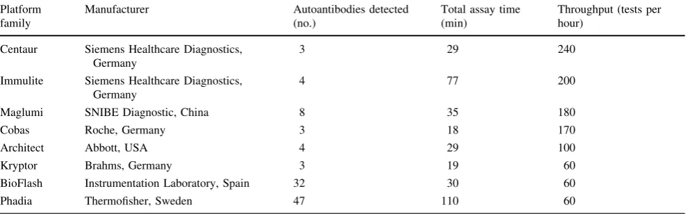
Furthermore, the availability of automated analyzers with reduced assay times (within 120 min) as well of manual point-of-care systems [41] enables real-time antibody measurement in the same day of the request or even instat mode, avoiding delay and improving compliance towards diagnosis, differ-ential diagnosis and therapy monitoring in the clinical con-ditions listed above [41]. This also responds to the increasing need for faster diagnosis owing to shorter period of hospital-ization. A partial list of automated platforms, with their assay times and throughput, is shown in Table3.
Automated multiplex IMAs and autoantibody
profiling
Multiplex proteomic technology is considered to be an opti-mal solution for the simultaneous detection of different autoantibodies related to AIDs. Some of these immunoassays
(planar and non-planar microarrays) may contribute to over-coming some drawbacks of the monoplex immunoassays (time expenditure, costs, lack of harmonization, volume of reagents and samples, turnaround time, etc.) [5].
The autoantibody profiling of AIDs patients provided by these systems may be useful for following the concentra-tion of specific autoantibodies, which may display different trends over time, both for diagnostic and prognostic pur-poses. This is the case, for instance, of celiac disease and anti-phospholipid syndrome which are characterized by the presence of multiple autoantibodies of different isotypes. Nowadays, multiplexed technology has achieved high analytical accuracy and provides results comparable to, if not superior to, the manual and automated monoplex technologies [42,43].
The autoimmunology laboratory of the near future
The referred changes have altered the scenario and strongly characterized the evolution of the immunology laboratory. But what implications do these new arrangements have for the future?
Avoiding the Ulysses syndrome for the patientsBecause the results of antibody tests can be available in a few hours, it can be expected that clinical reasoning will occur much faster than now and will involve not only the rheumatol-ogist but also many other specialists and even general practitioners. Signs of this are already visible [44]. If well organized and managed, this change could have positive effects—namely, to ensure greater efficiency of the diag-nostic filter, with the ability to identify during their first clinical visit those patients who need a specialist consul-tation. The advantages for patients, doctors and the health system overall are obvious (Table 4).
Reducing analytical variability of autoantibody tests Another prediction is that measurement of the
Table 3 Automated monoplex IMA platforms
Platform family
Manufacturer Autoantibodies detected (no.)
Total assay time (min)
Throughput (tests per hour)
Centaur Siemens Healthcare Diagnostics, Germany
3 29 240
Immulite Siemens Healthcare Diagnostics, Germany
4 77 200
Maglumi SNIBE Diagnostic, China 8 35 180
Cobas Roche, Germany 3 18 170
Architect Abbott, USA 4 29 100
Kryptor Brahms, Germany 3 19 60
BioFlash Instrumentation Laboratory, Spain 32 30 60
concentration of autoantibodies, which is still poorly standardized, will become more and more accurate. Sub-jective methods and semi-quantitative techniques will be replaced by more objective assays and with greater preci-sion, reducing the analytical variability to levels similar to those of other immunoassays. ELISA methods will disap-pear and blot methods will evolve to provide quantitative results. Measurement of therapeutic antibody levels for follow-up and customization of treatments will become increasingly popular. Costs will drop and autoimmune diagnostics will become part of the clinical profile for early and differential diagnosis.
In conclusion, 20 years after the statements of the pro-phets about the changes occurring in the management and organization of the clinical immunology laboratory, automation, integration and consolidation allow now for the alignment of the laboratory of autoimmune diseases to other specialized sectors within the general clinical labo-ratory. This improvement will enable greater harmoniza-tion of laboratory autoimmune tests, greater accuracy of test results, and a better outcome for patients with autoimmune diseases.
Conflict of interest The authors have no conflict of interest.
Informed consent For this type of study formal consent is not required.
Ethical approval This article does not contain any studies with human participants performed by any of the authors.
References
1. Hayter SM, Cook MC (2012) Updated assessment of the preva-lence, spectrum and case definition of autoimmune disease. Autoimmun Rev 11:754–765
2. Tozzoli R, Sorrentino MC, Bizzaro N (2013) Detecting multiple autoantibodies to diagnose autoimmune comorbidity (multiple autoimmune syndromes and overlap syndromes): a challenge for the autoimmunologist. Immunol Res 56:425–431
3. Damoiseaux J, Andrade LE, Fritzler MJ, Shoenfeld Y (2015) Autoantibodies 2015: From diagnostic biomarkers toward pre-diction, prognosis and prevention. Autoimmun Rev (epub ahead of print)
4. Bizzaro N, Tozzoli R, Villalta D (2015) Autoimmune diagnos-tics: the technology, the strategy and the clinical governance. Immunol Res 61:126–134
5. Tozzoli R, Bonaguri C, Melegari A, Antico A, Bassetti D, Biz-zaro N (2013) Current state of diagnostic technologies in the autoimmunology laboratory. Clin Chem Lab Med 51:129–138 6. Tozzoli R, Bizzaro N (2012) The clinical autoimmunologist and
laboratory autoimmunologist: the two sides of the coin. Autoimmun Rev 11:766–770
7. Nakamura RM, Bylund DJ (1994) Factors influencing changes in the clinical immunology laboratory. Clin Chem 40:2193–2204 8. Hamilton RG (1994) The clinical immunology laboratory of the
future. Clin Chem 40:2186–2192
9. Normansell DE (1996) The Erwin Neter Memorial lecture: looking to the future of medical immunology laboratory, a per-sonal view. Clin Diagn Lab Immunol 3:369–370
10. Tomar R (1999) Total laboratory automation and diagnostic immunology. Clin Diagn Lab Immunol 6:293–294
11. Bock JL (2000) The new era of automated immunoassay. Am J Clin Pathol 113:628–646
12. Wu AHB (2006) A selected history and future of immunoassay development and applications in clinical chemistry. Clin Chim Acta 369:119–124
13. Fritzler MJ (2006) Advances and applications of multiplexed diagnostic technologies in autoimmune diseases. Lupus 15:422– 427
14. Gonzalez-Buitrago JM, Gonzalez C (2006) Present and future of the autoimmunity laboratory. Clin Chim Acta 365:50–57 15. Villalta D, Tozzoli R, Tonutti E, Bizzaro N (2007) The laboratory
approach to the diagnosis of autoimmune diseases: is it time to change? Autoimmun Rev 6:359–365
16. Tozzoli R (2007) Recent advances in diagnostic technologies and their impact in autoimmune diseases. Autoimmun Rev 6:334–340 17. Armbruster DA, Overcash DR, Reyes J (2014) Clinical chemistry laboratory automation in the 21st century—Amat Victoria curam (victory loves careful preparation). Clin Biochem Rev 35:143– 153
18. Hoffmann GE (1998) Concepts for the third generation of labo-ratory systems. Clin Chim Acta 278:203–216
19. Hawker CD (2007) Laboratory automation: total and subtotal. Clin Lab Med 27:749–770
20. Plebani M (2013) Harmonization in laboratory medicine: the complete picture. Clin Chem Lab Med 51:741–745
21. Tozzoli R, Bizzaro N, Tonutti E, Villalta D, Bassetti D, Manoni F et al (2002) Guidelines for the laboratory use of autoantibody tests in the diagnosis and monitoring of autoimmune rheumatic diseases. Am J Clin Pathol 117:316–324
22. Agmon-Levin N, Damoiseaux J, Kallenberg C, Sack U, Witte T, Herold M et al (2014) International recommendations for the assessment of autoantibodies to celluar antigens referred to as anti-nuclear antibodies. Ann Rheum Dis 73:17–23
Table 4 Automated multiplex
IMA platforms Instrument Company Autoantibody menu (no.)
FIDIS Theradiag, France 25
Athena Multi-Lyte Zeus, USA 19
TruePlex Origene, USA 10
Bioplex 2200 Bio-Rad, USA 28
Microzyme ImmunoConcepts, USA 10
23. Csernok E, Jolle JU (2010) Twenty-eight years with antineu-trophil cytoplasmic antibodies (ANCA): how to test for ANCA— evidence-based immunology? Autoimmun Highlights 1:39–43 24. Tozzoli R, Antico A, Porcelli B, Bassetti D (2012) Automation in
indirect immunofluorescence testing: a new step in the evolution of the autoimmunology laboratory. Autoimmun Highlights 3:59– 65
25. Bizzaro N, Antico A, Platzgummer S, Tonutti E, Bassetti D, Pesente F et al (2013) Automated antinuclear immunofluores-cence antibody screening: a comparative study of six computer-aided diagnostic systems. Autoimmun Rev 13:292–298 26. Buzzulini F, Rigon A, Soda P, Onofri L, Infantino M, Arcarese L
et al (2014) The classification of Crithidia luciliae immunofluo-rescence test (CLIFT) using a novel automated system. Arthritis Res Ther 16:R71
27. Sowa M, Grossmann K, Knutter I, Hiemann R, Rober N, Anderer U et al (2014) Simultaneous automated screening and confirma-tory testing for vasculitis-specific ANCA. PLoS One 9:e107743 28. Bertin D, Jourde-Chiche N, Bongrand P, Bardin N (2013)
Original approach for automated quantification of antinuclear autoantibodies by indirect immunofluorescence. Clin Dev Immunol 2013:182172
29. Peng X, Tang J, Wu Y, Yang B, Hu J (2014) Novel method for ANA quantitation using IIF imaging system. J Immunol Methods 404:52–58
30. Maenhout TM, Bonroy C, Verfaillie C, Stove V, Devreese K (2014) Automated indirect immunofluorescence microscopy enables the implementation of a quantitative internal quality control system for anti-nuclear antibody (ANA) analysis. Clin Chem Lab Med 52:989–998
31. Pantanowitz L, Dickinson K, Evans AJ, Hassell LA, Hendricks WH, Lennerz JK et al (2014) American Telemedicine Associa-tion clinical guidelines for telepathology. J Pathol Inform 5:39 32. Thrall M, Pantanowits L, Khalbuss W (2011) Telecytology:
clinical applications, current challenges, and future benefits. J Pathol Inform 2:51
33. Wiik AS, Bizzaro N (2010) Missing links in high quality diag-nostics of inflammatory systemic rheumatic diseases. It is all about the patient! Autoimmun Highlights 3:35–49
34. Damoiseaux JGMC, Cohen Terwaert JW (2006) Fron ANA to ENA: how to proceed? Autoimmun Rev 5:10–17
35. Verboeket-van WPHG, de Venne KM, Aakre Watine J, Ooster-huis WP (2012) Reflective testing: adding value to laboratory testing. Clin Chem Lab Med 50:1249–1252
36. De Joode AAE, Roozendal C, van der Leij MJ, Bungener LB, Sanders JSF, Stegeman CA (2014) Performance of two strategy for urgent ANCA and anti-GBM analysis in vasculitis. Eur J Intern Med 25:182–186
37. Vaidya B, Pearce SHS (2014) Diagnosis and management of thyrotoxicosis. J Brit Med 349:g5128
38. Lynch MJ, Woodford NWF (2014) Sudden unexpected death in the setting of undiagnosed Graves’ disease. Forensic Sci Med Pathol 10:452–456
39. Quintero OL, Rojas-Villarraga A, Mantilla RD, Anaya J-M (2013) Autoimmune diseases in the intensive care unit. An update. Autoimmun Rev 12:380–395
40. Rangel ML, Alghamdi I, Contreras G, Harrington T, Thomas DB, Barisoni L et al (2013) Catastrophic antiphospholipid syndrome with concurrent thrombotic and hemorrhagic manifestations. Lupus 22:854–864
41. Kostantinov KN, Tzamaloukas A, Rubin RL (2013) Detection of autoantibodies in a point-of-care rheumatology setting. Autoim-mun Highlights 4:55–61
42. Tozzoli R, Villalta D (2014) Autoantibody profiling of patients with antiphospholipid syndrome using an automated multiplexed immunoassay system. Autoimmun Rev 13:59–63
43. Holding S, Wilson F, Spradbery D (2014) Clinical evaluation of the Bioplex 2200 celiac IgA and IgG kits—A novel multiplex screen incorporating an integral check for IgA deficiency. J Im-munol Methods 405:29–34