R E S E A R C H
Open Access
Abnormal daytime sleepiness in dementia with
Lewy bodies compared to Alzheimer
’
s disease
using the Multiple Sleep Latency Test
Tanis J Ferman
1*, Glenn E Smith
2, Dennis W Dickson
3, Neill R Graff-Radford
4, Siong-Chi Lin
5, Zbigniew Wszolek
4,
Jay A Van Gerpen
4, Ryan Uitti
4, David S Knopman
6, Ronald C Petersen
6, Joseph E Parisi
7, Michael H Silber
8and Bradley F Boeve
6,8Abstract
Introduction:Excessive daytime sleepiness is a commonly reported problem in dementia with Lewy bodies (DLB). We examined the relationship between nighttime sleep continuity and the propensity to fall asleep during the day in clinically probable DLB compared to Alzheimer’s disease (AD) dementia.
Methods:A full-night polysomnography was carried out in 61 participants with DLB and 26 with AD dementia. Among this group, 32 participants with DLB and 18 with AD dementia underwent a daytime Multiple Sleep Latency Test (MSLT). Neuropathologic examinations of 20 participants with DLB were carried out.
Results:Although nighttime sleep efficiency did not differentiate diagnostic groups, the mean MSLT initial sleep latency was significantly shorter in participants with DLB than in those with AD dementia (mean 6.4 ± 5 minutes vs 11 ± 5 minutes,P<0.01). In the DLB group, 81% fell asleep within 10 minutes compared to 39% of the AD
dementia group (P<0.01), and 56% in the DLB group fell asleep within 5 minutes compared to 17% in the AD dementia group (P<0.01). Daytime sleepiness in AD dementia was associated with greater dementia severity, but mean MSLT latency in DLB was not related to dementia severity, sleep efficiency the night before, or to visual hallucinations, fluctuations, parkinsonism or rapid eye movement sleep behavior disorder. These data suggest that abnormal daytime sleepiness is a unique feature of DLB that does not depend on nighttime sleep fragmentation or the presence of the four cardinal DLB features. Of the 20 DLB participants who underwent autopsy, those with transitional Lewy body disease (brainstem and limbic) did not differ from those with added cortical pathology (diffuse Lewy body disease) in dementia severity, DLB core features or sleep variables.
Conclusions:Daytime sleepiness is more likely to occur in persons with DLB than in those with AD dementia. Daytime sleepiness in DLB may be attributed to disrupted brainstem and limbic sleep–wake physiology, and further work is needed to better understand the underlying mechanisms.
Introduction
Daytime somnolence is commonly reported in patients with dementia with Lewy bodies (DLB) [1-3], and it is a major stressor for caregivers [4]. When daytime sleepiness is subjectively and objectively found in AD dementia, it is typically related to greater dementia severity [5,6]. In con-trast, DLB daytime sleepiness based on informant report
occurs early in the disease [2] and has been documented to occur in the Mild Cognitive Impairment stage of DLB [7]. Using the Multiple Sleep Latency Test (MSLT), we sought to objectively confirm whether patients with DLB have a greater propensity to fall asleep in a permissive set-ting compared to patients with AD dementia. If daytime sleepiness can be empirically confirmed in early DLB and distinguished from AD dementia, this has implications for the early clinical detection of DLB.
Since nighttime sleep debt is well known to increase the daytime drive to sleep in normal populations [8], we * Correspondence:ferman.tanis@mayo.edu
1
Department of Psychiatry and Psychology, Mayo Clinic, 4500 San Pablo Road, Jacksonville, FL 32224, USA
Full list of author information is available at the end of the article
investigated whether sleep fragmentation or poor sleep efficiency the night before was associated with subjective and objective daytime sleepiness. Moreover, sleep fragmen-tation due to respiratory and movement-related arousals can occur in DLB and Parkinson’s disease [9-12], but it is not known if these nocturnal arousals are sufficient to interfere with daytime alertness.
Methods Patients
Patients were consecutively recruited through the Neur-ology and NeuropsychNeur-ology clinics at the Mayo Clinic and enrolled as part of the Mayo Clinic’s Alzheimer’s Disease Research Center (ADRC; Jacksonville, FL, and Rochester, MN, USA). All patients had a reliable informant who provided a clinical history and completed symptom rating scales. The clinical diagnosis was determined by a consen-sus of neurologists and neuropsychologists. Patients were asked to participate if criteria were met for clinically probable DLB requiring dementia plus at least two of four clinical features (visual hallucinations, fluctuations, parkinsonism and rapid eye movement (REM) sleep behavior disorder (RBD)) [13]. Established diagnostic criteria for clinically probable AD dementia were used [14]. The determination of the presence of dementia was based on formal neurocognitive assessment requiring at least two areas of cognitive impairment and informant report of impaired instrumental activities of daily living that represented a decline from premorbid levels [15]. The termsdementia with Lewy bodiesand Alzheimer’s disease dementia are used to represent clinically probable DLB and AD dementia, respectively.
The study was approved by the Mayo Clinic Institutional Review Board, and informed consent for participation was obtained from every participant and a surrogate.
Clinical characterization
We administered the Global Deterioration Scale (GLDS) [16] and the Folstein Mini Mental State Examination score [17] to represent general ratings of dementia severity. A history of the presence or absence of RBD was docu-mented with the Mayo Sleep Questionnaire [18] and confirmed via informant interview. Each patient underwent a neurologic examination, which included the Unified Parkinson’s Disease Rating Scale for motor signs [19]. Patients were considered to have parkinsonism if two of the four cardinal features were present (bradykinesia, rigidity, resting tremor and/or postural instability). Fluc-tuations were deemed present based on a score of 3 or 4 on the Mayo Fluctuations Scale [1]. Informants completed a visual hallucinations questionnaire and were interviewed to obtain information about the presence, type and onset of visual hallucinations. The Epworth Sleepiness Scale (ESS) was administered to the informant, who was asked
to rate the patient’s likelihood to fall sleep in eight situations [20]. Measures of depression were obtained from self-report using the Geriatric Depression Scale– Short Form [21] and from informant report using the Neuropsychiatric Inventory Questionnaire–Short Form (NPI-Q) [22].
Procedures
A full-night polysomnography was carried out with 61 patients with DLB and 26 patients with AD dementia. The MSLT was undertaken with 32 DLB and 18 AD mentia, and there was no difference in demographics, de-mentia severity or core features from those who chose not to carry out the MSLT.
The MSLT was comprised of four daytime nap times with 2 hours of wakefulness between each nap oppor-tunity. Participants were asked to lie comfortably with lights out and were advised to try to fall asleep. Sleep onset was noted to occur when there were either three complete epochs of stage 1 sleep or any one epoch of unequivocal sleep. Once either sleep criterion was ob-served, the initial sleep latency was recorded, the subject was awakened and that nap session was terminated. If there was no sleep onset within a 20-minute period, then the nap session was terminated.
Scoring of sleep stages was carried out according to standard guidelines [23,24], and each polysomnogram was reviewed by a sleep medicine clinician certified by the American Board of Sleep Medicine. All polysomnography studies involved continuous videotaping synchronized to standard monitoring using the following montage: two electro-oculogram derivations, three electroence-phalography derivations f(Fz-Cz, Cz-Oz, C4-A1), an electrocardiogram, chin and at least two limb surface electromyographic electrodes, oronasal airflow, sonogram, oxyhemoglobin saturation, and chest and abdomen induct-ance plethysmography.
Sleep efficiency was defined as the total sleep time divided by the total time in bed multiplied by 100%. An
airflow and a reduction of thoracic and/or abnormal movement that led to an arousal. The respiratory disturb-ance index represents the sum of disordered breathing events related to obstructive apneas, central apneas, mixed apneas, hypopneas and respiratory effort–related changes averaged over the total sleep time, which represents a value of the sum per hour. Aperiodic limb movementwas defined as periodic contraction of the lower legs, either unilateral or bilateral, with a series of four consecutive movements separated by 4 to 90 seconds, with each movement lasting between 0.5 and 5 seconds and not associated with respiratory events. The mean number of periodic limb movements per hour associated with arousals was considered the movement-related arousals per hour. Aspontaneous arousalwas defined as an arousal not related to disordered breathing or movements, and the spontaneous arousal per hour reflects the number of spontaneous arousals per hour averaged over total sleep time. The finding of REM sleep without atonia was considered present if muscle tone during REM sleep was unequivocally abnormally increased and if no epileptiform discharges were noted on the record.
Neuropathological examination
Autopsy specimens were obtained for 20 patients with DLB and none of the patients with AD. Standardized neuropathologic assessments, including macroscopic and microscopic evaluations, were carried out with assignment of a pathologic diagnosis using established DLB criteria [13,25]. Lewy body distribution was determined on the basis of Lewy body counts using a polyclonal antibody to α-synuclein, with diffuse Lewy body disease (DLBD), in-cluding those with Lewy-related pathology in the neo-cortex and limbic and brainstem regions, and transitional Lewy body disease (TLBD), including those with Lewy-related pathology in the limbic and brainstem regions. Braak neurofibrillary tangle (NFT) stage was iden-tified using thioflavin-S microscopy or the Bielschowsky silver stain technique [26].
Statistical analysis
For each patient group, sleep efficiency and mean MSLT initial sleep latency showed normal distributions using the Kolmogorov-Smirnov test. Equality of variance was confirmed using the Levene test of homogeneity of vari-ance and Mauchly’s test of sphericity. Comparisons of continuous variables used the one-way analysis of vari-ance, and comparisons of categorical variables used the Chi-square test. A repeated measures analysis of covari-ance was used to compare the four MSLT nap latencies between DLB and AD dementia with the GLDS (a meas-ure of dementia severity) as the covariate. Two-tailed Pearson correlational analyses were carried out to exam-ine the associations between continuous variables. In an
effort to reduce type 1 error from multiple comparisons, the P-value for significance was set at ≤0.01. To deter-mine how nighttime sleep efficiency compared to community-dwelling older adults without an established dementia, individual z-scores were calculated based on data stratified by age and sex in a large community sample [27].
Results
Clinical characterization
In the DLB group, 23% had two core DLB features, 41% had three core DLB features and 36% had four core DLB features. In the AD dementia group, eight participants had one of the core DLB features. Demographic and clinical variables were compared between groups (see Table 1). The patient groups did not differ in age, education, dementia severity or duration of cognitive impairment. Parkinsonism severity, based on UPDRS scores, was greater in DLB compared to AD dementia. Informants provided higher ratings on the Epworth Sleepiness Scale for DLB compared to AD dementia. The DLB group had higher self-reported depression scores compared to the AD dementia group, though there was no difference in informant report of depression between groups using the NPI-Q. There was no difference between DLB and AD dementia in the frequency of cholinesterase inhibitor use (DLB 73% vs AD dementia 62%,X2= 1.2, p =0.27). Of the DLB group, 36% were taking carbidopa-levodopa. Of the four DLB patients prescribed pramipexole or ropinirole, two were also taking carbidopa-levodopa. In the AD dementia group, 7% were taking carbidopa-levodopa. None of the patients were taking amantadine, anti-cholinergic agents, or benzodiazepines at the time of the sleep study. There was no difference in demograph-ics, dementia severity, number or duration of core DLB features between patients who participated in the MSLT compared to those who opted out.
Full-night polysomnography
Overnight polysomnography data are presented in Table 2. There was no difference in total sleep time, sleep efficiency or arousals during sleep between the DLB and AD dementia groups. There was also no difference in the number or types of arousals per hour or in the percentage of sleep time affected by arousals between groups. Arousals from periodic limb movements did not distinguish groups and were relatively infrequent, with a movement-related arousal index≥15 in 8% of the DLB group and in 15% of the AD group (χ2= 0.95, P
Table 2 Overnight polysomnography in the dementia with Lewy bodies and Alzheimer’s disease dementia groupsa
DLB AD dementia F/χ2 P-value
No. of participants 61 26
Time in bed, min 488 ± 78 560 ± 65 17 <0.01
Total sleep time, min 348 ± 99 382 ± 77 2.43 0.12
Sleep efficiency, % 72 ± 18 69 ± 17 0.24 0.63
Initial sleep latency, min 27 ± 38 23 ± 20 0.26 0.61
Initial REM sleep latency, min 144 ± 101 140 ± 105 0.03 0.87
Stage N1, % total sleep time 12 ± 11 16 ± 12 1.9 0.17
Stage N2, % total sleep time 53 ± 16 53 ± 13 0.005 0.99
Stage N3, % total sleep time 22 ± 16 14 ± 9 5.6 0.02
Stage REM, % total sleep time 14 ± 12 17 ± 8 1.7 0.19
Arousal index/hr 25 ± 15 29 ± 17 0.88 0.35
Respiratory disturbance index/hr 10 ± 11 8 ± 12 0.55 0.46
Respiratory disturbance index≥15, % 18% 15% 0.11 0.74
Movement-related arousals/hr 4.3 ± 7 5.8 ± 6 1.28 0.28
Spontaneous arousals/hr 8.7 ± 8 9.8 ± 8 0.32 0.57
Oxygen saturation, % 94 ± 2 95 ± 2 0.52 0.47
REM sleep without atonia, % 71% 8% 43.4 <0.01
Absence of REM sleep, % 19% 0% – –
a
Values represent mean ± standard deviation or percentage per group. Index values/hour reflect the number of arousals per total hours of sleep time. AD dementia, Alzheimer’s disease dementia; DLB, Dementia with Lewy bodies; REM, Rapid eye movement. We used Fisher’s exact testforχ2
comparisons with less than five per cell, and no statistical comparisons were made with one or less per cell.
Table 1 Demographic and clinical variablesa
Variables DLB AD dementia F/χ2 P-value
No. of participants 61 26
Males, % 84% 62% 5.0 0.03
Age, yr 70.5 ± 7 70.8 ± 8 0.04 0.84
Education, yr 14.6 ± 3 14.8 ± 3 0.12 0.73
Estimated duration of cognitive impairment, yr 3.6 ± 2 4.6 ± 4 2.4 0.12
Mini Mental State Examination score 23.4 ± 5 23.4 ± 4 0.004 0.95
Global Deterioration Scale score 3.9 ± 1 3.7 ± 1 0.44 0.51
Visual hallucinations, % 59% 8% 19.5 <0.01
Estimated duration of visual hallucinations, yr 2.1 ± 2 2.0 ± 1 0.008 0.93
Parkinsonism, % 84% 0% – –
Estimated duration of parkinsonism, yr 2.7 ± 3 – – –
Unified Parkinson’s Disease Rating Scale score 10.4 + 8 0.35 + 1 29.0 <0.01
Probable RBD, % 90% 3.8% – –
Estimated duration of RBD, yr 10.2 ± 11 2 – –
Mayo Fluctuations Scale score >2, % 81% 21% 26.7 <0.01
Epworth Sleepiness Scale, informant 13.0 ± 6 8.3 ± 5 11.4 <0.01
Geriatric Depression Scale Score 4.6 ± 3.8 2.0 ± 1.5 9.3 <0.01
NPI-Q depression, informant report, % 36% 30% 0.19 0.66
a
Values represent mean ± standard deviation, except where indicated otherwise. AD dementia, Alzheimer’s disease dementia; DLB, Dementia with Lewy bodies; NPI-Q, Neuropsychiatric Inventory Questionnaire–Short Form; RBD, Rapid eye movement sleep behavior disorder. We used Fisher’s exact test inχ2
In the DLB group, there was no relationship between nighttime sleep efficiency and age, dementia severity, sex, fluctuations, parkinsonism severity, depression indices or the presence or duration of visual hallucinations, RBD or parkinsonism. When compared to age- and sex-stratified normative data for older adults [27], mean nighttime sleep efficiency was average for the DLB group (meanz-score =
−0.6 ± 1.7) and low average for the AD dementia group (meanz-score =−1.1 ± 1.6). In DLB, poor sleep efficiency was associated with longer nighttime initial sleep latency (r=−0.41,P<0.01), more time in N1 (r =−0.52,P<0.01), a greater arousal index (R=−0.33, P =0.01) and more spontaneous arousals (r=−0.37,P<0.01). Poor sleep effi-ciency in DLB was not associated with informant report of daytime sleepiness based on the Epworth Sleepiness Scale. In the AD dementia group, there was no relationship between nighttime sleep efficiency and age, sex, dementia severity, fluctuations, parkinsonism severity or depression indices. Poor sleep efficiency was associated with more time in N1 (r=−0.65,P<0.01) and a greater arousal index (r=−0.60, P <0.01). Poor sleep efficiency in AD dementia showed a nonsignificant trend with informant report of daytime sleepiness based on the Epworth Sleepiness Scale (r=−0.43,P<0.04).
Use of a cholinesterase inhibitor was not associated with differences in clinical, demographic or sleep variables be-tween the diagnostic groups. Patients with DLB who were taking carbidopa-levodopa had greater parkinsonism sever-ity compared to those not taking this agent (mean UPDRS =15 ± 7 vs 8 ± 7,F=13,P<0.01), but they also had a lower arousal index (mean arousal index =19 ± 11 vs 29 ± 17, F
=6.2,P=0.01) and fewer spontaneous arousals (mean spon-taneous arousal/hour =5 ± 5 vs 11 ± 9,F=8.3,P<0.01), sug-gesting a sleep benefit associated with such treatment. This effect did not change when the few patients taking dopa-mine agonists were included in the comparison.
A clinical history of RBD was present in 90% of the DLB group. REM sleep without atonia was confirmed in 71%, but REM sleep was not achieved in 19%, rendering it impossible to formally confirm RBD for them. In this group with a clinical history of RBD, the patients who did not achieve REM sleep had significantly less total sleep time (no REM sleep =265 ± 99 minutes vs REM sleep without atonia =370 ± 91 minutes,F=11.2,P<0.01) and lower sleep efficiency (no REM sleep =58% ±19 vs REM sleep without atonia =75% ± 16, F =12.3, P <0.01) than their counterparts who achieved REM sleep. REM sleep without atonia was found in two patients with AD dementia, despite the absence of a clinical history of dream enactment behavior during sleep.
Multiple Sleep Latency Test
Patients with DLB were more likely than those with AD dementia to have an abnormal mean MSLT initial sleep
latency <10 minutes (DLB =81% vs AD dementia =39%, χ2
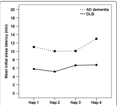
= 9.2, P <0.01) and <5 minutes (DLB =56% vs AD dementia =17%, χ2= 7.4, P =0.01). The mean MSLT initial sleep latency was shorter for DLB than AD de-mentia (DLB =6.4 ± 5 minutes vs AD dede-mentia =11.3 ± 5 minutes, F=12.6, P <0.01). Since dementia severity was associated with shorter mean MSLT initial sleep laten-cies in the AD dementia group (r=−0.59, P <0.01), we also carried out a repeated measures analysis of covari-ance with GLDS as the covariate. The results showed a significant between-subjects effect, confirming shorter MSLT mean initial sleep latencies across the four naps for the DLB group compared to the AD dementia group (F=14.5,P<0.001). There were no within-subjects effects, indicating no differences in mean initial sleep latencies between each of the four nap opportunities for either dementia group (see Figure 1).
In DLB, mean MSLT initial sleep latency was not asso-ciated with age, dementia severity, sex or use of antipar-kinsonian agents or cholinesterase inhibitors. There was also no relationship with clinical variables, such as the number of core DLB features, parkinsonism severity or dur-ation of visual hallucindur-ations, parkinsonism or RBD. In-formant ratings of fluctuations and Epworth Sleepiness Scale items were correlated in the DLB group (r=0.45, P
<0.01) and in the AD dementia group (r =0.62,P <0.01), but these data did not reach statistical significance when compared with mean MSLT initial sleep latency. In DLB, nighttime sleep efficiency and arousal index were unrelated to mean MSLT initial sleep latency (r=−0.05,P=0.78), in-dicating that daytime sleepiness occurred regardless of the degree of nighttime sleep fragmentation. When the sample
was limited to include only the very mild and mild stage of dementia, the differences between the DLB and AD de-mentia groups in informant Epworth Sleepiness Scale scores and mean MSLT initial sleep latencies were upheld. In the AD dementia group, there was a trend in the rela-tionship between nighttime sleep efficiency and mean MSLT initial sleep latency (r=−0.49,P<0.04) with a sub-set who had trouble sleeping at night who were also less likely to sleep during the day. This type of low sleepability and hyperarousability is well documented in primary insomnia [28,29], but our study did not have sufficient statistical power for us to examine this rela-tionship further in our AD dementia cohort.
Neuropathologic characterization
In the DLB group, 20 patients came to autopsy an aver-age of 4.1 ± 2 years following the formal sleep study. All had neuropathologic confirmation of intermediate or high likelihood DLB. Among these patients, eight had TLBD with predominantly brainstem/subcortical Lewy-related pathology and twelve had DLBD with additional cortical Lewy-related pathology. In the TLBD group, five had a Braak NFT stage less than IV and three had a Braak NFT stage of IV. In the DLBD group, two had a Braak NFT stage less than IV, seven had a Braak NFT stage of IV and three had a Braak NFT stage greater than IV.
Braak NFT stage, Lewy distribution (TLBD vs DLBD) and intermediate vs high likelihood DLB were not associated with demographic, clinical, sleep or dementia severity indices. As such, widespread cortical pathology does not appear to be a requirement for the dementia or other clinical or sleep features of DLB, including subject-ive or objectsubject-ive daytime sleepiness.
Discussion
Patients with DLB exhibited greater daytime somnolence than those with AD dementia of similar age, sex and de-mentia severity. On the MSLT, 81% of the DLB group fell asleep within a mean of 10 minutes in four daytime nap opportunities, compared to 39% of the AD group. Pathologic sleepiness, based on a mean MSLT initial sleep latency of less than 5 minutes, was evident in 56% of the DLB group compared to 17% of the AD dementia group. This was consistent with subjective informant ratings of a higher Epworth Sleepiness Scale score in DLB compared to AD dementia from the larger sample. Although daytime somnolence was associated with dementia severity in AD dementia, this was not the case in DLB. These data empirically confirm that daytime sleepiness is more likely to occur in patients with DLB than in those with AD dementia.
In DLB, the increased propensity to fall asleep during the day was not related to poor sleep quality the night
before. Mean nighttime sleep efficiency was average when individual scores were compared to published age- and sex-stratified norms [27]. Moreover, nighttime sleep efficiency in the DLB group was not associated with daytime sleepi-ness based on either objective measurement or informant ratings. Nonetheless, since poor sleep efficiency in the DLB group was associated with a higher number of spontaneous arousals, we examined whether extrapyr-amidal features, such as motor stiffness, which can disrupt sleep by restricting one’s ability to turn over [30], might have contributed to the daytime somnolence in our DLB group. This has particular relevance, because daytime sleepiness is often seen in persons with Parkinson’s disease [31-36]. In our sample, the patients with DLB who had more severe motor problems actually had fewer spontan-eous arousals, a sleep benefit related to their use of carbidopa-levodopa. In addition, parkinsonism severity or use of carbidopa-levodopa was not associated with objective and subjective measures of daytime sleepiness. Therefore, neither parkinsonism severity nor carbidopa-levodopa appears to be primarily responsible for the daytime somnolence in our DLB sample.
Sleep disorders, such as moderate to severe sleep apnea and periodic limb movement-related arousals, occurred in less than 20% of the entire sample, which is consistent with rates expected in normal community-dwelling older adults [37-39]. Although 81% of the DLB group had a mean MSLT initial sleep latency shorter than 10 minutes, nighttime arousals related to respiration or movement were not associated with mean MSLT initial sleep latency or with informant ratings of the Epworth Sleepiness Scale of the larger sample. Thus, the presence of these sleep disorders did not account for disturbed arousal in our DLB group.
sleepiness is a distinct feature of DLB that is not contin-gent on disease stage or on any one of the four core fea-tures of DLB. Further work is needed to determine if its presence helps improve diagnostic validity and reliable early detection of DLB.
Of the 20 patients with DLB who underwent patho-logic examination, all had confirmation of Lewy body dis-ease. There was no difference in demographics, clinical or sleep variables between the eight patients with TLBD (which includes brainstem and limbic Lewy pathology) and the twelve with DLBD (includes added cortical pathology). Similarly, there was no difference between those with intermediate likelihood DLB and high likeli-hood DLB, which also takes into account concomitant neurofibrillary tangle pathology. As such, neuronal loss and Lewy pathology in brainstem and limbic regions, without widespread cortical involvement, are sufficient to produce daytime sleepiness, dementia and the other core DLB features. This is consistent with the Braak sta-ging model of Lewy body disease, which suggests earlier involvement of brainstem and limbic regions relative to cortical regions [46].
We postulate that the mechanism underlying daytime sleepiness in DLB may be related to neuronal loss from the disease itself and triggered by the disruption of the brain regions responsible for sleep–wake physiology. In Lewy body disease, cell clusters that are particularly vul-nerable include the locus coeruleus, the raphe nucleus, the tuberomammillary nucleus of the hypothalamus, the periaqueductal gray and the basal forebrain [25,47]. These nuclei constitute a neuronal network composed of mul-tiple neurotransmitters known to regulate wakefulness that are referred to collectively as the ascending reticular activating system (ARAS) [48-52]. Saper and colleagues [53] have proposed a sleep–wake switch model based on the reciprocal relationship between the wakefulness neurons of the ARAS and the sleep neurons of the ventrolateral preoptic hypothalamus (VLPO) [53], with the hypocretin cells of the lateral hypothalamus serving as a modulator of sleep–wake transitions [54,55]. Fur-ther work is needed to determine wheFur-ther our finding of essentially normal nighttime sleep efficiency in DLB, but greater daytime sleepiness, may reflect a bias or imbalance between the VLPO and ARAS, and whether there is an inequity in their modulation by the lateral hypothalamic hypocretin neurons. Although cerebro-spinal fluid levels of hypocretin in DLB and Parkinson’s disease dementia show wide variability, ranging from very low to normal levels [56-58], there is pathologic evidence of greater hypocretin immunoreactive cell loss in DLB compared to AD [59,60], with one DLB study showing hypocretin cell loss correlating with hypersomno-lence andα-synuclein [60]. Whether hypocretin cell loss is directly related to Lewy-related pathology or is a
consequence of lost input due to damage to the ARAS neuronal network in Lewy body disease is not yet known. More detailed investigation of how the particular path-ways known to be involved in sleep and wakefulness are affected in DLB is clearly needed.
A clinical history of RBD was present in 90% of our DLB sample, but it could be confirmed in only the 71% who actually achieved REM sleep during polysomnogra-phy. Of the 19% with DLB and a history of RBD who did not achieve REM sleep, these patients had less total sleep time and lower nighttime sleep efficiency than their coun-terparts who did achieve REM sleep. Those with a longer documented clinical duration of RBD also spent less time in REM sleep, which may explain why RBD eventually be-comes quiescent in patients with very long histories of RBD [61].
Some limitations to the study deserve mention. Repli-cation with a larger sample size and with complete congruence between those who have undergone over-night polysomnography and daytime MSLT is needed. In addition, interpretation of the neuropathologic analysis is limited by the absence of AD autopsies and by the small number of DLB cases with sleep evaluations who have come to autopsy to date. In our effort to determine whether nighttime sleep efficiency was normal for age and sex, we calculated individual z-scores using a large normative data set that incorporated in-home overnight polysomnography. Although this setting may not be exactly comparable to the sleep laboratory, studies indicate good validity with in-home polysomnography, and discrepancies tend to be in the direction of better sleep efficiency at home relative to the sleep laboratory [62,63]. Under these conditions, we deemed it reasonable to provide this comparison, recognizing that our laboratory findings of average sleep efficiency in the DLB group and low aver-age sleep efficiency in the AD group may reflect an underestimate of true sleep efficiency for these groups.
In this study, we incorporated the MSLT, which is con-sidered the gold standard for the objective measurement of sleepiness and relies on the assessment of how quickly one falls asleep when asked to do so. Further study is needed to investigate whether patients with DLB also have trouble maintaining wakefulness when asked to do so.
Conclusions
from the other core DLB features, including fluctuations. If daytime sleepiness is a unique clinical feature of DLB, this has implications for improved early detection and differential diagnosis of DLB, for consideration of alter-nate treatment interventions, and for promoting our understanding of the pathologic and neuroanatomic in-volvement in DLB.
Note
This article is part of a series on Lewy Body Dementia, edited by Ian McKeith and James Galvin. Other articles in this series can be found at http://alzres.com/series/ LewyBodyDementia
Abbreviations
AD:Alzheimer’s disease; ARAS: Ascending reticular activating system; DLBs: Dementia with Lewy bodies; DLBD: Diffuse Lewy body disease; ESS: Epworth Sleepiness Scale; GLDS: Global Deterioration Scale; MMSE: Mini Mental State Examination; MSLT: Multiple Sleep Latency Test; NFT: Neurofibrillary tangle; NPI-Q: Neuropsychiatric Inventory Questionnaire–Short Form; RBD: Rapid eye movement sleep behavior disorder; RDI: Respiratory disturbance index; REM: Rapid eye movement; TLBD: Transitional Lewy body disease; UPDRS: Unified Parkinson’s Disease Rating Scale; VLPO: Ventrolateral preoptic hypothalamus.
Competing interests
This study was conducted independently of any pharmaceutical organization or corporate sponsorship. The authors declare that they have no competing interests. RCP serves on the data monitoring committee for Pfizer Inc and Janssen Alzheimer Immunotherapy and is a consultant for Merck Inc, Roch Inc and Genentech Inc. NGR has received funding from Lilly Inc and for a TauRX Therapeutics multicenter study, and is a consultant for Cytox Ltd. BFB has received funding from GE Healthcare. DSK serves on the data safety monitoring board for Lundbeck Pharmaceuticals and for the Dominantly Inherited Alzheimer’s Disease Treatment Unit.
Authors’contributions
TJF participated in the study conception, study design, acquisition of data, statistical analysis, data interpretation, manuscript preparation and coordination of the efforts related to the project. GES contributed to study design, subject recruitment, helped with statistical analysis and edited the manuscript. DWD and JP performed the neuropathologic assessment, provided critical feedback and edited the manuscript. NGR helped with subject recruitment, clinical assessment and edited the manuscript. SCL and MHS designed and ensured proper polysomnography methodology, oversaw the administration and scoring of the sleep studies and edited the manuscript. ZW, JAG, RU, DSK and RCP helped with subject recruitment, clinical assessment and edited the manuscript. BFB participated in study conception, subject recruitment, clinical assessment; designed and ensured proper sleep methodology and edited the manuscript. All authors read and approved the final manuscript.
Acknowledgements
We gratefully acknowledge support from the National Institutes of Health through grants R01AG15866, P50AG16574, U01AG06786 and P50NS072187 and also from The Mangurian Foundation for Lewy Body Dementia Research. We thank Jeremiah Aakre for his help with data management.
Author details
1Department of Psychiatry and Psychology, Mayo Clinic, 4500 San Pablo
Road, Jacksonville, FL 32224, USA.2Department of Psychiatry and Psychology, Mayo Clinic, 200 First Street SW, Rochester, MN 55905, USA.3Department of
Pathology, Mayo Clinic, 4500 San Pablo Road, Jacksonville, FL 32224, USA.
4Department of Neurology, Mayo Clinic, 4500 San Pablo Road, Jacksonville,
FL 32224, USA.5Center for Sleep Medicine, Mayo Clinic, 4500 San Pablo Road, Jacksonville, FL 32224, USA.6Department of Neurology, Mayo Clinic,
200 First Street SW, Rochester, MN 55905, USA.7Department of Laboratory Medicine and Pathology, Mayo Clinic, 200 First Street SW, Rochester, MN
55905, USA.8Center for Sleep Medicine, Mayo Clinic, 200 First Street SW,
Rochester, MN 55905, USA.
Received: 30 April 2014 Accepted: 14 October 2014
References
1. Ferman TJ, Smith GE, Boeve BF, Ivnik RJ, Petersen RC, Knopman D, Graff-Radford N, Parisi J, Dickson DW:DLB fluctuations: specific features that reliably differentiate DLB from AD and normal aging.Neurology
2004,62:181–187.
2. Rongve A, Boeve BF, Aarsland D:Frequency and correlates of caregiver reported sleep disturbances in a sample of persons with early dementia.
J Am Geriatr Soc2010,58:480–486.
3. Boddy F, Rowan EN, Lett D, O’Brien JT, McKeith IG, Burn DJ:Subjectively reported sleep quality and excessive daytime somnolence in Parkinson’s disease with and without dementia, dementia with Lewy bodies and Alzheimer’s disease.Int J Geriatr Psychiatry2007,22:529–535.
4. Lee DR, McKeith I, Mosimann U, Ghosh‐Nodyal A, Thomas AJ:Examining carer stress in dementia: the role of subtype diagnosis and neuropsychiatric symptoms.Int J Geriatr Psychiatry2013,28:135–141. 5. Bonanni E, Maestri M, Tognoni G, Fabbrini M, Nucciarone B, Manca ML, Gori
S, Iudice A, Murri L:Daytime sleepiness in mild and moderate Alzheimer’s disease and its relationship with cognitive impairment.J Sleep Res2005, 14:311–317.
6. Escandon A, Al-Hammadi N, Galvin JE:Effect of cognitive fluctuation on neuropsychological performance in aging and dementia.Neurology2010, 74:210–217.
7. Ferman TJ, Smith GE, Kantarci K, Boeve BF, Pankratz VS, Dickson DW, Graff-Radford NR, Wszolek Z, Van Gerpen J, Uitti R:Nonamnestic mild cognitive impairment progresses to dementia with Lewy bodies.
Neurology2013,81:2032–2038.
8. Dijk DJ, Groeger JA, Stanley N, Deacon S:Age-related reduction in daytime sleep propensity and nocturnal slow wave sleep.Sleep2010,33:211–223. 9. Pao WC, Boeve BF, Ferman TJ, Lin S-C, Smith GE, Knopman DS, Graff-Radford
NR, Petersen RC, Parisi JE, Dickson DW:Polysomnographic findings in dementia with Lewy bodies.Neurologist2013,19:1–6.
10. Terzaghi M, Arnaldi D, Rizzetti MC, Minafra B, Cremascoli R, Rustioni V, Zangaglia R, Pasotti C, Sinforiani E, Pacchetti C:Analysis of video‐ polysomnographic sleep findings in dementia with Lewy bodies.
Mov Disord2013,28:1416–1423.
11. Trotti LM, Bliwise DL:No increased risk of obstructive sleep apnea in Parkinson’s disease.Mov Disord2010,25:2246–2249.
12. Bliwise DL, Trotti LM, Yesavage JA, Rye DB:Periodic leg movements in sleep in elderly patients with Parkinsonism and Alzheimer’s disease.Eur J Neurol2012,19:918–923.
13. McKeith IG, Dickson DW, Lowe J, Emre M, O’Brien JT, Feldman H, Cummings J, Duda JE, Lippa C, Perry EK, Aarsland D, Arai H, Ballard CG, Boeve B, Burn DJ, Costa D, Del Ser T, Dubois B, Galasko D, Gauthier S, Goetz CG, Gomez-Tortosa E, Halliday G, Hansen LA, Hardy J, Iwatsubo T, Kalaria RN, Kaufer D, Kenny RA, Korczyn A:Diagnosis and management of dementia with Lewy bodies: third report of the DLB Consortium.Neurology2005,65:1863–1872. A published erratum appears inNeurology2005,65(12):1992.
14. Morris J, Blennow K, Froelich L, Nordberg A, Soininen H, Waldemar G, Wahlund LO, Dubois B:Harmonized diagnostic criteria for Alzheimer’s disease: recommendations.J Intern Med2014,275:204–213.
15. American Psychiatric Association:Diagnostic and Statistical Manual of Mental Disorders.4th edition. Washington, DC: American Psychiatric Association; 1994. 16. Reisberg B, Ferris SH, de Leon MJ, Crook T:Global Deterioration Scale
(GDS).Psychopharmacol Bull1988,24:661–663.
17. Folstein MF, Folstein SE, McHugh PR:“Mini-mental state”: a practical method for grading the cognitive state of patients for the clinician.
J Psychiatr Res1975,12:189–198.
18. Boeve BF, Molano JR, Ferman TJ, Smith GE, Lin SC, Bieniek K, Haidar W, Tippmann-Peikert M, Knopman DS, Graff-Radford NR:Validation of the Mayo Sleep Questionnaire to screen for REM sleep behavior disorder in an aging and dementia cohort.Sleep Med2011,12:445–453.
20. Johns MW:A new method for measuring daytime sleepiness: the Epworth sleepiness scale.Sleep1991,14:540–545.
21. Lesher EL, Berryhill JS:Validation of the Geriatric Depression Scale–Short Form among inpatients.J Clin Psychol1994,50:256–260.
22. Kaufer DI, Cummings JL, Ketchel P, Smith V, MacMillan A, Shelley T, Lopez OL, DeKosky ST:Validation of the NPI-Q, a brief clinical form of the
Neuropsychiatric Inventory.J Neuropsychiatry Clin Neurosci2000,12:233–239. 23. American Academy of Sleep Medicine Task Force:Sleep-related breathing
disorders in adults: recommendations for syndrome definition and measurement techniques in clinical research.Sleep1999,22:667–689. 24. Iber C, Ancoli-Israel S, Chesson A, Quan SF, for the American Academy of
Sleep Medicine:The AASM Manual for the Scoring of Sleep and Associated Events: Rules, Terminology and Technical Specifications.1st edition. Westchester, IL: American Academy of Sleep Medicine; 2007.
25. Dickson DW, Braak H, Duda JE, Duyckaerts C, Gasser T, Halliday GM, Hardy J, Leverenz JB, Del Tredici K, Wszolek ZK, Litvan I:Neuropathological assessment of Parkinson’s disease: refining the diagnostic criteria.Lancet Neurol2009,8:1150–1157. Published errata appear inLancet Neurol2010, 9:140, andLancet Neurol2010,9:29.
26. Braak H, Braak E:Neuropathological stageing of Alzheimer-related changes.Acta Neuropathol1991,82:239–259.
27. Unruh ML, Redline S, An MW, Buysse DJ, Nieto FJ, Yeh JL, Newman AB: Subjective and objective sleep quality and aging in the sleep heart health study.J Am Geriatr Soc2008,56:1218–1227.
28. Bonnet MH, Arand DL:Hyperarousal and insomnia: state of the science.
Sleep Med Rev2010,14:9–15.
29. Roehrs TA, Randall S, Harris E, Maan R, Roth T:MSLT in primary insomnia: stability and relation to nocturnal sleep.Sleep2011,34:1647–1652. 30. Pal P, Calne S, Samii A, Fleming J:A review of normal sleep and its
disturbances in Parkinson’s disease.Parkinsonism Relat Disord1999,5:1–17. 31. Arnulf I, Konofal E, Merino-Andreu M, Houeto J, Mesnage V, Welter M,
Lacomblez L, Golmard J, Derenne J, Agid Y:Parkinson’s disease and sleepiness: An integral part of PD.Neurology2002,58:1019–1024. 32. Rye DB, Bliwise DL, Dihenia B, Gurecki P:Daytime sleepiness in Parkinson’s
disease.J Sleep Res2000,9:63–69.
33. Shpirer I, Miniovitz A, Klein C, Goldstein R, Prokhorov T, Theitler J, Pollak L, Rabey JM:Excessive daytime sleepiness in patients with Parkinson’s disease: a polysomnography study.Mov Disord2006,21:1432–1438. 34. Peeraully T, Yong MH, Chokroverty S, Tan EK:Sleep and Parkinson’s
disease: a review of case‐control polysomnography studies.Mov Disord
2012,27:1729–1737.
35. Emre M, Aarsland D, Brown R, Burn DJ, Duyckaerts C, Mizuno Y, Broe GA, Cummings J, Dickson DW, Gauthier S, Goldman J, Goetz C, Korczyn A, Lees A, Levy R, Litvan I, McKeith I, Olanow W, Poewe W, Quinn N, Sampaio C, Tolosa E, Dubois B:Clinical diagnostic criteria for dementia associated with Parkinson’s disease.Mov Disord2007,22:1689–1707.
36. Ondo WG, Dat Vuong K, Khan H, Atassi F, Kwak C, Jankovic J:Daytime sleepiness and other sleep disorders in Parkinson’s disease.Neurology
2001,57:1392–1396.
37. Johansson P, Alehagen U, Svanborg E, Dahlström U, Broström A:Sleep disordered breathing in an elderly community-living population: relationship to cardiac function, insomnia symptoms and daytime sleepiness.Sleep Med2009,10:1005–1011.
38. Ancoli-Israel S, Kripke DF, Klauber MR, Mason WJ, Fell R, Kaplan O: Sleep-disordered breathing in community-dwelling elderly.Sleep1991, 14:486–495.
39. Ancoli-Israel S, Kripke D, Klauber M, Mason W, Fell R, Kaplan O:Periodic limb movements in sleep in community-dwelling elderly.Sleep1991,14:496–500. 40. Franco JG, Trzepacz PT, Meagher DJ, Kean J, Lee Y, Kim JL, Kishi Y, Furlanetto LM,
Negreiros D, Huang MC:Three core domains of delirium validated using exploratory and confirmatory factor analyses.Psychosomatics2013,54:227–238. 41. Inouye SK, van Dyck CH, Alessi CA, Balkin S, Siegal AP, Horwitz RI:Clarifying
confusion: the confusion assessment method: a new method for detection of delirium.Ann Intern Med1990,113:941–948. A published erratum appears in Ann Intern Med 1991,114(5):433.
42. Lee DR, McKeith I, Mosimann U, Ghosh-Nodial A, Grayson L, Wilson B, Thomas AJ:The Dementia Cognitive Fluctuation Scale, a new psychometric test for clinicians to identify cognitive fluctuations in people with dementia.
Am J Geriatr Psychiatry2013,22:926–935.
43. Ballard C, O’Brien J, Gray A, Cormack F, Ayre G, Rowan E, Thompson P, Bucks R, McKeith I, Walker M:Attention and fluctuating attention in
patients with dementia with Lewy bodies and Alzheimer disease.
Arch Neurol2001,58:977–982.
44. Walker MP, Ayre GA, Cummings JL, Wesnes K, McKeith IG, O’Brien JT, Ballard CG:The Clinician Assessment of Fluctuation and the One Day Fluctuation Assessment Scale: two methods to assess fluctuating confusion in dementia.Br J Psychiatry2000,177:252–256.
45. Bliwise DL, Scullin MK, Trotti LM:Fluctuations in cognition and alertness vary independently in dementia with Lewy bodies.Mov Disord2014,29:83–89. 46. Braak H, Ghebremedhin E, Rub U, Bratzke H, Del Tredici K:Stages in the
development of Parkinson’s disease-related pathology.Cell Tissue Res
2004,318:121–134.
47. Braak H, Del Tredici K, Rüb U, de Vos RA, Jansen Steur EN, Braak E:Staging of brain pathology related to sporadic Parkinson’s disease.Neurobiol Aging2003,24:197–211.
48. Moruzzi G, Magoun HW:Brain stem reticular formation and activation of the EEG.Electroencephalogr Clin Neurophysiol1949,1:455–473.
49. Buzsáki G, Bickford RG, Ponomareff G, Thal L, Mandel R, Gage FH:Nucleus basalis and thalamic control of neocortical activity in the freely moving rat.J Neurosci1988,8:4007–4026.
50. Saper CB, Scammell TE, Lu J:Hypothalamic regulation of sleep and circadian rhythms.Nature2005,437:1257–1263.
51. Aston-Jones G, Bloom FE:Activity of norepinephrine-containing locus coeruleus neurons in behaving rats anticipates fluctuations in the sleep-waking cycle.J Neurosci1981,1:876–886.
52. Panula P, Nuutinen S:The histaminergic network in the brain: basic organization and role in disease.Nat Rev Neurosci2013,14:472–487. 53. Saper CB, Fuller PM, Pedersen NP, Lu J, Scammell TE:Sleep state switching.
Neuron2010,68:1023–1042.
54. Lee MG, Hassani OK, Jones BE:Discharge of identified orexin/hypocretin neurons across the sleep-waking cycle.J Neurosci2005,25:6716–6720. 55. Saper CB:The neurobiology of sleep.Continuum (Minneap Minn)2013,19:19–31. 56. Drouot X, Moutereau S, Nguyen J, Lefaucheur J, Creange A, Remy P,
Goldenberg F, d’Ortho M:Low levels of ventricular CSF orexin/hypocretin in advanced PD.Neurology2003,61:540–543.
57. Fronczek R, Overeem S, Lee SY, Hegeman IM, van Pelt J, van Duinen SG, Lammers GJ, Swaab DF:Hypocretin (orexin) loss in Parkinson’s disease.
Brain2007,130:1577–1585.
58. Compta Y, Santamaria J, Ratti L, Tolosa E, Iranzo A, Muñoz E, Valldeoriola F, Casamitjana R, Ríos J, Marti MJ:Cerebrospinal hypocretin, daytime sleepiness and sleep architecture in Parkinson’s disease dementia.Brain2009, 132:3308–3317.
59. Kasanuki K, Iseki E, Kondo D, Fujishiro H, Minegishi M, Sato K, Katsuse O, Hino H, Kosaka K, Arai H:Neuropathological investigation of hypocretin expression in brains of dementia with Lewy bodies.Neurosci Lett2014,569:68–73. 60. Lessig S, Ubhi K, Galasko D, Adame A, Pham E, Remidios K, Chang M,
Hansen LA, Masliah E:Reduced hypocretin (orexin) levels in dementia with Lewy bodies.Neuroreport2010,21:756–760.
61. Claassen DO, Josephs KA, Ahlskog JE, Silber MH, Tippmann-Peikert M, Boeve BF:REM sleep behavior disorder preceding other aspects of synucleinopathies by up to half a century.Neurology2010,75:494–499. 62. Iber C, Redline S, Gilpin AK, Quan SF, Zhang L, Gottlieb DJ, Rapoport D,
Resnick HE, Sanders M, Smith P:Polysomnography performed in the unattended home versus the attended laboratory setting-Sleep Heart Health Study methodology.Sleep2004,27:536–540.
63. Bruyneel M, Sanida C, Art G, Libert W, Cuvelier L, Paesmans M, Sergysels R, Ninane V:Sleep efficiency during sleep studies: results of a prospective study comparing home‐based and in‐hospital polysomnography.J Sleep Res2011,20:201–206.
doi:10.1186/s13195-014-0076-z
Cite this article as:Fermanet al.:Abnormal daytime sleepiness in
dementia with Lewy bodies compared to Alzheimer’s disease using the