R E V I E W
Hematopoietic cell differentiation from embryonic
and induced pluripotent stem cells
Wai Feng Lim
1,2*†, Tomoko Inoue-Yokoo
1,3†, Keai Sinn Tan
1,2, Mei I Lai
2and Daisuke Sugiyama
1*Abstract
Pluripotent stem cells, both embryonic stem cells and induced pluripotent stem cells, are undifferentiated cells that can self-renew and potentially differentiate into all hematopoietic lineages, such as hematopoietic stem cells (HSCs), hematopoietic progenitor cells and mature hematopoietic cells in the presence of a suitable culture system. Establishment of pluripotent stem cells provides a comprehensive model to study early hematopoietic development and has emerged as a powerful research tool to explore regenerative medicine. Nowadays, HSC transplantation and hematopoietic cell transfusion have successfully cured some patients, especially in malignant hematological diseases. Owing to a shortage of donors and a limited number of the cells, hematopoietic cell induction from pluripotent stem cells has been regarded as an alternative source of HSCs and mature
hematopoietic cells for intended therapeutic purposes. Pluripotent stem cells are therefore extensively utilized to facilitate better understanding in hematopoietic development by recapitulating embryonic developmentin vivo, in which efficient strategies can be easily designed and deployed for the generation of hematopoietic lineages
in vitro. We hereby review the current progress of hematopoietic cell induction from embryonic stem/induced
pluripotent stem cells.
Introduction
Hematopoietic stem cells (HSCs) lay the foundation of hematopoiesis to generate all functional hematopoietic lineages, including erythrocytes, leukocytes (neutrophils, basophils, eosinophils, lymphocytes, monocytes and macrophages) and platelets [1]. Perturbations in the hematopoietic system have been reported to cause nu-merous diseases such as anemia, leukemia, lymphomas and thrombocytopenia. Currently, HSC transplantation and hematopoietic cell transfusion are useful treatments for some hematological diseases, such as thalassemia and leukemia. However, some conventional sources of
HSCs–such as cord blood, bone marrow and peripheral
blood–are limited in usage, owing to the limited
num-ber of transplantable cells as well as inefficient strategies
for expanding those cells. Additionally, problems such as shortage of human leukocyte antigen-matched donors, adverse effects from graft-versus-host disease, viral con-tamination and immunoreactions impede the utility of readily available HSCs. The use of alternative sources for these cells will therefore be a great advantage for regen-erative medicine.
Pluripotent stem cells are one of the potential sources
for HSCs and the in vitromodel for further elucidating
the regulatory mechanisms underlying embryonic
hematopoietic development. Embryonic stem (ES) cells are pluripotent cells established from the inner cell mass of blastocyst-stage embryos, in both mouse and human [2,3], and are capable of giving rise to three germ layers after directed differentiation in culture [3,4]. However, manipulation of human ES cells raises some ethical issues and immunoreactions. Induced pluripotent stem (iPS) cell technology has made a groundbreaking discov-ery to circumvent the problems of ethical and practical issues in using ES cells [5]. It is of great importance to develop efficient and controllable induction strategies to drive hematopoietic differentiation from ES/iPS cells in culture prior to the realization of pluripotent cell-derived therapies. To review current progress of
* Correspondence:[email protected];[email protected] †Equal contributors
1Department of Advanced Medical Initiatives, Division of Hematopoietic
Stem Cells, Advanced Medical Initiatives, Faculty of Medical Sciences, Kyushu University, Station for Collaborative Research 1, 4F, 3-1-1 Maidashi, Higashi-Ku, Fukuoka 812-8582, Japan
2Department of Pathology, Faculty of Medicine and Health Sciences,
Universiti Putra Malaysia, 43400 UPM, Serdang, Selangor Darul Ehsan, Malaysia
Full list of author information is available at the end of the article
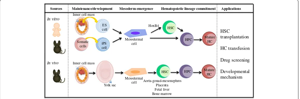
differentiation protocol from ES/iPS cells, we first summarize the knowledge of hematopoietic develop-ment during early mouse hematopoiesis followed by the manipulation of ES/iPS cells in hematopoietic cell induction (Figure 1).
Embryonic hematopoiesis
Studies of hematopoietic development during
embryo-genesis in vivo are important to gain insight into its
underlying mechanisms, whereby accumulated know-ledge will facilitate the induction of HSCs, hematopoietic progenitor cells (HPCs) and mature hematopoietic cells from pluripotent stem cells in culture. In mouse blasto-cyst, the inner cell mass at 3.5 days post coitum (dpc)
comprises a population of cells–which can give rise to
a derivative of three germ layers (endoderm, mesoderm
and ectoderm) – that eventually develop into both
intraembryonic and extraembryonic tissues as embryo develops [6]. The hematopoietic system that derives from the mesodermal germ layer can be classified into two waves. The first hematopoiesis (primitive hematopoiesis) begins to develop primitive erythroid and macrophage progenitors in the yolk sac (YS) blood islands at 7.0 dpc [7]. Para-aortic splanchnopleural regions that will develop
into aorta–gonad–mesonephros (AGM) already possess
hematopoietic precursors beginning at 8.5 dpc [8]. Before the establishment of circulation (8.0 dpc), both YS and para-aortic splanchnopleural-derived mesodermal cells acquire HSC activity after co-culturing with AGM-derived stromal cells [9]. After circulation commences,
CD34+c-Kit+ cells derived from both YS and para-aortic
splanchnopleura at 9.0 dpc were able to reconstitute the
hematopoietic system in newborn recipient pups, but not in adult recipient mice [10]. These findings demonstrate that both YS and para-aortic splanchnopleura possess HSC potential that can contribute to definitive hematopoiesis under a favorable microenvironment.
The first definitive HSCs that can reconstitute the adult hematopoietic system appear in the AGM region at 10.5 dpc followed by the YS, placenta and liver, span-ning from 11.0 to 11.5 dpc [11-13]. YS cells expressing
Runx1at 7.5 dpc progressed into fetal lymphoid progen-itors at 16.5 dpc in both fetal liver and thymus as well as adult HSCs in 9-month-old to 12-month-old mouse bone marrow [14]. In view of these results, both the YS and the AGM region contribute to HSC generation. However the extent of their contribution still remains
unclear. To address this issue, YS–YS chimeric embryos
were generated before blood circulation at 8.25 dpc, where no B-cell activity was detected, which is relevant to HSC activity in the early mouse embryo. As the chimeric embryos develop into 11.0 dpc equivalent in whole embryo culture, the grafted YS cells contributed to B-cell activity in the AGM region, but with low fre-quency [15]. This observation implies that the main source of HSCs is derived from the AGM region.
In addition to the YS and the AGM region, the pla-centa is another site for HSC generation. The plapla-centa exchanges oxygen and nutrient between mother and fetus, and is formed around 9.0 dpc after fusion of chor-ion and allantois. A fluorescent-labeled allantois regchor-ion at 8.25 dpc could be detected in the hematopoietic cell cluster after 42 hours of whole embryo culture, suggesting that allantois alone possesses HSC potential
Sources Hematopoietic lineage commitment
ES cell iPS cell
In vitro
In vivo
Maintenance/development Mesoderm emergence
HSC
HSC Yolk sac
Aorta-gonad-mesonephros Placenta Fetal liver Bone marrow
HPC
Applications
HSC transplantation
HC transfusion
Drug screening
Developmental mechanism
Somatic cells
Mesodermal cell
Mesodermal cell Inner cell mass
Inner cell mass
HoxB4
HPC Mature HC Mature
HC
Figure 1Schematic representations of hematopoietic development fromin vivoandin vitromodels.Both human and mousein vitro
models have been established for hematopoietic differentiation in a defined culture system from embryonic stem (ES) and adult cell-derived induced pluripotent stem (iPS) cells. For thein vivomodel, the mouse inner cell mass undergoes differentiation, later forming the yolk sac, which generates mesodermal cells and induces hematopoietic stem cells (HSCs), hematopoietic progenitor cells (HPCs) and mature hematopoietic cells (HCs). Successfully generated HSCs from bothin vitroandin vivomodels might be applied to HSC transplantation for hematopoietic disorders. Further differentiation of HSC in a cytokine-defined culture system produces hematopoietic cells for hematopoietic cell transfusion. Thorough understanding of molecular mechanism on these models will be beneficial for both drug screening as well as the mechanism of
and also proposing that the placenta is likely to be an ixndependent site of HSC generation, regardless of cells circulating from other hematopoietic organs [16]. Taken together, the YS, AGM region and placenta contribute to HSC generation prior to the HSC expansion in the fetal liver .
Gene and protein markers for mesodermal and hematopoietic lineages
During early embryonic development, Brachyurythat is
indispensable for mesodermal formation expresses
tran-siently [17]. Subsequently tyrosine kinaseFlk1for
blood-island formation and vasculogenesis expresses in
hemangioblasts, which is the common embryonic endo-thelial and hematopoietic precursor [18].
Transcription factors characterized to be involved in the hematopoietic lineages are shown below. In the
tran-sition of mesodermal to hematopoietic lineages,Sclis
in-dispensable for the development of all hematopoietic
lineages, in which a homozygous mutant Scl−/− model
showed undetectable hematopoietic lineages in both
in vivo andin vitrostudies [19].Runx1 plays an import-ant role in governing definitive hematopoiesis but not primitive hematopoiesis through the observation of blast colony-forming cells, which are both hematopoietic and endothelial cell precursors from ES cells and equivalent
of hemangioblast, using a Runx1−/− ES cell culture
model [20]. The GATA family of transcription factors, especially GATA-1 and GATA-2, are differentially regu-lated at multiple steps from the early development of hemangioblast to hematopoietic commitment [21].
Surface markers of hematopoietic lineages are briefly described. Cells expressing endothelial markers, such as CD34, PECAM-1 (CD31) and VE-cadherin, are closely associated with both hematopoietic and endothelial cell commitment [22]. Expression of CD41 initiates the primitive and definitive hematopoiesis, whereas HPCs are highly dependent on c-Kit expression [23]. Addition-ally, CD34 as a marker for HSCs and CD45 as a marker for pan-leukocyte and/or maturation of HSCs are uti-lized respectively [24,25].
Differentiation of embryonic stem/induced pluripotent stem cells and hematopoietic cell induction
Mouse ES/iPS cells can be maintained and propagated indefinitely on a layer of mitotically inactivated mouse embryonic fibroblasts (MEFs) and/or leukemia inhibitory factor in culture. On the contrary, human ES cells achieve an undifferentiated state on a layer of MEFs and/or basic fibroblast growth factor. After removal of leukemia inhibitory factor and basic fibroblast growth factor, these ES cells can differentiate into any kind of cell depending on appropriate culture conditions. One
of the ultimate goals of in vitrodifferentiation of ES/iPS cells is to generate HSCs with the acquisition of long-term reconstitution capacity.
By recapitulating mouse embryogenesis,in vitro
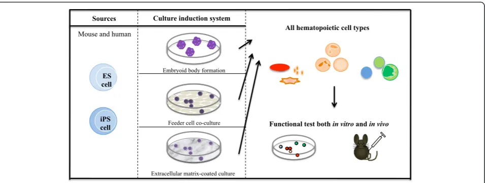
differ-entiation of ES/iPS cells toward hematopoietic develop-ment has been technically approached: through formation of a cystic-like structure that exhibits three germ layers that are able to generate mesodermal cells (embryoid body (EB) formation); through feeder cell co-culture that can provide ES/iPS cells an intimate cell contact with secreting factor(s) that promote cell proliferation and dif-ferentiation (feeder cell co-culture); and through an extra-cellular matrix-coated dish that supports differentiation of ES/iPS cells (Figure 2).
Embryoid body formation
EBs are three-dimensional cell aggregates formed in sus-pension culture and they mimic the spatial organization
of the embryo through enhancing cell–cell interactions
to form three germ layers [3,4]. There are a number of approaches to EB formation, such as methylcellulose or suspension culture medium either using a bacterial-grade dish, a methylcellulose-coated plate, a low-adherent micro-well plate or the hanging drops method. To specif-ically induce a hematopoietic lineage, single-cell suspen-sion of EBs are directed into methylcellulose culture medium that functions to support hematopoietic develop-ment in the presence of hematopoietic cytokines, such as stem cell factor (SCF), granulocyte colony-stimulating
factor (CSF), macrophage CSF, granulocyte–macrophage
CSF, erythropoietin (Epo) and interleukins (ILs) such as IL-1, IL-3, IL-4 and IL-6 [26]. In the absence of cytokines in culture, EB cells themselves reportedly possess the cap-ability to secrete proteins having macrophage CSF, IL-3 and Epo activity in order to support the early develop-ment of erythroid and macrophage precursors [27].
Using mouse ES cells, 75% of blast colonies were gen-erated from EBs of methylcellulose culture consisting of bipotential precursors (hematopoietic and endothelial cells) as blast colony-forming cells at day 2.5, and then decreased at day 4 (<15%) [28]. In mouse ES cells with
GFP targeted to the Brachyury, EB formation at day 2.5
in suspension culture gave rise to GFP–Flk1–, GFP+Flk1–
and GFP+Flk1+ distinct populations, which are
compar-able with the developmental progression ranging from pre-mesoderm to pre-hemangioblast mesoderm to the hemangioblast, respectively [29].
EBs derived from human ES cells formed three em-bryonic germ layers that can be identified using the
following markers: ζ-globin, neurofilament 68Kd and
α-fetoprotein [30]. By differentiating human ES cells
through EBs at day 3.0 to 4.0, blast colony-forming cells
were generated in liquid culture and expressed FLK1,
human iPS cells throughout a 21-day EB formation
dem-onstrated a sequential gene expression from
BRACHYURY (mesoderm) to GATA-2 and SCL
(hematopoietic commitment), followed by the emergence
of CD34+CD45+ cells that denote hematopoietic lineages
[32].
To improve the efficiency of EB formation using human ES cells, the modified spin-EB technique has been
devel-oped–in which a defined number of human ES cells are
aggregated by centrifugation in low-attachment 96-well plates, and which could reproduce the hematopoietic differentiation potential with more than 90% of the wells able to form hematopoietic cells. This spin-EB method efficiently accelerates hematopoietic differentiation and approximately 500 hematopoietic progenitors are induced from single ES cells [33]. Human iPS cells also differenti-ate into hematopoietic cells by the spin-EB method.
CD34+ cells derived from them are CD45+ (27 to 64%)
and CD43+(36 to 60%) after 13 to 17 days of culture [34].
Feeder cell co-culture
Co-culture is a method of culturing a layer of feeder cells together with ES/iPS cells to give support to the growing cells (germ layer cells) towards development of hematopoietic lineages in appropriate culture medium. A well-known feeder cell layer, OP9 stromal cells are de-rived from newborn bone marrow of the mouse calvaria model, which reveals osteopetrosis due to lack of macro-phage CSF [35]. In addition to OP9 co-cultures, stromal cells from the AGM region can be used to support hematopoiesis of both mouse and human ES cells in the culture system, suggesting that factors such as cytokines are secreted to regulate the early stem cell development and hematopoietic differentiationin vitro[36].
In mouse ES cells, the use of OP9 stromal cells could promote lymphohematopoietic differentiation by
minim-izing preferential differentiation towards monocyte–
macrophage lineages [37]. AGM stromal cell co-cultures
with mouse ES cells could induce CD45+ hematopoietic
cells, including cell differentiation from both Mac-1+
myeloid cells and B220+ B cells, suggesting that AGM
stromal cell lines support hematopoietic differentiation. To improve the efficiency of hematopoietic differenti-ation, a two-step co-culture system was designed, first by co-culturing of mouse ES cells with OP9 cells followed by AGM stromal cells at defined time points.
Approximately 90.6% of CD45+ hematopoietic cells
(23.6% in the OP9 system), 87% of Mac-1+cells (16% in
the OP9 system) and 19.1% of c-Kit+ cells (43% in the
OP9 system) could be detected using this system [36]. Using a colony-forming assay, Krassowska and col-leagues showed that ES cell-derived mesodermal cells could stimulate HPC production with 3.5-fold efficiency in primary AGM region co-cultures as compared with those derived from feeder-free culture. They also co-cultured differentiating ES cells on the dorsal aorta and mesenchyme-derived, urogenital ridge-derived and fetal liver-derived stromal cell lines that had previously been reported to support BM-derived HSCs [38]. The time-course of ES/iPS cell differentiation towards hematopoietic cells does not synchronize with regular hematopoietic differentiation, implying that hematopoietic differentiation does not occur via HSCs. In OP9 cell co-culture, mouse iPS cells initially formedBrachyury-expressing mesodermal cells and later co-cultured again onto fresh OP9 cells supplemented with hematopoietic cocktails were induced to form hematopoietic lineages, including erythroid and myeloid lineage cells but not lymphoid lineage cells [39].
All hematopoietic cell types Sources Culture induction system
Functional test both in vitro and in vivo ES
cell
iPS cell Mouse and human
Embryoid body formation
Feeder cell co-culture
Extracellular matrix-coated culture
Human ES/iPS cells co-cultured with OP9 cells
gener-ated HPCs defined by CD34+CD43+expression and
eval-uated by the clonogenic progenitor cell assay [40]. Using a co-culture system with mouse fetal liver-derived stro-mal cells, human ES cells could generate the erythroid progenitors as determined by colony-forming cells, followed by enucleated erythrocytes with oxygen-carrying capacity [41]. To examine the properties among stromal cells, primary cells and cell lines derived from the mouse AGM region and fetal liver were compared in supporting hematopoietic differentiation from human ES cells. Eight-een days after co-culturing, primary cells derived from the AGM region exhibited the highest number of both
CD34+ and CD45+ cells among the cells, suggesting that
cell lines probably lose their ability to support during the passages [42].
Extracellular matrix-coated dish
Dishes coated with extracellular matrixes, such as colla-gen and fibronectin, are used as monolayer cultures to differentiate ES/iPS cells. In mouse ES cells, the use of collagen IV-coated dishes directs the generation of E-cadherin–Flk1+VE-cadherin–-expressing mesodermal cells and further accelerate differentiation into hemangioblast (VE-cadherin+Flk1+CD45–), HPCs (c-Kit+CD45+) and ma-ture hematopoietic cells (c-Kit–CD45+or Ter119+) [43].
In human ES/iPS cells, matrixes such as human fibronectin, human collagen IV and mouse collagen IV are utilized to generate hematopoietic progenitors (CD43+CD34+), which could give rise to several lineages of differentiated blood cells including erythroid cells
(CD71+CD235a+), megakaryocytes (CD41a+CD42b+),
neutrophils (CD15+CD66b+), macrophages (CD14+CD68+) and dendritic cells (HLA-DR+CD1a+) phenotypically [44]. Human ES/iPS cells could differentiate into mesodermal cells in the presence of the major matrix components such as laminin, collagen I, entactin and heparin-sulfate proteo-glycan as well as growth factors and several other un-defined compounds. These mesodermal cells were able to induce hematopoietic cells after substitution with hematopoietic cocktail culture medium [45].
These studies suggested that monolayer culture system could commit hematopoietic potential in differentiating ES/iPS cells. Combination of this system with xenogeneic-free culture has been widely considered a more suitable approach for future clinical applications.
Embryonic stem/induced pluripotent stem cell-derived hematopoietic lineages
As mechanisms of hematopoietic development in both mouse and human are being established, manipulation of ES/iPS cells for hematopoietic differentiation is also improving. ES/iPS cells possess embryonic natures and are likely to be differentiated into primitive hematopoietic cells.
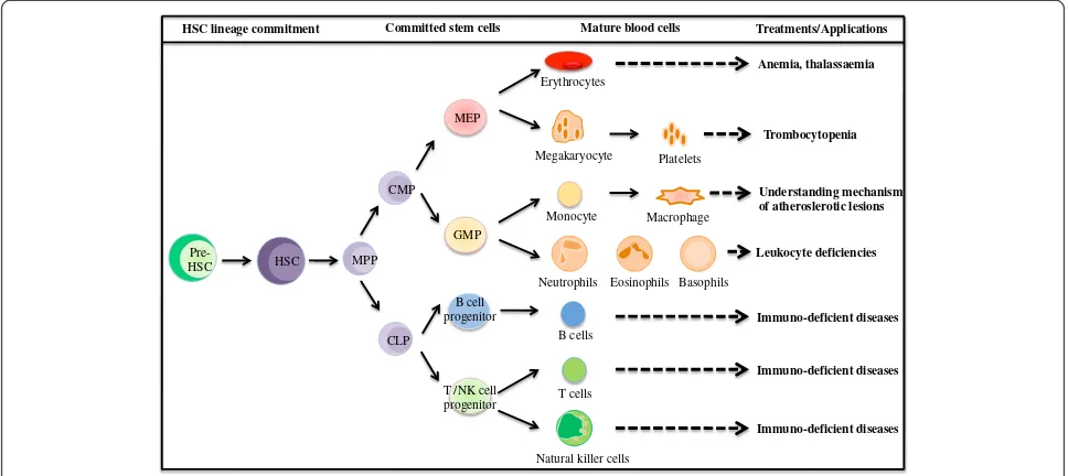
Exogenous cytokines, feeder cells and extracellular matrix-coated dishes enable ES/iPS cells to differentiate into definitive hematopoietic cells in addition to primitive types. In the following section, induction of hematopoietic cell types from both mouse and human ES/iPS cells is described (Figure 3).
Hematopoietic stem cells and hematopoietic progenitor cells
HSCs are the cells that possess self-renewing capacity to maintain the stem cell pool and multipotency to differ-entiate into all lineages of mature hematopoietic cells, while HPCs do not self-renew and their differentiation capacity is limited only to certain types of cells. HSCs/ HPCs can be defined by the surface marker expressions, which enables us to investigate the molecular
mecha-nisms underlying HSC/HPC development.
Mouse-derived HSCs/HPCs are commonly defined by lineage
marker (Lin–, a cocktail of lineage markers including
B220, Ter119, Mac-1, Gr-1, CD3, CD4 and CD8), stem
cell antigen (Sca-1+, a putative marker of stem and
pro-genitor cells) [46], c-Kit+ (a receptor for SCF) [47],
CD34low [48] and Tie-2+ (a tyrosine kinase receptor
expressed in endothelial and hematopoietic cells) [49]. However, unlike in mouse, CD34 is the representative marker for human HSCs [50].
In mouse ES cells, after co-culturing them with the bone marrow stromal cell line with a combination of IL-3, IL-6, fetal calf serum and cell-free supernatants of
fetal liver stromal cell line culture, the PgP-1+ (CD44,
phagocytic glycoprotein-1) Lin– cells (B220–Mac-1–
JORO75–Ter119– (JORO75 is a marker of T-cell
pro-genitors)) are generated and transplanted to repopulate the lymphoid, myeloid and erythroid lineages of primary
adult irradiated mice 15 to 18 weeks
post-transplantation. The PgP-1+Lin– cells also possess self-renewal potential as examined in secondary adult irradi-ated mice after 16 to 20 weeks post-transplantation [51].
Primitive HPCs were generated from EB-derived mouse ES cells and injected into lethally irradiated adult mice to reconstitute the hematopoiesis system, showing a survival rate of more than 3 weeks and subsequently
lymphoid and myeloid engraftment from CD45+
ES-derived cells was detected 12 weeks post-transplantation [52]. Another group demonstrated the acquisition of long-term multilineage capacity in lethally irradiated
mice after injection with c-Kit+CD45+ HSCs, which was
In addition, ectopic expression of certain genes into ES/iPS cells has been used to induce development of
HSCs/HPCs in an in vitrosystem. HoxB4 (a homeobox
transcription factor) was retrovirus-transduced into EB-derived mouse ES cells followed by co-cultures in OP9 stromal cells to induce hematopoietic development. This has resulted in a population of definitive HSCs that could repopulate lymphoid and myeloid cell lineages in both primary and secondary transplanted irradiated
adult mice [54]. On the other hand, Wang’s group
exploited the fact that ectopic expression of Cdx4 (a family of caudal-related homeobox-containing transcrip-tion factors) in mouse ES cells induced mesodermal spe-cification together with increased HPC production. With this notion, they also observed the synergistic effect of
HoxB4 and Cdx4 on HPC production after EB-derived ES cells grown on OP9 stromal cells, and those cultured cells could support the engraftment of all hematopoietic lineages in irradiated adult mice [55]. EB-derived mouse
ES cells are able to generate c-Kit+CD41+CD45– cells
without HoxB4 regulation, but their ability to reconsti-tute adult hematopoiesis is only seen after those cells undergo maturation in OP9 co-cultures in the presence of HoxB4, which was measured by total chimerism
16 weeks post-transplantation of fractionated
co-cultured cells, such as CD41+ cells, c-Kit+ cells, CD34+
cells and CD45–cells [56], suggesting that HoxB4
prob-ably regulates HSC maturation through upregulating the CD34 surface marker expression.
Although the production of HSCs/HPCs is possible using a HoxB4-expressing approach, problems such as an inhibitory effect on lymphoid cell differentiation due to long-term constitutive HoxB4 expression [54] as well as the risk of leukemogenesis in dogs and monkeys after
transducing with HOXB4-expressing retroviral vector
[57] have been reported. To alleviate these adverse
ef-fects, EB-derived CD41+c-Kit+cells derived from ES/iPS
cells were transduced with adenoviral vector of human HOXB4 and then co-cultured on OP9 stromal cells together with hematopoietic cytokines (SCF, fms-like tyrosine kinase 3 receptor ligand (Flt-3 ligand), thrombopoietin, IL-3 and IL-6) and fetal bovine serum. Although human HOXB4 expression was transiently expressed (expression level decreased 6 days
post-transduction), those transduced CD41+c-Kit+ cells could
proliferate up to 20 days with traceable CD45+, CD41+
and Sca-1+, indicating the existence of immature
hematopoietic cells with hematopoietic differentiation potential in a colony-forming assay [58]. However,
whether these HPCs possess in vivo repopulation
cap-acity remains an ongoing experiment. Nevertheless, with the use of iPS cell technologies, the humanized sickle
Treatments/Applications HSC lineage commitment Committed stem cells Mature blood cells
Pre-HSC MPP CMP
CLP
GMP MEP
T /NK cell progenitor
Megakaryocyte Monocyte Erythrocytes
Natural killer cells B cells T cells
Neutrophils Eosinophils Basophils B cell
progenitor HSC
Platelets Macrophage
Anemia, thalassaemia
Trombocytopenia
Immuno-deficient diseases Leukocyte deficiencies
Understanding mechanism of atheroslerotic lesions
Immuno-deficient diseases Immuno-deficient diseases
cell anemic mouse has been successfully treated with the
injection of HoxB4-induced mouse iPS cells (from tail
tip fibroblast of this mouse), whereby the human sickle globin gene has been corrected through gene-specific targeting beforehand [59]. This finding suggests a valu-able tool to approach human gene therapy.
In humans, ES cell-derived hematopoietic cells are engrafted successfully by direct femoral injection but not intravenous transplant (due to cell aggregation in
re-sponse to rodent serum), enforce expression ofHOXB4
in human ES-derived hematopoietic cells, but confer no effect in promoting repopulation capacity [60], suggesting that different mechanisms control HSC regu-lation between human and mouse ES cells. After human ES cell co-cultures with mouse fetal liver stromal cells, the derivation of multipotential HPCs from the
cobblestone-like CD34+ cell populations displayed
hematopoietic potential in a colony-forming assay [61]; however, the reconstitution capacity remains uninvesti-gated. On the other hand, Ji and colleagues used an OP9 stromal cell co-culture with human ES cells; al-though they found that OP9 stromal cells could prevent
CD45+CD34+ HPCs from apoptosis, those HPCs failed
to engraft both adult and newborn NOD/SCID mice [62], suggesting that this model did not improve the re-populating capacity in HPCs, unlike in the mouse model. In OP9-free and serum-free culture conditions, ES/iPS cell differentiation on extracellular matrix-coated dishes
could cultivate HSCs/HPCs, defined as CD34+CD45+
cells that could support myeloid, erythroid and megakar-yocyte cell lineages in the presence of respective hematopoietic cytokines [45]. As an effort to overcome the shortage of HSC/HPC sources for transplantation, several attempts in generating them from ES/iPS cells
have been reported; however, some of theirin vivo
recon-stitution capacity still remains to be determined.
Mature hematopoietic cells
In this section, we review the current progress in lineage-restricted mature hematopoietic cells of pluripo-tent stem cell differentiation in culture.
Erythrocytes
Erythrocytes are differentiated sequentially from HSCs, BFU-E, CFU-E (erythroid progenitors) and later into erythroblasts and reticulocytes (immature erythrocytes) through a process known as erythropoiesis. Erythropoi-esis involves a multistep regulation of cytokines such as Epo.
After EB formation of mouse ES cells, erythroid cells
expressing βH1 globin, βmajor globin and Gata1 are
generated in the presence of Epo and SCF in methylcel-lulose culture medium [63]. Co-culture of ES cells with OP9 stromal cells in the presence of Epo and IL-3 could
generate erythroid cells that are characterized by Ter119 (glycophorin A-associated protein) [64]. By co-culturing
of mouse iPS cells with OP9 stromal cells, c-Kit+CD41+
HPCs were generated and transplanted into sickle cell anemia mice in which the anemic status was amelio-rated, suggesting that HPCs derived from iPS cells differ-entiated into erythrocytesin vivo[59].
Human ES-derived erythrocytes principally express CD235a (glycophorin A) and both embryonic and fetal
globins such as ξ-globin and γ-globin by EB formation
and extracellular matrix culture [65]. Continuous co-culture of human ES cells with fetal liver-derived
stro-mal cells enables the induction of adult type β-globin,
while suppressingξ-globin expression [41]. On the other
hand, Olivier and colleagues reported a three-step cul-ture system to obtain large-scale production of erythroid cells, in which they co-cultured human ES cells with immortalized human liver cells, followed by culture of
CD34+ cells with cytokines, and subsequently co-culturing
with mouse bone marrow stromal cells. Consequently,
ES-derived CD34+ cells could amplify the erythroid cells
efficiently (5,000-fold to 10,000-fold in number), which are hemoglobinized and expressed embryonic-globin and fetal-globin, but notβ-globin [66].
In human iPS cells, EB-derived CD34+CD45+ HPCs
established from patients with polycythemia vera could
pro-duce both CFU-E and CD235a+CD45– erythroid lineage
cells in the presence of SCF, IL-3 and Epo [34]. Both human embryonic and fetal mesenchymal-reprogrammed iPS cells recapitulate early human erythropoiesis that are char-acterized by embryonic (ζ2ε2 and α2ε2) and fetal (α2γ2)
hemoglobin, by co-culturing iPS cells with human fetal liver-derived feeder layer, followed by liquid culture in the presence of IL-3, SCF, Epo, bone morphogenic protein 4 and insulin-like growth factor-1 [67].
Granulocytes
Granulocytes can be classified into neutrophils, baso-phils and eosinobaso-phils, which are differentiated from HPCs. Granulocyte differentiation antigen 1 (Gr-1) is commonly used as a marker for granulocytes. Both CD15 and CD11b are neutrophil and monocyte markers while CD16 is the mature neutrophil marker.
In mouse, Gr-1+ neutrophils are generated from
EB-derived ES cells and after 4 to 20 days co-culture with OP9 cells in the presence of granulocyte CSF,
granulo-cyte–macrophage CSF and IL-6. These neutrophils
com-prise granules of lactoferrin and gelatinase, and exhibit chemotactic responses and superoxide production [68].
In human ES cells, neutrophils expressing CD15+,
CD11b+ and CD16+ exhibiting equivalent phenotype to
with granulocyte CSF, and exhibited oxidative burst function and phagocytic activityin vitro[69]. Culture of human iPS cells established from bone marrow mesen-chymal stem cells (from a patient with X-linked chronic granulomatous disease) on OP9 stromal cells produced oxidase-deficient neutrophils. They were also successful in rescuing oxidase deficiency by gene modification using zinc finger nuclease-mediated safe harbor targeting [70]. This finding demonstrated that precise gene targeting might be applied to correct a disease-causing mutation in patient iPS cells.
Lymphocytes
Several kind of lymphocytes play important roles in regulating immune response, such as T lymphocytes/T cells (CD3, CD4, CD8), B lymphocytes/B cells (CD10, CD19) and natural killer cells (CD56, CD94), which can be stimulated from mouse and human pluripotent stem cells.
In mice, mature CD8+ T cells expressing γδ and αβ
T-cell receptors were generated from ES cells after co-culture on OP9-expressing Notch ligand, delta like 1 (OP9-DL1). Additionally, T-cell progenitors generated after stimulation by Flt-3 ligand and IL-7 were capable of reconstituting the T-cell compartments in sublethally
irradiated Rag2−/− mice [71]. OP9-DL1 co-cultures with
both iPS cell lines derived from murine splenic B cells and MEF also resulted in T-cell development with trace-able CD44 and CD24 in addition to CD4 and CD8
markers, but are not committed to the CD19+ B-cell
lineage in the presence of Flt-3 ligand and IL-7 [72]. In the presence of Flt-3 ligand, IL-15, IL-6, IL-7 and SCF, co-cultures of mouse ES cells with OP9 stromal cells generated natural killer cells with CD94/NKG2 receptors to combat certain tumor cell lines and major histocom-patibility complex (MHC) class I-deficient lymphoblasts [73].
Co-culturing of human ES cells with OP9 cells
in-duced CD34highCD43lowcells, and subsequent culture of
CD34highCD43lowcells in OP9-DL1 cells in the presence
of Flt-3 ligand, IL-7 and SCF generate functional T cells that have a response to phytohemagglutinin stimulation [74]. After 10-day co-cultures of OP9 stromal cells with iPS cells derived from adult human dermal fibroblasts,
CD34+ cells were harvested and subsequently
co-cultured on MS-5 stromal cells for another 21 days in the presence of SCF, Flt-3 ligand, IL-7 and IL-3, which
were then capable of generating CD45+CD19+CD10+
pre-B cells [75]. Additionally, using human pluripotent stem cells, Ni and colleagues demonstrated that the
gen-eration of CD45+CD56+ and CD117–CD94+ natural
killer cells can inhibit HIV-1 infection [76], a possible potential to treat immunologic diseases in humans.
Monocytes and macrophages
Macrophages are differentiated from monocytes and function to regulate both innate and adaptive immunity to combat foreign particles including pathogens by stimulating the response of immune cells, such as lym-phocytes. The putative surface markers for macrophages are CD11b (Mac-1), CD14 (ligand receptor of lipopoly-saccharide), CD115 (colony-stimulating factor 1 recep-tor) and F4/80 (a highly glycosylated proteoglycan extracellular antigen).
In mice, after EB formation of CCEG2 and D3 ES cell lines, the generated HPCs drive the development of macrophage that expresses F4/80 marker in the presence of Epo, IL-1, IL-3 and macrophage CSF [27]. From bone marrow-derived iPS cells, macrophages were generated after co-culture with OP9 stromal cells and further in-duced differentiation in the presence of fetal calf serum and macrophage CSF. These iPS-derived macrophages showed similar expression of F4/80 and CD11b surface markers and phagocytic capacity with those bone marrow-derived macrophages [77].
In humans, co-culture of ES cells with S17 cells, a mouse bone marrow-derived stromal cell, were able to generate CD15-expressing macrophage progenitor cells [78]. After differentiating human ES cells by EB forma-tion, monocytes and macrophages were induced in cul-ture medium containing macrophage CSF and IL-3 [79]. EB formation of bone marrow mesenchymal stem
cell-reprogrammed iPS cells reportedly produce monocyte–
macrophage lineage cells after stimulation with cytokine cocktails including macrophage CSF and RANKL, evidenced by surface marker expression such as CD14, CD18, CD11b and CD115 [80].
Megakaryocytes and platelets
Platelets are particles fragmented from megakaryocytes that function in modulating hemostasis and vascular repair through cell aggregation and adhesion. CD41, also
known asαIIb integrin, is reportedly expressed on
mega-karyocytes and platelets. This is a platelet glycoprotein relating to a complex of receptors such as fibronectin, fibrinogen, von Willebrand factor and thrombin that regulate platelet aggregation and attachment to extracel-lular matrix.
In mice, ES-derived megakaryocytes were formed after OP9 stromal cell co-cultures with thrombopoietin [81]. The proplatelets, which fragment into blood platelets, were developed from ES cells with integrinαIIbβ3
-medi-ated signaling after OP9 stromal cell co-cultures followed by supplementation of thrombopoietin, IL-6 and IL-11 [82].
In humans, CD41+ megakaryocyte progenitors were
generated after co-culturing the ES cells with S17 cells
be generated from human ES cells after co-cultures with OP9 stromal cells [83]. Platelets were induced from cell populations that expressed CD34, VE-cadherin, CD31, CD41a and CD45 surface antigen markers from both human ES cells and adult fibroblast-derived iPS cells after OP9 co-cultures [78].
Variation of hematopoietic differentiation in embryonic stem/induced pluripotent stem cells
In hematopoietic differentiation from mouse ES cells, CCE and D3 cell lines have been frequently used due to their high capability for hematopoietic differentiation, suggesting variation of hematopoietic potential among ES cell lines [37,39,43]. Previously, our group compared the differentiation potential of iPS cells derived from MEFs and adult somatic cells (hepatocytes and gastric epithelial cells). Among them, we found that MEF-derived iPS cells were more efficient in generating Flk1-expressing mesodermal cells and hematopoietic cells compared with adult-derived iPS cells [84]. In addition, we observed that each iPS cell line exhibits different mesodermal and hematopoietic potentials, although those iPS cell lines are derived from the same origin of tail tip fibroblasts [85]. Other groups also demonstrated that variation of mesodermal and hematopoietic poten-tial is observed among mouse iPS cell lines depending on the origins of the cells [39,86].
In humans, hematopoietic potential was examined in several ES and iPS cells. In vitro differentiation capabil-ities of myeloid and erythroid cells are not identical among human ES and iPS cells based on surface marker expression of CD45 (myeloid) and CD235 (erythroid) by flow cytometry [45,87]. Concerning the HPC potential, variation of HPC generation was observed based on
CD34 expression by flow cytometry and in vitrocolony
formation by culture [45,67]. Taken together, such vari-ation should be considered in hematopoietic differenti-ation for clinical purpose.
Conclusion
For clinical use of hematopoietic cells derived from ES/iPS cells, benefits and risks for patients should be considered. For transplantation and transfusion of hematopoietic cells, infections, rejection and donor risks have been addressed. Although ES/iPS technology potentially improves these issues, other problems such as cost of the products, efficiency of hematopoietic differentiation and quality of differentiated cells emerge as research progresses. Recently, the reprogramming capacity of hematopoietic cells at different stages of differentiation was compared, and HSCs/HPCs could be reprogrammed into iPS cells more frequently than ma-ture lymphoid and myeloid cells [88]. Taken together with the variation of hematopoietic potential among iPS
cells, it will be necessary to choose appropriate cells for reprogramming and to standardize the methods in iPS cell generation and differentiation for future regenerative medicine.
Abbreviations
AGM:Aorta–gonad–mesonephros; CSF: Colony-stimulating factor; dpc: Days post coitum; EB: Embryoid body; Epo: Erythropoietin; ES: Embryonic stem; GFP: Green fluorescent protein; HPC: Hematopoietic progenitor cell; HSC: Hematopoietic stem cell; IL: Interleukin; iPS: Induced pluripotent stem; MEF: Mouse embryonic fibroblast; SCF: Stem cell factor; YS: Yolk sac.
Competing interests
The authors declare that they have no competing interests.
Acknowledgements
The authors are grateful for the grant support from the Ministry of Education, Culture, Sports, Science and Technology, the Ministry of Health, Labor and Welfare, the Japan Society for the Promotion of Science, and SENSHIN Medical Research Foundation, and thank Dr Kulkeaw for helpful discussion. Wai Feng Lim is the recipient of a fellowship supported by Takeda Scientific Foundation.
Author details
1Department of Advanced Medical Initiatives, Division of Hematopoietic
Stem Cells, Advanced Medical Initiatives, Faculty of Medical Sciences, Kyushu University, Station for Collaborative Research 1, 4F, 3-1-1 Maidashi, Higashi-Ku, Fukuoka 812-8582, Japan.2Department of Pathology, Faculty of Medicine and Health Sciences, Universiti Putra Malaysia, 43400 UPM, Serdang, Selangor Darul Ehsan, Malaysia.3Department of Medicine and Biosystemic Science, Graduate School of Medical Sciences, Kyushu University, Fukuoka 812-8582, Japan.
Published: 18 June 2013
References
1. Dzierzak E, Speck NA:Of lineage and legacy: the development of mammalian hematopoietic stem cells.Nat Immunol2008,9:129–136. 2. Evans MJ, Kaufman MH:Establishment in culture of pluripotential cells
from mouse embryos.Nature1981,292:154–156.
3. Thomson JA, Itskovitz-Eldor J, Shapiro SS, Waknitz MA, Swiergiel JJ, Marshall VS, Jones JM:Embryonic stem cell lines derived from human blastocysts. Science1998,282:1145–1147.
4. Doetschman TC, Eistetter H, Katz M, Schmidt W, Kemler R:The in vitro development of blastocyst-derived embryonic stem cell lines: formation of visceral yolk sac, blood islands and myocardium.J Embryol Exp Morphol1985,87:27–45.
5. Takahashi K, Yamanaka S:Induction of pluripotent stem cells from mouse embryonic and adult fibroblast cultures by defined factors.Cell2006,
126:663–676.
6. Gardner RL:Cell lineage and differentiation during growth of the early mammalian embryo.Proc Nutr Soc1990,49:269–279.
7. Palis J, Robertson S, Kennedy M, Wall C, Keller G:Development of erythroid and myeloid progenitors in the yolk sac and embryo proper of the mouse.Development1999,126:5073–5084.
8. Godin I, Dieterlen-Lievre F, Cumano A:Emergence of multipotent hemopoietic cells in the yolk sac and paraaortic splanchnopleura in mouse embryos, beginning at 8.5 days postcoitus.Proc Natl Acad Sci USA
1995,92:773–777.
Note:This article is part of a thematic series onStem cell
research in the Asia-Pacificedited by Oscar Lee, Songtao Shi, Yufang Shi and Ying Jin. Other articles in the series can be
9. Matsuoka S, Tsuji K, Hisakawa H, Xu M, Ebihara Y, Ishii T, Sugiyama D, Manabe A, Tanaka R, Ikeda Y, Asano S, Nakahata T:Generation of definitive hematopoietic stem cells from murine early yolk sac and paraaortic splanchnopleures by aorta-gonad-mesonephros region-derived stromal cells.Blood2001,98:6–12.
10. Yoder MC, Hiatt K, Dutt P, Mukherjee P, Bodine DM, Orlic D:
Characterization of definitive lymphohematopoietic stem cells in the day 9 murine yolk sac.Immunity1997,7:335–344.
11. Medvinsky A, Dzierzak E:Definitive hematopoiesis is autonomously initiated by the AGM region.Cell1996,86:897–906.
12. Kumaravelu P, Hook L, Morrison AM, Ure J, Zhao S, Zuyev S, Ansell J, Medvinsky A:Quantitative developmental anatomy of definitive haematopoietic stem cells/long-term repopulating units (HSC/RUs): role of the aorta-gonad-mesonephros (AGM) region and the yolk sac in colonisation of the mouse embryonic liver.Development2002,
129:4891–4899.
13. Rhodes KE, Gekas C, Wang Y, Lux CT, Francis CS, Chan DN, Conway S, Orkin SH, Yoder MC, Mikkola HK:The emergence of hematopoietic stem cells is initiated in the placental vasculature in the absence of circulation.Cell Stem Cell2008,2:252–263.
14. Samokhvalov IM, Samokhvalova NI, Nishikawa S:Cell tracing shows the contribution of the yolk sac to adult haematopoiesis.Nature2007,
446:1056–1061.
15. Sugiyama D, Ogawa M, Nakao K, Osumi N, Nishikawa S, Arai K, Nakahata T, Tsuji K:B cell potential can be obtained from pre-circulatory yolk sac, but with low frequency.Dev Biol2007,301:53–61.
16. Sasaki T, Mizuochi C, Horio Y, Nakao K, Akashi K, Sugiyama D:Regulation of hematopoietic cell clusters in the placental niche through SCF/Kit signaling in embryonic mouse.Development2010,137:3941–3952. 17. Herrmann BG:Expression pattern of the Brachyury gene in whole-mount
TWis/TWis mutant embryos.Development1991,113:913–917. 18. Shalaby F, Rossant J, Yamaguchi TP, Gertsenstein M, Wu XF, Breitman ML,
Schuh AC:Failure of blood-island formation and vasculogenesis in Flk-1 -deficient mice.Nature1995,376:62–66.
19. Porcher C, Swat W, Rockwell K, Fujiwara Y, Alt FW, Orkin SH:The T cell leukemia oncoprotein SCL/tal-1 is essential for development of all hematopoietic lineages.Cell1996,86:47–57.
20. Lacaud G, Gore L, Kennedy M, Kouskoff V, Kingsley P, Hogan C, Carlsson L, Speck N, Palis J, Keller G:Runx1 is essential for hematopoietic
commitment at the hemangioblast stage of development in vitro.Blood
2002,100:458–466.
21. Orkin SH:GATA-binding transcription factors in hematopoietic cells. Blood1992,80:575–581.
22. Ema M, Yokomizo T, Wakamatsu A, Terunuma T, Yamamoto M, Takahashi S:
Primitive erythropoiesis from mesodermal precursors expressing VE-cadherin, PECAM-1, Tie2, endoglin, and CD34 in the mouse embryo. Blood2006,108:4018–4024.
23. Ferkowicz MJ, Starr M, Xie X, Li W, Johnson SA, Shelley WC, Morrison PR, Yoder MC:CD41 expression defines the onset of primitive and definitive hematopoiesis in the murine embryo.Development2003,130:4393–4403. 24. Sanchez MJ, Holmes A, Miles C, Dzierzak E:Characterization of the first
definitive hematopoietic stem cells in the AGM and liver of the mouse embryo.Immunity1996,5:513–525.
25. Mizuochi C, Fraser ST, Biasch K, Horio Y, Kikushige Y, Tani K, Akashi K, Tavian M, Sugiyama D:Intra-aortic clusters undergo endothelial to
hematopoietic phenotypic transition during early embryogenesis.PLoS One2012,7:e35763.
26. Schmitt RM, Bruyns E, Snodgrass HR:Hematopoietic development of embryonic stem cells in vitro: cytokine and receptor gene expression. Genes Dev1991,5:728–740.
27. Wiles MV, Keller G:Multiple hematopoietic lineages develop from embryonic stem (ES) cells in culture.Development1991,111:259–267. 28. Choi K, Kennedy M, Kazarov A, Papadimitriou JC, Keller G:A common
precursor for hematopoietic and endothelial cells.Development1998,
125:725–732.
29. Fehling HJ, Lacaud G, Kubo A, Kennedy M, Robertson S, Keller G, Kouskoff V:
Tracking mesoderm induction and its specification to the hemangioblast during embryonic stem cell differentiation.Development2003,
130:4217–4227.
30. Itskovitz-Eldor J, Schuldiner M, Karsenti D, Eden A, Yanuka O, Amit M, Soreq H, Benvenisty N:Differentiation of human embryonic stem cells into
embryoid bodies compromising the three embryonic germ layers.Mol Med2000,6:88–95.
31. Kennedy M, D'Souza SL, Lynch-Kattman M, Schwantz S, Keller G:
Development of the hemangioblast defines the onset of hematopoiesis in human ES cell differentiation cultures.Blood2007,109:2679–2687. 32. Lengerke C, Grauer M, Niebuhr NI, Riedt T, Kanz L, Park IH, Daley GQ:
Hematopoietic development from human induced pluripotent stem cells.Ann N Y Acad Sci2009,1176:219–227.
33. Ng ES, Davis RP, Azzola L, Stanley EG, Elefanty AG:Forced aggregation of defined numbers of human embryonic stem cells into embryoid bodies fosters robust, reproducible hematopoietic differentiation.Blood2005,
106:1601–1603.
34. Ye Z, Zhan H, Mali P, Dowey S, Williams DM, Jang YY, Dang CV, Spivak JL, Moliterno AR, Cheng L:Human-induced pluripotent stem cells from blood cells of healthy donors and patients with acquired blood disorders.Blood2009,114:5473–5480.
35. Yoshida H, Hayashi S, Kunisada T, Ogawa M, Nishikawa S, Okamura H, Sudo T, Shultz LD:The murine mutation osteopetrosis is in the coding region of the macrophage colony stimulating factor gene.Nature1990,
345:442–444.
36. Weisel KC, Gao Y, Shieh JH, Moore MA:Stromal cell lines from the aorta-gonado-mesonephros region are potent supporters of murine and human hematopoiesis.Exp Hematol2006,34:1505–1516.
37. Nakano T, Kodama H, Honjo T:Generation of lymphohematopoietic cells from embryonic stem cells in culture.Science1994,265:1098–1101. 38. Krassowska A, Gordon-Keylock S, Samuel K, Gilchrist D, Dzierzak E,
Oostendorp R, Forrester LM, Ansell JD:Promotion of haematopoietic activity in embryonic stem cells by the aorta–gonad–mesonephros microenvironment.Exp Cell Res2006,312:3595–3603.
39. Niwa A, Umeda K, Chang H, Saito M, Okita K, Takahashi K, Nakagawa M, Yamanaka S, Nakahata T, Heike T:Orderly hematopoietic development of induced pluripotent stem cells via Flk-1(+) hemoangiogenic progenitors. J Cell Physiol2009,221:367–377.
40. Choi KD, Yu J, Smuga-Otto K, Salvagiotto G, Rehrauer W, Vodyanik M, Thomson J, Slukvin I:Hematopoietic and endothelial differentiation of human induced pluripotent stem cells.Stem Cells2009,27:559–567. 41. Ma F, Ebihara Y, Umeda K, Sakai H, Hanada S, Zhang H, Zaike Y, Tsuchida E,
Nakahata T, Nakauchi H, Tsuji K:Generation of functional erythrocytes from human embryonic stem cell-derived definitive hematopoiesis.Proc Natl Acad Sci USA2008,105:13087–13092.
42. Ledran MH, Krassowska A, Armstrong L, Dimmick I, Renstrom J, Lang R, Yung S, Santibanez-Coref M, Dzierzak E, Stojkovic M, Oostendorp RA, Forrester L, Lako M:Efficient hematopoietic differentiation of human embryonic stem cells on stromal cells derived from hematopoietic niches.Cell Stem Cell2008,3:85–98.
43. Nishikawa SI, Nishikawa S, Hirashima M, Matsuyoshi N, Kodama H:
Progressive lineage analysis by cell sorting and culture identifies FLK1
+VE-cadherin+cells at a diverging point of endothelial and hemopoietic
lineages.Development1998,125:1747–1757.
44. Salvagiotto G, Burton S, Daigh CA, Rajesh D, Slukvin II, Seay NJ:A defined, feeder-free, serum-free system to generate in vitro hematopoietic progenitors and differentiated blood cells from hESCs and hiPSCs.PLoS One2011,6:e17829.
45. Niwa A, Heike T, Umeda K, Oshima K, Kato I, Sakai H, Suemori H, Nakahata T, Saito MK:A novel serum-free monolayer culture for orderly
hematopoietic differentiation of human pluripotent cells via mesodermal progenitors.PLoS One2011,6:e22261.
46. Okada S, Nakauchi H, Nagayoshi K, Nishikawa S, Miura Y, Suda T:In vivo and in vitro stem cell function of c-kit- and Sca-1-positive murine hematopoietic cells.Blood1992,80:3044–3050.
47. Ogawa M, Matsuzaki Y, Nishikawa S, Hayashi S, Kunisada T, Sudo T, Kina T, Nakauchi H:Expression and function of c-kit in hemopoietic progenitor cells.J Exp Med1991,174:63–71.
48. Matsuoka S, Ebihara Y, Xu M, Ishii T, Sugiyama D, Yoshino H, Ueda T, Manabe A, Tanaka R, Ikeda Y, Nakahata T, Tsuji K:CD34 expression on long-term repopulating hematopoietic stem cells changes during developmental stages.Blood2001,97:419–425.
49. Iwama A, Hamaguchi I, Hashiyama M, Murayama Y, Yasunaga K, Suda T:
50. Satterthwaite AB, Burn TC, Le Beau MM, Tenen DG:Structure of the gene encoding CD34, a human hematopoietic stem cell antigen.Genomics
1992,12:788–794.
51. Palacios R, Golunski E, Samaridis J:In vitro generation of hematopoietic stem cells from an embryonic stem cell line.Proc Natl Acad Sci USA1995,92:7530–7534. 52. Hole N, Graham GJ, Menzel U, Ansell JD:A limited temporal window for the
derivation of multilineage repopulating hematopoietic progenitors during embryonal stem cell differentiation in vitro.Blood1996,88:1266–1276. 53. Burt RK, Verda L, Kim DA, Oyama Y, Luo K, Link C:Embryonic stem cells as
an alternate marrow donor source: engraftment without graft-versus -host disease.J Exp Med2004,199:895–904.
54. Kyba M, Perlingeiro RC, Daley GQ:HoxB4 confers definitive lymphoid– myeloid engraftment potential on embryonic stem cell and yolk sac hematopoietic progenitors.Cell2002,109:29–37.
55. Wang Y, Yates F, Naveiras O, Ernst P, Daley GQ:Embryonic stem cell-derived hematopoietic stem cells.Proc Natl Acad Sci USA2005,
102:19081–19086.
56. Matsumoto K, Isagawa T, Nishimura T, Ogaeri T, Eto K, Miyazaki S, Miyazaki J, Aburatani H, Nakauchi H, Ema H:Stepwise development of hematopoietic stem cells from embryonic stem cells.PLoS One2009,4:e4820.
57. Zhang XB, Beard BC, Trobridge GD, Wood BL, Sale GE, Sud R, Humphries RK, Kiem HP:High incidence of leukemia in large animals after stem cell gene therapy with a HOXB4-expressing retroviral vector.J Clin Invest
2008,118:1502–1510.
58. Tashiro K, Kawabata K, Omori M, Yamaguchi T, Sakurai F, Katayama K, Hayakawa T, Mizuguchi H:Promotion of hematopoietic differentiation from mouse induced pluripotent stem cells by transient HoxB4 transduction.Stem Cell Res2012,8:300–311.
59. Hanna J, Wernig M, Markoulaki S, Sun CW, Meissner A, Cassady JP, Beard C, Brambrink T, Wu LC, Townes TM, Jaenisch R:Treatment of sickle cell anemia mouse model with iPS cells generated from autologous skin. Science2007,318:1920–1923.
60. Wang L, Menendez P, Shojaei F, Li L, Mazurier F, Dick JE, Cerdan C, Levac K, Bhatia M:Generation of hematopoietic repopulating cells from human embryonic stem cells independent of ectopic HOXB4 expression.J Exp Med2005,201:1603–1614.
61. Ma F, Wang D, Hanada S, Ebihara Y, Kawasaki H, Zaike Y, Heike T, Nakahata T, Tsuji K:Novel method for efficient production of multipotential hematopoietic progenitors from human embryonic stem cells.Int J Hematol2007,85:371–379.
62. Ji J, Vijayaragavan K, Bosse M, Menendez P, Weisel K, Bhatia M:OP9 stroma augments survival of hematopoietic precursors and progenitors during hematopoietic differentiation from human embryonic stem cells.Stem Cells2008,26:2485–2495.
63. Keller G, Kennedy M, Papayannopoulou T, Wiles MV:Hematopoietic commitment during embryonic stem cell differentiation in culture.Mol Cell Biol1993,13:473–486.
64. Motoyama N, Kimura T, Takahashi T, Watanabe T, Nakano T:bcl-x prevents apoptotic cell death of both primitive and definitive erythrocytes at the end of maturation.J Exp Med1999,189:1691–1698.
65. Chang KH, Nelson AM, Cao H, Wang L, Nakamoto B, Ware CB, Papayannopoulou T:Definitive-like erythroid cells derived from human embryonic stem cells coexpress high levels of embryonic and fetal globins with little or no adult globin.Blood2006,108:1515–1523. 66. Olivier EN, Qiu C, Velho M, Hirsch RE, Bouhassira EE:Large-scale production
of embryonic red blood cells from human embryonic stem cells.Exp Hematol2006,34:1635–1642.
67. Chang CJ, Mitra K, Koya M, Velho M, Desprat R, Lenz J, Bouhassira EE:
Production of embryonic and fetal-like red blood cells from human induced pluripotent stem cells.PLoS One2011,6:e25761.
68. Lieber JG, Webb S, Suratt BT, Young SK, Johnson GL, Keller GM, Worthen GS:
The in vitro production and characterization of neutrophils from embryonic stem cells.Blood2004,103:852–859.
69. Yokoyama Y, Suzuki T, Sakata-Yanagimoto M, Kumano K, Higashi K, Takato T, Kurokawa M, Ogawa S, Chiba S:Derivation of functional mature neutrophils from human embryonic stem cells.Blood2009,
113:6584–6592.
70. Zou J, Sweeney CL, Chou BK, Choi U, Pan J, Wang H, Dowey SN, Cheng L, Malech HL:Oxidase-deficient neutrophils from X-linked chronic granulomatous disease iPS cells: functional correction by zinc finger nuclease-mediated safe harbor targeting.Blood2011,117:5561–5572.
71. Schmitt TM, de Pooter RF, Gronski MA, Cho SK, Ohashi PS, Zuniga-Pflucker JC:Induction of T cell development and establishment of T cell competence from embryonic stem cells differentiated in vitro.Nat Immunol2004,5:410–417.
72. Wada H, Kojo S, Kusama C, Okamoto N, Sato Y, Ishizuka B, Seino K:Successful differentiation to T cells, but unsuccessful B-cell generation, from B-cell-derived induced pluripotent stem cells.Int Immunol2011,23:65–74. 73. Lian RH, Maeda M, Lohwasser S, Delcommenne M, Nakano T, Vance RE,
Raulet DH, Takei F:Orderly and nonstochastic acquisition of CD94/NKG2 receptors by developing NK cells derived from embryonic stem cells in vitro.J Immunol2002,168:4980–4987.
74. Timmermans F, Velghe I, Vanwalleghem L, De Smedt M, Van Coppernolle S, Taghon T, Moore HD, Leclercq G, Langerak AW, Kerre T, Plum J,
Vandekerckhove B:Generation of T cells from human embryonic stem cell-derived hematopoietic zones.J Immunol2009,182:6879–6888. 75. Carpenter L, Malladi R, Yang CT, French A, Pilkington KJ, Forsey RW,
Sloane-Stanley J, Silk KM, Davies TJ, Fairchild PJ, Enver T, Watt SM:Human induced pluripotent stem cells are capable of B-cell lymphopoiesis. Blood2011,117:4008–4011.
76. Ni Z, Knorr DA, Clouser CL, Hexum MK, Southern P, Mansky LM, Park IH, Kaufman DS:Human pluripotent stem cells produce natural killer cells that mediate anti-HIV-1 activity by utilizing diverse cellular mechanisms. J Virol2011,85:43–50.
77. Senju S, Haruta M, Matsunaga Y, Fukushima S, Ikeda T, Takahashi K, Okita K, Yamanaka S, Nishimura Y:Characterization of dendritic cells and macrophages generated by directed differentiation from mouse induced pluripotent stem cells.Stem Cells2009,27:1021–1031.
78. Kaufman DS, Hanson ET, Lewis RL, Auerbach R, Thomson JA:
Hematopoietic colony-forming cells derived from human embryonic stem cells.Proc Natl Acad Sci USA2001,98:10716–10721.
79. Karlsson KR, Cowley S, Martinez FO, Shaw M, Minger SL, James W:
Homogeneous monocytes and macrophages from human embryonic stem cells following coculture-free differentiation in M-CSF and IL-3.Exp Hematol2008,36:1167–1175.
80. Grigoriadis AE, Kennedy M, Bozec A, Brunton F, Stenbeck G, Park IH, Wagner EF, Keller GM:Directed differentiation of hematopoietic precursors and functional osteoclasts from human ES and iPS cells.Blood2010,115:2769–2776. 81. Era T, Takagi T, Takahashi T, Bories JC, Nakano T:Characterization of
hematopoietic lineage-specific gene expression by ES cell in vitro differentiation induction system.Blood2000,95:870–878.
82. Eto K, Murphy R, Kerrigan SW, Bertoni A, Stuhlmann H, Nakano T, Leavitt AD, Shattil SJ:Megakaryocytes derived from embryonic stem cells implicate CalDAG-GEFI in integrin signaling.Proc Natl Acad Sci USA2002,99:12819–12824. 83. Gaur M, Kamata T, Wang S, Moran B, Shattil SJ, Leavitt AD:Megakaryocytes
derived from human embryonic stem cells: a genetically tractable system to study megakaryocytopoiesis and integrin function.J Thromb Haemost2006,4:436–442.
84. Kulkeaw K, Horio Y, Mizuochi C, Ogawa M, Sugiyama D:Variation in hematopoietic potential of induced pluripotent stem cell lines.Stem Cell Rev2010,6:381–389.
85. Inoue T, Kulkeaw K, Okayama S, Tani K, Sugiyama D:Variation in mesodermal and hematopoietic potential of adult skin-derived induced pluripotent stem cell lines in mice.Stem Cell Rev2011,7:958–968. 86. Reimer A, Seiler K, Tornack J, Tsuneto M, Melchers F:Reprogramming to
iPS cells and their subsequent hematopoietic differentiation is more efficient from MEFs than from preB cells.Immunol Lett2012,143:70–76. 87. Maclean GA, Menne TF, Guo G, Sanchez DJ, Park IH, Daley GQ, Orkin SH:
Altered hematopoiesis in trisomy 21 as revealed through in vitro differentiation of isogenic human pluripotent cells.Proc Natl Acad Sci USA
2012,109:17567–17572.
88. Eminli S, Foudi A, Stadtfeld M, Maherali N, Ahfeldt T, Mostoslavsky G, Hock H, Hochedlinger K:Differentiation stage determines potential of hematopoietic cells for reprogramming into induced pluripotent stem cells.Nat Genet2009,41:968–976.
doi:10.1186/scrt222