• No results found

Imaging assessment of penetrating injury of the neck and face

N/A
N/A
Protected

Academic year: 2020

Share "Imaging assessment of penetrating injury of the neck and face"

Copied!
13
0
0

Loading.... (view fulltext now)

Full text

Figure

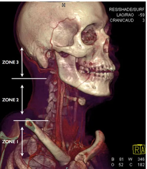
Fig. 1 Three-dimensional volume rendering technique (3D VRT) ofMDCTA performed on a young male victim of a stab wound to theneck demonstrating the anatomical trauma zones in penetrating injuryof the neck
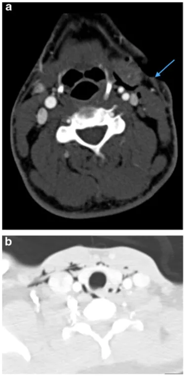
Fig. 2 a Axial image of MDCTA performed on a 33-year-old manwho sustained a stab injury to left zone 2
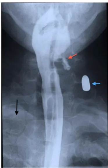
Fig. 3 a Coronal reformatted non-contrast CT brain image of a 34-year-old man who sustained a penetrating knife injury to the left zone 2of the neck requiring emergency surgical neck exploration prior toimaging assessment because of catastrophic haemorrhag
Fig. 4 a Photograph of full-metal jacket bullets (9-mm pistol bullets)exhibiting the appearances of an unfired bullet and the relative defor-mation exhibited by discharged bullets which have struck targets ofvarying density
+7

References

Related documents