R E S E A R C H
Open Access
Polydatin improves osteogenic
differentiation of human bone
mesenchymal stem cells by stimulating
TAZ expression via BMP2-Wnt/
β
-catenin
signaling pathway
Ying-Shan Shen
1†, Xiao-Jun Chen
1†, Sha-Na Wuri
1, Fan Yang
1, Feng-Xiang Pang
1, Liang-Liang Xu
2, Wei He
3,4and
Qiu-Shi Wei
3,4,5,6,7*Abstract
Objectives:Polydatin (PD), extracted fromPolygonum cuspidatum, has shown potential therapeutic applications due to its antiosteoporotic and anti-inflammatory activities. Our previous study suggested that PD promotes the osteogenesis of human bone marrow stromal cells (hBMSCs) via the BMP2-Wnt/β-catenin pathway. The aim of our present study was to further explore the role of PD-mediated regulation of Tafazzin (TAZ), a transcriptional
coactivator with a PDZ-binding motif, in osteogenesis.
Materials and methods:hBMSCs were isolated and treated with PD at various concentrations. Alizarin red staining
and RT-qPCR were performed to identify calcium complex deposition in hBMSCs as well as the expression of specific osteoblast-related markers, respectively, in each group. Next, TAZ-silenced hBMSCs were generated by lentivirus-produced TAZ shRNA. After treatment with PD, the osteogenic abilities of the TAZ-silenced and control hBMSCs were estimated by ALP activity assay, and expression of the TAZ protein was detected by Western blot analysis and immunofluorescence staining. In vitro, an ovariectomized (OVX) mouse model was established and used to evaluate the effect of PD on bone destruction by micro-CT, immunohistochemistry, and ELISA.
(Continued on next page)
© The Author(s). 2020Open AccessThis article is licensed under a Creative Commons Attribution 4.0 International License, which permits use, sharing, adaptation, distribution and reproduction in any medium or format, as long as you give appropriate credit to the original author(s) and the source, provide a link to the Creative Commons licence, and indicate if changes were made. The images or other third party material in this article are included in the article's Creative Commons licence, unless indicated otherwise in a credit line to the material. If material is not included in the article's Creative Commons licence and your intended use is not permitted by statutory regulation or exceeds the permitted use, you will need to obtain permission directly from the copyright holder. To view a copy of this licence, visithttp://creativecommons.org/licenses/by/4.0/. The Creative Commons Public Domain Dedication waiver (http://creativecommons.org/publicdomain/zero/1.0/) applies to the data made available in this article, unless otherwise stated in a credit line to the data.
* Correspondence:[email protected]
†Ying-Shan Shen and Xiao-Jun Chen contributed equally to this work.
3Hip Preserving Ward, No. 3 Orthopaedic Region, The First Affiliated Hospital
of Guangzhou University of Chinese Medicine, Guangzhou, Guangdong, China
4No. 3 Orthopaedic Region and Institute of the Hip Joint, The First Affiliated
Hospital of Guangzhou University of Chinese Medicine, Guangzhou, Guangdong, China
(Continued from previous page)
Results:In vitro, 30μM PD significantly improved the proliferation and calcium deposition of hBMSCs and markedly stimulated the expression of the mRNAsRUNX2,Osteopontin,DLX5,β-catenin,TAZ, andOsteocalcin(OCN).
Osteogenic differentiation induced by PD was blocked by lentivirus-mediated TAZ shRNA. Furthermore, Noggin (a regulator of bone morphogenic protein 2 (BMP2)) and DKK1 (an inhibitor of the Wnt/β-catenin pathway) were found to inhibit the increase in TAZ expression induced by PD. In vivo, PD prevented estrogen deficiency-induced bone loss in the OVX mouse model.
Conclusion:Taken together, our findings suggest that PD improved the osteogenic differentiation of hBMSCs and
maintained the bone matrix in the OVX mouse model through the activation of TAZ, a potential target gene of the BMP2-Wnt/β-catenin pathway.
Keywords:Polydatin, Human bone marrow stromal cells, Osteogenic differentiation, Transcriptional coactivator with PDZ-binding motif
Introduction
Osteoporosis (OP) is a bone metabolic disease character-ized by reduced bone density and an increased risk of bone fracture [1,2]. More than 20% of males and 50% of females older than 50 have experienced a decrease in bone density; thus, the annual medical expense for hip fracture in the USA is expected to rise to $67.7 billion by 2020 [3]. To date, the pathogenesis of OP is still un-clear, but multiple important pathogenic factors, includ-ing age, sex, and steroid treatment, have been identified. Additionally, growing evidence indicates that defective osteogenic differentiation of bone marrow stromal cells (BMSCs) might lead to the occurrence of OP [4, 5]. Thus, promoting the osteogenesis of BMSCs may be a promising strategy for the treatment of OP.
In recent years, cell therapy has emerged as an effect-ive approach for bone regeneration. BMSCs, the pro-genitor cells in skeletal tissues, play a critical role in cell differentiation in cell types such as osteoblasts, adipo-cytes, and chondrocytes [6,7] through specific signaling pathways. The Wnt/β-catenin and BMP pathways have been shown to be essential in the osteogenic differenti-ation of BMSCs [4, 8]. When Wnt ligands bind the re-ceptors LRP5/6 and Frizzled rere-ceptors, the classical Wnt/β-catenin pathway is activated, after which forma-tion of the Wnt-fz-lrp6 complex (including Axin, APC, GSK3, and CK1) promotes the release of β-catenin [9– 13]. Ultimately,β-catenin migrates to the nucleus, where it forms a complex with TCF/LEF to stimulate the expression of osteogenic genes, such as RUNX2, ALP,
DLX5, andOCN [14, 15]. Wnt3a, a ligand of Wnt, has been proven to bind BMP proteins to activate a series of downstream reactions and promote osteogenic differen-tiation of BMSCs [16]. BMP2, one of the most important cytokines of the TGFβ1 superfamily [17], can promote the expression of Wnt3a and FZ and the activity of TCF/LEF and increase the expression of Wnt3a protein [18, 19]. Moreover, BMP2 has the capacity to stimulate
osteogenic differentiation of BMSCs by increasing the expression and phosphorylation of β-catenin [15]. All these findings support the critical roles of the BMP2 and Wnt/β-catenin pathways and their crosstalk in inducing osteoblastic differentiation of BMSCs.
As a transcriptional coactivator in the Hippo signaling pathway, transcriptional coactivator with Tafazzin (TAZ) plays a regulatory role in the Wnt signaling pathway [20]. A previous study confirmed the functions of TAZ, such as its mediation of the osteogenic differentiation of adipose-derived stem cells [21]; remarkably, TAZ obvi-ously accelerated the osteogenesis of BMSCs through in-creasing the expression of RUNX2, a key transcription factor in the BMP and Wnt/β-catenin pathways [22,23]. All these findings indicate that TAZ serves as a vital osteogenesis mediator in BMSCs. In addition, our former study highlighted the pivotal role of TAZ in the osteogenic differentiation of BMSCs stimulated by the natural compound icariin via the estrogen receptor (Era) and Wnt/β-catenin signaling pathways [24].
Polydatin (PD), an important Chinese compound fam-ous for its effects against inflammation, oxidation, and scar hyperplasia, can also improve the migration of BMSCs through the ERK 1/2 signaling pathway [25–29]. PD was recently found to alleviate osteoporosis symp-toms in the ovariectomized (OVX) mouse model by up-regulating the expression of β-catenin [30]. Previously, we showed the osteogenetic effect of PD on hBMSCs through the BMP-Wnt/β-catenin pathway [31].
by the effect of shTAZ, which reversed the effect of TAZ on osteogenesis. Therefore, TAZ may serve as a decisive factor involved in the osteogenic effect of PD in hBMSCs, as well as the antiosteoporosis effect of PD, through the BMP2-Wnt/β-catenin signaling pathway.
Materials and methods Reagents and antibodies
hBMSCs were obtained from Cyagen Bioscience (Guangzhou, China). PD (purity ≥94%) was purchased from the National Institutes for Food and Drug Control (Beijing, China). Recombinant human Dickkopf-related protein 1 (DKK1) and Noggin were obtained from PeproTech (Rocky Hill, NJ, USA). Ficoll medium to gen-erate a Ficoll density gradient was purchased from GE Healthcare (Silverwater, Australia). Fetal bovine serum (FBS), low-glucose Dulbecco minimum essential medium (LG-DMEM), and penicillin-streptomycin were obtained from Gibco-BRL (Gaithersburg, MD, USA). An MTT assay kit, β-glycerophosphate, dexamethasone, di-methyl sulfoxide (DMSO), and L-ascorbic acid-2-phosphate were all purchased from Sigma (Steinheim, Germany). Alizarin red was obtained from Aladdin Company, and an alkaline phosphatase activity measure-ment kit was obtained from Nanjing Jiancheng Company (Nanjing, China). pLent-U6-GFP-Puro vector was ob-tained from GenePharma Company (China). SYBR® Pre-mix Ex TaqTM II and Prime Script TM RT Master Mix were purchased from Takara Biotechnology Company (Dalian, China). Anti-β-catenin antibody, anti-β-actin antibody, secondary antibodies, and phosphor-β-catenin (p-β-catenin) were obtained from Santa Cruz (Paso Robles, CA, USA). Chemiluminescence reagents were purchased from Pierce (Rockford, IL, USA).
Cell extraction and culture
hBMSCs were separated and expanded following a pre-viously described method [32]. Briefly, the human bone marrow was isolated using a Ficoll density gradient. Then, suspended cells were seeded into culture flasks after washing the MSC-enriched fraction. All flasks were maintained in an incubator with a humidified atmos-phere at 37 °C and 5% CO2, and the medium was
re-placed every 4 days. After reaching confluence, the cells were passaged to the third generation. hBMSC surface markers (CD44 and HLA-DR) were identified by flow cytometry. Third-generation hBMSCs were used in the following experiments.
Cells were treated with PD at four different concentra-tions (0, 10, 30, or 100μM) in the presence of osteogenic induction medium (OIM) consisting of L-ascorbic acid-2-phosphate (50 mM), dexamethasone (10−8mol/L), and β-glycerophosphate (10−2mol/L) to determine the opti-mal concentration of PD to increase the proliferation
and osteogenic differentiation of hBMSCs. The following four different groups of cells were set to ascertain the ef-fects of PD, Noggin, and DKK1 on the osteogenic differ-entiation of hBMSCs: group 1, which was cultured with PBS and OIM; group 2, which was cultured with PD and OIM; group 3, which was cultured in a mixture of OIM, PD, and 10−7M Noggin; and group 4, which was cul-tured in a mixture of OIM, PD, and 2 mg/mL DKK1 [33].
Cell proliferation assay
The MTT assay was used to determine the effect of PD on the proliferation of hBMSCs. Cells (1 × 10⁴per well) were plated in 96-well plates and maintained in basic medium for 24 h. When the hBMSCs were 80% conflu-ent, they were starved for 12 h without serum and then incubated in OIM with or without PD at different con-centrations (0, 30, 10, 100μM) for 1, 2, 3, 7, and 14 days. In addition, hBMSCs were cultured in a medium con-taining MTT (5 mg/mL) for 3 h at each of these time points at 37 °C. After the formazan crystals had dissolved in DMSO, the absorbance at 570 nm was measured with a microplate reader (Thermo Scientific, Waltham, MA, USA).
Alizarin red staining
hBMSCs were seeded into 24-well plates and cultured in OIM with PD at different doses (0, 30, 10, and 100μM) at 37 °C and 5% CO2 for 21 days. Then, the cells were
washed with PBS twice and fixed with 97% ethanol for 10 min. The hBMSCs were then washed three times with deionized water and stained with a 0.1% Alizarin red staining solution (pH 8.3) for 30 min at room temperature. After the Alizarin red solution was re-moved, the hBMSCs were rinsed with deionized water and PBS twice each and then dried at room temperature. The cells were observed using a 450 fluorescent inverted phase contrast microscope (Nikon Corporation, Tokyo, Japan).
Real-time quantitative PCR analysis
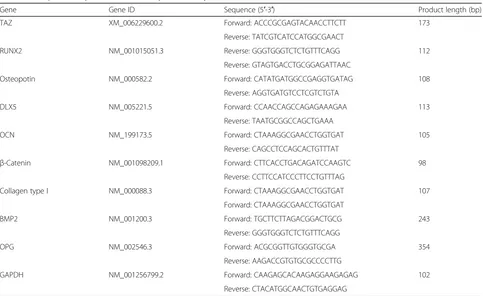
All analyses were carried out in triplicate. The primer sequences are shown in Table1.
Amplification and purification of lentivirus-mediated TAZ shRNA
Short hairpin RNA (shRNA) duplex targeting TAZ mRNA (shTAZ) was synthesized, and a nontargeting oligo duplex (shCON) served as a negative control. The pLent-U6-GFP-Puro vector was used to prepare the lentiviral vector (LV). shTAZ was cloned into the lentiviral vector accord-ing to the manufacturer’s protocol. The lentiviral vector (LvshTAZ), pHelper 1.0 plasmid, and pHelper 2.0 helper plasmid were cotransfected into HEK293T cells to prepare lentiviral particles. Viral supernatants were harvested at 48 h after transfection and applied to hBMSCs inα-MEM containing 10% FBS and 8 mg/mL polybrene. Cells were incubated for 24 h with 2 mg/mL puromycin to remove uninfected cells. After 1 week of selection, the cells were used in the differentiation experiment. TAZ levels were certified by Western blot analysis and qPCR. The follow-ing are the shTAZ and shCON sequences:
shTAZ#1: 5′-GATCCGCTTCTGGACCAAGTATAT GAACTTCAAGAGAGTTCATATACTTGGTCCAGAA GCTTTTTTA-3′, 5′-CGCGTAAAAAAGCTTCTGGA CCAAGTATATGAACTCTCTTGAAGTTCATATACT TGGTCCAGAAGC-3′
shTAZ#2: 5′-GATCCGCCACATCTGGAACCTGAA GTTGTTCAAGAGACAACTTCAGGTTCCAGATGTG GCTTTTTTA-3′, 5′-CGCGTAAAAAAGCCACATCT GGAACCTGAAGTTGTCTCTTGAACAACTTCAGGT TCCAGATGTGGC-3′
shTAZ#3: 5′-GATCCGGCAAATGTGTGCCTGTAT GTCGTTCAAGAGACGACATACAGGCACACATTTG CCTTTTTTA-3′, 5′-CGCGTAAAAAAGGCAAATGT GTGCCTGTATGTCGTCTCTTGAACGACATACAGG CACACATTTGCC-3′
shCON: 5′-GATCCGCCTAAGGTTAAGTCGCCCTC GCTCGAGCGAGGGCGACTTAACCTTAGGTTTTTA -3′, 5′-CGCGTAAAAACCTAAGGTTAAGTCGCCCT CGCTCGAGCGAGGGCGACTTAACCTTAGG-3′
ALP activity assay
hBMSCs were seeded into 48-well plates (2 × 10⁴ per well) and incubated at 37 °C and 5% CO2. After reaching
80% confluence, the cells were cultured in OIM without or with PD at different concentrations (10, 30, and 100μM) for 1, 2, 3, 7, and 14 days. hBMSC lysates were subjected to ALP activity examination by a commercial ALP assay kit (Abcam, Cambridge, MA, USA).
Western blot analysis
At the indicated time points (3, 7, and 14 days), hBMSCs were separately lysed with RIPA lysis buffer consisting of
Table 1The primer sequences used in the present study
Gene Gene ID Sequence (5′-3′) Product length (bp)
TAZ XM_006229600.2 Forward: ACCCGCGAGTACAACCTTCTT 173
Reverse: TATCGTCATCCATGGCGAACT
RUNX2 NM_001015051.3 Reverse: GGGTGGGTCTCTGTTTCAGG 112
Reverse: GTAGTGACCTGCGGAGATTAAC
Osteopotin NM_000582.2 Forward: CATATGATGGCCGAGGTGATAG 108
Reverse: AGGTGATGTCCTCGTCTGTA
DLX5 NM_005221.5 Forward: CCAACCAGCCAGAGAAAGAA 113
Reverse: TAATGCGGCCAGCTGAAA
OCN NM_199173.5 Forward: CTAAAGGCGAACCTGGTGAT 105
Reverse: CAGCCTCCAGCACTGTTTAT
β-Catenin NM_001098209.1 Forward: CTTCACCTGACAGATCCAAGTC 98
Reverse: CCTTCCATCCCTTCCTGTTTAG
Collagen type I NM_000088.3 Forward: CTAAAGGCGAACCTGGTGAT 107
Forward: CTAAAGGCGAACCTGGTGAT
BMP2 NM_001200.3 Forward: TGCTTCTTAGACGGACTGCG 243
Reverse: GGGTGGGTCTCTGTTTCAGG
OPG NM_002546.3 Forward: ACGCGGTTGTGGGTGCGA 354
Reverse: AAGACCGTGTGCGCCCCTTG
GAPDH NM_001256799.2 Forward: CAAGAGCACAAGAGGAAGAGAG 102
Tris-HCl (50 mM, pH 8.0), NaCl (150 mM), 1% NP-40, 0.5% sodium chloride, and 0.1% sodium dodecyl sulfate. The lysate protein concentration was measured with a Bradford protein assay kit. All samples were separated by 10% SDS-PAGE and electrotransferred to the PVDF membranes. The membranes were blocked with 5% fat-free milk and incubated overnight at 4 °C with the primary antibodies anti-TAZ, anti-pTAZ Ser89, and anti-β-actin. The membranes were incubated with sec-ondary antibody for 1 h after rinsing with PBS contain-ing Tween-20 three times. Immunoreaction signals were detected by an enhanced chemiluminescence detection kit, and the images were analyzed using the Quantity One software. Band intensity was quantified and normal-ized againstβ-actin.
Immunofluorescence staining
After 14 days of treatment, hBMSCs (groups A–D) were fixed with cold acetone for 15 min at room temperature, permeabilized (0.25% Triton X-100 in PBS for 15 min), and then blocked in 0.1% Triton X for 20 min. To block nonspecific antibody binding, cells were incubated for 20 min with 1% bovine serum albumin (BSA) and then incubated with primary anti-β-catenin antibody (1:200) at 4 °C overnight. The next day, the cells were incubated
with a 1:500 dilution of rabbit secondary antibody la-beled with APC for 1 h. After the cells were rinsed with PBS, the nuclear translocation of β-catenin was visual-ized by staining with 4′-6-diamidino-2-phenylindole (DAPI). Stained hBMSCs were examined under a fluor-escence microscope, and images were captured with the Image Manager software.
OVX mouse model
All experiments were authorized by the Institutional Animal Ethics Committee of Guangzhou University of Chinese Medicine and performed under the guidelines set forth by the National Institutes of Health Guidelines for the Care and Use of Laboratory Animals. Eighteen 7-week-old specific pathogen-free C57BL/6J female mice were randomly divided into three groups: (1) sham group (served as controls without any treatment, n= 6), (2) OVX group (mice whose ovaries were removed,n= 6), and (3) OVX+PD group (ovariectomized mice treated with 3 mg/kg PD by intraperitoneal injection,n= 6). The mice underwent standard feeding in cages (three mice per cage) under a 12-h light/dark cycle. After anesthetization, mice in the OVX group and OVX+PD group underwent ovariectomy to induce estrogen defi-ciency, while mice in the sham group underwent
ovariectomy in the same manner, except a small amount of fat tissue beside the ovaries was removed. Each ovary removed from mice in the OVX and OVX+PD groups was completely removed, including its capsule and part of the oviduct. Mice in all of the groups had 1 week for recovery and incision healing. After that, mice in the OVX+PD group were given an intraperitoneal injection of 3 mg/kg PD every 2 days, instead of the 1% DMSO administered to mice in the sham and OVX groups. After predetermined durations (4, 8, and 12 weeks), the femurs of the mice were removed after sacrifice for sub-sequent histological and micro-CT examinations. The serum was extracted from the abdominal aorta of the mice in every group, and the protein levels of osteoclas-togenesis inhibitory factor (OPG), RANKL, OPG/ RANKL, andβ-CTX were evaluated using ELISA kits ac-cording to the manufacturers’instructions.
Micro-CT analysis
The femurs were fixed overnight in 10% neutral buffered formalin and analyzed by SkyScan 1176 micro-CT equipment (SkyScan, Aartselaar, Belgium). The scanner was set at a resolution of 9μm per pixel. The proximal femur beginning at 0.5 mm above the bottom of the growth plate and extending for 1 mm was chosen as the region of interest (ROI) used to determine the trabecular thickness (Tb.Th), trabecular separation (Tb.Sp), bone volume per tissue volume (BV/TV), bone surface area per bone volume (BS/BV), structure model index (SMI), and trabecular number (Tb.N).
Histological and immunohistochemical analyses
The samples were fixed in 4% paraformaldehyde (PFA) for 24 h and then decalcified. All samples were cut into 5-μm sections and stained using hematoxylin and eosin
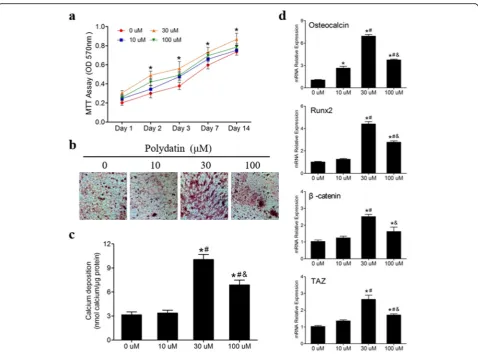
Fig. 2Polydatin promoted the proliferation and osteogenesis of hBMSCs.aPD with various concentrations notably stimulated hBMSCs to proliferate at 1, 2, 3, 7, and 14 days, particularly the 30μM, according to the MTT assay results (n= 6).b,chBMSCs were cultured in OIM with different concentrations PD (0, 10, 30, and 100μM) for 21 days. The 30μM PD significantly promoted the mineralization of hBMSCs (n= 3).d Following 3 days of PD treatments with various concentrations,OCN,RUNX2,β-catenin, andTAZmRNA levels were measured by qPCR. *p< 0.05 versus the 0μM group at the same time point,#p< 0.05 versus the 10
μM group, and&p< 0.05 versus the 30
(H&E) for histological observation. The extent of osteo-genesis was evaluated by immunohistochemical (IHC) analysis of TAZ and OCN. Images were acquired with the Aperio ScanScope (Leica Biosystem, Buffalo Grove, IL, USA), and bone histomorphometric analyses were performed using the BIOQUANT OSTEO software (Bioquant Image Analysis Corporation, Nashville, TN, USA).
Statistical analysis
The results are shown as the mean ± standard deviation (SD). One-way ANOVA was used to analyze data from multiple groups using SPSS version 19.0 (SPSS, Inc., USA). Apvalue of less than 0.05 indicated statistical significance.
Results
Phenotype identification of hBMSCs
hBMSCs at passages 2 and 3 were 90% confluent after 3 days of culture (Fig. 1a). As shown in Fig.1b, passage 2
cells were tested by flow cytometry. The results indi-cated that 94.4% of cells were positive for CD44, while 0.4% were negative for HLA-DR. Hence, the majority of the isolated and purified cells expressed markers charac-teristic of hBMSCs.
Polydatin promoted the proliferation and osteogenic differentiation of hBMSCs
hBMSCs were cultured in a basal medium with or with-out PD at various concentrations (10, 30, and 100μM) for 1–14 days. The results of the MTT assay showed that 30μM PD treatment significantly improved hBMSC pro-liferation at each time point (p< 0.05) (Fig. 2a). The ef-fect of PD on the osteogenesis of hBMSCs was estimated by Alizarin red staining and qPCR. hBMSCs were cultured in OIM or OIM with PD for 21 days. As shown in Fig. 2b, PD, particularly at a concentration of 30μM, increased calcium deposition at day 21, as shown by Alizarin red staining, which was consistent with the
results of the Alizarin red staining (Fig. 2c). We further conducted qPCR to examine the expression of osteo-genic marker genes (OCN,RUNX2, β-catenin, andTAZ) in hBMSCs induced with PD for 3 days. PD significantly promoted osteogenic gene expression in hBMSCs com-pared to that in cells treated with only OIM (Fig.2d). In summary, 30μM PD notably increased the proliferation and osteogenic differentiation of hBMSCs. Thus, this concentration was chosen for the following experiments.
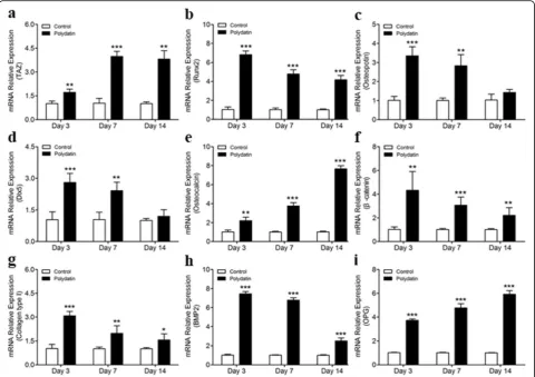
Effect of polydatin on the expression of osteogenic genes
We tested the effects of 30μM PD on TAZ, RUNX2,
Osteopontin, DLX5, OCN, β-catenin, Collagen type I,
BMP2, andOPGexpression during osteogenic differenti-ation of hBMSCs cultured in OIM at days 3, 7, and 14. The qPCR results showed that PD stimulation for 3, 7, and 14 days increased TAZ, OCN, RUNX2, β-catenin,
Collagen type I, BMP2, and OPG mRNA levels (Fig. 3a, b, e–i), and PD stimulation for 3 and 7 days upregulated
OsteopontinandDLX5mRNA levels (Fig.3c, d).
Lentivirus-mediated TAZ shRNA offsets the effects of polydatin on osteogenic differentiation
Depletion of TAZ in hBMSCs was evaluated by using lentivirus-produced TAZ shRNA. The results of both qPCR and Western blot analysis showed that transfec-tion with shTAZ2# significantly reduced the TAZ expression compared with that in the control and shCON-transfected groups (Fig. 4a, b). Moreover, shTAZ transfection blocked the PD-induced osteogenic differentiation of hBMSCs. This was supported by the results of the ALP activity assay and determination of the mRNA expression of osteogenic genes. The ALP ac-tivity was obviously decreased in the shTAZ+PD group compared with the shCON+PD group, and there were no significant differences in ALP activity among the shCON, shTAZ, and shTAZ+PD groups (Fig.4c). Com-pared with the shCON+POL group, the shTAZ+POL group exhibited reduced mRNA expression of RUNX2,
Osteopontin,β-catenin, andDLX5at day 3;TAZ expres-sion at 7 days; andOCNexpression at day 14 (Fig.4d).
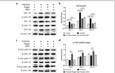
Polydatin increased total TAZ protein expression in hBMSCs mediated by the BMP2 and Wnt/β-catenin signaling pathways
Noggin and DKK1 are inhibitors of BMP2 and Wnt/β -catenin, respectively. Western blot analysis indicated that PD enhanced the expression of the TAZ protein on days 3 and 7 compared with that in the control group. However, this increase in expression induced by PD was blocked by Noggin and DKK1 (Fig.5a, b). However, the change in p-TAZ Ser89 expression was contrary to that in TAZ expression; the level of p-TAZ Ser89 on days 3 and 14 was lower in the PD group than in the control group. However, the corresponding levels on days 3, 7, and 14 in the Noggin-treated groups were significantly higher than those in the PD group (Fig. 5c, d). In conclusion, our results showed that PD increased TAZ expression through the BMP2 and Wnt/β-catenin signal-ing pathways.
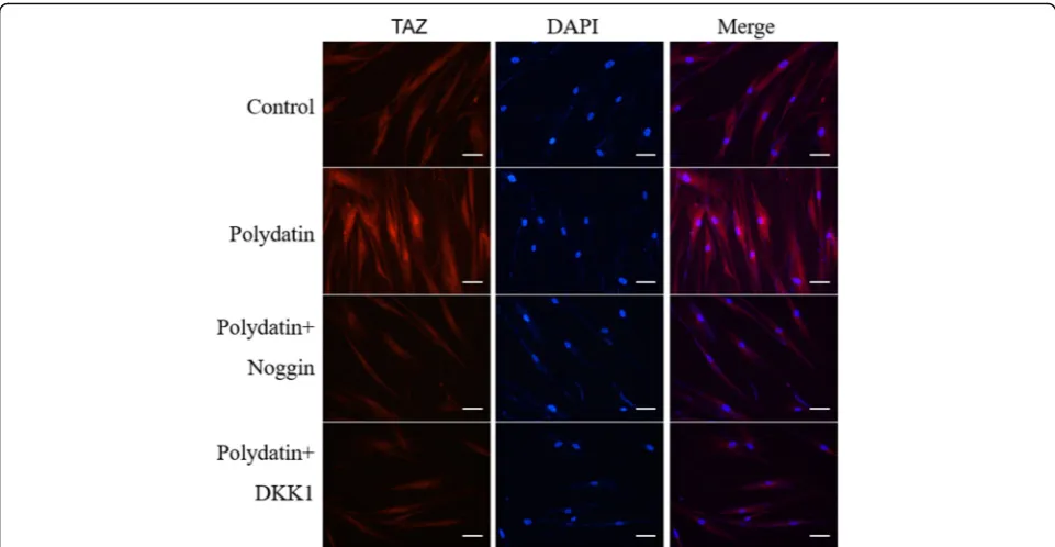
Polydatin-induced TAZ nuclear translocation in hBMSCs
To determine whether the effect of PD in activating hBMSC osteogenic differentiation was related to an
increase in TAZ nuclear translocation, we conducted immunolabeling and fluorescence microscopy and found that 30μM PD promoted the nuclear (blue) transloca-tion of TAZ (red) in hBMSCs, which was hampered by Noggin and DKK1 (Fig.6).
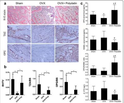
PD inhibited estrogen deficiency-induced bone loss in OVX mice
To explore the effect of PD on estrogen deficiency-induced bone loss, OP model mice were treated with PD (3 mg/kg) or an equal volume DMSO alone as a control. The mice were sacrificed after 4, 8, or 12 weeks of treat-ment (Fig. 7a). The micro-CT scanning results revealed that PD significantly reduced ovariectomy-induced bone loss, as shown by the increased BV/TV, Tb.N, and Tb.Th in the OVX+PD group. Trabecular separation (Tb.Sp) was also decreased in the OVX+PD group com-pared with the OVX group (Fig.7b, c). The H&E stain-ing results also confirmed the improved trabecular structure in different groups after PD intervention (Fig. 8a). Immunohistochemistry proved that PD signifi-cantly stimulated the expression of TAZ and OPG in the
bone (Fig. 8a, b). As shown by ELISA, PD suppressed the serum expression of the osteoclastic markers RANKL and β-CTX. The change in the expression of serum OPG was opposite that of the other two markers (Fig.8c).
Discussion
OP is a common bone disease characterized by low bone mass and subsequent elevated fracture risk [34]; there-fore, antiresorptive agents, anabolic agents, and bone/ mineral drugs have been used to maintain normal bone mass. However, their limitations and side effects, such as an increased risk of breast cancer, jaw necrosis, or atyp-ical femur fracture, have gradually been found [35, 36]. Thus, the discovery of an alternative OP treatment with fewer side effects would be promising. Disordered pro-liferation and differentiation of BMSCs are the main causes of osteoporosis [37, 38], especially the impaired ability of BMSCs to differentiate into osteoblasts [39]. Therefore, the promotion of osteogenic differentiation can help inhibit the development of osteoporosis. Ac-cording to the literature, herbal extracts, acting as stimu-lants of the proliferation and differentiation of hBMSCS, have outstanding effects, and their osteogenic, vascular, chondrogenic, and neurogenic potential should be fur-ther studied [40]. We therefore investigated the exact
antiosteoporotic mechanism of a prominent Chinese compound, PD, in vitro and in vivo.
As described by our results, PD increased the pro-liferation and osteogenic differentiation of hBMSCs without cytotoxicity, as proven by the significant in-creases in calcium nodules and expression of osteo-blast markers, which is consistent with the results of our previous study [31]. In our previous study, we also found that the PD-induced expression of BMP2 was not blocked by a specific Wnt signal inhibitor. Furthermore, it is illustrated that PD activated the BMP2-induced Wnt signaling pathway through en-hancing the accumulation and nuclear translocation of β-catenin and subsequently promoted the osteo-genesis of hBMSCs [31]. Further experiments found that knockdown of TAZ hampered the effect of PD on osteogenesis through the BMP2-Wnt/β-catenin signaling pathway. The increased bone mass of OVX mice also supported that the osteogenesis effect of PD is closely related to TAZ. Thus, we demonstrated that PD improves the osteogenic differentiation of hBMSCs by stimulating TAZ expression via the BMP2-Wnt/β-catenin signaling pathway.
The BMP and Wnt/β-catenin pathways engage in substantial crosstalk in preosteoblasts, and the Wnt pathway acts as a downstream regulator of BMPs to regulate the progression of proliferation, bone
differentiation, and mineralization [41–43]. RUNX2 is the main regulatory factor that activates osteogenic differentiation through the BMP and Wnt/β-catenin signaling pathways. It can induce the nuclear trans-location of β-catenin [44] and promote BMP2-induced osteogenic differentiation [45] and the binding of BMP2 with Wnt/β-catenin [16, 46]. Thus, the central mechanism of osteogenesis is executed by the BMP and Wnt signaling pathways [47]. More spe-cifically, BMP2 stimulates bone formation through ac-tivating RUNX2 [48], and the Wnt/β-catenin pathway activates RUNX2 in MSCs through the transcriptional regulator TCF/LEF [49], while RUNX2 also promotes the expression of β-catenin [50].
Recently, TAZ was found to serve as a regulatory transcription coactivator for RUNX2-stimulated OCN
gene expression and thus promote osteogenic differ-entiation of MSCs [51]. TAZ is widely known as a regulator of the Hippo pathway and a novel regulator of typical Wnt signaling [20, 52], but the specific mechanism by which TAZ promotes osteogenic differentiation has not been fully clarified. Azzolin et al. [53] found that Wnt can stabilize TAZ and β -catenin and stimulate their nuclear transfer, thus promoting osteoblastic differentiation. In addition, the presence of Wnt3a facilitates the interaction between TAZ and RUNX2, which significantly increases RUNX2-mediated osteogenic gene transcription [54]. Several studies have elaborated that TAZ stimulates the osteogenic differentiation of MSCs by increasing the expression of RUNX2 [22, 55, 56]. In addition, BMP2 not only increases TAZ expression but also
promotes subsequent TAZ-stimulated expression of the OCN gene by RUNX2, which is essential for osteoblast differentiation [51]. Therefore, RUNX2 plays a vital role in the crosstalk between the BMP2 and Wnt/β-catenin pathways to regulate BMSC osteo-genic differentiation, which is closely related to the interaction of RUNX2 with TAZ [57–60]. Consistent with these previous studies, we found that the knock-down of TAZ tremendously impeded the osteogenic effect of PD through the BMP2-Wnt/β-catenin pathway.
This hypothesis was confirmed by the increased bone mass of OVX mice after PD treatment.
Remarkably, PD intervention significantly increased the expression of TAZ and OPG and decreased that of RANKL. The OPG/RANKL ratio plays a crucial role in OP. RANKL activates the receptor RANK to induce osteoclast differentiation, which is blocked by OPG, leading to the inhibition of osteoclast growth and bone resorption [61–63]. PD has been proven to alleviate the symptoms of OP in OVX mice by up-regulating the expression of OPG and β-catenin and downregulating that of RANKL [30]. Combined with our in vitro results, these findings demonstrate that TAZ plays a key role in the process by which PD improves hBMSC osteogenic differentiation.
Conclusion
In conclusion, PD could promote the proliferation and osteogenic differentiation of hBMSCs and maintain the bone matrix in an OVX mouse model through the regulation of BMP2 and the Wnt/β-catenin pathway, and TAZ plays a pivotal role in regulating this process (Fig.9).
Abbreviations
OP:Osteoporosis; ALP: Alkaline phosphatase; BMP2: Bone morphogenetic protein 2; DKK1: Dickkopf-related protein 1; DMSO: Dimethyl sulfoxide; OVX: Ovariectomized; PD/POL: Polydatin; hBMSCs: Human bone marrow stromal cells; OIM: Osteogenic induction medium; PBS: Phosphate-buffered saline; PFA: Paraformaldehyde PDZ-binding motif: TAZ; RUNX2: Runt-related transcription factor 2; DLX5: Distal-less homeobox 5; OPG: Osteoclastogenesis inhibitory factor; OCN: Osteocalcin
Acknowledgements
We thank the team for their cooperation.
Authors’contributions
Conceived and designed the experiments: Qiu-Shi Wei Performed the experiments: Ying-Shan Shen and Xiao-Jun Chen Analyzed the data: Sha-Na Wuri, Fan Yang, and Feng-Xiang Pang Contributed funders/reagents/materials/analysis tools: Wei He and Liang-Liang Xu
The authors read and approved the final manuscript.
Funding
This work was supported by National Natural Science Foundation of China (Grant NO. 81873327 and 81573996) from China, and Guangdong Province Natural Science Fund Project (2017A030313698).
Availability of data and materials
The datasets used and/or analyzed during the current study are available from the corresponding author on reasonable request.
Ethics approval and consent to participate
The mouse model testing involved in our study followed the Basel Declaration outlines fundamental principles and was approved by the Guangzhou University of Chinese Medicine Institutional Animal Ethics Committee.
Consent for publication Not applicable.
Competing interests
The authors declare that they have no competing interests.
Author details 1
Guangzhou, Guangdong, China.5Third Clinical Medical College, Guangzhou University of Chinese Medicine, Guangzhou, Guangdong, China.6The Third Affiliated Hospital of Guangzhou University of Chinese Medicine, Guangzhou, Guangdong, China.7Institute of orthopedics of Guangzhou University of Chinese Medicine, Guangzhou, Guangdong, China.
Received: 14 December 2019 Revised: 21 April 2020 Accepted: 5 May 2020
References
1. Ranuccio N, Luisa BM, Giovanni C, Ombretta DM, Ligia D, et al. Guidelines for the management of osteoporosis and fragility fractures. Intern Emerg Med. 2019;14(1):85–102.
2. Black DM, Rosen CJ. Clinical practice. Postmenopausal osteoporosis. N Engl J Med. 2016;374(3):254–62.
3. Ward RJ, Roberts CC, Bencardino JT, Arnold E, Baccei SJ, Cassidy RC, et al. ACR appropriateness criteria® osteoporosis and bone mineral density. J Am Coll Radiol. 2017;14(5):S189–202.
4. Li K, Zhang X, He B, Yang R, Zhang Y, Shen Z, et al. Geraniin promotes osteoblast proliferation and differentiation via the activation of Wnt/β -catenin pathway. Biomed Pharmacother. 2018;99:319–24.
5. Yang A, Yu C, You F, He C, Li Z. Mechanisms of zuogui pill in treating osteoporosis: Perspective from bone marrow mesenchymal stem cells. Evidence-based Complement Altern Med. 2018;3717391.https://doi.org/10. 1155/2018/3717391.
6. Derubeis AR, Cancedda R. Bone marrow stromal cells (BMSCs) in bone engineering: limitations and recent advances. Ann Biomed Eng. 2004;32(1): 160–5.
7. Zhang RF, Wang Q, Zhang AA, Xu JG, Zhai LD, Yang XM, Liu XT. Low-level laser irradiation promotes the differentiation of bone marrow stromal cells into osteoblasts through the APN/Wnt/β-catenin pathway. Eur Rev Med Pharmacol Sci. 2018;22(9):2860–8.
8. Açil Y, Ghoniem AA, Wiltfang J, Gierloff M. Optimizing the osteogenic differentiation of human mesenchymal stromal cells by the synergistic action of growth factors. J Cranio Maxillofacial Surg. 2014;42(8):2002–9. 9. Dong M, Jiao G, Liu H, Wu W, Li S, Wang Q, et al. Biological silicon
stimulates collagen type 1 and osteocalcin synthesis in human osteoblast-like cells through the BMP-2/Smad/RUNX2 signaling pathway. Biol Trace Elem Res. 2016;173(2):306–15.
10. MacDonald BT, Tamai K, He X. Wnt/β-catenin signaling: components, mechanisms, and diseases. Dev Cell. 2009;17(1):9–26.
11. Little RD, Carulli JP, Del Mastro RG, Osborne M, Folz C, Manning SP, et al. A mutation in the LDL receptor-related protein 5 gene results in the autosomal dominant high-bone-mass trait. Am J Hum Genet. 2002; 70(1):11–9.
12. Boland GM, Perkins G, Hall DJ, Tuan RS. Wnt 3a promotes proliferation and suppresses osteogenic differentiation of adult human mesenchymal stem cells. J Cell Biochem. 2004;93(6):1210–30.
13. Mahajan G, Kaur H, Jain S, Kaur N, Sehgal NK, Gautam A. To compare the gingival melanin repigmentation after diode laser application and surgical removal. J Indian Soc Periodontol. 2017;21(2):112–8.
14. Xing Y, Zhang Y, Jia L, Xu X. Lipopolysaccharide from Escherichia coli stimulates osteogenic differentiation of human periodontal ligament stem cellsthrough Wnt/β-catenin–induced TAZ elevation. Molecular oral microbiology. 2019;34(1).https://doi.org/10.1111/omi.12249.
15. Li J, Zhang Y, Zhao Q, Wang J, He X. MicroRNA-10a influences osteoblast differentiation and angiogenesis by regulatingβ-catenin expression. Cell Physiol Biochem. 2015;37(6):2194–208.
16. Zhang R, Oyajobi BO, Harris SE, Chen D, Tsao C, Deng HW, et al. Wnt/β -catenin signaling activates bone morphogenetic protein 2 expression in osteoblasts. Bone. 2013;52(1):145–56.
17. Koseki T, Gao Y, Okahashi N, Murase Y, Tsujisawa T, Sato T, et al. Role of TGF-βfamily in osteoclastogenesis induced by RANKL. Cell Signal. 2002; 14(1):31–6.
18. Yang L, Yamasaki K, Shirakata Y, Dai X, Tokumaru S, Yahata Y, et al. Bone morphogenetic protein-2 modulates Wnt and frizzled expression and enhances the canonical pathway of Wnt signaling in normal keratinocytes. J Dermatol Sci. 2006;42(2):111–9.
19. Itasaki N, Hoppler S. Crosstalk between Wnt and bone morphogenic protein signaling: a turbulent relationship. Dev Dyn. 2010;239(1):16–33.
20. Azzolin L, Zanconato F, Bresolin S, Forcato M, Basso G, Bicciato S, et al. Role of TAZ as mediator of wnt signaling. Cell. 2012;151(7):1443–56.
21. Zhu Y, Wu Y, Cheng J, Wang Q, Li Z, Wang Y, et al. Pharmacological activation of TAZ enhances osteogenic differentiation and bone formation of adipose-derived stem cells. Stem Cell Res Ther. 2018;9(1):1–16.
22. Cui CB, Cooper LF, Yang X, Karsenty G, Aukhil I. Transcriptional coactivation of bone-specific transcription factor Cbfa1 by TAZ. Mol Cell Biol. 2003;23(3): 1004–13.
23. Byun MR, Jeong H, Bae SJ, Kim AR, Hwang ES, Hong JH. TAZ is required for the osteogenic and anti-adipogenic activities of kaempferol. Bone. 2012; 50(1):364–72.
24. Wei QS, He MC, Chen MH, Chen ZQ, Yang F, Bin WH, et al. Icariin stimulates osteogenic differentiation of rat bone marrow stromal stem cells by increasing TAZ expression. Biomed Pharmacother. 2017;91:581–9. 25. Yang B, Zhao S. Polydatin regulates proliferation, apoptosis and autophagy
in multiple myeloma cells through mTOR/p70s6k pathway. Onco Targets Ther. 2017;10:935–44.
26. Tang S, Tang Q, Jin J, Zheng G, Xu J, Huang W, et al. Polydatin inhibits the IL-1β-induced inflammatory response in human osteoarthritic chondrocytes by activating the Nrf2 signaling pathway and ameliorates murine osteoarthritis. Food Funct. 2018;9(3):1701–12.
27. Hanna DA, Khalaf MM, Abo-Saif AA. Polydatin protects against ovalbumin-induced bronchial asthma in rats; involvement of urocortin and surfactant-D expression. Immunopharmacol Immunotoxicol. 2019;41(3):403–12. 28. Dong M, Ding W, Liao Y, Liu Y, Yan D, Zhang Y, et al. Polydatin
prevents hypertrophy in phenylephrine induced neonatal mouse cardiomyocytes and pressure-overload mouse models. Eur J Pharmacol. 2014;746:186–97.
29. Chen ZQ, Wei QS, Hong GJ, Chen D, Liang J, He W, et al. Polydatin induces bone marrow stromal cells migration by activation of ERK1/2. Biomed Pharmacother. 2016;82:49–53.
30. Zhou QL, Qin RZ, Yang YX, Huang KB, Yang XW. Polydatin possesses notable anti-osteoporotic activity via regulation of OPG, RANKL andβ -catenin. Mol Med Rep. 2016;14(2):1865.
31. Chen XJ, Shen YS, He MC, Yang F, Yang P, Pang FX, et al. Polydatin promotes the osteogenic differentiation of human bone mesenchymal stem cells by activating the BMP2-Wnt/β-catenin signaling pathway. Biomed Pharmacother. 2019;112:108746.
32. Nie B, Ao H, Zhou J, Tang T, Yue B. Biofunctionalization of titanium with bacitracin immobilization shows potential for anti-bacteria, osteogenesis and reduction of macrophage inflammation. Colloids Surf B Biointerfaces. 2016;145:728–39.
33. Morsczeck C, Reck A, Reichert TE. WNT3A and the induction of the osteogenic differentiation in adipose tissue derived mesenchymal stem cells. Tissue Cell. 2017;49(4):489–94.
34. Letarouilly JG, Broux O, Clabaut A. New insights into the epigenetics of osteoporosis. Genomics. 2018;11(4):793–8.
35. Einhorn TA, Bogdan Y, Tornetta P III. Bisphosphonate-associated fractures of the femur: pathophysiology and treatment. J Orthop Trauma. 2014;28(7): 433–8.
36. Tabatabaei-Malazy O, Salari P, Khashayar P, Larijani B. New horizons in treatment of osteoporosis. Daru. 2017;25(1):2.
37. Zhou S, Greenberger JS, Epperly MW, Goff JP, Adler C, Leboff MS, et al. Age-related intrinsic changes in human bone-marrow-derived mesenchymal stem cells and their differentiation to osteoblasts. Aging Cell. 2008;7(3):335–43.
38. Astudillo P, Ríos S, Pastenes L, Pino AM, Rodríguez JP. Increased adipogenesis of osteoporotic human-mesenchymal stem cells (MSCs) is characterized by impaired leptin action. J Cell Biochem. 2008;103(4):1054–65.
39. Hu L, Yin C, Zhao F, Ali A, Ma J, Qian A. Mesenchymal stem cells: cell fate decision to osteoblast or adipocyte and application in osteoporosis treatment. Int J Mol Sci. 2018;19(2):1–19.
40. Lankika UV, Dilumi JC, Vidya UP. Potential role of herbal remedies in stem cell therapy: proliferation and differentiation of human mesenchymal stromal cells. Stem Cell Res Ther. 2016;7(1):110.
41. Park KH, Kang JW, Lee EM, Kim JS, Rhee YH, Kim M, et al. Melatonin promotes osteoblastic differentiation through the BMP/ERK/Wnt signaling pathways. J Pineal Res. 2011;51(2):187–94.
43. Tamura M, Nemoto E, Sato MM, Nakashima A, Shimauchi H. Role of the Wnt signaling pathway in bone and tooth. Front Biosci. 2010;2:1405–13.https:// doi.org/10.2741/e201.
44. Kushwaha P, Khedgikar V, Gautam J, Dixit P, Chillara R, Verma A, et al. A novel therapeutic approach with Caviunin-based isoflavonoid that en routes bone marrow cells to bone formation via BMP2/Wnt-β-catenin signaling. Cell Death Dis. 2014;5(9):e1422–16.
45. Yun HM, Park KR, Quang TH, Oh H, Hong JT, Kim YC, et al. 2,4,5-Trimethoxyldalbergiquinol promotes osteoblastic differentiation and mineralization via the BMP and Wnt/β-catenin pathway. Cell Death Dis. 2015;6(7):1–9.
46. Rodríguez-Carballo E, Ulsamer A, Susperregui ARG, Manzanares-Céspedes C, Sánchez-García E, Bartrons R, et al. Conserved regulatory motifs in osteogenic gene promoters integrate cooperative effects of canonical Wnt and BMP pathways. J Bone Miner Res. 2011;26(4):718–29.
47. Lian JB, Stein GS, Javed A, Van Wijnen AJ, Stein JL, Montecino M, et al. Networks and hubs for the transcriptional control of osteoblastogenesis. Rev Endocr Metab Disord. 2006;7(1–2):1–16.
48. Byun MR, Kim AR, Hwang JH, Kim KM, Hwang ES, Hong JH. FGF2 stimulates osteogenic differentiation through ERK induced TAZ expression. Bone. 2014; 58:72–80.
49. Deng Z, Sharff KA, Tang N, Song W, Luo J, Luo X, et al. Regulation of osteogenic differentiation during skeletal development. Front Bioence. 2008; 13(6):2001–21.
50. Gaur T, Lengner CJ, Hovhannisyan H, Bhat RA, Bodine PVN, Komm BS, et al. Canonical WNT signaling promotes osteogenesis by directly stimulating Runx2 gene expression. J Biol Chem. 2005;280(39):33132–40.
51. Hong J-H, McManus MT, Amsterdam A, Kalmukova R, Sharp PA, Hopkins N, et al. TAZ, a transcriptional modulator of mesenchymal stem cell differentiation. Science. 2005;309(5737):1074–8.
52. Azzolin L, Panciera T, Soligo S, Enzo E, Bicciato S, Dupont S, et al. YAP/TAZ incorporation in theβ-catenin destruction complex orchestrates the Wnt response. Cell. 2014;158(1):157–70.
53. Kida J, Hata K, Nakamura E, Yagi H, Takahata Y, Murakami T, et al. Interaction of LEF1 with TAZ is necessary for the osteoblastogenic activity of Wnt3a. Sci Rep. 2018;8(1):1–8.
54. Byun MR, Hwang JH, Kim AR, Kim KM, Hwang ES, Yaffe MB, et al. Canonical Wnt signalling activates TAZ through PP1A during osteogenic
differentiation. Cell Death Differ. 2014;21(6):854–63.
55. Xue P, Wu X, Zhou L, Ma H, Wang Y, Liu Y, et al. IGF1 promotes osteogenic differentiation of mesenchymal stem cells derived from rat bone marrow by increasing TAZ expression. Biochem Biophys Res Commun. 2013;433(2):226–31. 56. Hong JH, Yaffe MB. Aβ-catenin-like molecule that regulates mesenchymal
stem cell differentiation. Cell Cycle. 2006;5(2):176–9.
57. Byun MR, Kim AR, Hwang JH, Sung MK, Lee YK, Hwang BS, et al. Phorbaketal A stimulates osteoblast differentiation through TAZ mediated Runx2 activation. FEBS Lett. 2012;586(8):1086–92.
58. Byun MR, Sung MK, Kim AR, Lee CH, Jang EJ, Jeong MG, et al.
(−)-Epicatechin gallate (ECG) stimulates osteoblast differentiation via runt-related transcription factor 2 (RUNX2) and transcriptional coactivator with PDZ-binding motif (TAZ)-mediated transcriptional activation. J Biol Chem. 2014;289(14):9926–35.
59. Merrick D, Mistry K, Wu J, Gresko N, Baggs JE, Hogenesch JB, et al. Polycystin-1 regulates bone development through an interaction with the transcriptional coactivator TAZ. Hum Mol Genet. 2019;28(1):16–30. 60. Li N, Lu N, Xie C. The Hippo and Wnt signalling pathways: crosstalk during
neoplastic progression in gastrointestinal tissue. FEBS J. 2019;286(19):3745–56. 61. Hofbauer LC, Schoppet M. Clinical implications of the osteoprotegerin/
RANKL/RANK system for bone and vascular diseases. J Am Med Assoc. 2004; 292(4):490–5.
62. Hsu H, Solovyev I, Colombero A, Timms E, Elliott G, Wang L, et al. Tumor necrosis factor receptor family member RANK mediates osteoclast differentiation and activation induced by osteoprotegerin ligand. Proc Natl Acad Sci U S A. 1999;96(7):3540–5.
63. Simonet WS, Lacey DL, Dunstan CR, Kelley M, Chang MS, Lüthy R, et al. Osteoprotegerin: a novel secreted protein involved in the regulation of bone density. Cell. 1997;89(2):309–19.
Publisher’s Note