R E S E A R C H
Open Access
Preconditioning of bone marrow-derived
mesenchymal stem cells highly strengthens
their potential to promote IL-6-dependent
M2b polarization
Denise Philipp, Laura Suhr, Thorsten Wahlers, Yeong-Hoon Choi and Adnana Paunel-Görgülü
*Abstract
Background:During the last decade, mesenchymal stem cells (MSCs) have gained much attention in the field of
regenerative medicine due to their capacity to differentiate into different cell types and to promote immunosuppressive effects. However, the underlying mechanism of MSC-mediated immunoregulation is not fully understood so far. Macrophages are distinguished in classical activated, pro-inflammatory M1 and alternatively activated M2 cells, which possess different functions and transcriptional profiles with respect to inflammatory responses. As polarization is not fixed, macrophage functional plasticity might be modulated by the microenvironment allowing them to rapidly react to danger signals and maintaining tissue homeostasis.
Methods:Murine MSCs were preconditioned with IL-1ß and IFN-ɣto enhance their immunosuppressive capacity regarding macrophage polarization under M1- and M2a-polarizing conditions. Macrophage polarization was analyzed by real-time PCR, flow cytometry, and cytokine detection in culture supernatants. The role of MSC-derived nitric oxide (NO), prostaglandin E2 (PGE2), and IL-6 in this process has been evaluated using siRNA transfection and IL-6 receptor-deficient macrophages, respectively.
Results:Preconditioned, but not unprimed, MSCs secreted high levels of NO, IL-6, and PGE2. Co-culture with
macrophages (M0) in the presence of M1 inducers (LPS + IFN-ɣ) led to significant reduction of CD86 and iNOS protein in macrophages and diminished TNF-αsecretion. Additionally, CD86 and iNOS protein expression as well as NO and IL-10 secretion were markedly increased under M2a-polarizing culture conditions (IL-4). MSC-dependent macrophage polarization did not depend on direct cell-cell contact. Co-culturing in the presence of LPS and IFN-ɣresulted in the upregulation of M2a, M2b, and M2c marker genes, whereas in the presence of IL-4 only M2b markers were significantly increased. In turn, IL-10-producing regulatory M2b cells significantly inhibited IFN-ɣexpression in CD4+T lymphocytes. Finally, we show that MSC-mediated macrophage polarization strongly depends on IL-6, whereas a minor role for NO and PGE2 was found.
Conclusions:Preconditioning of MSCs highly strengthens their capacity to regulate macrophage features and to promote immunosuppression. Repression of M1 polarization during inflammation and M2b polarization under anti-inflammatory conditions strongly depend on functional IL-6 signaling in macrophages. The potential benefit of preconditioned MSCs and IL-6 should be considered for future clinical treatment.
Keywords:Mesenchymal stem cells, Macrophage polarization, IL-6, Preconditioning, Immunosuppression
* Correspondence:[email protected]
Department of Cardiothoracic Surgery, Heart Center of the University of Cologne, Cologne, Germany
Background
Bone marrow-derived mesenchymal cells (MSCs) are pluripotent adult stromal cells able to differentiate into different cell types such as osteoblasts, chondrocytes, adipocytes, myocytes, and neurons [1–3]. MSCs prefer-entially recruit to sites of tissue damage, and several studies have demonstrated that these cells are capable of producing a wide range of growth factors such as transforming growth factor-β(TGF-β), hepatocyte growth factor (HGF), epidermal growth factor (EGF), fibroblast growth factor (FGF), vascular endothelial growth factor (VEGF), platelet-derived growth factor (PDGF), insulin-like growth factor 1 (IGF-1), stromal cell-derived factor 1 (SDF-1), and angiopoietin-1 [4] which support tissue re-generation and repair. Due to their ability of self-renewal and to differentiate into multiple tissues, MSCs have attracted most scientific attention as potential therapeutic tools for cell-based therapy. Recently, it became evident that MSCs exert potent immunomodulatory capacities. In general, MSC-mediated immunosuppression requires pre-liminary activation of MSCs by immune cells through the secretion of interferon gamma (IFN-ɣ) as part of the inflammatory milieu [5]. The important role of the IFN-ɣ-triggered, essential activation step is highlighted by the finding that MSCs from IFN-ɣ−/− mice do not have immunosuppressive activity [6, 7]. The mechanisms underlying these immunosuppressive effects mediated by MSCs are actually not fully understood, but they appear to be mediated by soluble factors, produced constitutively or in response to paracrine signals, by MSCs. These sol-uble mediators include, e.g., transforming growth factor beta 1 (TGF-β1), prostaglandin E2 (PGE2), indoleamine 2,3-dioxygenase (IDO), nitric oxide (NO), heme oxygen-ase, interleukin (IL)-6, and IL-10 [8–10]. The effects of MSCs on immune cells are multifarious. MSCs have been demonstrated to inhibit monocyte differentiation into dendritic cells (DCs), to suppress the maturation of DCs as well as the production of IFN-ɣ by NK cells and to exert strong immunosuppressive effects on T [9] and B cells [11], among others. Several studies reported func-tional interaction between MSCs and macrophages as well as regulation of macrophage function by MSCs. Macro-phages can acquire distinct functional phenotypes via undergoing different phenotypic polarization. Classically activated M1 macrophages show pro-inflammatory prop-erties because they express high levels of inducible nitric oxide synthase (iNOS) activity and release large amounts of NO, tumor necrosis factor alpha (TNF-α), IL-1ß, and IL-12. Within the anti-inflammatory M2 set, several sub-sets characterized by unique functions are distinguished depending on the polarizing stimuli such as M2a, M2b, M2c, and M2d (only in mice) [12]. M2 macrophages are observed in healing-type circumstances promoting wound healing and tissue remodeling and generate
anti-inflammatory cytokines such as IL-10 and relatively low levels of TNF-α or IL-12, respectively [13]. Through the actions of MSCs, monocytes are directed to differentiate into alternative anti-inflammatory M2 phenotype. There-fore, MSCs have the potential to suppress uncontrolled immune responses, by in situ downregulation of the inflammatory response. In this study, we have explored the immunosuppressive effects of bone marrow-derived MSCs in respect to their ability to promote a shift in macrophage phenotype in dependence of their precondi-tioning with pro-inflammatory cytokines. We hypothesize that pretreatment by inflammatory cytokines should be a better strategy for future application of MSCs in clinic.
Methods
Animals
In this study, adult 8–12-week-old C57/Bl6 and IL-6Rα-deficient [14] C57/Bl6 mice were used. Mice were maintained in the local animal facility at a 12-h light/ dark cycle with free access to food and water.
Isolation and characterization of bone marrow-derived MSCs
MSCs were isolated from femurs of 10–12-week-old C57Bl/6 mice. Bone marrow cells were obtained by flushing out femurs with PBS. Cells were cultured in a density of 1.5–2 × 10⁶/cm2 in MSC culture medium (PAN-Biotech) supplemented with 2.5 ng/ml human basic fibroblast growth factor FGF (FGF-b, Peprotech), 100 U/ml penicillin, and 10 μg/ml streptomycin (Sigma Aldrich) at 37 °C until a proliferative and homogenous MSC population was obtained. To prove MSC status, cells were differentiated along adipogenic, chondrogenic, and osteogenic lineages by means of differentiation medium as previously described [15].
In addition, MSC-specific surface markers were analyzed by incubation of cells with monoclonal fluorochrome-la-beled antibodies against stem-cell antigen 1 (PE rat anti-mouse Sca-1, clone E13-161.7, BD), CD29 (PE hamster anti-mouse, clone HM β1-1, BD), CD44 (FITC rat anti-mouse, clone IM7, BD), and CD49e (PE rat anti-mouse, clone 5H10-27, BD) for 20 min at 4 °C in cell-wash buffer (BD) supplemented with 3% FCS. Antibodies against CD11b (PE rat anti-mouse, clone M1/70, BD) and CD45 (FITC rat anti-mouse, clone 30-F11, BD) were used to exclude the expression of these molecules on our MSC population. After washing with 3% FCS in cell wash buffer (BD), cells were analyzed by flow cytometry using a MACS-Quant Analyzer (Miltenyi Biotec).
2.5 ng/ml FGF-b for 24 h. MSCs without cytokine treat-ment served as controls.
Isolation, in vitro polarization, and characterization of murine macrophages
Bone marrow cells were isolated by flushing femurs of 8–12-week-old C57/Bl6 or IL-6Rα-deficient C57/Bl6 mice, respectively. Cells (1.7–2 × 10⁶) were cultured in six-well plates in RPMI medium supplemented with 20% FCS containing 20 ng/ml recombinant murine M-CSF (Peprotech), 100 U/ml penicillin, and 10 μg/ml strepto-mycin (Sigma Aldrich) in a humidified incubator at 37 °C. On day 3 and day 6, the medium was changed. Differenti-ated macrophages (M0) were obtained after 7 days of cul-ture. Expression of the macrophage markers CD11b and F4/80 was proven by flow cytometry using PE- and FITC-labeled antibodies (PE rat anti-mouse, clone M1/70, BD; APC rat anti-mouse, clone BM8, Bio Legend). For in vitro polarization, M0 macrophages were cultured in RPMI medium with 10% FCS supplemented with 20 ng/ ml recombinant murine IFN-ɣ(Peprotech) and 100 ng/ml LPS (Sigma Aldrich) to induce a M1-like phenotype or in medium supplemented with 20 ng/ml recombinant murine IL-4 (Peprotech) to induce M2a-like macrophages, respectively. In some experiments, macrophages were additionally treated with 25 ng/ml recombinant murine IL-6 (Peprotech).
After 24 h, the phenotype of macrophages was analyzed by flow cytometry (MACSQuant Analyzer, Miltenyi Biotec). The following fluorochrome-labeled antibodies were used: CD11b (PE rat anti-mouse, clone M1/70, BD), F4/80 (APC rat anti-mouse, clone BM8, Bio Legend), CD86 (FITC rat anti-mouse, clone GL1, BD), CD206 (Alexa Fluor 647 rat anti-mouse, clone MR5D3, BD), and IL6Rα (PE anti-mouse CD126 (IL-6Rα chain), clone D7715A7, Bio Legend). The macrophage phenotypes were further examined by real-time PCR and ELISA.
Isolation of CD4+T lymphocytes
CD4+-positive lymphocytes were isolated from spleens of C57/Bl6 wild type mice. Single-cell suspensions were obtained by using gentle MACS C tubes and gentle MACS dissociator according to the protocol provided by the company (Miltenyi Biotec). Spleen isolation and cell labeling were performed under sterile conditions. The spleens were isolated and transferred into gentle MACS C tubes containing sterile PBS supplemented with 0.5% BSA and 2 mM EDTA. Using the gentle MACS dissocia-tor, the spleens were segregated. The cell suspension was then transferred into a 50-ml tube using a 70-μm filter and centrifuged at 1000 rpm for 2 min. After that, the cells were washed using 5 ml PBS supplemented with 0.5% BSA and 2 mM EDTA and centrifuged at 400×gfor 10 min. Following, residual erythrocytes were removed
by resuspending the cell pellet in 6 ml 0.2% NaCl for 45 s. The lysis was then stopped by adding 14 ml 1.2% NaCl. The cell suspension was filtered, centrifuged, and counted, and cells were labeled for negative depletion of CD4+ cells. According to the protocol provided by Miltenyi Biotec, the cells were labeled with biotin-anti-body cocktail and incubated for 5 min at 4 °C. After that, anti-biotin conjugated microbeads were added and incu-bated for 10 min at 4 °C. Finally, the CD4+-positive lym-phocytes were separated by negative depletion using autoMACS (Miltneyi Biotec). Purity of CD4+ cells was confirmed by flow cytometry using PE-conjugated CD4 antibody (clone YTS 191.1.2, ImmunoTools).
Co-culture of bone marrow-derived MSCs and macrophages
MSCs and macrophages (M0) were suspended in RPMI medium supplemented with 10% FCS, 100 U/ml penicil-lin, 10μg/ml streptomycin, and M1 or M2 activating cy-tokines, respectively. Cells were cultured in six-well plates at a MSC:M ratio of 1:2 (2.5 × 10⁵ MSC and 5 × 10⁵ M) for 24 h. Controls of macrophages and MSCs cultured alone were included. In parallel experiments, MSCs were preconditioned with 30 ng/ml IFN-γ and 3 ng/ml IL-1β for 24 h before culturing with macro-phages. After co-culture, cells were separated using mag-netic separation (autoMACS, Miltneyi Biotec) by following the manufacturer’s instructions. In brief, mac-rophages were labeled with a biotin-conjugated F4/80 antibody (Miltenyi Biotec) for 10 min at 4 °C and then further incubated with monoclonal anti-biotin microbe-ads UltraPure (Miltenyi Biotec) for 15 min at 4 °C. After washing of cells, cells were loaded onto AutoMACS col-umns (Miltneyi Biotec) and non-labeled cells (MSCs) were collected at the outlet port “negative” whereas la-beled macrophages were eluted at the positive outlet. Cells were immediately examined by flow cytometry.
For transwell experiments, bone marrow-derived cells were seeded into six-well plates and allow to differenti-ate into M0 macrophages as described above. At day 7, MSCs were placed into 0.4 μm inserts (MSC:M ratio 1:2) and cells were further cultured in the presence of M1 and M2 inducers for 24 h at 37 °C. Supernatants were collected and stored at−80 °C for further analyses. Cells were immediately analyzed by flow cytometry.
Co-culture of macrophages with CD4+T lymphocytes
non-adherent cells were harvest from the co-culture super-natants by gentle pipetting and lysed for RNA extraction.
siRNA transfection
To knockdown iNOS and COX-2 expression, 5 × 10⁴ MSCs were seeded in six-well plates 2 days before trans-fection. Transfection has been performed using Opti-MEM medium (Gibco) and Lipofectamine RNAiMAX Reagent (Invitrogen) according to manufacturer’s instructions. Cells were transfected with 5.5 nM Silencer Select iNOS siRNA, Silencer Select COX-2 siRNA, and Silencer Select negative control siRNA (Ambion), re-spectively, and incubated for 24 h at 37 °C and 5% CO2.
MSCs were further activated by treatment with 30 ng/ml recombinant murine IFN-γ (Peprotech) and 3 ng/ml recombinant murine IL-1β (Peprotech) for additional 24 h. Transfected, preconditioned cells were used for co-culture experiments. Transfection efficiency was proven by real-time PCR, ELISA, and Griess Assay.
Real-time PCR
Total RNA was extracted using RNeasy Mini Kit (Qia-gen) according to the manufacturer’s instructions. Con-taminating DNA was removed by DNA-free Kit DNA Removal Kit (Ambion). RNA was reverse transcribed using High-Capacity cDNA Reverse Transcription Kit (Applied Biosystems). For real-time PCR, gene-specific primers for iNOS [16], Arg I [17], COX-2 [18], TNF-α [19], Fizz1/RELMα, Ym-1 [20], SPHK1, LIGHT [21],
MertK [22], IFN-ɣ, IL-2, IL-4 [23], IL-10 [24], and
GATA-3 [25] were used. To detect IL-6 and 18S RNA expression, the following primers were used: IL-6 for-ward 5′-CCACTTCACAAGTCGGAGGCTTA-3′; IL-6 re-verse 5′-GCAAGTGCATCATCGTTGTTCATAC-3′; 18S RNA forward 5′-CGGCTACCACATCCAAGGAA-3′, 18S RNA reverse 5′-GCTGGAATTACCGCGGCT-3′.
All samples were run in triplicates. Relative gene ex-pression levels were determined using Power SYBR Green PCR Master Mix (Applied Biosystems) according to the manufacturer’s recommended protocol with following thermal cycling conditions: 10 min 95 °C, 40 cycles of 15 s 95 °C and 60 s 60 °C, and 4 °C hold (StepOnePlus Real-Time PCR System, Applied Biosys-tems). Expression of target genes was normalized to the endogenous control18SRNA gene. Fold expression was calculated using the 2−ΔΔCTmethod [26].
Immunoblot analysis
Cells were lysed in RIPA buffer supplemented with pro-tease inhibitor cocktail and phosphatase inhibitor cock-tail (Cell Signaling Technology). Cell lysates were loaded on 10% polyacrylamide gel and blotted onto nitrocellu-lose membrane. Membranes were further blocked with blocking buffer and incubated with pSTAT3(Tyr705)
antibody (clone D3A7, Cell Signaling Technology) ac-cording to the manufacturer’s instructions. Afterwards, membranes were incubated with a HRP-conjugated goat anti-rabbit secondary antibody (Dako) and developed with UptiLight HRP Blot Chemiluminescent ECL Sub-strate (Uptima). Blots were further stripped and reincu-bated with anti-STAT3 antibody (clone 9D8, Abcam) and HRP-conjugated goat mouse secondary anti-body (Dako).
Analysis of cytokine production by ELISA
For cytokine quantification in culture supernatants, the following ELISA kits have been used: Human/mouse TGF beta 1 2nd Generation ELISA Ready-SET-Go (eBioscience), Mouse IL-6 DuoSet ELISA (R&D Sys-tems), Mouse IL-10 DuoSet ELISA (R&D SysSys-tems), Mouse TNF alpha ELISA Ready-SET-Go (eBioscience), and Prostaglandin E2 ELISA Kit-Monoclonal (Cayman
Chemical) following the manufacturers’instructions.
Detection of nitric oxide (NO)
NO production was quantified by measuring nitrite (NO2) in culture supernatants, using a modified Griess
reagent (Sigma Aldrich). In brief, 80 μl supernatant was mixed with 80 μl Griess reagent and incubated at room temperature for 15 min. Absorbance at 540 nm was measured using a microplate reader (FLUOstar Omega, BMG Labtech) and nitrite concentrations were esti-mated using a standard nitrite curve (range 0 μM– 100μM).
Statistical analyses
Data were analyzed with GraphPad Prism 5 software. Experimental data are presented as means with standard error of the mean (SEM). Unpaired data of two groups were analyzed using unpaired t test. One-sample t test was used when samples were compared with reference control sample (set as 1). Normally distributed unpaired data of multiple groups were analyzed using one-way ANOVA with Newman Keuls post hoc test. pvalue less than 0.05 was considered as statistically significant.
Results
Characterization of bone marrow-derived MSCs and polarized macrophages
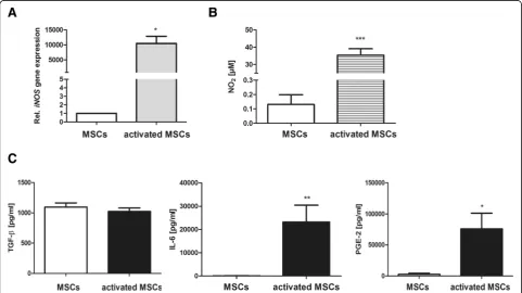
capacities, MSCs were initially stimulated with IL-1ß and IFN-ɣfor 24 h. As displayed in Fig.1, cytokine stimulation strongly increasediNOSgene expression in MSCs (Fig.1a), accompanied by increased amounts of secreted NO (Fig.1b) in the culture supernatant. However, the secretion of TGF-ß, which was previously demonstrated to be a major mediator of MSC-mediated immunosuppression [27], was not influ-enced by the cytokine treatment. Hence, significant upregu-lation of IL-6 and PGE2 production by preconditioned MSCs could be observed (Fig.1c)
Preconditioned MSCs efficiently suppress the polarization toward the M1 phenotype
Monocyte-derived differentiated M0 macrophages were characterized in respect to the expression of F4/80 and CD11b (Additional file2: Figure S2a). To generate differ-ent macrophage phenotypes, M0 macrophages were treated with cytokines as described under “Methods”. M1-like macrophages showed strong increase in iNOS expression as well as TNF-α and IL-6 secretion compared to M2a-like cells. In turn, upregulation of
Arg I expression was detected in M2a macrophages (Additional file 2: Figure S2b, c, d).
We next examined whether preconditioning of MSCs might improve their ability to impair M1 polarization. To
this end, co-culture experiments under M1-polarizing conditions were performed (M:MSC ratio 2:1). Transwell experiments were conducted in parallel to avoid direct cross-talk between MSCs and macrophages. Percentage of cells expressing co-stimulatory CD86 was significantly downregulated in the presence of MSCs, whereas the expression of the M2a marker CD206 was not influenced in co-cultures (Fig. 2a). Although the number of iNOS-positive cells did not change in the presence of pre-conditioned MSCs, a significant diminution of intracellu-lar protein levels could be detected. More importantly, co-cultured preconditioned MSCs strongly suppressed TNF-α secretion indicating diminished M1 polarization (Fig. 2b). Because NO secretion is significantly increased in activated MSCs, no reduction in NO2levels in culture
supernatants was found (Fig. 2b), despite reduced iNOS expression in macrophages. Suppressive effects on M1 differentiation mediated by MSCs exclusively depended on soluble factors as cell separation by transwell mem-branes did not alter macrophage characteristics.
Preconditioned MSCs increase CD86, iNOS, and IL-10 expression in macrophages under M2a-polarizing conditions
To elaborate MSC-triggered effects on M2 polarization, same experiments as described above were performed in
the presence of IL-4, which favors M2a polarization. The number of CD86 positive cells was significantly upregu-lated by primed MSCs when compared to unstimuupregu-lated MSCs, but no alterations regarding CD206 expression were found (Fig. 3a). In addition, upregulation of intra-cellular iNOS protein expression in macrophages
(Fig.3a) and increase in NO secretion (Fig.3b) could be observed in the presence of primed MSCs indicating M0-M2b transition. This assumption was further sup-ported by increased IL-10 secretion found in co-cultures (Fig.3b). Again, these effects did not depend on physical contact between cells.
Different regulation of macrophage marker genes by preconditioned MSCs under M1- and M2a-polarizing culture conditions
In order to prove whether preconditioned MSCs indeed promote the polarization toward M2b cells in our ex-perimental setting, gene expression of individual M2
subtype markers was analyzed by real-time PCR. As MSC-derived effects were found to be mediated by soluble mediators, cells were cultured in transwells. According to our assumption, significant suppression of
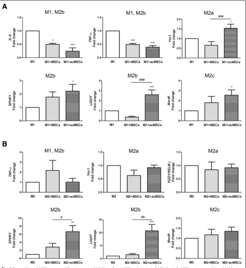
TNF-α and IL-6 gene expression was confirmed under M1-polarizing conditions (Fig.4a). Additionally, significant
upregulation of the M2a marker Ym-1 [28], the M2b markers SPHK1 and LIGHT [13] and also of MertK, which is highly expressed in M2c macrophages [29], has been observed under these conditions. In turn, under M2a-polarizing conditions, MSCs forced spe-cific M2b polarization by upregulating SPHK1 and
LIGHT gene expression (Fig. 4b). Notably, without preconditioning, MSCs failed to modulate macrophage polarization although significant inhibition of IL-6 and
TNF-α expression could be detected. Only weak, ir-regular expression of FIZZ1/RELMα [30] was detected in the presence IFN-ɣ and LPS (M1 polarization) whereas IL-6 expression was not detected in cultures supplemented with IL-4 (M2a) (not shown).
Lack of IL-6 receptor in macrophages, but not limited availability of NO and PGE2, prevents M2b polarization in the presence of preconditioned MSCs and IL-4
Immunosuppressive effects of MSCs were previously reported to occur through the secretion of NO, TGF-ß, IL-6, and PGE2, among others. In order to better evaluate the role of these soluble mediators, we used macrophages deficient in IL-6Rα and cultured them with preconditioned MSCs. In addition, iNOS and COX-2 expression in MSCs was downregulated by siRNA transfection before co-culturing with mac-rophages under M1- and M2a-polarizing conditions. Since preconditioned MSCs did not secrete higher levels of TGF-ß, the role of TGF-ß was considered as negligible.
Absence of IL-6Rαon macrophages [14] has been con-firmed by flow cytometry (Additional file 3: Figure S3a). Additionally, macrophages isolated from IL-6Rα-deficient C57Bl6 mice displayed widely same characteristics after polarization as wild type macrophages. However, SPHK1 gene expression in M1-polarized cells was significantly in-creased (Additional file3: Figure S3b). Cells deficient in IL-6Rα further showed strong suppression of FIZZ1/
RELMαexpression after stimulation with IL-4 when com-pared to wild type macrophages wherebyYm1, which rep-resents an additional M2a signature gene, did not change (Additional file 3: Figure S3c). Data presented in Fig. 5 demonstrate that secreted IL-6 profoundly suppresses
IL-6 and TNF-α expression in macrophages under M1-polarizing conditions, as the expression of both genes was significantly upregulated in macrophages deficient in IL-6Rαcompared to wild type cells (Fig.5a). Having found that IL-6R-deficient M1-polarized cells express higher levels of SPHK1 mRNA, this gene does not seem to be regulated by co-cultured MSCs. We also found that lack of IL-6Rα under M2a-polarizing conditions strongly affects the expression ofSPHK1,LIGHT, andMertK indi-cating that MSC-derived IL-6 represents an important mediator of M2b and probably M2c polarization (Fig.5b).
To validate the role of IL-6 signaling for MSC-mediated M2b polarization, we performed Western blot analysis to detect phosphorylated STAT3, which is implicated in the IL-6 transduction pathway. Indeed, cells deficient in IL-6Rαfailed to upregulate pSTAT3 upon incubation with IL-6-secreting MSCs, confirming an impaired IL-6 signal-ing pathway in these cells (Fig.6a). Of note, recombinant IL-6 widely mimicked the effects mediated by precondi-tioned MSCs. As displayed in Fig. 6b, in the presence of IL-4 and IL-6, macrophages significantly upregulated
SPHK1 expression and to a lesser, not significant, extent
LIGHTexpression. Thus, as M2b polarization was stron-gest in the presence of preconditioned MSCs, it is likely that M2b polarization does not solely depend on IL-6 expression.
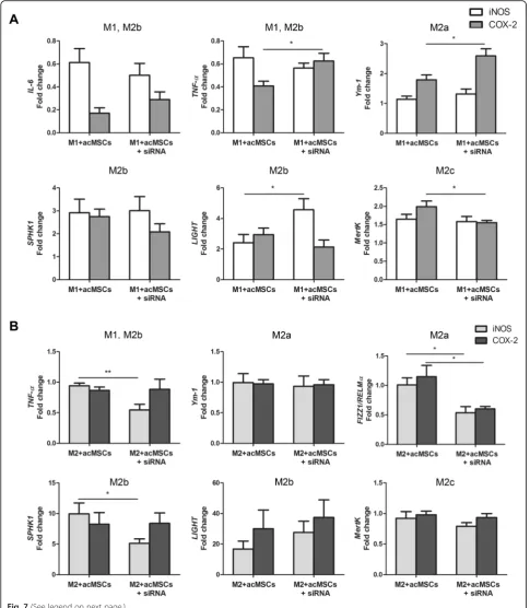
To further elaborate the role of MSC-derived NO and PGE2 for macrophage polarization, MSCs were trans-fected withiNOSas well asCOX-2specific siRNA before preconditioning. Knockdown efficiency of both genes in transfected, preconditioned MSCs was verified by real-time PCR (Additional file 4: Figure S4a). COX-2 knockdown did not influence NO nor IL-6 secretion by MSCs. Additionally, PGE2 as well as IL-6 secretion were not altered after transfection of MSCs withiNOSsiRNA. Consistently, significant reduction of NO and PGE2 could be detected in iNOSorCOX-2 siRNA-transfected MSCs, respectively (Additional file 4: Figure S4b). Im-paired NO availability under M1-polarizing conditions re-sulted in increasedLIGHTexpression, indicating that this gene is usually downregulated by NO. Similarly, PGE2 was found to downregulate TNF-α and Ym-1 (M2a marker) under same conditions. However, MSC-derived PGE2 seemed to promote M2c polarization, asMertKwas significantly reduced in macrophages co-cultured with
COX-2transfected MSCs (Fig.7a). NO was further found to be an inducer ofTNF-αexpression in the presence of IL-4 and to further upregulate FIZZ1/RELMα (M2a) (Fig.7b). The last one was also identified to be reduced in the absence of PGE2. Thus, although both NO and PGE2 drive the expression of the M2a-specific gene FIZZ/
RELMα, they play a subordinate role in MSC-mediated M2b polarization.
M2b macrophages suppress IFN-ɣproduction by CD4+T lymphocytes more efficiently than M2a macrophages
expressed higher levels ofIL-10compared to M2a-polar-ized control macrophages. Recombinant IL-6 also in-creased the expression of IL-10 in macrophages; however, this increase was significantly reduced when compared to MSC-mediatedIL-10upregulation. Corres-pondingly, macrophages from IL-6Rα-deficient mice showed strong reduction in IL-10 expression after co-culturing with preconditioned MSCs (versus wild type macrophages), arguing against an M2b phenotype.
We next questioned if macrophages cultured in the presence of IL-4 and preconditioned MSCs might influ-ence Th1/Th2 differentiation of naïve CD4+ T lympho-cytes. To this end, we cultured M2a-polarized and macrophages pre-cultured with preconditioned MSCs (M2b phenotype) with CD4+ T cells (Additional file 5) for 24 h in the presence of CD3 and CD28 antibodies. Then, gene expression ofGATA-3,IL-4,IFN-ɣ, andIL-2 was analyzed by real-time PCR to discriminate between Th1 and Th2 lymphocytes. As shown in Fig. 8b, T cells co-cultured with M2b macrophages showed significantly reduced IFN-ɣ expression compared to cells cultured in the presence of M2a macrophages. Further on, higher not significantGATA-3andIL-2expression could be de-tected, indicating that M2b cells significantly suppress pro-inflammatory pathways presumably favoring Th2 differentiation.
Discussion
In the recent past, the promising immunomodulatory ef-fects of MSCs and their therapeutic potential have become evident. However, beside first encouraging results in clin-ical studies, most MSC-based clinclin-ical trials occur in an early phase (phase I-II), demonstrating that the thera-peutic effectiveness of MSCs needs to be investigated. Among all MSC-based studies, there is a substantial pro-portion dealing with the treatment of cardiovascular dis-eases. Because ischemic heart diseases and heart failure remain major causes of morbidity and mortality, much ef-fort is expanded to repair cardiac tissue with stem cell transplantation. The therapeutic properties of MSCs are largely related to their anti-inflammatory and immuno-modulatory abilities which have been confirmed by in vivo and in vitro studies [8,10,32]. However, low survival rate
in vivo and undefined mechanisms underlying the benefi-cial properties of MSCs represent crubenefi-cial reasons account-ing for the impeded cardiac repair effect of MSCs.
Impaired pro-inflammatory response of macrophages and changes in the M1/M2 balance in the presence of MSCs has already been evidenced. Macrophage polarization is critical for the resolution of inflammation and tissue remodeling [33]. During the first days after myocardial infarction, neutrophils and M1 macrophages dominate in the infarcted area, whereas M2 transition during the late stage triggers Th2 responses and reso-lution of inflammation. Prolonged presence of M1 mac-rophages extends the pro-inflammatory environment and causes expansion of the infarcted area in the myo-cardium post-acute myocardial infarction (AMI) [34]. In turn, macrophage polarization toward the M2 phenotype has been shown to promote the resolution of inflamma-tion and improve infarct healing post-AMI [35]. There-fore, elucidation of MSC-triggered mechanisms and improvement of their anti-inflammatory capacities rep-resent an attractive strategy for MSC-based therapies.
In this study, we found that preconditioning of murine MSCs with IFN-ɣ and IL-1ß highly improves their po-tential to promote immunomodulation. In fact, precon-ditioning of MSCs resulted in significantly elevated NO, IL-6, and PGE2 production, which have been already de-scribed to represent prominent immunosuppressive mol-ecules [36, 37], although their mode of action remains obscure. Preconditioned MSCs markedly reduced CD86, iNOS protein expression, and TNF-α secretion by mac-rophages when cultured in the presence of pro-inflam-matory IFN-ɣ and LPS (M1 polarization). On the other hand, under M2a-polarizing culture conditions (IL-4), upregulation of CD86, iNOS, and increased NO and IL-10 secretion could be found. According to previously published results, these effects largely depended on para-crine factors and not on direct cell-to-cell contact [38]. Our data strongly suggest that immunoregulation by MSCs under pro-inflammatory conditions, especially after preconditioning with IFN-ɣ and IL-1ß, is based on a shift from the inflammatory M1 phenotype toward the anti-inflammatory M2 phenotype. In this regard, it was already reported that different M2 macrophage subtypes,
(See figure on previous page.)
namely M2a, M2b, and M2c, exhibiting different func-tions exist. Whereas M2a/c macrophages were found to be beneficial in early inflammatory stages, they have been found to impair tissue remodeling [39]. M2b cells have been proposed to be immunoregulatory diminish-ing immune responses with minor damage to local tissue
[40]. Having found that M2-polarized macrophages upregulated CD86 expression and NO and IL-10 pro-duction, we propose that preconditioned MSCs are effective in M2b polarization in the presence of anti-inflammatory IL-4. Indeed, by analyzing gene expression of selected macrophage markers after
Fig. 6Role of IL-6 for macrophage polarization toward M2b phenotype.aPreconditioned MSCs (acMSCs) were co-cultured with wild type macrophages or macrophages isolated from IL-6Rα-deficient (IL6R-KO) mice in the presence of IL-4 (20 ng/ml). After 18 h, the expression of pSTAT3 (Tyr705) was verified by Western blot. STAT3 served as loading control. One representative Western blot of three independent experiments is depicted.
co-culture with preconditioned MSCs, we found specific significant upregulation of the M2b markersSPHK1and
LIGHT in macrophages in the presence of the M2a in-ducer IL-4. In turn, all M2 markers were upregulated under M1-polarizing co-culture conditions. Notably, al-though unprimed MSCs were able to reduce the expression of pro-inflammatoryIL-6andTNF-αin mac-rophages, these cells failed to modulate M2 macrophage marker expression, thus being in line with recently re-ported data [41].
We also showed that macrophages co-cultured with preconditioned MSCs in the presence of IL-4 displayed highest IL-10 production, which could be attributed to
the M2b phenotype initiated by preconditioned MSCs. In this regard, when compared with other macrophage subtypes, M2b cells are known to represent the main cellular source for IL-10, playing an important role in the resolution of inflammation [31]. Direct co-culture of IL-10-producing M2b macrophages resulted in signifi-cant downregulation of IFN-ɣ expression in CD4+ T lymphocytes and also slight increase in expression of the Th2 markerGATA-3[42], indicating polarization toward a Th2 phenotype and suppression of Th1 responses.
Previous studies argue for a prominent role of PGE2 as a key immunosuppressive mediator derived from MSCs [36, 41, 43]. Additionally, both PGE2 and NO
(See figure on previous page.)
Fig. 7NO and PGE2 are only partially involved in MSC-mediated macrophage polarization. MSCs were transfected withiNOS-orCOX-2-specific siRNAs to reduce NO and PGE2 secretion and further stimulated with IFN-ɣand IL-1ß. Transfected, activated MSCs (acMSCs+siRNA) were co-cultured with M0 macrophages under M1- (20 ng/ml IFN-gamma + 100 ng/ml LPS,a) and M2a- (20 ng/ml IL-4,b) polarizing culture conditions for 24 h. Preconditioned MSCs (acMSCs) served as controls. Gene expression ofIL-6,TNF-α, M2a markers (Ym-1,FIZZ1/RELMα), M2b markers (SPHK1,LIGHT), and the M2c marker MertKin macrophages was analyzed by real-time PCR. Results are representative for six to eight independent experiments and are expressed as fold change vs. expression found in macrophages. *p< 0.05, **p< 0.01. FIZZ1/RELMα, found in inflammatory zone 1/resistin-like molecule alpha, IL interleukin, LIGHT lymphotoxin-like, exhibits inducible expression and competes with HSV glycoprotein D for herpes virus entry mediator, a receptor expressed by T lymphocytes, M macrophage, MertK tyrosine-protein kinase MER, MSCs mesenchymal stem cells, acMSCs activated, preconditioned mesenchymal stem cells, SPHK1, sphingosine kinase 1, TNF-αtumor necrosis factor alpha, Ym-1 chitinase-like protein
production are positively regulated by IL-6 and their se-cretion is strongly reduced in the absence of IL-6. In light of these considerations, we further elucidated the role of IL-6, NO, and PGE2 for MSC-mediated M2 or M2b polarization, respectively. Using IL-6Rα-deficient macrophages, we demonstrate here that IL-6 is the main MSC-derived mediator of IL-6 and TNF-α suppression under M1-polarizing conditions. Hence, MSCs failed to upregulate almost all M2 marker genes in cells cultured in the presence of M1 inducers lacking IL-6 signaling. Most importantly, IL-6Rα-deficient macrophages showed up-regulated TNF-α expression and significantly reduced
SPHK1, LIGHT, and MertK expression after co-culturing with preconditioned MSCs, suggesting that IL-6 repre-sents an essential regulator of M2b and probably M2c polarization. Indeed, after co-culture with preconditioned MSCs, macrophages deficient in IL-6Rα expressed strongly suppressed levels of IL-10 when compared to wild type cells, most likely due to impaired IL-6 signaling as evidenced by suppressed STAT3 activation. Although there is already evidence for the role of IL-6 for the polarization of alternatively activated macrophages [44, 45], in our knowledge, this study is the first showing that MSC-derived IL-6 triggers polarization toward the M2b phenotype. Thus, our results strongly underline the re-cently postulated anti-inflammatory role of IL-6 [44, 46]. Nevertheless, our data with recombinant IL-6 implicate that additional MSC-derived soluble factors might also be involved in M2b polarization. In contrast to previously published work [31,43], PGE2 was not found to influence macrophage polarization or to favor M2b polarization.
COX-2knockdown indeed strongly reduced PGE2 secre-tion, without affecting NO or IL-6 secretion by precondi-tioned MSCs, but this impaired PGE2 production did not diminish the expression of M2 marker genes, except for
FIZZ/RELMα. These discrepancies between results might be based on different experimental setups used. In this re-gard, in vitro polarized macrophage subsets [43] and com-bination of PGE2 with LPS [31] have been proposed for the induction of regulatory macrophages. Further on, the capacity of MSCs to regulate diverse macrophage features was demonstrated to differ between various strains of mice [36]. In addition, our data confirmed that MSC-mediated generation of regulatory macrophages is largely independent on NO secretion [36].
Altogether, our results presented here strongly suggest that MSCs, beside their potency to inhibit T cell prolifera-tion [47] and to promote generaprolifera-tion of Treg [48], signifi-cantly influence the plasticity of macrophages promoting a regulatory phenotype. Unpublished data of our group revealed pro-proliferative effects of preconditioned MSCs on macrophages, although the impact if IL-6 has not been further evaluated. Also, a possible link between M2b polarization and increased proliferative activity should be
considered in further studies. The modulation of macro-phage plasticity represents an important therapeutic strat-egy [49]. In the early stages of inflammation, MSCs might limit M1 responses and trigger M2 polarization, whereas during the late anti-inflammatory response, they might promote the generation of a regulatory M2b phenotype via IL-6 secretion. In view of these results, we propose that therapeutic use of preconditioned MSCs might be beneficial during different stages of inflammation in car-diovascular diseases. Indeed, in a very recent study, trans-plantation of M2b macrophages significantly ameliorated myocardial ischemia/reperfusion injury in mice [40]. Further in vivo studies based on preconditioned MSCs should be conducted in the future to enhance immuno-suppressive capacity of MSCs and M2b polarization and to validate the results of this study.
Conclusions
Preconditioning of MSCs with IL-1ß and IFN-ɣ strongly improves the capacity of MSCs to modulate macrophage plasticity, mainly by IL-6 secretion. MSC-dependent polarization toward the M2 phenotype in a pro-inflamma-tory microenvironment and toward the regulapro-inflamma-tory M2b phenotype in the presence of anti-inflammatory IL-4 should be considered for future therapeutic approaches to treat cardiovascular diseases.
Additional files
Additional file 1:Figure S1.Characterization of bone-marrow derived MSCs. (a) As assessed by flow cytometry, MSCs were positive for the well-established MSCs markers CD29, CD44, CD49e and Sca-1 and negative for CD11b and CD45. (b) Adipogenic, chondrogenic and osteogenic differen-tiation of MSCs. Scale bar, 100μm. (TIF 9562 kb)
Additional file 2:Figure S2.Characterization of M0, M1- and M2a-like macrophages. (a) CD11b and F4/80 expression on bone marrow-derived differentiated M0 macrophages. M0 macrophages were further polarized in vitro to M1-like and M2a-like cells by treatment with 20 ng/ml IFN-ɣ and 100 ng/ml LPS (M1) or 20 ng/ml IL-4 (M2), respectively, for 24 h. (b) Gene expression ofiNOSandArg Iwas determined by real-time PCR.n= 3. (c) TNF-αand IL-6 levels in culture supernatants were quantified by Elisa.n= 5, *p< 0.05, **p< 0.001. (TIF 1040 kb)
Additional file 3:Figure S3.Characterization of IL-6Rα-deficient macro-phages. (a) IL-6Rαexpression on bone marrow-derived M0 macrophages from WT and IL-6Rα-deficient mice.n= 3, **p< 0.01. (b) IL-6Rα-deficient and wild type macrophages were differentiated into M1- and M2a-like cells as described in Methods. Gene expression ofIL-6,TNF-αand the subtype-specific markersYm-1,FIZZ1/RELMα(M2a markers),SPHK1,LIGHT (M2b markers) andMertK(M2c marker) was determined by real-time PCR. n= 4–6, *p< 0.05, ***p< 0.001. (TIF 2221 kb)
Additional file 4:Figure S4.siRNA knockdown ofiNOSandCOX-2 expression in preconditioned MSCs. MSCs were transfected with siRNA specific foriNOS,COX-2or control siRNA (each 5.5 nM), respectively. After 24 h, cells were preconditioned with IFN-ɣand IL-1ß. (a) Transfection effi-ciency was proven by real-time PCR.n= 5, **p< 0.01 vs. control siRNA (Ctrl). ##p< 0.01, ###p< 0.001. (b) Levels of NO, IL-6 and PGE2 secreted by transfected MSCs were determined by Griess assay and Elisa.n= 5–6, *p < 0.05, ***p< 0.001, n.s. not significant (TIF 1510 kb)
Abbreviations
AMI:Acute myocardial infarction; EGF: Epidermal growth factor; FGF: Fibroblast growth factor; FIZZ1/RELMα: Found in inflammatory zone 1/resistin-like molecule; HGF: Hepatocyte growth factor; IFN-ɣ: Interferon gamma; IGF: Insulin-like growth factor; IL: Interleukin; IL-6Rα: Interleukin 6 receptor alpha; iNOS: Inducible nitric oxide synthase; LIGHT: Lymphotoxin-like, exhibits inducible expression and competes with HSV glycoprotein D for herpes virus entry mediator, a receptor expressed by T lymphocytes; LPS: Lipopolysaccharides; MertK: Tyrosine-protein kinase MER; MSCs: Mesenchymal stem cells; PDGF: Platelet-derived growth factor; PGE2: Prostaglandin E2; SDF: Stromal cell-derived factor; SPHK1: Sphingosine kinase 1; STAT3: Signal transducer and activator of transcription 3;
pSTAT3: Phosphorylated STAT3; TGF-ß: Transforming growth factor beta; TNF-α: Tumor necrosis factor alpha; VEGF: Vascular endothelial growth factor; Ym-1: Chitinase-like protein
Acknowledgements
The authors would like to thank Meike Lauer and Sabine Schmitt for excellent technical assistance. We are grateful to Dr. Thomas Wunderlich from the Max-Planck-Institute for Metabolism Research, CECAD and Institute for Genetics, Cologne, for providing IL-6Rα-deficient mice.
Funding
This study was supported by a grant from the German Heart Research Foundation (to A.P.G. Y.H.C., T.W., #F/06/16).
Availability of data and materials
The datasets used and/or analyzed during the current study are available from the corresponding author on reasonable request.
Authors’contributions
DP performed the experiments, interpreted the data, and contributed to the writing of the manuscript. LS performed transfection experiments. TW and YHC helped supervise the study and edited the manuscript. APG conceived and supervised the study, interpreted the data, and wrote the manuscript. All authors read and approved the final manuscript.
Ethics approval and consent to participate
Cell isolation from mice was approved by the local ethics committee (Bezirksregierung Köln; Germany; No: 4.16.002; 4.16.027).
Consent for publication
Not applicable.
Competing interests
The authors declare that they have no competing interests.
Publisher’s Note
Springer Nature remains neutral with regard to jurisdictional claims in published maps and institutional affiliations.
Received: 14 June 2018 Revised: 16 August 2018 Accepted: 7 October 2018
References
1. Pittenger MF, Mackay AM, Beck SC, Jaiswal RK, Douglas R, Mosca JD, Moorman MA, Simonetti DW, Craig S, Marshak DR. Multilineage potential of adult human mesenchymal stem cells. Science. 1999;284(5411):143–7. 2. Tropel P, Platet N, Platel JC, Noel D, Albrieux M, Benabid AL, Berger F.
Functional neuronal differentiation of bone marrow-derived mesenchymal stem cells. Stem Cells. 2006;24(12):2868–76.
3. Ferrari G, Cusella-De Angelis G, Coletta M, Paolucci E, Stornaiuolo A, Cossu G, Mavilio F. Muscle regeneration by bone marrow-derived myogenic progenitors. Science. 1998;279(5356):1528–30.
4. English K. Mechanisms of mesenchymal stromal cell immunomodulation. Immunol Cell Biol. 2013;91(1):19–26.
5. Crisostomo PR, Wang Y, Markel TA, Wang M, Lahm T, Meldrum DR. Human mesenchymal stem cells stimulated by TNF-alpha, LPS, or hypoxia produce growth factors by an NF kappa B- but not JNK-dependent mechanism. Am J Physiol Cell Physiol. 2008;294(3):C675–82.
6. Krampera M, Cosmi L, Angeli R, Pasini A, Liotta F, Andreini A, Santarlasci V, Mazzinghi B, Pizzolo G, Vinante F, et al. Role for interferon-gamma in the
immunomodulatory activity of human bone marrow mesenchymal stem cells. Stem Cells. 2006;24(2):386–98.
7. Ren G, Zhang L, Zhao X, Xu G, Zhang Y, Roberts AI, Zhao RC, Shi Y. Mesenchymal stem cell-mediated immunosuppression occurs via concerted action of chemokines and nitric oxide. Cell Stem Cell. 2008;2(2):141–50. 8. Nauta AJ, Fibbe WE. Immunomodulatory properties of mesenchymal
stromal cells. Blood. 2007;110(10):3499–506.
9. Keating A. How do mesenchymal stromal cells suppress T cells? Cell Stem Cell. 2008;2(2):106–8.
10. Uccelli A, Moretta L, Pistoia V. Mesenchymal stem cells in health and disease. Nat Rev Immunol. 2008;8(9):726–36.
11. Franquesa M, Hoogduijn MJ, Bestard O, Grinyo JM. Immunomodulatory effect of mesenchymal stem cells on B cells. Front Immunol. 2012;3:212. 12. Wang Q, Ni H, Lan L, Wei X, Xiang R, Wang Y. Fra-1 protooncogene
regulates IL-6 expression in macrophages and promotes the generation of M2d macrophages. Cell Res. 2010;20(6):701–12.
13. Edwards JP, Zhang X, Frauwirth KA, Mosser DM. Biochemical and functional characterization of three activated macrophage populations. J Leukoc Biol. 2006;80(6):1298–307.
14. Wunderlich CM, Delic D, Behnke K, Meryk A, Strohle P, Chaurasia B, Al-Quraishy S, Wunderlich F, Bruning JC, Wunderlich FT. Cutting edge: inhibition of IL-6 trans-signaling protects from malaria-induced lethality in mice. J Immunol. 2012;188(9):4141–4.
15. Drey F, Choi YH, Neef K, Ewert B, Tenbrock A, Treskes P, Bovenschulte H, Liakopoulos OJ, Brenkmann M, Stamm C, et al. Noninvasive in vivo tracking of mesenchymal stem cells and evaluation of cell therapeutic effects in a murine model using a clinical 3.0 T MRI. Cell Transplant. 2013;22(11):1971– 80.
16. Jin Y, Liu Y, Nelin LD. Extracellular signal-regulated kinase mediates expression of arginase II but not inducible nitric-oxide synthase in lipopolysaccharide-stimulated macrophages. J Biol Chem. 2015;290(4):2099– 111.
17. Davis MJ, Tsang TM, Qiu Y, Dayrit JK, Freij JB, Huffnagle GB, Olszewski MA. Macrophage M1/M2 polarization dynamically adapts to changes in cytokine microenvironments in Cryptococcus neoformans infection. MBio. 2013;4(3): e00264–13.
18. Wu F, Chakravarti S. Differential expression of inflammatory and fibrogenic genes and their regulation by NF-kappaB inhibition in a mouse model of chronic colitis. J Immunol. 2007;179(10):6988–7000.
19. Yeh CH, Cho W, So EC, Chu CC, Lin MC, Wang JJ, Hsing CH. Propofol inhibits lipopolysaccharide-induced lung epithelial cell injury by reducing hypoxia-inducible factor-1alpha expression. Br J Anaesth. 2011;106(4):590–9. 20. Tatano Y, Shimizu T, Tomioka H. Unique macrophages different from M1/
M2 macrophages inhibit T cell mitogenesis while upregulating Th17 polarization. Sci Rep. 2014;4:4146.
21. Clark K, MacKenzie KF, Petkevicius K, Kristariyanto Y, Zhang J, Choi HG, Peggie M, Plater L, Pedrioli PG, McIver E, et al. Phosphorylation of CRTC3 by the salt-inducible kinases controls the interconversion of classically activated and regulatory macrophages. Proc Natl Acad Sci U S A. 2012;109(42):16986–91. 22. Galvan MD, Foreman DB, Zeng E, Tan JC, Bohlson SS. Complement
component C1q regulates macrophage expression of Mer tyrosine kinase to promote clearance of apoptotic cells. J Immunol. 2012;188(8):3716–23. 23. Tellechea M, Buxade M, Tejedor S, Aramburu J, Lopez-Rodriguez C.
NFAT5-regulated macrophage polarization supports the proinflammatory function of macrophages and T lymphocytes. J Immunol. 2018;200(1):305–15. 24. Horber S, Hildebrand DG, Lieb WS, Lorscheid S, Hailfinger S, Schulze-Osthoff
K, Essmann F. The atypical inhibitor of NF-kappaB, IkappaBzeta, controls macrophage Interleukin-10 expression. J Biol Chem. 2016;291(24):12851–61. 25. Onodera A, Yamashita M, Endo Y, Kuwahara M, Tofukuji S, Hosokawa H,
Kanai A, Suzuki Y, Nakayama T. STAT6-mediated displacement of polycomb by trithorax complex establishes long-term maintenance of GATA3 expression in T helper type 2 cells. J Exp Med. 2010;207(11):2493–506. 26. Schmittgen TD, Livak KJ. Analyzing real-time PCR data by the comparative
C(T) method. Nat Protoc. 2008;3(6):1101–8.
27. Shi Y, Hu G, Su J, Li W, Chen Q, Shou P, Xu C, Chen X, Huang Y, Zhu Z, et al. Mesenchymal stem cells: a new strategy for immunosuppression and tissue repair. Cell Res. 2010;20(5):510–8.
29. Zizzo G, Hilliard BA, Monestier M, Cohen PL. Efficient clearance of early apoptotic cells by human macrophages requires M2c polarization and MerTK induction. J Immunol. 2012;189(7):3508–20.
30. Murray PJ, Wynn TA. Protective and pathogenic functions of macrophage subsets. Nat Rev Immunol. 2011;11(11):723–37.
31. MacKenzie KF, Clark K, Naqvi S, McGuire VA, Noehren G, Kristariyanto Y, van den Bosch M, Mudaliar M, McCarthy PC, Pattison MJ, et al. PGE(2) induces macrophage IL-10 production and a regulatory-like phenotype via a protein kinase A-SIK-CRTC3 pathway. J Immunol. 2013;190(2):565–77.
32. Dazzi F, Horwood NJ. Potential of mesenchymal stem cell therapy. Curr Opin Oncol. 2007;19(6):650–5.
33. Eggenhofer E, Hoogduijn MJ. Mesenchymal stem cell-educated macrophages. Transplant Res. 2012;1(1):12.
34. Nahrendorf M, Swirski FK, Aikawa E, Stangenberg L, Wurdinger T, Figueiredo JL, Libby P, Weissleder R, Pittet MJ. The healing myocardium sequentially mobilizes two monocyte subsets with divergent and complementary functions. J Exp Med. 2007;204(12):3037–47.
35. Zhou LS, Zhao GL, Liu Q, Jiang SC, Wang Y, Zhang DM. Silencing collapsin response mediator protein-2 reprograms macrophage phenotype and improves infarct healing in experimental myocardial infarction model. J Inflamm (Lond). 2015;12:11.
36. Bouffi C, Bony C, Courties G, Jorgensen C, Noel D. IL-6-dependent PGE2 secretion by mesenchymal stem cells inhibits local inflammation in experimental arthritis. PLoS One. 2010;5(12):e14247.
37. Maggini J, Mirkin G, Bognanni I, Holmberg J, Piazzon IM, Nepomnaschy I, Costa H, Canones C, Raiden S, Vermeulen M, et al. Mouse bone marrow-derived mesenchymal stromal cells turn activated macrophages into a regulatory-like profile. PLoS One. 2010;5(2):e9252.
38. Lee RH, Oh JY, Choi H, Bazhanov N. Therapeutic factors secreted by
mesenchymal stromal cells and tissue repair. J Cell Biochem. 2011;112(11):3073–8. 39. Ma Y, Halade GV, Zhang J, Ramirez TA, Levin D, Voorhees A, Jin YF, Han HC,
Manicone AM, Lindsey ML. Matrix metalloproteinase-28 deletion exacerbates cardiac dysfunction and rupture after myocardial infarction in mice by inhibiting M2 macrophage activation. Circ Res. 2013;112(4):675–88. 40. Yue Y, Yang X, Feng K, Wang L, Hou J, Mei B, Qin H, Liang M, Chen G, Wu
Z. M2b macrophages reduce early reperfusion injury after myocardial ischemia in mice: a predominant role of inhibiting apoptosis via A20. Int J Cardiol. 2017;245:228–35.
41. Lin T, Pajarinen J, Nabeshima A, Lu L, Nathan K, Jamsen E, Yao Z, Goodman SB. Preconditioning of murine mesenchymal stem cells synergistically enhanced immunomodulation and osteogenesis. Stem Cell Res Ther. 2017; 8(1):277.
42. Lee HJ, Takemoto N, Kurata H, Kamogawa Y, Miyatake S, O'Garra A, Arai N. GATA-3 induces T helper cell type 2 (Th2) cytokine expression and chromatin remodeling in committed Th1 cells. J Exp Med. 2000;192(1):105–15. 43. Kudlik G, Hegyi B, Czibula A, Monostori E, Buday L, Uher F. Mesenchymal
stem cells promote macrophage polarization toward M2b-like cells. Exp Cell Res. 2016;348(1):36–45.
44. Braune J, Weyer U, Hobusch C, Mauer J, Bruning JC, Bechmann I, Gericke M. IL-6 regulates M2 polarization and local proliferation of adipose tissue macrophages in obesity. J Immunol. 2017;198(7):2927–34.
45. Sanmarco LM, Ponce NE, Visconti LM, Eberhardt N, Theumer MG, Minguez AR, Aoki MP. IL-6 promotes M2 macrophage polarization by modulating purinergic signaling and regulates the lethal release of nitric oxide during Trypanosoma cruzi infection. Biochim Biophys Acta. 2017;1863(4):857–69. 46. Mauer J, Chaurasia B, Goldau J, Vogt MC, Ruud J, Nguyen KD, Theurich S,
Hausen AC, Schmitz J, Bronneke HS, et al. Signaling by IL-6 promotes alternative activation of macrophages to limit endotoxemia and obesity-associated resistance to insulin. Nat Immunol. 2014;15(5):423–30. 47. Carrade Holt DD, Wood JA, Granick JL, Walker NJ, Clark KC, Borjesson DL.
Equine mesenchymal stem cells inhibit T cell proliferation through different mechanisms depending on tissue source. Stem Cells Dev. 2014;23(11):1258–65. 48. Luz-Crawford P, Kurte M, Bravo-Alegria J, Contreras R, Nova-Lamperti E,
Tejedor G, Noel D, Jorgensen C, Figueroa F, Djouad F, et al. Mesenchymal stem cells generate a CD4+CD25+Foxp3+ regulatory T cell population during the differentiation process of Th1 and Th17 cells. Stem Cell Res Ther. 2013;4(3):65.