© 2016 The Author(s). Published by The Geological Society of London for The Micropalaeontological Society. All rights reserved. For permissions: http://www.geolsoc.org.uk/permissions. Publishing disclaimer: www.geolsoc.org.uk/pub_ethics
Published online November 19, 2015 doi:10.1144/jmpaleo2014-026 | Vol. 35 | 2016 | pp. 26–53
Journal of Micropalaeontology
JM-TMS
Neogene radiolarians are generally abundant and well preserved in Antarctic sediments, which have been recovered by both pis-ton coring and by the various phases of the deep-sea drilling pro-grammes. Early studies of the radiolarian assemblages concentrated either on describing the more common species of the Recent faunas for use in Quaternary palaeoceanographic research, or on identifying a small number of species in older sediments which could be used for biostratigraphy. The majority of the species present in these sediments, if not common or clearly useful for biostratigraphic zonation remained unde-scribed. In order to better use these faunas in studies of biodiver-sity dynamics (Renaudie & Lazarus 2013b) we have carried out a fuller description of the faunas, including many of the rarer forms that typically make up a large fraction of total species rich-ness. Many of the species encountered were new to science. In this article, the last of a series of four publications, we describe these new forms. In our three prior papers (Renaudie & Lazarus 2012, 2013a, 2015) we described a total of 70 new species, to which we add 25 more in this paper. Although a very substantial number, our studies do not fully exhaust the diversity of taxa found in Antarctic Neogene sediments. There remain many only partially resolved species clusters, or groups with difficult mor-phologies (e.g. Actinommidae, Litheliidae) whose diversity remains to be fully diagnosed.
Material and methods
All studied samples (c. 350) come from DSDP and ODP deep-sea drilling sections from the Southern Ocean, mostly from the Kerguelen-Heard Plateau in the Indian sector of the Southern Ocean (Leg 119 Sites 737, 738, 744, 745 and 746; Leg 120 Sites 747, 748 and 751; Leg 183 Site 1138) with the addition of samples from the Atlantic sector (Leg 113 Sites 689, 690 and 693) and from the Pacific sector (Leg 29 Site 278) (Fig. 1). Prepared slides were drawn from the junior author’s collection or the MRC (Micropaleontological
Reference Center) radiolarian collection hosted by the Museum für Naturkunde in Berlin (Lazarus 2006). Samples were prepared to ran-dom strewn slides using standard methods (Moore 1973) using either 38, 45 or 63 µm sieves.
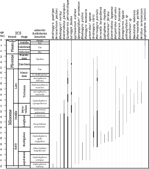
The radiolarian biozonation follows Lazarus (1992) and Abelmann (1992). The age estimates used for the range chart (Fig. 2) are inferred linearly from an age model based on Gersonde et al. (1990) for Leg 113, Barron et al. (1991) for Leg 119, Harwood et al. (1992) for Leg 120 and Bohaty et al. (2003) for Leg 183, with all ages adjusted to the Berggren et al. (1995) time-scale. The relative abundances given in the range chart are drawn from counts made on 45 µm strewn slides for 119 of the c. 350 samples. Measurements were made on specimen pictures using ImageJ (Abramoff et al. 2004): the range of variation and the mean (between brackets) are both given in microns (µm) under the Dimensions section for each species.
Higher-level classification largely follows that of Riedel (1967), with a few subsequent emendations as individually noted below.
The terminology used here follows mostly Jørgensen (1905) and Petrushevskaya (1965, 1968) for nassellarian internal structure (Fig. 3), Goll (1968) for features specific to the family Trissocyclidae and Boltovskoy (1998) for general external charac-ters. The notation for connecting arches in nassellarians follows generally De Wever et al. (1979), Dumitrica (1991) and Funakawa (1995a) in which they are named after a combination of the initials of the spines they originate from (i.e. arch AV would be an arch connecting spine A and spine V, see Fig. 3a), or, when necessary, follows Petrushevskaya (1965, 1968) in which they are named after the apophyses they are joining (i.e. arche mj joins apophyses m on spine A and j on spine V, see Fig. 3b).
All holotypes are deposited in the micropaleontology collection of the Museum für Naturkunde, Berlin and are indentified by their accession numbers (ECO-xxx) in the descriptions. Specimens are identified by a circle on the slide.
New species of Neogene radiolarians from the
Southern Ocean – part IV
Johan Renaudie
*& David B. Lazarus
Museum für Naturkunde, Leibniz-Institut für Evolutions- und Biodiversitätsforschung an der Humboldt-Universität zu Berlin, Invalidenstraße 43, 10115 Berlin, Germany
* Correspondence: johan.renaudie@mfn-berlin.de
Abstract: In this last paper in our planned series, we describe 25 new radiolarian species from the Antarctic Neogene: 6 spumellarians (Lithatractus? floridus, Spongopylidium? aerostatum, Haeckeliella hederacia, Larcopyle faustae, Excen-trodiscus planangulus and E. lappaceus) and 19 nassellarians (Anthocyrtidium sp., Artostrobus? oganeae, Botryostrobus exstructus, Ceratospyris clarki, Cornutella burgundiensis, Cystophormis petrushevskayae, Dendrospyris quadripes, Enne-aphormis tippula, Euscenarium funakawai, Lophophaena kamikurii, L. rhopalica, Lychnocanium andreae, Periarachnium pauliani, Peridium tortonianicum, Phormacantha garbela, Phormospyris punnulis, Rhodospyris? morleyi, Rhodospyris pulchra and Tholospyris tautessares). We also report the finding of fragments of an unknown Middle Miocene phaeodarian (Conchellium? sp.).
Supplementary Material: List of species of polycystine Radiolarians and of Phaeodarians encountered during our study of the Antarctic Neogene is available at http://www.geolsoc.org.uk/SUP18854
Keywords: Radiolaria; Polycystinea; Phaeodarea; Antarctic; Cenozoic; taxonomy
Received 18 October 2014; accepted 20 January 2015
Systematic palaeontology
Phylum Rhizaria Cavalier-Smith, 2002
Class Cercozoa Cavalier-Smith, 1998, emend. Adl et al., 2005 Subclass Phaeodarea Haeckel, 1879
Family Conchariidae Haeckel, 1879
Genus Conchellium Haeckel, 1887 Type species.Conchellium tridaena Haeckel, 1887
Conchellium? sp. (Pl. 1, figs 7A–8)
Material. Ten fragments observed in ODP Sites 689, 690 and 751.
Occurrence. Sporadic from the Actinomma golownini Zone to the Acrosphaera australis Zone (Middle to Late Miocene).
Remarks. The fragments found are those of a hemispherical shell of c. 150–200 µm diameter. It bears numerous, small, perfectly round, tube-like pores whose external rims extend laterally to form a second shell layer, seemingly (Pl. 1, fig. 7B), evoking what can be seen in Acrosphaera australis Lazarus, 1990. All these pores are arranged in quincuncial rows. The brownish tint of the shell indicates that it is probably a phaeodarian.
Specimen in Plate 1, figure 7A–B is the most complete frag-ment found to date.
The only other Conchariidae we recognized in the Neogene fos-sil record of the Southern Ocean is ?Conchellium capsula Borgert, 1907, which differs quite clearly from these fragments in its large polygonal pores.
Class Radiolaria Müller, 1858
Superorder Polycystinea Ehrenberg, 1839 emend. Riedel, 1967
Order Spumellaria Ehrenberg, 1876
Family Actinommidae Haeckel, 1862 emend. Sanfilippo & Riedel, 1980
Genus Lithatractus Haeckel, 1887
Type species.Stylosphaera fragilis Haeckel, 1887
Lithatractus? floridus n. sp (Pl. 1, figs 1A–3)
Derivation of name. From the Latin adjective floridus meaning ‘flowering’, for the flower-shape pore arrangement.
Diagnosis. Unequal polar spines, pores arranged in flower-shaped packs of seven, themselves arranged in a hexagonally-packed pat-tern.
Holotype. Plate 1, figure 2A–B; sample 119-744A-8H-3, 53–55 cm (Early Miocene); ECO-083.
Material. 97 specimens from DSDP Site 278 and ODP Site 744.
Description. Ellipsoidal shell with two polar spines: one long (more than the length of the shell) and tribladed, the other short, triangular and tribladed at its base. Pores on the shell are small and round, closely packed, arranged by group of seven in a petaloid pattern. This pattern is repeated and itself arranged in a hexagonal pattern. There is c. 6 of such petaloid groups of pores in a half-equator. Bars between the pores seem (based on observation of half-dissolved specimens, see Pl. 1, fig. 3) to be thinner between the pores of one such group and thicker between each group of pores. Both types of bars are very narrow. No medullary shell was observed to date.
737
738 744
745−746 747
748 751
690 689
693
1138
278
60°S 80°S
30°W 0°E 30°E
60°E
90°E
120°E
150°E 180°E
150°W
Weddell Sea
Ross Sea
Kerguelen Islands
Dimensions. Based on 3 specimens. Length of cortical shell main axis: 77–86 (83); of cortical shell minor axis: 70–79 (74); of long spine: 88–97 (94); of short spine: 16–21 (19).
Occurrence. Rare to common in the Cycladophora golli regipi-leus Zone (Early Miocene).
Remarks.Lithatractus? floridus n. sp. is significantly smaller, has a smaller number of relatively large main pores on the cortical shell,
and has a noticeably thinner cortical shell in comparison to Sphaerostylus rosetta Blueford, 1982, Stylatractus neptunus Haeckel, 1887, or any of the several ‘stylosphaerid’ species of Ehrenberg, as illustrated in Suzuki et al. (2009b) and Ogane et al. (2009). The overall outline of this species is very similar to that of Lithatractus timmsi Campbell & Clark, 1944 (and, to some extent, to that of the more robust form Stauroxiphos communis Carnevale, 1908) but it differs from it in possessing only one small spine instead
! !" #$ % & $ ' $ ( ) *+ & ,-" *" ./! !
01
2*#
#!3 ' ! " ! 4 # 4 ! ! 4 " $ " % ! 4 4
of several small spines on the opposite pole of the large spine and in its peculiar pore arrangement. It differs in general from most other stylosphaerids because of its 7-fold petaloid subpore arrangement, but in addition to that it also differs from Druppatractus irregularis Popofsky, 1912 and D. nanus Blueford, 1982 in having one long spine (at least longer than the cortical shell) and from Druppatractus hastatus Blueford, 1982 in the latter having a flattened cortical shell. Species of the genus Druppatractus also have a pyriform medullary shell but no medullary shell was observed in Lithatractus? floridus n. sp. to date. As this absence is likely to be purely taphonomical, the generic assignment is based only on overall similarity of the cortical shell shape and is therefore only tentative.
Genus Spongopylidium Dreyer, 1889 emend. Suzuki et al., 2009a
Type species.Spongopyle (Spongopylidium) ovata Dreyer, 1889.
Spongopylidium? aerostatum n. sp. (Pl. 1, figs 4–6B)
Derivation of name. From the modern Latin aerostatum meaning ‘hot-air balloon’.
Diagnosis. Spherical outline; narrow pylome with peristome; crested pore frame.
Holotype. Plate 1, figures 6A–B; sample 120-751A-9H-5, 98–102 cm (Late Miocene); ECO-084.
Material. 27 specimens from DSDP Site 278 and ODP Sites 744, 748 and 751.
Description. Large spherical single cortical shell. The shell wall is lat-ticed and bears many circular pores of similar size. The bars between the pores are thick and crested, giving the whole shell a honeycomb aspect. The pores are arranged in an irregular hexagonal pattern.
A small pylome protrudes from the shell. It is surrounded by a hyaline, narrow, fairly long and thick-walled peristome. Its termi-nation can be smooth (see Pl. 1, fig. 4) or ragged (see Pl. 1, fig. 5B). The pore pattern around the pylome is particularly irregular. No medullary shell was observed.
Dimensions. Based on 5 specimens. Diameter of shell: 174–249 (210); width of pylome: 18–30 (26).
Occurrence. Sporadic from the Cycladophora antiqua Zone to the Cycladophora spongothorax Zone (Early to Late Miocene). Remarks. The complete lack of medullary shell in S.? aerostatum n. sp. may very well be taphonomical. All comparisons can
therefore be based only on the elements of the cortical shell (which hinders proper assignment to a genus), however, S.? aerostatum does seem to show a unique combination of these elements.
Lithocarpium monikae Petrushevskaya, 1975 differs from this species in having an elliptical instead of a circular outline, in its thinner cortical shell having tinier, more numerous pores arranged randomly in a 3D meshwork and in having a spongy medullary shell linked to the cortical shell by relatively thick radial beams; even if the absence of medullary shell in our species is likely to be taphonomical, the junction of the radial beams with the cortical shell could have been seen. Prunopyle frakesi Chen, 1975 has a narrow pylome but it is surrounded by a cluster of spines instead of being prolonged by a peristome; additionally the pores in this spe-cies are more numerous, smaller, more closely packed and, more importantly, tube-shaped. Finally the shell wall of P. frakesi is smooth and its shell outline elliptical. Enamelon inemmestrum Sugiyama, 1992 shares with S.? aerostatum many characteristics: its spherical shell outline and its narrow pylome with a fairly long hyaline peristome. However, they differ in their shell wall which is, in E. inemmestrum, smooth and covered by a multitude of small pores. The size range of the last two species is also considerably larger than that of S.? aerostatum (270 to 342 µm for P. frakesi and 290 to 430 for E. inemmestrum). The Paleocene specimen illus-trated in Nishimura (1992, pl. 11, fig. 10) as Diploplegma (?) sp. aff. D. somphum Sanfilippo & Riedel, 1973 is also very similar to S.? aerostatum for the same reasons invoked in the comparison with E. inemmestrum but they differ in that Nishimura’s specimen possesses a thick spongy outer layer over the cortical shell. Larcopyle titan (Campbell & Clark, 1944) differs from S.? aero-statum in its larger pylome, its elliptical outline, its shell thickness and in having smaller, irregular pores with at most only weak frames. L. titan also possesses internal shell structures, although these are very delicate and difficult to visualize.
Sphaeropyle langii Dreyer, 1889, S. antarctica (Dreyer, 1889), S. tetrapila (Hays, 1965) and S. robusta Kling, 1973 all differ from this new species in the shape of their pylome: in all these species, the pylome is wide and surrounded by several spines, whereas in S.? aerostatum it is narrow and prolonged by a hyaline peristome.
Finally, Spongopylidium pyloma (Reynolds, 1980) is a species apparently devoid of medullary shell, having a thick latticed wall and a pylome very similar to that of S.? aerostatum. However, it differs from S.? aerostatum in its small size, elliptical outline, the smoothness of its cortical shell and the random arrangement of the pores. We ten-tatively assign our new species to the genus Spongopylidium because of the similarity between the pylome of S. pyloma and the one of S.? aerostatum coupled with the apparent absence, in both forms, of a
medullary shell. Enamelon inemmestrum also shared a similar pylome but its dense spongiose inner shell makes it stand out compared to those two prior forms. Suzuki et al. (2009a) in their emendation of the genus Spongopylidium state that this genus includes forms with a spongy inner structure as well, meaning that E. inemmestrum may very well belong to that genus. It is interesting to note here that the three forms also happen to be more or less contemporaneous.
Genus Haeckeliella Hollande & Enjumet, 1960
Type species.Haliomma macrodoras Haeckel, 1887
Haeckeliella hederacia n. sp. (Pl. 3, figs 1A–3B)
Derivation of name. From the Latin adjective hederacius (liter-ally ‘of ivy’), after the trellis-like lattice climbing over the spines of this species, as ivy on a tree.
Diagnosis. Smooth, single layer cortical shell with large spines surrounded by a secondary wiring, regularly connected to them.
Holotype. Plate 3, figure 1A–B; sample 113-690B-6H-4, 22– 24 cm (Early Miocene); ECO-082.
Material. 48 specimens, including fragments of spines, observed in DSDP Site 278 and ODP Sites 689, 690, 747, 758 and 751.
Description. Large single-shelled skeleton. The cortical shell bears numerous large, irregularly-shaped (mostly elliptical or cir-cular) pores. The bars between the pores are narrow and smooth. They do, however, exhibit some thorns now and then (see Pl. 3, figs 1B and 3A–B) but not at bar nodes. Some of the thorns are oriented toward the pores and not toward the exterior. The cortical shell also bears a few (6–10?) main radial spines. These spines are long and tribladed. They are attached to the cortical shell in a com-plex way: arches connect the blades of the spine to the bars of the cortical shell a few micrometres above the surface and again a few micrometres below the surface. The specimen on Plate 3, figure 3A–B shows that these spines continue inside the cortical shell as thin rods directed toward the shell centre. Unfortunately no speci-men seems to have these rods or beams completely preserved and hence no medullary shell or spicule has been observed to date. At regular intervals along the spines, apophyses protrude from each blade, extend perpendicularly to the spine axis and then branch out and join one another to form eventually a secondary trellis-like lattice surrounding the spines. The bars that constitute this mesh-work are very thin and cylindrical.
Dimensions. Based on two complete specimens. Diameter of shell: 271–276; diameter of pores: 4–32 (18).
Occurrence. Rare in the Actinomma golownini Zone (Middle Miocene), although some specimens were observed sporadically from the Cycladophora golli regipileus Zone to the Cycladophora spongothorax Zone (Early to Late Miocene).
Remarks. The attachment of the spines to the cortical shell seems identical to the one described for Haeckeliella macrodoras (Haeckel,
1881) in Hollande & Enjumet (1960) and in Blueford (1982); how-ever, the latter lacks the trellis surrounding the spines and has a large robust medullary shell. The trellis surrounding the main spines evokes what can be seen in Cleveiplegma boreale (Cleve, 1900) in Dumitrica (2013); however, the latter has a completely different cor-tical shell and its spines are considerably more delicate.
The assignment of this species to Haeckeliella is questionable because the inner structure is unknown.
Family Heliodiscidae Haeckel, 1881
Genus Excentrodiscus Hollande & Enjumet, 1960
Type species. Excentrodiscus echinatus Hollande & Enjumet, 1960
Excentrodiscus planangulus n. sp. (Pl. 2, figs 1A–3, 5A–6) ?1978 Excentrodiscus sp. Dumitrica: pl. 4, figs 5–6. ?1990 Actinomma medusa Abelmann: pl. 1, fig. 6.
Derivation of name. From the contraction of the Latin adjective planus (flat) and the noun angulus (corner), for this species’ some-what flattened areas.
Diagnosis. Eccentric inner microsphere, spherical to ovoid outer medullary shell, globular, somewhat flattened in spots, thin corti-cal shell.
Holotype. Plate 2, figure 1A–B; sample 119-744A-7H-3, 53–55 cm (Middle Miocene); ECO-075 (circle 2).
Material. 1548 specimens encountered in DSDP Site 278 and ODP Sites 744, 748, 751 and 1138.
Description. Three-shelled spumellarian with two medullary shells and one cortical shell. The cortical shell is subspherical to globular, with most specimens having at least a couple of somewhat flattened areas. The shell wall is rather thin yet bars are crested, often with raised apices that can be, rarely, expressed as small thorns. Pore size varies but they are generally medium-sized, closely packed, arranged in an irregular hexagonal pattern. Pores are generally elliptical. The outer medullary shell size varies between half and a third of the size of the cortical shell. Its shape is spherical to ovoid (with the elongated part where the inner medullary shell is situated). The shell wall is thicker than that of the cortical shell. Circular to elliptical pores are closely packed on the shell wall, but the pore diameter to bar width ratio is smaller than that of the cortical shell. Pores covering the side where the inner medullary shell lies are smaller than the others. The microsphere (i.e. inner medullary shell) is small, spherical and closely fixed to one pole of the outer medullary shell by a few robust radial beams. No beams connecting the microsphere with the distal half of the outer medullary shell were observed (see Pl. 2, fig. 3). The outer medullary shell and the cortical shell are connected by many thin, cylindrical radial beams. They do not protrude outside the cortical shell wall. The connections to the cortical shell wall tend to create characteristic small flattened areas or even depressions on the shell outline. The radial beams also seem to widen near the connection.
Explanation of Plate 2.figs 1–3, 5–6.Excentrodiscus planangulus n. sp.: 1A, B, sample 119-744A-7H-3, 53–55 cm, holotype – (A) focus on cortical shell, (b) focus on medullary shell; 2, sample 119-744A-7H-2, 53–55 cm; 3, sample 120-751A-13H-2, 98–102 cm – isolated medullary shell; 5A, B, sample 119-744A-8H-1, 60–62 cm – (A) focus on cortical shell, (B) focus on medullary shell; 6, sample 119-744A-7H-2, 53–55 cm. figs 4, 7, 8, 10.
Excentrodiscus lappaceus n. sp.: 4A, B, sample 120-751A-13H-2, 98–102 cm – isolated medullary shell, (A) focus on outer medullary shell, (B) focus on inner medullary shell; 7A, B, sample 119-744A-8H-1, 60–62 cm, holotype – (A) focus on cortical shell, (B) focus on medullary shell; 8, sample 120-751A-17H-CC; 10A, B, sample 120-748B-6H-7, 45–47 cm – (A) focus on medullary shell, (B) focus on cortical shell. figs 9, 11.Excentrodiscus japonicus (Nakaseko & Nishimura, 1974): 9A, B, sample 119-744A-4H-4, 53–55 cm – isolated medullary shell, (A) focus on outer medullary shell, (B) focus on inner medullary shell; 11, sample 113-689B-3H-5, 136–138 cm. Scale bars represent 50 µm.
Dimensions. Based on 8 specimens. Diameter of inner medullary shell: 11–24 (16); of outer medullary shell: 43–69 (57); of cortical shell: 103–157 (128).
Occurrence. Common from the Stylosphaera radiosa to the Cycladophora humerus Zone (Late Oligocene to Middle Miocene), then rare until the lower Cycladophora spongothorax Zone (Middle Miocene).
the same microsphere structure with Excentrodiscus echinatus and Excentrodiscus japonicus (Nakaseko & Nishimura, 1974) but dif-fers from the former in lacking external spines and from the latter in its thin, globular cortical shell. Additionally, the species we rec-ognize as Excentrodiscus japonicus in the Southern Ocean (see Pl. 2, figs 9A–B and 11) was observed from the upper Cycladophora spongothorax Zone (Late Miocene) to the Tau Zone (Early Pliocene), thus its range is not overlapping with that of our new species in the region.
Excentrodiscus planangulus n. sp. also differs from Excentrodiscus lappaceus n. sp. in its shell outline (at most globu-lar but never anguglobu-lar), lack of external spines, the shape of the outer medullary shell and the size ratio between outer and inner medullary shell. The specimen illustrated as Excentrodiscus sp. in Dumitrica (1978, pl. 4, figs 5–6) might be conspecific with Excentrodiscus planangulus as it shares its shell ratio, shape of the outer and inner medullary shells, numerous radial beams and glob-ular cortical shell; however, the cortical shell of that specimen seems thicker, with a more regular pore pattern.
Excentrodiscus lappaceus n. sp. (Pl. 2, figs 4A–B, 7A–8, 10)
Derivation of name. From the Latin adjective lappaceus meaning ‘resembling a burr’, after the many spines covering this species’ cortical shell.
Diagnosis. Microsphere excentric, outer medullary shell spherical and cortical shell spinose and with an angular, often indented out-line.
Holotype. Plate 2, figure 7A–B; sample 119-744A-8H-1, 60–62 cm (Early/Middle Miocene); ECO-027, circle 2.
Material. Thirteen specimens were observed in DSDP Site 278 and ODP Sites 744, 746, 748 and 751.
Description. Three-shelled spumellarian (two medullary shells and one cortical). The cortical shell has an angular, partially indented outline (more or less hexagonal on most specimens). The pores on the cortical shell are large, polygonal to circular, separated by nar-row, crested bars. The outer medullary shell is spherical to globular, with smaller circular pores separated by thick bars. The microsphere (i.e. inner medullary shell) is eccentric, attached to the wall of the outer medullary shell. It is small and more or less spherical. It is also heteropolar as, on the hemisphere that is closer to the outer medul-lary shell, it bears some small bars joining it to the outer medulmedul-lary shell wall. On the other hemisphere, one long rod (which could be homologous with the antapical beam seen in the species of the genus Pentactinosphaera; see Nakaseko et al. (1983) and Renaudie & Lazarus (2013a); or indeed with the rod seen on the specimen illus-trated as Heliodiscus sp. in De Wever et al. (2001, pl. 69, fig. 4)) seems to join the other side of the outer medullary shell (see speci-mens on Pl. 2, figs 4A–B and 7A). Multiple rod-like, radial beams join the outer medullary shell and the cortical shell. They protrude outside the cortical shell wall as short but robust radial spines. These radial spines have a large tribladed base. They seem to be more numerous than the radial beams, meaning that some are probably by-spines, yet they are indistinguishable from the radial spines.
Dimensions. Based on two specimens. Diameter of inner medul-lary shell: 14–23, of outer medulmedul-lary shell: 51–54; of cortical shell: 107–120.
Occurrence. Rare from the Cycladophora golli regipileus Zone to the Eucyrtidium punctatum Zone (Early Miocene).
Remarks. As for the previous species, this species differs from Excentrosphaerella sphaeroconcha, E. kamchatica, E. kovalenki,
E. sukhovi, E. spinulosa and Excentrococcus annulatus in possess-ing only one cortical shell. It also differs from Excentrodiscus japonicus (see, in addition to Nakaseko & Nishimura (1974), Kamikuri (2010) and herein in Pl. 2, figs 9 and 11) in its shell out-line and the presence of external spines but also in the shape of its outer medullary shell, and from E. echinatus in the shape and length of the spines but also primarily in the cortical shell outline.
Thecosphaerella glebulenta Sanfilippo & Riedel, 1973 shares with Excentrodiscus planangulus and E. lappaceus its shell out-line; however, its inner medullary shell does not seem to be similar and, if the specimen of T. glebulenta illustrated by Jackett et al. (2008) indeed belongs to this species, it actually seems to be a regular concentric inner medullary shell and, consequently, this species is not a heliodiscid. Additionally the shell ratio is clearly different in this species.
Family Litheliidae Haeckel, 1862
Genus Larcopyle Dreyer, 1889 emend. Lazarus et al., 2005
Type species. Larcopyle buetschlii Dreyer, 1889
Larcopyle faustae n. sp. (Pl. 1, figs 9A–12B)
2005 Caltanisetta radiolarians Lazarus et al.: pl. 1, figs 1–6, non 7–9.
Derivation of name. Named after after Karoline Faust, in honour of her contributions to the study of litheliid taxonomy.
Diagnosis. Globular, lumpy cortical shell, with a spiraling medul-lary shell.
Holotype. Plate 1, figure 12A–B; sample 119-737A-27X-2, 53–55 cm (Late Miocene); ECO-085.
Material. 1669 specimens were encountered in DSDP Site 278 and ODP Sites 689, 690, 693, 737, 744, 745, 746, 747, 748, 751 and 1138.
Description. Globular shell, with a roughly spherical to elliptical outline, deformed by numerous bumps. The shell wall is rather thin and covered with many, closely packed, round to elliptical pores of various sizes. The bars between the pores are smooth. The inner part of the shell seems to be a loose whorl that spirals from its centre and ends up merging with the cortical shell (relatively clear in spec-imens on Pl. 1, figs 10A to 12B). Each coil is linked to the next one (and eventually to the cortical shell) by several relatively thick radial rods (seen on Pl. 1, figs 9B, 10B and 11B, particularly). The depressions between the cortical shell bumps seem to correspond to the connection of these radial rods (see e.g. Pl. 1, figs 9B and 11B).
Some specimens (Pl. 1, figs 10A–B and 12A–B) have numerous small needle-like thorns extending from the shell’s bar nodes. No osculum or pylome was observed; there was no obvious sign of shell polarity.
Dimensions. Based on five specimens. Diameter of shell: 129–189 (145).
Occurrence. Rare throughout the whole Neogene and Quaternary, common in the Actinomma golownini Zone (Middle Miocene), the upper Cycladophora spongothorax Zone (Late Miocene) and the upper Tau Zone (early Pliocene).
Lazarus et al., 2005 and in Larcopyle eccentricum Lazarus et al., 2005 primarily in lacking an osculum but also in its almost spheri-cal outline. Larcopyle peregrinator Lazarus et al., 2005 shares its outline (though being actually rounder than L. faustae) but differs in its smooth, not lumpy, thick shell wall, with raised frames around its pores. Larcopyle buetschlii is more elliptical, does not have a lumpy cortical shell, and has a denser medullary shell sur-rounding a central polygonal structure.
The specimens illustrated as ‘Caltanisetta radiolarians’ from the Ehrenberg Collection in Lazarus et al. (2005, pl. 1, figs 1–6, non 7–9) are probably conspecific with this new species.
Finally, L. faustae differs from the Eocene Lithelius foremanae Sanfilippo & Riedel, 1973 in its lumpy, thinner shell.
Order Nassellaria Ehrenberg, 1876
Family Artostrobiidae Riedel, 1967 emend. Foreman, 1973
Genus Botryostrobus Haeckel, 1887 emend. Nigrini, 1977
Type species.Lithostrobus botryocyrtis Haeckel, 1887
Botryostrobus exstructus n. sp. (Pl. 3, figs 9–12)
Derivation of name. From the Latin exstruo meaning ‘to pile up, to stack’, because of the impression that this species’ segments are randomly stacked on top of one another.
Diagnosis. Wavy post-thoracic segments with numerous trans-verse pore rows; row of wider pores at the lumbar stricture.
Holotype. Plate 3, figure 12; sample 119-745B-23H-2, 53–55 cm (Late Miocene); ECO-086.
Material. Fifty-three specimens were observed in ODP Sites 693, 757, 745, 746, 747, 748, 751 and 1138.
Description. Multi-segmented nassellarian with its spherical cephalis embedded in its campanulate thorax. The cephalis bears a short, needle-like apical horn and an upward-directed ventral tube (see Pl. 3, figs 9 and 12). The thorax is separated from the post-thoracic segments by a lumbar stricture expressed externally, in most specimens, by a narrower hyaline ring. All post-thoracic seg-ments are short, of various widths, sometimes giving the shell a wavy outline. Although the last segment termination is ragged, hinting at the possibility that the shell is not complete, most speci-mens’ last segment differs from the previous segment in being flared (see Pl. 3, figs 10–12). The pores on the cephalothorax are round and arranged in transversal rows. The last row before the lumbar constriction is usually composed of larger pores (see Pl. 3, figs 9–10, 12). The pores on the post-thoracic segments are also arranged in transversal rows, but are smaller and, in most cases, elliptical to quadrate and elongated in the direction of the rows. Some specimens have a shape gradient from circular pores to rec-tangular pores from their most proximal to the most distal post-thoracic segment (see typically Pl. 3, fig. 9–10). The wider segment is usually the first or second post-thoracic segment.
Dimensions. Based on the four specimens figured. Total height: 118–144 (121); maximum width: 54–68 (56); height of the cepha-lothorax: 38–41 (39); maximum width of the cephacepha-lothorax: 41– 52 (45).
Occurrence. This species was encountered sporadically from the Cycladophora golli regipileus Zone (Early Miocene) to the Tau Zone (Early Pliocene).
Remarks. Because of its upward-directed ventral tube (visible in the specimens illustrated in Pl. 3, figs 9 and 12), its apical horn, its numerous segments and its transversal rows of pores, this species is
clearly congeneric with other species of the genus Botryostrobus. It, however, differs from Botryostrobus bramlettei (Campbell & Clark, 1944) in the outline and size of the post-thoracic segments (they are at least as long as the thorax in B. bramlettei); from Botryostrobus miralestensis (Campbell & Clark, 1944) and from Botryostrobus acquilonaris (Bailey, 1856) in their fusiform outline and the uniform height of their post-thoracic segments; from Botryostrobus auritus-australis (Ehrenberg) group sensu Nigrini, 1977 and Botryostrobus? sp. in Nigrini (1977, pl. 1, fig. 6) primarily in lacking a wide con-striction between each post-thoracic segments. It also differs from Artostrobus? oganeae n. sp. described herein in possessing a ventral tube, in the shape of its post-thoracic pores and in the size differen-tiation between the thoracic pores and the post-thoracic pores.
Family Carpocaniidae Haeckel, 1881 emend. Riedel, 1967
Genus Cystophormis Haeckel, 1887
Type species.Cystophormis pyla Haeckel, 1887
Cystophormis petrushevskayae n. sp. (pl. 4, figs 5A–6, 9–12)
1972 Carpocanistrum sp. aff. Sethocorys odysseus (Haeckel); Petrushevskaya & Kozlova: pl. 22, fig. 16.
1975 Cystophormis brevispina (Vinassa de Regny) group; Petrushevskaya: 588; pl. 13, figs 3–7; pl. 44, figs 1–2; non Carpocanistrum brevispina Vinassa de Regny, 1900: 579–580; pl. 2, fig. 23.
1989 Cystophormis brevispina? (Vinassa de Regny); Lazarus & Pallant: 363; pl. 5, figs 14–15.
Derivation of name. Named after Maria G. Petrushevskaya who first illustrated the species.
Diagnosis. Dicyrtid with a three-lobed cephalis separated from the thorax by a change in contour, a thorax with longitudinally-aligned pores, and a hyaline peristome.
Holotype. Plate 4, figure 9; sample 119-737B-6R-2, 54–56 cm (Middle Miocene); ECO-087.
Material. 3163 specimens were encountered in DSDP Site 278 and ODP Sites 689, 690, 737, 744, 746, 747, 748, 751 and 1138.
Description. Thick-walled, two-segmented nassellarian. The cepha-lis and the thorax are clearly distinguished by a change in contour. The thorax is subspherical to barrel-shaped. It bears more or less regular longitudinal rows (6–10 rows on a hemisphere) of round pores (4–6 per row). The rows, transversally, are arranged quincun-cially. The bars between the pores are large, thick and crested. In most specimens, the crests appear as longitudinal wavy ridges. The thorax terminates in a hyaline peristome, often bearing ridges at its base (where the pore rows end). Externally the peristome is inverted-truncated-conical and internally cylindrical. The opening is usually narrower than the cephalis width but wider than the eucephalic lobe. The peristome ends without any teeth or ornamentation, with a smooth rim. The internal length of the peristome is roughly similar to the height of the cephalis. The cephalis is dome-shaped with three clear lobes. The antecephalic and the eucephalic lobe are separated by spine A which, in some rare specimens, protrudes as a small, needle-like spine. The eucephalic lobe is higher than the two others and, in many specimens, there is a change of contour between the lobes (though less abrupt than the one in Cystophormis ob Petrushevskaya, 1975). The postcephalic lobe is separated from the eucephalic lobe by a spine V or V'. Externally the cephalis is very rough, with a few, small, randomly arranged pores.
maximum width: 77–93 (84); width of cephalis at collar stricture: 47–53 (49); width of peristome (inside): 26–31 (28); width of per-istome (outside): 40–57 (51).
Occurrence. Rare to common from the Stylosphaera radiosa Zone to the Amphymenium challengerae Zone (Late Oligocene to Late Miocene), then sporadic until the lower Upsilon Zone (Early Pliocene). Funakawa & Nishi (2005) reported a first occurrence for the Cystophormis brevispina (Vinassa de Regny) group Petrushevskaya, 1975 down in the Early Oligocene (in palaeomag-netic Chron C12r).
Remarks. This species is classically found in the literature as Cystophormis brevispina (Vinassa de Regny, 1900) group Petrushevskaya, 1975; however, we do not believe this species is the one described and illustrated by Vinassa de Regny as it differs sig-nificantly from its diagnosis: it is described as possessing a multi-tude of tiny spines around its peristome (‘Appendici basali numerose, piccolissime, simili a minute spine’ (Vinassa de Regny 1900, p. 580), also shown in his drawing) which this species completely lacks. Furthermore his drawing of the species does not show any separa-tion or shell wall differentiasepara-tion between the cephalis and the thorax when this separation is very clearly marked in C. petrushevskayae.
This species shares its trilobed cephalis with Cystophormis ob Petrushevskaya, 1975 and Cystophormis gargantua Renaudie & Lazarus, 2012 but it is easily differentiated from these two species in the former having very distinctly separated cephalic lobes, a short, thin, subspherical thorax with a few large, closely packed pores, and the latter being at least twice as large, with a large, subspherical to barrel-shaped thorax and consequently a very small cephalis to thorax size ratio. It differs from Sethocorys odysseus Haeckel, 1887 and from species of genera Carpocanium and Carpocanistrum primarily in its distinctly trilobed cephalis and the change in contour at the collar stric-ture. It also differs from species of the genus Plannapus O’Connor, 1997, and from Carpocanium? uburex Renaudie & Lazarus, 2012 in the cephalic lobes being arranged in a row in the sagittal plane. It finally differs from Cystophormis pulchrum (Carnevale, 1908) – illus-trated here on Plate 4, figure 14 – in the cephalis to thorax ratio which is higher in the latter, in the change in contour at the collar stricture (absent in C. pulchrum) and in the latter having a longer peristome (more than half the length of its thorax) ending in shovel-shaped teeth.
Family Theoperidae Haeckel, 1881, emend. Riedel, 1967
Genus Artostrobus Haeckel, 1887
Type species. Cornutella? annulata Bailey, 1856
Artostrobus? oganeae n. sp. (Pl. 4, figs 1–3B)
2009aEurystomoskevos sp. 1 Suzuki et al.: 265, pl. 22, fig. 8A–B.
Derivation of name. Named after Kaoru Ogane for her contribu-tion to radiolarian taxonomy.
Diagnosis. Characterized by a campanulate thorax, a clear lumbar stricture and several irregular post-thoracic segments with pores aligned transversally.
Holotype. Plate 4, figure 2A–B; sample 120-751A-12H-3, 98– 102 cm (Middle Miocene); ECO-088.
Material. Forty specimens were observed from ODP Sites 689, 748, 751 and 1138.
Description. Multi-segmented nassellarian with a spherical cepha-lis separated from a long campanulate thorax by furrows along
arches DL and LL, and post-thoracic segments separated from the thorax by a distinct lumbar stricture. The cephalis wall bears sev-eral circular and randomly distributed pores. Spine A is free in the cephalic chamber but is close to the dorsal side of the wall. It pro-trudes subapically as a short triangular horn. Spines D, Ll and Lr protrude at the collar stricture as downward-directed, conical wings, no longer than the thorax. Arches AL are visible on the cephalic wall of some specimens. The thorax bears three to four rows of round pores aligned transversally. Pores on post-thoracic segments are usually larger and more closely packed than those of the thorax. The abdomen is usually short with only a couple of pore rows, but some specimens have longer abdomens. The post-thoracic segments are usually irregularly shaped, from somewhat campanulate to annular and are not systematically aligned with one another, giving the shell a somewhat wavy outline. The last segment has a ragged termination.
Dimensions. Based on three specimens. Height of cephalis: 12–17 (15); of thorax: 24–28 (26); of abdomen: 10–34 (20); of post-abdominal segments: 16–28 (22); maximum width: 48–56 (52); width of cephalis: 16–21 (19); of thorax: 36–42 (40).
Occurrence. Sporadic to rare from the Cycladophora antiqua Zone (Early Miocene) to Acrosphaera australis Zone (Late Miocene). Suzuki et al. (2009a) reported a specimen that seems to be conspecific with our new species in the Middle Eocene, although the basal segment in the Eocene form is considerably more flared than is typical for our species.
Remarks. This species was assigned to the genus Artostrobus because, just as in A. annulatus, A. joergenseni Petrushevskaya, 1971 and A. quadriporus Bjørklund, 1976, it possesses a cephalis clearly separated from the thorax, an apical horn and up to three wings and multiple post-thoracic segments with transversal rows of pores. However, in contrast with these species, it has, as does A.? semazen Renaudie & Lazarus, 2012, a long campanulate thorax that accounts for at least a third of the specimen height, and a clearly expressed lumbar stricture. It differs from the latter in its shorter apical horn and in the latter having a flared abdomen and no post-abdominal seg-ments. It differs from artostrobiids, such as Siphostichartus corona (Haeckel, 1887) or Botryostrobus auritus-australis (Ehrenberg, 1844) group Nigrini, 1977, primarily in lacking a ventral tube and in the cephalis and the thorax being clearly differentiated.
Genus Cornutella Ehrenberg, 1839
Type species.Cornutella clathrata Ehrenberg, 1839
Cornutella burgundiensis n. sp. (Pl. 3, figs 4–8B)
Derivation of name. Named after the shape of the Burgundy wine bottle (from the Latin burgundiensis meaning ‘from Burgundy’). Diagnosis. Large Cornutella with a flaring upper thorax and an almost cylindrical lower thorax.
Holotype. Plate 3, figure 8A–B; sample 120-748B-7H-2, 45–47 cm (Early Miocene); ECO-030, circle 2.
Material. Eleven specimens were observed in DSDP Site 278 and ODP Sites 744 and 748.
these, spines D, Ll and Lr are not visible. The uppermost thorax is also hyaline (yet it seems less smooth than the cephalis and actu-ally looks like it bears infilled pores), and is conical with a narrow angle. It seems to be separated internally in a series of two to three narrow, transversal chambers (see Pl. 3, fig. 8B). The rest of the upper thorax flares widely. Finally the lower thorax (i.e. more than two-thirds of the shell) is almost cylindrical. Pores, in general, are large, and tend to get larger as they approach the thorax termina-tion. They are longitudinally aligned but not transversally aligned. Their shape is almost round in the upper thorax and quadrate with rounded angles afterwards. Occasionally pore rows split in two (see Pl. 3, fig. 6) or merge (see Pl. 3, fig. 5). Thorax termination is ragged, with the bars between the pore rows appearing sometimes as teeth (see Pl. 3, fig. 4). Generally the thoracic shell wall is rather thin (Pl. 3, figs 6 and 7, for instance) but when it is thicker, the bars between the pore rows can look like ribs (see Pl. 3, fig. 4).
Dimensions. Based on four specimens. Total length: 253–323 (287); width at basal opening: 124–137 (130).
Occurrence. Rare in the Cycladophora golli regipileus Zone and the beginning of the Eucyrtidium punctatum Zone (Early Miocene). Remarks. This species resembles Cornutella clathrata and Cornutella profunda Ehrenberg, 1854 in its simple spherical hyaline cephalis and its uppermost hyaline thorax. However, it differs from these two species in its size, its thorax outline, its lack of an apical horn and its large squarish pores. Cornutella trochus Ehrenberg, 1873 (Suzuki et al. 2009b, pl. 73) has a similarly flared shell shape but the lattice wall is a hexagonal meshwork rather than vertical rows of quadrate-rounded pores as in our species. It also differs from Plectopyramis dodecomma Haeckel, 1887 and Peripyramis circum-texta Haeckel, 1887 in its upper/lower thorax differentiation, in its thinner shell wall and in its smaller pores with rounded corners. Litharachnium tentorium Haeckel, 1862 also has a flaring upper tho-rax and longitudinal rows of pores splitting in half, but its lower thorax also flares widely, to end up flat instead of cylindrical.
Genus Lychnocanium Ehrenberg, 1847 emend. Nishimura, 1990
Type species.Lychnocanium falciferum Ehrenberg, 1854
Lychnocanium andreae n. sp. (Pl. 5, figs 3A–4B)
1992 Lychnocanoma sp. C Abelmann: pl. 5, figs 1–2.
Derivation of name. Named after Andrea Abelmann who first illustrated the species.
Diagnosis. No external collar constriction; thick, campanulate tho-rax; three inward-directed blade-like feet.
Holotype. Plate 5, figure 3; sample 120-748B-8H-3, 45–47 cm (Early Miocene); ECO-089.
Material. Six specimens were observed from ODP Sites 748 and 751.
Description. Large dicyrtid nassellarian with a spherical, hyaline cephalis and a large campanulate thorax bearing three feet. The cephalis seems to be half-sunken into the thoracic cavity (Pl. 5, fig. 4A). The inner spicule was not observed, which might indicate that it is either minute or more probably embedded in the cephalic wall. The apical horn is large, conical to blade-shaped, leaning toward the dorsal side; it has a large base. No pore was observed on the cephalic wall. The thoracic wall is very thick, campanulate with its maximum width in its lower half, and bears closely packed, large circular to hexagonal pores, arranged in a hexagonal pattern. The
thorax terminates in a large hyaline rim, from which three feet depart. The feet are shorter than the length of the thorax and curved inwards. The feet are flat (i.e. with a rectangular cross-section) and at their base seem thicker on the inwards and outwards margin than at their centre (see Pl. 5, figs 3B and 4B); they seem also to termi-nate without tapering to their tip, as is common in other species of Lychnocanium.
Dimensions. (of the two specimens illustrated) Length of the shell (without the apical horn and the feet): 161–170; maximum width of the shell: 156–168; length of the feet: 95–110(103).
Occurrence. Found in the Stylosphaera radiosa Zone and the Cycladophora antiqua Zone (Early Miocene). Some specimens were observed from the Cycladophora humerus Zone to the Cycladophora spongothorax Zone (Middle Miocene). The speci-men illustrated in Plate 5, figure 4A–B, possibly reworked, was found in a sample belonging probably to the earliest Tau Zone (lat-est Miocene). Abelmann (1992) reported this species in the Cycladophora golli regipileus Zone (Early Miocene) of site 748. Remarks. This species differs from Lychnocanoma amphitrite Foreman, 1973 primarily in lacking an abdomen, although the type specimen of L. amphitrite also has a more globular inflated thorax shape than our species. In addition, the size range of these two spe-cies does not overlap, as L. amphitrite is considerably larger than our new species: according to Foreman (1973), its overall length, with-out the horn, is between 190 and 280 µm, while the speciemens we measured for our new species lie between 161 and 170 µm. Foreman notes that very late specimens of L. amphitrite tend to have reduced abdomens, suggesting that our species is possibly a descendent form of the former species. It also differs from the specimen illustrated as Lychnocanoma sp. cf. L. bellum (Clark & Campbell) in Foreman (1973) in its larger and thicker thorax bearing more numerous, more compactly arranged pores. Bekoma bidarfensis Riedel & Sanfilippo, 1971 differs from this new species primarily in its peculiar cephalic structure, its two horns and its long neck.
Other species of Lychnocanium and Lychnocanoma, such as L. grande Campbell & Clark, 1944, L. nipponicum Nakaseko, 1963 and Lychnocanoma magnacornuta Sakai, 1980 and L. parallelipes Motoyama, 1996, differ from this species in their size and in their long tribladed feet.
Genus Periarachnium Haeckel, 1881 emend. Nishimura, 1990
Type species.Periarachnium periplectum Haeckel, 1887
Periarachnium pauliani n. sp. (Pl. 4, figs 4, 7–8, 15–17; ?Pl. 4, fig. 13) ?1975 Sethoconus sp. Chen: 462; pl. 10, fig. 5 non 6.
1978 Ceratocyrtis cf. histricosa Jørgensen in Dumitrica: pl. 6, figs 9, 19.
Derivation of name. Named after Paulian Dumitrica who first illustrated this species.
Diagnosis. Dicyrtid with small, thick cephalis covered by thorns; conical-campanulate thorax, ragged termination, thoracic subcir-cular, irregular pores more or less aligned longitudinally, increas-ing in size distally.
Holotype. Plate 4, figure 16; Sample 120-748B-6H-7, 45–47 cm (Early Miocene); ECO-090.
Material. Twenty-one specimens in DSDP Site 278 and ODP Sites 690, 744 and 748.
Explanation of Plate 4.figs 1–3.Artostrobus? oganeae n. sp.: 1, sample 120-751A-12H-3, 98–102 cm; 2A, B, sample 120-751A-12H-3, 98–102 cm, holotype – (A) focus on shell, (B) focus on cephalic inner structure; 3A, B, sample 120-751A-12H-3, 98–102 cm – (A) focus on shell, (B) focus on cephalic inner structure. figs 4, 7, 8, 15–17.Periarachnium pauliani n. sp.: 4, sample 119-744A-11H-3, 60–62 cm; 7, sample 120-748B-6H-7, 45–47 cm; 8, sample 120-751A-15H-CC; 15, sample 113-690B-6H-6, 22–24 cm, 16, sample 120-748B-6H-7, 45–47 cm, holotype; 17, sample 120-748B-6H-7, 45–47 cm. fig. 13.Periarachnium pauliani?, sample 120-748B-7H-6, 45–47 cm. figs 5, 6, 9–12.Cystophormis petrushevskayae n. sp.: 5A, B, sample 119-737B-5R-2, 53–55 cm – (A) focus on shell, (B) focus on cephalic inner structure; 6, sample 120-748B-6H-5, 45–47 cm; 9, sample 119-737B-6R-2, 54–56 cm, holotype; 10, sample 119-737B-6R-2, 54–56 cm; 11, sample 119-737B-5R-2, 53–55 cm; 12, sample 119-737B-6R-2, 54–56 cm. fig. 14.Cystophormis pulchrum (Carnevale, 1908), sample 119-737B-6R-2, 54–56 cm. Scale bars represent 50 µm.
rough and thick and bears at most one or two small, round pores (see Pl. 4, figs 7 and 16). It, however, bears multiple short triangu-lar to conical horns. The thorax is thinner than the cephalis and bears many closely packed elliptical to polygonal pores arranged, longitudinally, in rows and, transversally, quincuncially. The size of the pores increases (see Pl. 4, figs 8, 15) toward the thorax ter-mination, which is ragged. The upper, flaring part of the thorax bears smaller pores that are more irregularly distributed and shaped, and several short, triangular to conical byspines similar to those borne by the cephalis. It also bears three ribs (see Pl. 4, figs 7, 15, 16) extending from spines D, Ll and Lr that can eventually protrude as short wings (see Pl. 4, fig. 15 and? Pl. 4, fig. 13). Spine
A seems to be free in the cephalic cavity but close to the dorsal side of the wall. It protrudes as an apical horn but is not discernable from the other additional horns. The specimen on Plate 4, figure 8 seems to bear apophyses m on A in the cephalic cavity close to the apex.
Dimensions. Based on eight specimens. Height of cephalis: 21–26 (23); width at collar stricture: 28–34 (32); total height: 82–139 (100); width at thorax opening: 72–102 (82).
Remarks. This species resembles forms like Periarachnium peri-plectum and P. anthocyrtis (Haeckel, 1887) in the longitudinal align-ment of the thoracic pores, in the cephalis and thorax separation, in the thorns on cephalis and upper thorax, in spines D, Ll and Lr being present as ribs on the upper thorax and, according to Nishimura (1990), in sharing their cephalic structure, hence the generic assign-ment. It differs from them, however, in being considerably shorter, in the thickness and shortness of the cephalis and the fact that the tho-racic pore size increases distally. Periarachnium pauliani n. sp. also shares its longitudinal pore alignment and its thorns on the cephalis and upper thorax with Sethoconus tabulatus (Ehrenberg 1873) but differs in its cephalis being clearly separated by a change in contour from the thorax. It also differs from Lophocorys polyacantha Popofsky, 1913 in the wavy thorax of the latter and in the shape of the cephalis; from Phlebarachnium sp. aff. Periarachnium periplectum in Nishimura & Yamauchi (1984) in the shape of the cephalis, the length of the thorax, the thickness of the shell and the pore diameter/ bar width ratio; from Ceratocyrtis? morawanensis Funakawa, 1995b and C.? cantharoides Sugiyama & Furutani, 1992 in these species having well-expressed, tribladed apical and ventral horns; from Ceratocyrtis histricosus (Jørgensen, 1905) and Ceratocyrtis stoer-meri Goll & Bjørklund, 1989 primarily in their size but also in the
separation between cephalis and thorax being expressed in those spe-cies by furrows along arches DL and VL; from Ceratocyrtis mashae Bjørklund, 1976 in the latter’s cephalis being incorporated into the thoracic cavity and in the thorax of the latter tapering distally; from Gondwanaria reshetnjakae (Petrushevsakaya, 1967) and G. cam-panulaeformis (Campbell & Clark, 1944) (= Sethoconus dogieli Petrushevskaya, 1967) primarily in the first one having no distinct collar stricture and in the second having an apically elongated cepha-lis with well-expressed arches ap (see Funakawa 2000).
Family Pterocorythidae Haeckel, 1881 emend. Riedel, 1967 emend. Moore, 1972
Genus Anthocyrtidium Haeckel, 1881
Type species.Anthocyrtis ophirensis Ehrenberg, 1873
Anthocyrtidium sp. (Pl. 5, figs 1A–2B)
Material. Six specimens observed in DSDP Site 278 and ODP Sites 751 and 1138.
Explanation of Plate 5.figs 1, 2.Anthocyrtidium sp.: 1A–C, sample 120-751A-14H-CC – (A) focus on thoracic wall, (B) focus on cephalic wall, (C) focus on lateral lobes; 2A, B, sample 120-751A-14H-CC – (A) focus on thoracic wall, (B) focus on cephalic wall. figs 3, 4.Lychnocanium andreae
Description. Two-segmented nassellarian with an apically elon-gated cephalis and a long, barrel-shaped to spindle-shaped thorax. As for the type species of the genus, the cephalis is separated in an elongated eucephalic lobe that extends far up on the apical horn, and two lateral lobes separated from the former by arches ap (Pl. 5, fig. 1A–C) and situated at the base of the cephalis, below the euce-phalic chamber. The cephalis bears a few randomly distributed small circular pores, mostly on the basal part of the cephalis. The apical horn is fairly long, blade-shaped with a thickened base (because of the eucephalic chamber extending over it). Spines Ll and Lr seem particularly thick (see Pl. 5, fig. 1C). The collar is characterized by a change in contour but no clear stricture. The thorax is long, barrel-shaped to spindle-shaped. Its termination is a relatively wide hyaline, tapering peristome, not demarcated from the rest of the thorax by any change in contour or any thickening. Several small, triangular teeth extend from its base, at regular intervals. The pores on the lower part of the thorax are aligned longitudinally, small, circular and are rather widely spaced. These pore rows are separated by thin ridges (see Pl. 5, fig. 1A). Pores are arranged, transversally, in quincunx. On the upper part of the tho-rax, though, the pores are more irregularly spaced, with occasion-ally large areas of smooth, hyaline wall between them, and their longitudinal alignment is less regular. One specimen (Pl. 5, fig. 2A–B) exhibits a thin line on the shell wall between the upper and the lower part of the thorax (as delimited by the different pore pat-tern). Although this might be purely taphonomical it could also be the mark of the fusion between a true thorax (which we referred to earlier as the ‘upper thorax’) and an abdomen (the ‘lower thorax’). Other specimens (Pl. 5, fig. 1A–C), however, do not show such a line.
Dimensions. (of the two specimens illustrated) Total length: 168– 190; maximum width: 81–95; length of cephalis (including apical horn): 79–80.
Occurrence. A few specimens have been observed in the Eucyrtidium punctatum Zone (Middle Miocene) of Sites 278 and 751 and some others in the Amphymenium challengerae Zone (Late Miocene) of Site 1138.
Remarks. The cephalis of Anthocyrtidium sp. corresponds exactly to that described for the genus in Sanfilippo & Riedel (1992). It is, in particular, almost identical to that of Anthocyrtidium stenum Sanfilippo & Riedel, 1992, with the exception that the two lateral lobes are somewhat narrower in our specimens. Apart from this detail, they differ in A. sp. termination and in A. stenum abdomen. Anthocyrtidium adiaphorum Sanfilippo & Riedel, 1992 differs also from A. sp. for the same two reasons.
Anthocyrtidium ehrenbergi (Stöhr, 1880), A. ophirensis (Ehrenberg, 1873), A. zanguebaricum (Ehrenberg, 1873), A. angu-lare Nigrini, 1971, A. euryclathrum Nigrini & Caulet, 1988, A. jenghisi Streeter, 1988 and A. michelinae Caulet, 1979 all differ from our specimens in the shape of their thorax (generally cam-panulate) and their thorax porosity (mainly their higher pore density but also in the pore’s shape and size). A. nosicaae Caulet, 1979 has a more elongated thorax, more similar to that of our specimens, but its pores are larger and compactly arranged. The same can be said for A. prolatum Nigrini & Caulet, 1988 and A. pliocenica Seguenza, 1880, which have a thorax outline roughly similar to that of A. sp. but differ in their porosity and, in the case of A. pliocenica, in hav-ing a second row of teeth above the basal openhav-ing. The shell outline is reminiscent of that of Lamprocyrtis junonis (Haeckel, 1887) (=? Lamprocyrtis hannai (Campbell & Clark, 1944)) but they differ in their porosity and in the latter being separated into two segments by a clear lumbar stricture. Similarly, it differs from Pterocorys clau-sus (Popofsky, 1913) in the latter’s lumbar structure, its row of ter-minal teeth and in the cephalic structure.
The patchy occurrence pattern observed for A. sp. is similar to the observed overall patchiness of Pterocorythidae occurrences in the Southern Ocean: it is probable that A. sp was not, in fact, a typically Antarctic species but rather a Subantarctic or even, per-haps, Subtropical species.
The two illustrated specimens are the only complete specimens observed to date, hence we have left this species in open nomen-clature.
Family Plagiacanthidae Hertwig, 1879 emend. Petrushevskaya, 1971
Genus Enneaphormis Haeckel, 1881 emend. Petrushevskaya, 1971
Type species.Enneaphormis rotula Haeckel, 1881
Enneaphormis tippula n. sp. (Pl. 6, figs 1A–4)
Derivation of name. From the Latin tippula, water bug.
Diagnosis. Cephalic cap; three smooth spines joined by a smooth ring.
Material. Fourteen specimens in DSDP Site 278 and ODP Sites 689, 746, 748 and 751.
Holotype. Plate 6, figure 4A-B; sample 119-746A-5H-1, 53–55 cm (Late Miocene); ECO-070, circle 2.
Description. Three large, cylindrical spines arising from spines D, Ll and Lr constitute the frame of that species’ skeleton. Proximally, the cephalic wall diverges from these spines to form a network of anasto-mosed bars forming a hemispherical cap. Pores delimited by these bars are polygonal, irregular in size and shape and closely packed. Distally, arches diverge also from these spines to form a ring. This ring is usually circular but can, in some specimens, be somewhat hexago-nal (as in the type species of the genus). The three spines continue after the ring. Under the cephalic cap, in addition to the well-developed spines D, Ll and Lr, spines MB and A are clearly distinguished. MB is relatively long – which explains why the three main spines do not join at the centre of the cephalic cap but that their junction is some-what eccentric; see for example Plate 6, fig. 4A–B – and aligned with spine D. Spine A is short, directed vertically, joins the cephalic cap subapically and protrudes as a very short, needle-like horn.
Dimensions. Based on three specimens. Diameter of cephalic cap: 44–52 (47); diameter of arch ring: 74–88 (82).
Occurrence. Sporadic from the Cycladophora golli regipileus Zone to the Cycladophora spongothorax Zone (Early to Late Miocene). Remarks. Its circular ring and the hemispherical latticed cephalic cap seem characteristic enough to unambiguously differentiate this form from related forms such as Enneaphormis rotula Haeckel, 1881, Sethophormis aurelia Haeckel, 1881 and Enneaphormis sp. Renaudie & Lazarus, 2013a. In particular, Enneaphormis rotula has a cephalic velum (when present) that is attached to the ring rather than halfway between the centre and the ring, and usually possesses additional spines arising from the ring, between the three main ones.
Genus Euscenarium Haeckel, 1887 emend. Petrushevskaya, 1981
Type species.Euscenium tricolpium Haeckel, 1887