www.fm.viamedica.pl
Address for correspondence: K. Bogus-Nowakowska, Department of Comparative Anatomy, University of Warmia and Mazury in Olsztyn, Plac Łódzki 3, 10–727 Olsztyn, Poland, tel: +48 89 523 43 05, fax: +48 89 523 43 01, e-mail: [email protected]
A morphometric study of the preoptic area
of the guinea pig
K. Bogus-Nowakowska, A. Robak, S. Szteyn, M. Równiak, B. Wasilewska, J. Najdzion
Department of Comparative Anatomy, University of Warmia and Mazury in Olsztyn, Poland
[Received 16 December 2009; Accepted 31 January 2010]
The aim of the study was to provide the topography and morphometric chara-cteristics of the preoptic area (POA) of the guinea pig. The study was carried out on the brains of sexually mature guinea pigs of both sexes. A uniform procedure was followed in the study of the paraffin-embedded brain tissue blocks of males and females. The blocks were cut in the coronal plane into 50 mm sections and stained according to the Nissl method. The guinea pig POA consists of four parts: the medial preoptic area (MPA), lateral preoptic area (LPA), periventricular preoptic nucleus (PPN), and median preoptic nucleus (MPN). The topography and general structure of POA parts are similar in males and females. However, the PPNa cells of females are more intensely stained and are more densely packed than the PPNa cells of males. For morphometric analysis, the MPA and LPA as well as PPN and MPN were considered respec-tively as uniform structures, namely MPA-LPA and PPN-MPN. The statistical analysis showed that the volume of the PPN-MPN was larger in males than in females, whereas the MPA-LPA volume did not differ between the sexes. More-over, the numerical density and the total number of neurons were statistically larger in males than in females in both the MPA-LPA and PPN-MPN. The pa-rameters describing POA neurons were larger for MPA-LPA neurons in compar-ison with the PPN-MPN neurons. However, in this respect no sex differences were observed in both studied complexes. (Folia Morphol 2010; 69, 1: 15–23)
Key words: morphometry, male, female, sex differences
INTRODUCTION
The preoptic area (POA), which is sometimes de-scribed as a part of the telencephalon, seems to be structurally and functionally continuous with the hy-pothalamus. It sends and receives input to and from many hypothalamic nuclei [2, 21, 28]. The preoptic area influences behavioural, neuroendocrine, and autonomic nervous system activity associated with reproduction. It controls male sexual behaviour [23, 24, 26] and plays a critical role in regulating the cyclic release of pituitary gonadotropins in fe-males [36]. Furthermore, it takes part in the regula-tion of parental behaviour [33].
mor-terstitial nuclei of the preoptic-anterior hypothalamic area were quantitatively analysed in humans [1, 11]. Some more data refer to the morphometric param-eters of the POA in rodents. For example, quantita-tive analysis of the anteriorly and centrally placed subnuclei of the medial preoptic nucleus [9] and the sexually dimorphic region of the medial preoptic area (MPA) [20] was performed in the guinea pig, and in the magnocellular subdivision of the medial preoptic nucleus in the hamster [19]. The total cell number [13] and the cell density per unit area [17] were estimated in the rat sexually dimorphic nucle-us of the POA.
In the existing literature almost all the quantita-tive data, although not numerous, refer only to cho-sen components of the preoptic region, and there is a lack of studies concerning such parameters as, for example, volume, number of neurons, or their size in all POA components in the guinea pig. Moreover, some reports related to the dimorphic region of the POA, for example in rodents [4, 9, 20] and humans [25, 36], present divergent results. Therefore, tak-ing into consideration all the above-mentioned rea-sons, the aim of our study was to provide a topo-graphy and the morphometric characteristics of the guinea pig preoptic region in males and females.
MATERIAL AND METHODS
The study was performed on the brains of sexu-ally mature guinea pigs (Cavia porcellus). The mate-rial consisted of 10 brains, namely 5 of males and 5 of females. The study was conducted according to the rules of the Local Ethical Committee of the Uni-versity of Warmia and Mazury. All efforts were tak-en to limit animal suffering and the number of ani-mals used to get reliable data. The male and female brains were treated according to the same proce-dure. All animals were given a lethal dose of Nem-butal (80 mg/kg) intraperitoneally and were then de-capitated. The brains were removed from the skulls and placed into 4% neutralised formalin for immer-sion fixation for at least three months. Then the
a digital camera (CM40P, VideoTronic, Germany). Brain sections were coded so that the analyses were done without knowledge of the specimen sex. Each series of (animal) brain sections was treated uniform-ly according to the following procedure.
Computer reconstructions of the preoptic area
Microscope images (512 ¥ 512 pixels each) of each of the brain sections were digitally recorded using a camera combined with a microscope and a computer. The recorded images from one section were joined to form one digital section that con-tained the preoptic area and neighbouring struc-tures. All sections containing the POA were digitally reconstructed.
Volumetric analysis. The volume of the POA parts was calculated according to the formula de-scribed by deVito et al. [14]. The total volume (V0 )
of a structure consists of the sum of the volumes of all sections (Vn ) containing the structure.
V0 = SVn
The volumes (Vn) were calculated according to
the following formula:
An — area of the examined structure on the nth
sec-tion; An+1 — area of the examined structure on the
nth + 1 section; Zn — distance between the two
sections.
In the digital sections, the boundaries of the pre-optic structures were outlined on the monitor with a cursor.
Because the lateral side of the MPA adjoins the medial lateral preoptic area (LPA) side and the cell structure of the two areas is similar, it is very difficult to outline this boundary in cresyl violet stained sec-tions. For these reasons, the MPA and LPA were con-sidered as one part — MPA-LPA. Similarly, the dorso-lateral border of the periventricular preoptic nucleus
Zn
Vn = ¥ (An+An+1+÷An ¥ An+1)
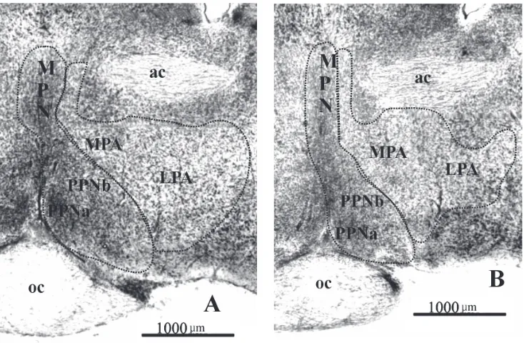
(PPN) closely adjoins the median preoptic nucleus (MPN), and these nuclei cannot be distinguished from each other, so in the morphometric analysis they were also considered as one complex — PPN-MPN. The bor-ders of the structures are shown in Figure 1. After outlining the MPA-LPA and the PPN-MPN boundaries their areas were calculated using a computer equipped with Multi-Scan 8.2 software.
Numerical density. The number of neurons in 1 mm3 was estimated according to the optical
dissec-tor method described by West and Gundersen [40]. Total number of neurons. The total number of neurons in the morphometrically examined POA parts was calculated according to the formula de-scribed by West and Gundersen [40]. The density and numbers of cells in the manuscript and in the graphs are presented in thousands.
N0 = V0 ¥ Nmm3
V0 — volume of structure; Nmm3 — numerical
densi-ty of structure.
Morphometric parameters of neurons. The analysis of neuronal parameters was carried out with a 40¥ objective lens, and every second section was analysed for each individual: 5 females and 5 males. For each of the 10 individuals, more than 2000 neu-rons were measured. The first section within a
se-ries was selected randomly. Perikarya with distinct nuclei were outlined digitally and measured using a calibrated system and image-analyzing Multi-Scan 8.2 software. The following neuronal parameters were measured: area of soma, soma circumference, soma length (long axis), and soma width (short axis).
Statistical analysis
The data were analysed by using Statistica 6.0 software. The data are presented as mean value ± ± standard error of the mean. Significant differences between the sexes were determined using a one-way analysis of variance. The comparison of two means was made using Student’s t- test. The statis-tical significance level was set at p < 0.05.
RESULTS
The preoptic area of the guinea pig is situated in front of the hypothalamus, bilaterally from the su-praoptic recess of the third ventricle, and reaches the anterior hypothalamus. It consists of four parts: the medial preoptic area, the lateral preoptic area, the median preoptic nucleus, and the periventricular pre-optic nucleus (Figs. 1, 2).
The most rostral part of the POA is made up of the MPA, the anterior pole of which appears in front of the decussation of the optic nerves. Just behind
it is the lateral preoptic area. The similar cell struc-ture of the LPA and the adjoining part of MPA, as well as their loose arrangement, cause these areas to be indistinctly separated from each other. The areas constitute quite extensive cell groups that are arranged parallel to the anterior commissure, which, together with the nucleus of stria termina-lis, create their dorsal border. Medially, the MPA is bound in front by the diagonal band of Broca, whereas in the middle and posterior sections it is bound by the PPN and MPN. From the lateral side, the MPA is restricted by the LPA, whereas the lat-eral border of the LPA is adjacent to the globus pallidus and substantia innominata. Ventrally, the areas are limited by the basal nucleus of Meynert. Small groups of cells — the posterior poles of the MPA and LPA — pass into the anterior and lateral hypothalamic area, respectively, without any dis-tinct border on the level of the anterior sector of the supraoptic nucleus.
The PPN and MPN appear slightly behind the LPA anterior pole and are small clusters of tightly packed cells. The PPN is located ventrally to the MPA and laterally to the supraoptic recess, whereas the MPN is an unpaired band of grey matter placed in the
mid-division is more distinct in the female than in the male. The median preoptic nucleus forms a kind of PPN continuation in the dorsal direction. From the ven-tral side the MPN adjoins the PPN, from the lateral it is encircled by the MPA, and dorsally it reaches the fornix. Posteriorly, an elongated stripe of cells creating the MPN lengthens considerably in the dor-sal direction where it connects the columns of the fornix. More caudally, the MPN is divided by the commissura anterior into two parts: dorsal and ven-tral. The dorsal part has the form of a triangle the base of which is located above and parallel to the commissura anterior, and its sides are bounded by the columns of the fornix. The ventral part has the form of a vertical strip, lying under the commissura anterior, which widens significantly ventrolaterally so that the cells of the MPN enclose the dorsal and lateral borders of the PPN. The similar cell structures of the MPN and PPN, as well as their adjacent loca-tion, cause the border between these nuclei to be indistinct. In the caudal sections the PPN and MPN decrease and are separated from each other. The MPN posterior pole forms the posterior pole of the POA and disappears at the anterior level of the periventricular nucleus of the hypothalamus.
Morphometric analysis
Volume. The mean overall volume of the whole preoptic area for males was 0.85 ± 0.02 mm3
com-pared to the mean POA volume in females of 0.79 ± ± 0.02 mm3
. The results of volumetric analysis of the individual POA parts are shown in Figures 3 and 4. The analysis of variance (ANOVA) indicated that the mean volume occupied by the MPA-LPA (Fig. 3) was not statistically different between males and females (p > 0.05) whereas the PPN-MPN mean vol-ume (Fig. 4) was significantly larger in males than in females (p < 0.05).
Density of neurons. The mean number of neu-rons in 1 mm3 in the whole POA for males was 124 ±
± 2.9 compared to the mean number of 98 ± 2.3 in females. In both males and females the mean
ron density was higher in the PPN-MPN than in the MPA-LPA (Figs. 5, 6). Moreover, the numerical den-sity was sex-dependent: neurons within the MPA--LPA as well as in the PPN-MPN (Fig. 5, 6) were sig-nificantly more densely packed in males than in fe-males (p < 0.05).
Total number of neurons. The sex differenc-es observed in the numerical density and in the volume correspond to the sex differences in the total number of POA neurons. The mean total neu-ron number of the whole POA in males was 87 ± ± 3.2 compared to 60 ± 1.6 in females. Thus, in the whole male POA there were approximately 31% more neurons than in the female POA. The statis-tical analysis indicated that the mean total neu-ron number of MPA-LPA (Fig. 7), as well as of the PPN-MPN (Fig. 8), was significantly larger in males than in females (p < 0.05).
Area of neuronal cell bodies. The morphomet-ric parameters of neurons for the individual POA
parts are shown in Table 1. In both sexes the mean neuronal area of the MPA-LPA was larger than the area of the PPN-MPN neurons. The statistical analy-sis indicated that the neuronal area of the two exa-mined parts differed significantly (p < 0.05). How-ever, there was no significant sex difference in the neuronal area in the MPA-LPA (p > 0.56) or in the PPN-MPN (p > 0.32).
Neuronal body circumference. The mean neu-ron circumference of the MPA-LPA was larger than the parameter of PPN-MPN neurons in both males and females (Table 1). The two parts of the POA differed significantly in terms of this parameter (p < 0.05). However, statistical analysis demonstrat-ed no significant differences between males and fe-males in the neuron circumference in the MPA-LPA (p > 0.60) or in the PPN-MPN (p > 0.35).
Neuronal body length. The mean neuron length of the MPA-LPA was always larger than the mean neuron length of the PPN-MPN in both sexes (Table 1).
Figure 3. Mean overall volume (± SEM) of the medial and lateral preoptic area (MPA-LPA).
Figure 4. Mean overall volume (± SEM) of the periventricular and median preoptic nuclei (PPN-MPN) in the male and female guinea pig; *p< 0.05, in comparison to the female group.
Figure 5. Mean estimated numerical density ± SEM ¥ 103 within
the medial and lateral preoptic area (MPA-LPA) in the male and female guinea pig; *p < 0.05, in comparison to the female group.
Figure 6. Mean estimated numerical density ± SEM ¥ 103 within
The length of the MPA-LPA neurons differed in a statistically significant way from the length of PPN-MPN neurons (p < 0.05). The neuron length was not sexually differentiated in the MPA-LPA (p > 0.64) or in the PPN-MPN (p > 0.50).
Neuronal body width. The mean width of the MPA-LPA soma was always larger than that of the soma of PPN-MPN neurons (Table 1) in both males
in other rodents [4–6, 15], insectivores [31], and sheep [30]. Even though some authors present oth-er POA divisions, it is possible to carry out a com-parative analysis. For example, Broadwell and Bleier [6] distinguished the triangular nucleus of the mouse that probably corresponds to the dorsal part of the median preoptic nucleus of the guinea pig. On the other hand, the medial preoptic nucleus of the rat and mouse [5, 6] seems to correspond to part of the guinea pig MPA (present study). The PPN nucleus of the guinea pig in the middle and posterior sectors contains two subgroups: the medial PPNa and the lateral PPNb. The PPNa, which is more intensely stained and more densely packed in the female guin-ea pig, appguin-ears to correspond to the MPNa [9] and the medial preoptic nucleus of the guinea pig [4]. According to Bleier et al. [4], the medial preoptic nu-cleus of the female guinea pig, just as the PPNa in our study, is also more tightly packed in the female than in the same region of the male.
Our study is the first concerning the morphome-try of the whole preoptic area in the guinea pig. Although a few morphometric reports of guinea pig POA providing some volumetric measurements are available, they concern only some nuclei, or even subnuclei, usually showing sexual dimorphism.
The volumetric analysis of the guinea pig POA revealed that the PPN-MPN complex differed signif-icantly between the male and female, whereas the volume of the MPA-LPA did not show sexual differ-ences in this parameter. In our study we did not
dis-Figure 8. Mean estimated total number of neurons ± SEM ¥ 103
within the periventricular and median preoptic nuclei (PPN-MPN) in the male and female guinea pig; *p < 0.05, in comparison to the female group.
Table 1. Mean estimated parameters (± SEM) of neurons of the medial and lateral preoptic area (MPA-LPA) and the
periventricular and median preoptic nuclei (PPN-MPN) in the male and female guinea pig
Nucleus Sex Neuron area Neuron circumference Neuron length Neuron width
[µm²] [µm] [µm] [µm]
MPA-LPA Male 106.91 ± 2.41 41.00± 0.65 15.08 ± 0.31 9.97 ± 0.12
Female 110.00 ± 3.25 41.60 ± 0.72 15.00 ± 0.26 10.27 ± 0.16
PPN-MPN Male 76.59 ± 1.83 34.25 ± 0.55 11.93 ± 0.21 8.75 ± 0.09
Female 80.02 ± 1.96 35.04 ± 0.5 12.17 ± 0.19 9.01 ± 0.10 Figure 7. Mean estimated total number of neurons ± SEM ¥ 103
tinguish a group of cells corresponding to the sexu-ally dimorphic nucleus of the preoptic area (SDN--POA), which was described by Gorski and Harlan [18] in the rat as several times larger in males than in females. The cytoarchitectonic study of the sexu-ally dimorphic nuclear complex of the medial pre-optic-anterior hypothalamic area (SDNC-MPAH) in rodents (guinea pig, rat, hamster, mouse) showed that the entire configuration was similar in size and contours in the two sexes, but that the dimorphism of the region was related to patterns of cell distri-bution and density. The medial preoptic nucleus was larger and, except in the mouse, appeared to have higher cellular density in females than in males [4]. According to Hines et al. [20], the darkly stain-ing portion of the medial preoptic area of the guin-ea pig is approximately four times larger in the male than in the female. However, Byne, and Bleier [9] observed sex differences in two components of the medial preoptic nucleus of the guinea pig. The an-teriorly placed subnucleus corresponding to the SDN--POA of the rat [18] was twice as large in females as in males, and a centrally placed subnucleus had about ten-times greater volume in males than in females. On the other hand, Bleier et al. [4] identi-fied a group of cells corresponding to the SDN-POA described by Gorski and Harlan [18] in the guinea pig only in the male medial preoptic nucleus while in the female such a cell group could not be distin-guished. However, in the study of the guinea pig preoptic region [9] a cell subgroup that resembles the SDN-POA of the rat [18] was observed in 9 out of 20 females. In the study by Hines et al. [20], a group of cells corresponding to the rat SDN-POA was distinguished in the male and female guinea pig. It should be noted, however, that in the studies of Byne and Bleier [9] and Hines et al. [20] an ante-rior hypothalamic nucleus showing sexual differenc-es has been ddifferenc-escribed. In addition, such discrepan-cies between the observations may result from the differences in technical or histological procedures [4, 9, 20]. Apart from rodents, sexual dimorphism relating to the volume of the POA nuclei has also been reported in a number of other species, includ-ing the sheep [32], ferret [12], rhesus monkey [8], and humans [1, 25, 36].
The ovine sexually dimorphic nucleus (oSDN), which is a part of the medial preoptic area/anterior hypothalamus, is three times larger in volume in rams than in ewes [32]. In addition to the sex differ-ence, the study of Roselli et al. [32] revealed that the oSDN is two-times larger in female-oriented rams
than in male-oriented rams. Because the preoptic area is known to control the expression of the male sexual behavior such findings may suggest that naturally occurring variations in sexual partner preferences may be related to differences in brain anatomy [32].
Like in rodents, quantitative studies of the hu-man preoptic region have also produced discrepant results. Swaab and Flier’s [36] analysis of the SDN revealed that the nucleus is larger in men than in women. Allen et al. [1] did not confirm these find-ings. Nevertheless, they found two other nuclei, the interstitial nuclei of the anterior hypothalamus INAH-2 and INAH-3, that were larger in males than in fe-males. In INAH-1, which corresponds to the SDN of Swaab and Fliers [36], and INAH-4 Allen et al. [1] found no sexual dimorphism. The sex-related differ-ence in the volume of the human INAH-3 was con-firmed by LeVay [25] and Byne et al. [10]. They stat-ed that the human INAH-3 occupistat-ed a greater vol-ume in males than in females. However, they did not find any sex differences in other parts of INAH. According to Allen et al. [1] and Byne et al. [10], such discrepancies among the nuclei of the preop-tic region may be attributed to methodological dif-ferences. For example, studies that detected no sex-ual dimorphism in INAH-1 [1, 25] were conducted with the use of much thicker sections than those of Swaab and Fliers [36] and Swaab and Hofman [37]. Larger volume of INAH-3 in men [10] is associat-ed with an increase in the number of neurons with-in this nucleus, but not with sexual differences with-in neuron size or density. Our results support these findings to some extent. As regards the size of neu-rons, we agree with Byne et al. [10], as neither in the MPA-LPA nor in the PPN-MPN were sex differ-ences in any parameter describing the size of neu-rons observed, as in the POA neuneu-rons of the ham-ster [19]. However, Brown et al. [7] found that POA neurons located beneath the anterior commissure were significantly larger in females than in males of the 129SvEv strain mouse, but not in other exam-ined strains. Furthermore, cells from another group of the POA were larger in males than in females of the C57BL/6J and SF-1 gene-disrupted wild-types.
Taking into consideration the number of neurons in the human INAH-3, we agree with Byne et al. [10] that a higher number of neurons occurs in the male nucleus. Contrary to their findings, however, we found that the neuronal density differed between sexes, and there were more neurons in 1 mm3
thalamic area on day E41, whereas on day E33 the density of cells in both sexes was similar. Our find-ings indicating that the total number of neurons was higher in the male preoptic area are similar to the results of studies on different nuclei within the pre-optic region of the rat [18, 27], gerbil [22], hamster [19], and humans [36]. In hamster MPN [19], the same as in the MPA-LPA of the guinea pig, sex dif-ferences in the neuron number are compensated for by sex differences in cell density, although the vol-ume is not sexually dimorphic.
According to Swaab and Hofman [37] and Swaab et al. [35], sex differences concerning the neuron number may vary in different stages of human life, and even in adulthood their magnitude does not remain constant. At the time of birth, the total cell number of the SDN-POA is similar in boys as it is in girls and increases equally in both sexes until the age of four years. From that age, the cell number starts to decrease in girls. In boys, the cell number of the SDN-POA remains stable until approximately 50 years of age, when it starts to decrease, which results in much less pronounced sex difference in the cell number in this period. In women, the sec-ond phase of marked cell loss takes place after the age of 70 [37]. Beck et al. [3] suggest that the struc-ture of limbic brain regions is dynamic in adulthood and the process of neuronal aging and changes in sex hormone levels during aging seem to be instru-mental in the changes occurring in dimorphic brain regions [34]. The latest study of the adult rat indi-cates considerable plasticity of the brain dimorphic regions in which circulating androgens are required to maintain soma size, but not regional volume in males. However, ovarian steroids maintain both soma size and regional volume in females [16].
The study carried out on the guinea pig confirms that the preoptic area is a dimorphic structure, al-though our attention was not focused on the di-morphic areas of the POA. The guinea pig, as a pre-cocial animal [39], may provide a better model of particular developmental and neuroendocrine
pro-REFERENCES
1. Allen LS, Hines M, Shryne JE, Gorski R (1989) Two sexual-ly dimorphic cell groups in the human brain. J Neuros-ci, 9: 497–506.
2. Anderson CH, Shen CL (1980) Efferents of the medial preoptic area in the guinea pig: an autoradiographic study. Brain Res Bull, 5: 257–265.
3. Beck LA, O’Bryant EL, Wade JS (2008) Sex and seasonal differences in morphology of limbic forebrain nuclei in the green anole lizard. Brain Res, 1227: 68–75. 4. Bleier R, Byne W, Siggelkow I (1982) Cytoarchitectonic
sexual dimorphisms of the medial preoptic and ante-rior hypothalamic areas in guinea pig, rat, hamster, and mouse. J Comp Neurol, 212: 118–130.
5. Bleier R, Cohn P, Siggelkow IR (1979) A cytoarchitectonic atlas of the hypothalamus and hypothalamic third ven-tricle of the rat. In: Morgane P, Panksepp J eds. Hand-book of the hypothalamus. Vol. 1. Anatomy of the hypo-thalamus. Marcel Deker, New York, Basel, pp. 137–219. 6. Broadwell RD, Bleier R (1976) A cytoarchitectonic atlas of the mouse hypothalamus. J Comp Neurol, 167: 315–340. 7. Brown AE, Mani S, Tobet SA (1999) The preoptic area/ /anterior hypothalamus of different strains of mice: sex dif-ferences and development. Dev Brain Res, 115: 171–182. 8. Byne W (1998) The medial preoptic and anterior hypo-thalamic regions of the rhesus monkey: cytoarchitec-tonic comparison with the human and evidence for sexual dimorphism. Brain Res, 793: 346–350. 9. Byne W, Bleier R (1987) Medial preoptic sexual
dimor-phisms in the guinea pig. I. An investigation of their hormonal dependence. J Neurosci, 7: 2688–2696. 10. Byne W, Lasco MS, Kemether E, Shinwari A, Edgar MA,
Morgello S, Jones LB, Tobet S (2000) The interstitial nuclei of the human anterior hypothalamus: an inves-tigation of sexual variation in volume and cell size, number and density. Brain Res, 856: 254–258. 11. Byne W, Tobet S, Mattiace LA, Lasco MS, Kemether E,
Edgar MA, Morgello S, Buchsbaum MS, Jones LB (2001) The interstitial nuclei of the human anterior hypotha-lamus: an investigation of variation with sex, sexual orientation, and HIV status. Horm Behav, 40: 86–92. 12. Cherry JA, Basham ME, Weaver CE, Krohmer RW, Baum MJ
(1990) Ontogeny of the sexually dimorphic male nu-cleus in the preoptic/anterior hypothalamus of ferrets and its manipulation by gonadal steroids. J Neurobiol, 21: 844–857.
14. deVito JL, Graham J, Sackett GP (1989) Volumetric growth of the major brain divisions in fetal Macaca nemestrina. J Hirnforsch, 30: 479–487.
15. Diepen R (1962) Der Hypothalamus. In: Handbuch der mikroskopischen Anatomie des Menschen. Nervensys-teme. 7 Teil. Springer, Berlin-Göttingen-Heidelberg. 16. Dugger BN, Morris JA, Jordan CL, Breedlove SM (2008)
Gonadal steroids regulate neural plasticity in the sexu-ally dimorphic nucleus of the preoptic area of adult male and female rats. Neuroendocrinology, 88: 17–24. 17. Gorski RA, Gordon JH, Shryne JE, Southam AM (1978) Evidence for a morphological sex difference within the medial preoptic area of the rat brain. Brain Res, 148: 333–346.
18. Gorski RA, Harlan RE (1980) Evidence for the existence of a sexually dimorphic nucleus in the preoptic area of the rat. J Comp Neurol, 193: 529–539.
19. Govek EK, Wang J, Swann JM (2003) Sex differences in the magnocellular subdivision of the medial preoptic nucleus in Syrian hamsters. Neuroscience, 116: 593–598. 20. Hines M, Davis FC, Coquelin A, Goy RW, Gorski RA (1985) Sexually dimorphic regions in the medial pre-optic area and the bed nucleus of the stria terminalis of the guinea pig brain: a description and an investiga-tion of their relainvestiga-tionship to gonadal steroids in adult-hood. J Neurosci, 5: 40–47.
21. Hoffman GE, Dohanics J, Watson RE, Wiegand SJ (1996) The hypothalamic ventromedial nucleus sends a me-tenkephalin projection to the preoptic area’s periventri-cular zone in the female rat. Mol Brain Res, 36: 201–210. 22. Holman SD, Collado P, Skepper JN, Rice A (1996) Postna-tal development of a sexually dimorphic, hypothalamic nucleus in gerbils: a stereological study of neuronal num-ber and apoptosis. J Comp Neurol, 376: 315–325. 23. Hurtazo HA, Parades RG, Agmo A (2008) Inactivation
of the medial preoptic area/anterior hypothalamus by lidocaine reduces male sexual behavior and sexual in-centive motivation in male rats. Neuroscience, 152: 331–337.
24. Klaric JS, Hendricks SE (1986) Effects of two-stage le-sions of the medial preoptic area on sexual behavior of male rats. Physiol Behav, 37: 539–542.
25. LeVay S (1991) A difference in hypothalamic structure between heterosexual and homosexual men. Science, 253: 1034–1037.
26. Liu YC, Salamone JD, Sachs BD (1997) Lesions in medial preoptic area and bed nucleus of stria terminalis: differ-ential effects on copulatory behavior and noncontact erection in male rats. J Neurosci, 17: 5245–5253.
27. Madeira MD, Leal S, Paula-Barbosa MM (1999) Stereolo-gical evaluation and Golgi study of the sexual dimorphisms in the volume, cell numbers, and cell size in the medial preoptic nucleus of the rat. J Neurocytol, 28: 131–148. 28. Pompolo S, Ischenko O, Pereira A, Iqbal J, Clarke IJ
(2005) Evidence that projections from the bed nucleus of the stria terminalis and from the lateral and medial regions of the preoptic area provide input to gonado-tropin releasing hormone (GNRH) neurons in the fe-male sheep brain. Neuroscience, 132: 421–436. 29. Raisman G, Field PM (1971) Sexual dimorphism in the
preoptic area of the rat. Science, 173: 731–733. 30. Rajtová V (1979) The preoptic region of the merino
sheep. Folia Morphol, 27: 329–333.
31. Robak A, Szteyn S (1989) The topography and cytoar-chitectonics of the nuclei of supraoptic and preoptic areas in insectivores. Folia Morphol, 48: 201–218. 32. Roselli CE, Larkin K, Resko JA, Stelleflug JN, Stormshak F
(2004) The volume of a sexually dimorphic nucleus in the ovine medial preoptic area/anterior hypothalamus varies with sexual partner preference. Endocrinology, 145: 478–483.
33. Sturgis JD, Bridges RS (1997) N-methyl-DL-aspartic acid lesions of the medial preoptic area distrupt on-going parental behavior in male rats. Physiol Behav, 62: 305–310.
34. Swaab DF, Chung WCJ, Kruijver FPM (2001) Structural and functional sex differences in the human hypotha-lamus. Horm Behav, 40: 93–98.
35. Swaab DF, Chung WCJ, Kruijver FPM, Hofman MA, Hestiantoro A (2003) Sex differences in the hypothala-mus in the different stages of human life. Neurobiol Aging, 24: 1–16.
36. Swaab DF, Fliers E (1985) A sexually dimorphic nucleus in the human brain. Science, 228: 1112–1115. 37. Swaab DF, Hofman MA (1988) Sexual differentiation
of the human hypothalamus: ontogeny of the sexually dimorphic nucleus of the preoptic area. Brain Res Dev Brain Res, 44: 314–318.
38. Vasey PL, Pfaus JG (2005) A sexually dimorphic hypo-thalamic nucleus in a macaque species with frequent female-female mounting and same-sex sexual partner preference. Behav Brain Res, 157: 265–272.
39. Weaver LT, Landymore-Lim AE, Hudson GJ (1988) The guinea pig as a model for the study of the effects of milk on growth and development. Growth Dev Aging, 52: 91–96.