Sikander et al. World Journal of Pharmaceutical and Life Sciences
LACTATE DEHYDROGENASE: STRUCTURE, FUNCTIONS AND ITS SIGNIFICANCE
Rabia Iqbal Khan and Sikander Ali*
Institute of Industrial Biotechnology (IIB), Government College University, Lahore.
Article Received on 15/12/2018 Article Revised on 06/01/2019 Article Accepted on 27/01/2019
INTRODUCTION
Lactate dehydrogenase (E.C. 1.1.1.27) is an oligomeric enzyme of class oxidoreductase (Wong, 1998). Its systematic name is L-Lactate: NAD+ Oxidoreductase and is distributed in all human tissues. It catalyzes the conversion of Lactate to pyruvate with production of reduced coenzyme, Nicotinamide adenine dinucleotide. Glycolysis produced oxidized form of NAD which later on became reduced along with lactate to pyruvate conversion in the presence of Lactate dehydrogenase to be used again by glycolysis (Di Stefano et al., 2016; Wong, 1998).
It has been long known that end product of anaerobic glycolysis is lactic acid. In 1932, it was first obtained that lactate to pyruvate conversion could be catalyzed by cell free extracts. The above reaction was discovered for the first time by Warburg and his colleagues and they associated it to be chemical properties of a coenzyme. In 1950 it was demonstrated, that proton is released in this reaction, by Racker. In 1940, Straub purified the lactate dehydrogenase for the first time. Kubowitz and Ott reported the first micrograph of lactate dehydrogenase crystals. Enzyme is trimer or tetramer was stated by Markert and Moller in their classic paper. Different scientists had exhibited LDH molecules in several species. Market and Moller combine this work with their own work and finally showed that lactate dehydrogenase was a tetrameric molecule. In 1964, x-ray diffraction
results, of pig lactate dehydrogenase, were shown for the first time according to which 140,000 MW of tetramer LDH was confirmed. In 1970, complete three dimensional structure of LDH was reported (Holbrook et al., 1975).
Structure of lactate dehydrogenase
Lactate dehydrogenase is an oligomeric enzyme as it is composed of two polypeptide chains which join together to form a functional unit. Homo-tetramers and hetero-tetramers of human LDH is composed of two polypeptide chains encoded by two genes LDH-A and LDH-B which join together to form 5 Isoenzymes of LDH (Dempster et al., 2014). Apo structure of human LDH- A was reported in 2014 in 2.1 A ° resolution. In this active site of human LDH-A was studied in the absence of any inhibitor or cofactor at the binding site. In free form, it was shown that N- terminal tail, active site loop and number of regions have increased flexibility than bound enzyme. In comparison to bound form, Apo structure has specific conformational changes in active site loop constituting 99- 110 residues. Apo-structure of human LDH-A is shown in Figure 1 taken from PDB. This loop is also called substrate specificity loop (Dempster et al., 2014).
World Journal of Pharmaceutical and Life Sciences
WJPLS
www.wjpls.org SJIF Impact Factor: 5.008
*Corresponding Author: Sikander Ali
Institute of Industrial Biotechnology (IIB), Government College University, Lahore.
ABSTRACT
Lactate dehydrogenase (E.C. 1.1.1.27) is an oligomeric enzyme of class oxidoreductase with systematic name, L-Lactate: NAD+ Oxidoreductase. It catalyzes the conversion of Lactate to pyruvate with production of reduced coenzyme, Nicotinamide adenine dinucleotide. It is a tetramer of 140,000 MW and its three dimensional structure and active site has been described. Its isoenzymes LDH-1, LDH-2, LDH-3, LDH-4, LDH-5 and LDH-C has been distributed in different human tissues which include skeletal muscle, heart, kidney, liver, spleen, lungs, erythrocytes, platelets, white blood cells and brain. Oncogenic signals has increased the expression of ldh-a genes in cancerous cells. Lactate dehydrogenase inhibition can be used as an anticancer therapy. LDH is an important diagnostic marker for hemolytic anemia and a couple of other diseases. It has its applications in lactate biosensors as well.
Figure 1: Apo structure of LDH-A taken from PDB reference 4l4r.
In this, the loop is in open conformation and its electron density is well ordered. Type II β- turn conformation is stabilized due to salt bridge interaction between Glu103 and Arg111. Glu103 interacts with Asn107 side chains via hydrogen bonded interactions. It also interacts with the amine of main chain of Gln100. Another important interaction i.e. Leu108 is ordered against Gln99 and Ala97 side chains. In the crystal lattice interaction of neighboring monomers also contribute towards stabilization of such conformation. Arg105 and Arg98 are not well defined and contribute towards flexibility of Apo enzyme while Arg168, His192 (catalytic) and Thr247 have well defined electron density. Contrary to this, on binding to substrate and cofactor active site loop gains a closed conformation over the active site and formed a desolated ternary complex. For the catalytic function of this enzyme firstly NADH binds to cofactor binding site and then pyruvate binds to substrate binding site followed by reduction of pyruvate and the coenzyme (Dempster et al., 2014).
Function of LDH
In aerobic respiration, glucose is converted into pyruvate by glycolysis. Pyruvate enter into citric acid cycle after its conversion into acetyl co-A. In oxidative phosphorylation, 36 net adenosine triphosphates per molecule of glucose are generated. In the absence of oxygen, cell cannot generate ATP by oxidative phosphorylation. In such cases cell depends upon glycolysis to meet the ATP demand. Glycolysis generates 2 ATP per molecule of glucose. To carry out sixth step of glycolysis glyceraldehyde phosphate dehydrogenase needs NAD+ to convert glyceraldehyde 3- phosphate into 1,3- Bisphosphoglycerate while NAD+ is reduced into NADH. This NAD+ is regenerated by oxidative phosphorylation by electron transport chain but in the absence of oxygen it is regenerated from NADH
by Lactate dehydrogenase along with conversion of pyruvate into lactate. This process is termed as anaerobic glycolysis (Adeva-Andany et al., 2014). Aerobic and anaerobic glycolysis is shown in Figure 2. Anaerobic glycolysis is 100 times faster than aerobic glycolysis. Faster rate of anaerobic glycolysis is to meet the energy requirements but it uses large amount of glucose (Valvona et al., 2015).
Stereo chemical specificity of the enzyme is for L- isomer, it cannot act upon D- isomer (Elliot et al., 1962). Binding affinity of NADH is 400 times stronger than NAD+ while 50 times stronger than NADPH. NAD+ is the coenzyme but NADP+ sometimes does serve as coenzyme because specificity of enzyme is for Nicotinamide ring’s A side (Wong and Wong, 1983). On binding of substrate and coenzyme, conformational changes took place in the enzyme. Substrate binds to enzyme in such a way to react with imidazole of his195 and nicotinamide of coenzyme. This carries out acid base catalysis as it removes proton from lactate and donate it to pyruvate in the backward reaction shown in Fig 1 (Holbrook et al., 1975).
Lactate dehydrogenase can be inhibited by a variety of inhibitors. It can be inhibited by lactate and pyruvate both. LDH also exhibit substrate inhibition mechanism of reversible inhibition but this can be reduced by increasing pH (Everse and Kaplan, 1973: Holbrook et al., 1975). Mercuric ion and p- chloromercuricbenzoate are inhibitors of LDH. Some other reagents like malonate, tartronate, borate and oxalate compete lactate for binding to the enzyme while oxamate competes pyruvate for binding thus shown competitive inhibition of LDH (Everse and Kaplan, 1973; Holbrook et al.,
Figure 2: Anaerobic glycolysis: Lactate dehydrogenase regenerates NAD+ and form lactate as by product.
Isoenzymes and their distribution in human tissues
Lactate dehydrogenase (hLDH) is a tetramer composed of two subunits M (muscle) and H (heart) which are encoded by two genes LDH-A and LDH-B, respectively. These two subunits give rise to five Isoenzymes hLDH-1 (H4), hLDH-2 (MB3), HLDH-3 (M2 H2), HLDH-4
(M3H), HLDH-5 (M4). In addition to this, another
isoenzyme is also discovered which is found in
spermatozoa named HLDHC4 (Di Stefano et al., 2016).
LDH-A and LDH-B subunits have 75% sequence identity and are similar in size. Both of these subunits have different catalytic properties as M catalyzes pyruvate to lactate conversion while H catalyzes lactate to pyruvate. Isoenzymes and their subunits are shown in Figure 3.
Figure 3: Isoenzymes of Lactate Dehydrogenase.
Glucose
Fructose 1, 6-bisphosphate
2 (Glyceraldehyde- 3- phosphate
2 (1, 3- Bisphosphoglycerate) Glyceraldehyde phosphate
dehydrogenase
2 (ATP)
2 (NAD+)
2 (NADH)
2 (Lactate)
2 (Pyruvate) LDH
LDH is found in almost all cells of the body, invariably in cytoplasm. It is not found in mitochondrion. Highest concentration of LDH is found in skeletal muscle, heart, kidney, liver, spleen, lungs, erythrocytes, platelets, white blood cells and brain. Different Isoenzymes are present in different tissues in different proportions (Wong, 1998). Different scientists displayed different quantitative values regarding presence of a particular isoenzyme in a particular tissue. These differences are because of differences in techniques, variations in treatment of specimen and loss of slow moving labile Isoenzymes.
In addition to these post mortem autolysis and contamination of tissues with blood. In spite of these problems, there is agreement that LDH-1 and LDH-2 predominate in erythrocytes, heart and kidney while LDH-$ and LDH-5 are present in large amounts in liver and skeletal muscle. LDH-3 predominates in spleen, pancreas, lymph nodes and adrenals. Earlier on absence of LDH-5 in erythrocyte is found but it was against the random association of enzyme subunit (Wilkinson, 1970). Later on, it was proved that erythrocytes loses LDH-5 with aging as it is less stable and erythrocytes have no mechanism of protein synthesis so it cannot be replaced (Valvona, et al., 2015). Some amount of isoenzyme is leaked into the circulatory blood. Electrophoretic analysis of serum shows that LDH-2 is present there in large amount (Lott and Turner, 1982).
Mobility of LDH isoenzymes is dependent on their B- subunit content as displayed by fastest moving LDH-1 and slowest moving LDH-5. Different kinetic properties are displayed by LDH-A and LDH-B subunits. Higher pyruvate concentrations are required by LDH-A to reach to its maximum activity. Km for LDH-A is 158 µM while
for LDH-B it is 58 µM for pyruvate (Eszes et al., 1996; Hewitt et al., 1999). In addition to this, LDH-B is inhibited by the substrate concentration required by LDH-A to show its maximum activity. Ki of LDH-A for
pyruvate is 3900 µM while for LDH-B it is 770 µM (Eszes et al., 1996; Hewitt et al., 1999). These observations compelled Kaplan and colleagues in 1975 to propose a hypothesis regarding physiological functions of LDH. According to his aerobic- anaerobic theory, LDH-A is present in oxygen limiting tissues while LDH-B is present in tissues with sufficient oxygen supply. Later on, further research has questioned this theory because of the experimental evidence of varying isoenzyme pattern in different tissues of the body (Fiume
et al., 2014).
Lactate dehydrogenase and cancer
In 1926, Warburg had observed that lactate was produced by cancerous cells in high amounts even during sufficient oxygen supply (Warburg, 1926). He named this process aerobic glycolysis especially attributed to impaired cell respiration of neoplastic cells. He proposed it be the mechanism with which neoplastic cells fulfill their high energy requirements by increasing the amount
of glucose uptake. On the basis of this observation, occult tumor masses had been detected by using F- deoxyglucose as a tracer in position emission tomography (Gillies et al., 2008). Impaired cell respiration to be the primary cause of cancer was also proposed by Warburg but later on his theory was questioned because it failed to explain the molecular mechanism of uncontrolled cell growth.
Later on with the advent of modern molecular techniques and discovery of molecular mechanism behind uncontrolled proliferation of cancerous cells, this theory had lost its authenticity. Not only this, the concept that cancer is a metabolic disease was replaced by cancer as a genetic disease. It was only the last decade, that the need to study metabolism of cancer cell was revived. According to the results of molecular biology experiments it was demonstrated that metabolic changes in cancerous cells are due to abnormalities in oncogenes and oncosuppressors (Levine and Puzio- Kuter, 2010; DeBerardinis et al., 2008; Buchakjian). Uncontrolled cell division could not be carried out without sufficient energy and building blocks. Oncogenic signals reprogrammed the catabolic metabolism of normal cells into anabolic metabolism of cancerous cells. Normally cells completely degrade glucose to carbon dioxide with the maximum generation of energy while cancerous cells need numerous building blocks for continuous division which is only possible by the alteration of catabolism to anabolism (Levine and Puzio- Kuter, 2010).
Activation of Ras, Akt, Myc and pI3K growth factor receptors (oncogenes) and inactivation of p53 (oncosuppressors) induce enhanced uptake of glucose by glycolysis. As a result of increased glycolysis levels, high levels of glycolytic intermediates gave rise to different numerous anabolic pathways which resulted in production of amino acids, lipids and nucleotide precursors (Christofk et al., 2008). Increased rate of glycolysis resulted in excess production of NADH which needs to oxidize continuously to prevent cell from oxidative damage and to maintain glycolytic flux. In cancerous cells, oncogenic signals increased the expression of ldh-a genes which resolves the problem of concomitant oxidation of NADH (Levine and Puzio- Kuter, 2010).
LDH Inhibition a Therapeutic Approach against Cancer
target ldh-a gene thus playing key role in tumor development (Levine and Puzio- Kuter, 2010; DeBerardins et al., 2008; Buchakjian Dang et al., 2009). Constant up- regulation of LDH-A levels in tumor is related to increased tumor size (Levine and Puzio- Kuter, 2010; DeBerardins et al., 2008; Buchakjian and Kornbluth, 2010; Dang et al., 2009). In vivo tumorigenesis, migration and cell growth was inhibited by siRNA or shRNA silencing of LDH-A expression (Rong et al., 2013; Yao et al., 2013; Langhammer et al.,
2011; Wang et al., 2012; Arseneault et al., 2013; Zhang
et al., 2012). While increased cleavage of PARP and caspases, decreased activation of Akt and expression of cyclin D1 (Yao et al., 2013) and Oct4 down regulation also produces the same affects (Zhang et al., 2012).
Knockdown of LDH-A had produced increased level of mitochondrial ROS (Wang et al., 2012; Arseneault et al.,
2013) which causes cell death. In addition to this cytoskeletal structures was also affected by oxidative stress conditions (Arseneault et al., 2013). Resistance of breast cancer cells against Taxol and Trastuzumab (two chemotherapeutic drugs) was proved to overcome by inhibition of LDH-A (Zhou et al., 2010; Zhao et al.,
2011). It was observed by scientists that resistant cells were more dependent upon glycolysis for their survival in comparison to sensitive parent cells. Thus therapeutic effects of Taxol and Trastuzumab were increased by combining them with LDH inhibition. Protein synthesis and proliferation in normal cells were not impaired with inhibition of LDH-A (Kim and Lee, 2007; Jeong et al.,
2006).
As far as LDH-B is concerned regarding tumor development, different scientists have found conflicting results in the same tumor. For instance, in a recent study loss of LDH-B expression is found to contribute to the development of breast cancer (Brown et al., 2013) while in another study, high level of LDH-B expression was found in aggressive forms breast cancer cells (McCleland
et al., 2012), clearly conflicting the previous one. LDH-B was proved to be an essential factor in mTOR mediated tumorigenesis (Zha et al., 2011), growth of maxillary sinus squamous carcinoma (Kinoshita et al.,
2012) and K-Ras dependent lung adenocarcinomas (McCleland et al., 2012). Claudin-1 mediated invasiveness of hepatocellular carcinoma cells was increased by reduced expression of LDH-B (Kim et al.,
2011). Though conflicting results regarding LDH-B role in cancer development is evidently leading towards missing links in this mechanism but encouraging evidences in favor of inhibiting LDH-A to control uncontrolled proliferation has lead this as a therapeutic approach against cancer cells.
LDH as diagnostic marker Hemolytic Marker
Erythrocyte destruction referred as hemolysis is the symptom of a number of physiological and pathological conditions of the body. The conditions in which
erythrocytes losses their half-life, due to autoimmune, chemical or mechanical problems of the body, are also referred as hemolysis.as a result of RBC destruction hemoglobin level falls below the normal level, such condition is referred as hemolytic anemia. Laboratory markers and treatment strategies of this type of anemia are different from anemia caused by impairment of bone marrow erythrocyte production (Barcellini and Fattizzo, 2015).
Direct antiglobulin test (DAT) is vital for diagnosis of acquired hemolytic anemia. DAT can also distinguish between different forms of autoimmune hemolytic anemia (AIHA); warm form, cold agglutinin disease (CAD) and mixed form. Almost 70 % cases of AIHA are warm forms and they are DAT positive for IgG or IgG + C. almost 20 % cases are of CAD which depict DAT positive for C while less than 10 % are of mixed forms which show DAT positive for IgG and C. In mixed form warm antibodies and cold agglutinin coexists (Barcellini and Fattizzo, 2015).
Lactate dehydrogenase is present in cytoplasm of cell of various organs and serum. LDH-1 and LDH-2 isoenzymes predominate in erythrocytes. LDH is increased slightly in extravascular form of AIHA while 4-5 fold in intravascular form than normal level. Extravascular form includes warm form of AIHA and congenital form and intravascular type includes PNH and prosthetic valve hemolysis. Table 1 shows different hemolytic diseases in which LDH can be used as hemolytic marker. At about 37 ° C LDH levels are increased in warm and CAD forms of AIHA. Complement activation is the cause of intravascular type and it is also related to thrombotic events (Barcellini et al., 2014). LDH levels are in significant increase in patients with mechanic than biologic prosthetic valve and in patients with double than single prosthetic valve replacement (Mecozzi et al., 2002). As the hemolytic rate decreases LDH levels decreases thus clearly depicting important evaluating measure of treatment of AIHA, HUS, PNH (Petz and Garratty et al., 2004; De Latour et al., 2008) and microangiopathic hemolytic anemia (Kaiser et al., 2012).
Table 1: +/++/+++ signs shows the increase in strength for using LDH as hemolytic marker in different hemolytic diseases.
Hemolytic diseases LDH as marker of hemolysis
AIHA autoimmune hemolytic
anemia +/++
Membrane/enzyme defects + CDA congenital dyserythropoietic
anemia +
PNH paroxysmal nocturnal
hemoglobinuria +++ TMA thrombotic micro
Diagnostic Markers Other Than Hemolytic Anemia
Lactate Dehydrogenase is widely distributed in different tissues so its increased levels could be increased in several other conditions. Among such conditions are cellular necrosis, myocardial infarction, heart failure, hepatitis of all types, high muscular exertion and hematologic tumors, LDH can be used as diagnostic markers in all such diseases. Vitamin B12 and folic acid deficiencies could be detected by high LDH levels due to premature death of erythrocytes and inefficient erythropoiesis. Diseases could be distinguished from one another in diagnostic methods by comparing different ratios of LDH with other enzymes in different diseases. In a study it was shown that above 22.12 ration of LDH: aspartate aminotransferase distinguishes TTP and other thrombotic microangiopathies. Such strategies are used for diagnosis before carrying out ADAMTS 13 activity test (Pourrat et al., 2015).
LDH in Lactate Biosensors
Biosensor is a detection device which monitor analyte’s concentration by interacting with biological entity, which is a part of bio sensing assembly. Biological element and transducer element are components of biosensor. Like biological systems, biosensing devices contain bioreceptors which interact with analyte of interest. The whole assembly is connected to output device called sensor which projects a measurable signal in proportion to analyte concentration. Wide range of bio elements can be used in such devices like enzymes, antibodies, nucleic acids, lectins, cell organelles etc. biosensors manipulate sensitivity and specificity of such molecules. Interaction between bio elements and analyte generates electrical, optimal and thermal signals for detection. These signals are transformed into current or voltage by specific transducer element. Sensitivity of biosensor is based on transducer while selectivity is based on bio element (Rathee et al., 2015). Basic principle of biosensors is shown in Figure 4.
Figure 4: Basic principles of Biosensors.
Working principle of biosensors involved exploitation of current which produced as a result of redox reaction reactions at electrode. Amount of current generated is in direct proportionality with concentration of analyte in sample. The current is measured by applying voltage between two types of electrodes (working and reference electrode) which gave rise to electro catalytic reactions. Enzyme involved in this mechanism have the ability to catalyze the reaction involving electro active reactant
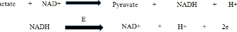
which then give rise to electro active product. This is the way analyte concentration is measured by this process. Mostly these biosensors are either based on oxidases which consume oxygen and generated hydrogen peroxide or dehydrogenases which break down substrate and reduce nicotinamide adenine dinucleotide (Mamas and Miltiades, 2002). L- Lactate biosensors based on dehydrogenases uses LDH, the reaction shown in Figure 5 occurs in these biosensors.
Figure 5: enzyme catalyzed reaction at the electrode of LDH based lactate biosensors.
LDH is widely used as bio sensing element in biosensors (Gue et al., 2002; Rahmann et al., 2009; Arnaldo et al.,
2006). Improved transducer element and increased purity of enzyme lead toward better L- Lactate biosensor. LDH uses NAD as mediator to transfer electrons between electrode and enzyme. LDH carried out conversion of lactate to pyruvate along with reduction of NAD into NADH. NADH is oxidized on the surface of electrode
friendly. It also requires no sample preparation (Rathee
et al., 2015).
CONCLUSION
LDH is an oligomeric enzyme which forms 5 isoenzymes. It plays significant role in diagnostics of different diseases, anticancer target and in biosensors. LDH plays a key role in cancer cell metabolism. Mechanism of LDH-A is somewhat clear but controversies do exist regarding LDH-B. A lot of scope exists in studying the mechanism of LDH-B in cancerous cells. Implantable lactate biosensors have great demand for checking continuous lactate concentration. Great research is needed in this regard.
REFERENCES
1. Adeva-Andany, M., López-Ojén, M., Funcasta-Calderón, R., Ameneiros-Rodríguez, E., Donapetry-García, C., Vila-Altesor, M. and Rodríguez-Seijas, J. Comprehensive review on lactate metabolism in human health. Mitochondrion, 2014;17C: 76–100. 2. Arnaldo, C., Marina, R., Alexandre, K., Denise, V.,
Lauro, T. Amperometric biosensor for lactate based on lactate dehydrogenase and Meldol a Blue co-immobilized on multi-wall carbon-nanotube.
Sensors and Actuators B: chemical-Journal, 2006; B124: 269–276.
3. Arseneault, R., Chien, A. and Newington, J.T. Attenuation of LDHA expression in cancer cells leads to redox-dependent alterations in cytoskeletal structure and cell migration. Cancer Letters, 2013, 338: 255–266.
4. Barcellini, W. and Fattizzo, B. Clinical applications of hemolytic markers in differential diagnosis of hemolytic anemia. Disease markers, 2015.
5. Barcellini, W., Fattizzo, B. and Zaninoni, A. Clinical heterogeneityand predictors of outcome in primary autoimmune hemolytic anemia: aGIMEMAstudy of 308 patients. Blood,2014; 124(19): 2930–2936.
6. Brown, N.J., Higham, S.E. and Perunovic, B. Lactate dehydrogenase-B is silenced by promoter methylation in a high frequency of human breast cancers. PLoS ONE, 2013; 8.
7. Buchakjian, M.R., and Kornbluth, S. The engine driving the ship: metabolic steering of cell proliferation and death. Nature Reviews Molecular Cell Biology, 2010; 11; 715–727.
8. Christofk, H.R., Vander Heiden, M.G. and Wu, N. Pyruvate kinase M2 is a phosphotyrosine-binding protein. Nature, 2008;452: 181–186.
9. Dang, C. V., Le, A. Gao, P. MYC-induced cancer cell energy metabolism and therapeutic opportunities. Clinical Cancer Research, 2009; 15; 6479–6483.
10. De Latour, R.P., Mary, J.Y. and Salanoubat, C. Paroxysmal nocturnal hemoglobinuria: natural history of disease subcategories. Blood, 2008; 112(8): 3099–3106.
11. DeBerardinis, R.J., Lum, J.J., Hatzivassiliou, G.The biology of cancer: metabolic reprogramming fuels cell growth and proliferation. Cell Metabolism,
2008;7: 11–20.
12. Dempster, S.S., Harper, J.E., Moses and Dreveny, I. Structural characterization of the apoform and NADH binary complex of human lactate dehydrogenase. Acta. Crystallographica, 2014;D70: 1484-1490.
13. Di Stefano, G., Manerba, M., Di Lanni, L. and Fiume. L. Lactate dehydrogenase inhibition: exploring possible applications beyond cancer treatment. Future Medical Chemistry, 2016; 8(6): 713-725.
14. Elliot, B.A., Jepson, E.M. and Wilkinson, J.H. Serum alpha-hydroxybutyrate dehydrogenase: A new test with improved specificity for myocardial lesions. Clinical Science, 1962; 23: 305-316. 15. Eszes, C. M., Sessions, R.B., Clarke, A.R. Removal
of substrate inhibition in a lactate dehydrogenase from human muscle by a single residue change.
FEBS Letters, 1966 399: 193–197.
16. Everse, J. and Kaplan. Lactate dehydrogenase. Structure and function. Advances in Enzymology, 1973; 61-133.
17. Fiume, L., Manerba, M., Vettraino, M. and Di Stefano, G. Inhibition of Lactate dehydrohenase activity as an approach to cancer therapy. Future Medicinal Chemistry, 2014; 6(4): 429-445.
18. Gillies, R.J., Robey, I. and Gatenby, R.A. Causes and consequences of increased glucose metabolism of cancers. Journal of Nuclear Medicine, 2008; 49: 24–42.
19. Gue, A.M., Tap, H., Gros, P., Maury, F. Miniaturized silicon based enzymatic biosensor: towards a generic structure and technology formulti-analytes assays. Sensors and Actuators B: chemical-Journal, 2002; B82: 227–232.
20. Hewitt, C.O., Eszes, C.M. and Sessions, R.B. A general method for relieving substrate inhibition in lactate dehydrogenases. Protein Engineering, Design and Selection, 1999; 12: 491–496.
21. Holbrook, J.J., Li1jas, A., Steindel, S.J. and Rossmann, M.G. Lactate dehydrogenase. In: The Enzymes. 3rd ed. New York, 1975; 111: 191-292. 22. Jeong, D.W., Cho, I.T. and Kim, T.S. Effects of
lactate dehydrogenase suppression and glycerol-3-phosphate dehydrogenase overexpression on cellular metabolism. Molecular and Cellular Biochemistry,
2006; 284: 1–8.
23. Kaiser, C., Gembruch, U., Janzen, V. and Gast, A-S. Thrombotic thrombocytopenic purpura. Journal of Maternal-Fetal and Neonatal Medicine, 2012; 25(10): 2138–2140.
25. Kim, S.H., and Lee, G.M. Down- regulation of lactate dehydrogenase-A by siRNA for reduced lactic acid formation of Chinese hamster ovary cells producing thrombopoietin. Applied. Microbiology and Biotechnology, 2007; 74: 152–159.
26. Langhammer, S., Najjar, M., Hess-Stumpp, H. LDH-A influences hypoxia-inducible factor 1a (HIF1 a) and is critical for growth of HT29 colon carcinoma cells in vivo. Targeted. Oncology, 2011; 6: 155–162.
27. Latner, A.L. and Skillen, A.W. Isozymes in biology and medicine. New York, Academic. M. Panteghini, R. Bonora, and F. Pagani. 1990. Evaluation of a new commercial assay kit for quantification of isoenzyme 1 in serum. Journal of clinical chemistry and Biochemistry, 1968; 28: 545-548.
28. Levine, A. J., and Puzio-Kuter, A.M. The control of the metabolic switch in cancers by oncogenes and tumor suppressor genes. Science, 2010; 330: 1340– 1344.
29. Lott, J.A. and Nemesanszky, E. Lactate dehydrogenase (LD). In: Clinical enzymology. A case-oriented approach, J.A. Lott and P.L. Wolf, Field and Richland Year Book, New York, 1986; 213-244.
30. Lott, J.A. and Turner, K., Lactate dehydrogenase in serum. In: Selected methods for the small clinical chemistry laboratory, S. Meites and W. Faulkner. (Eds) Washington, DC, American Association for Clinical Chemistry, 1982; 271.
31. Mamas, P., Miltiades, IK. Enzyme based amperometric biosensors for food analysis.
Electroanalysis, 2002; 14: 241–261.
32. McCleland, M.L., Adler, A.S. and Shang, Y. An integrated genomic screen identifies LDHB as an essential gene for triple-negative breast cancer.
Cancer Research, 2012; 72: 5812–5823.
33. McCleland, M.L., Adler, A.S., Deming, L. Lactate dehydrogenase B is required for the growth of KRAS-dependent lung adenocarcinomas. Clinical Cancer Research, 2012; 19: 773–784.
34. Mecozzi, G., Milano, A.D., De Carlo, M. Intravascular hemolysis in patients with new-generation prosthetic heart valves: a prospective study. Journal of Thoracic and Cardiovascular Surgery, 2002; 123(3): 550–556.
35. Origbinde, T.A., Wu, A.H.B., Wu, Y-S., Simmons, M.J. and Wong, S.S. Mechanism of differential inhibition of lactate dehydrogenase isoenzymes in the BMC LD 1 assay. ClinicalBiochemistry, 1992; 25: 425-429.
36. Petz, L.D. and Garratty, G., Immune Hemolytic Anemias, Churchill Livingstone, Philadelphia, Pa,
U.S.A., 2nd edition, 2004.
37. Pourrat, O., Coudroy, R. and Pierre, F. Differentiation between severe HELLP syndrome and thrombotic microangiopathy, thrombotic thrombocytopenic purpura and other imitators.
European Journal of Obstetrics & Gynecology and ReproductiveBiology, 2015; 189: 68–72.
38. Rahman, M.M., Shiddiky, M.J. Rahman, M.A. Shim, Y.B. A lactate biosensor based on lactate dehydrogenase / nictotinamide adenine dinucleotide (oxidized form) immobilized on a conducting polymer / multiwall carbon nano-tube composite film, Analytical Biochemistry, 2009; 384: 159–165. 39. Rathee, K., Dhull, V., Dhull, R. and Singh, S.
Biosensors based on electrochemical lactate detection: A comprehensive review. Biochemistry and Biophysics, reports, 2015; 5: 35-54.
40. Rathee, K., Dhull, V., Dhull, R. and Singh, S. Biosensors based on electrochemical lactate detection: A comprehensive review. Biochemistry and Biophysics, reports, 2015;5: 35-54.
41. Rong, Y., Wu, W., Kuang, T. Lactate dehydrogenase A is overexpressed in pancreatic cancer and promotes the growth of pancreatic cancer cells. Tumor Biology, 2013; 34: 1523–1530.
42. Valvona, C.J., Filmore, H.L., Nunn, P.B., Pilkington, G.J. The regulation and function of lactate dehydrogenase A: therapeutic potential in brain tumor. Brain pythology, 2015; 26: 3-17. 43. Wang, Z.Y., Loo, T.Y., and Shen, J.G. LDH-A
silencing suppresses breast cancer tumorigenicity through induction of oxidative stress mediated mitochondrial pathway apoptosis. Breast Cancer Research and Treatment, 2012; 131: 791–800. 44. Warburg, O. Uber den Stoffwechsel der Tumoren.
Verlag Springer, Berlin (1926). Translated into English by Dickens F [The Metabolism of Tumours.] Constable, London, UK, 1930).
45. Wilkinson, J.H. (1970) Lactate Dehydrogenase Isoenzymes. In: Isoenzymes. Springer, Boston. 46. Wong S.S., Lactate Dehydrogenase and its
Isoenzymes. In: Wu A.H.B. Cardiac Markers. Pathology and Laboratory Medicine. (eds). Humana Press, Totowa, 1998.
47. Wong, S.S. and Wong, L-G.C. A one-step PMR determination of hydrogen transfer stereospecificity of NADP+-linked oxidoreductases. International Journal of Biochemistry, 1983; 15: 147-150.
48. Yao, F., Zhao, T. and Zhong, C.LDHA is necessary for the tumorigenecity of esophageal squamous cell carcinoma. Tumor Biology, 2013; 34: 25–31. 49. Zhang, Y., Zhang, X. and Wang, X. Inhibition of
LDH-A by lentivirus-mediated small interfering RNA suppresses intestinal-type gastric cancer tumorigenecity through the down regulation of Oct4.
Cancer Letters, 2012; 321: 45–54.
50. Zhao. Y., Liu, H. and Liu, Z. Overcoming trastuzumab resistance in breast cancer by targeting dysregulated glucose metabolism. Cancer Research,
2011; 71: 4585–4597.
51. Zhou, M., Zhao, Y. and Ding, Y. Warburg effect in chemo sensitivity: targeting lactate dehydrogenase-A re-sensitizes taxol-resistant cancer cells to taxol.