*Corresponding author: Alzahraa A.M. Fergany
Genetics and Genetic Engineering, Department of Animal Husbandry and Animal Wealth Development, Faculty of Veterinary ISSN: 0976-3031
Research Article
POLYMORPHISM AND EXPRESSION OF SOME MYOGENIC GENES AT EMBRYONIC STAGES AND 37
DAYS AGE OF COBB BROILER CHICKENS AND THEIR IMPACT ON THE MARKETING WEIGHTS
Alzahraa A.M. Fergany*., Shabaan A. Hemeda., Abeer F.El-Nahas
and Walaa S.H. Abd El Naby
Genetics and Genetic Engineering, Department of Animal Husbandry and Animal Wealth Development,
Faculty of Veterinary Medicine, Alexandria University
DOI: http://dx.doi.org/10.24327/ijrsr.2017.0808.0692
ARTICLE INFO ABSTRACT
Background: Myostatin gene (MSTN) and myogenic regulatory factors (MRFs) have an important role in muscle development. The aim of this study is investigate the expression level of MSTN gene and myogenic regulatory factors (MyoD and MyoG) at embryonic days 7, 13, and 16 and 3days post hatch in Cobb broiler chickens as well as the difference in their expression at the marketing age in higher and lower body weight birds. Also, to screen nucleotide polymorphism in a whole translated region of MSTN gene among the two different weights.
Methods: Quantitative real-time PCR was used for assessment of the target genes expression at different embryonic stages of Cobb broiler and at marketing age. Cobb MSTN exons were sequenced for their characterization.
Results: The expression profile of MSTN and MyoG at different embryonic stages showed the highest significant increase at day 7 (125±0.06, 25±0.99 fold), and their expression decrease at E13, 16 and 3 days post hatch. The relative expression level of MyoD was significantly high (p≤ 0.05) at E7 (42.79±2.03 fold) and reach its peak at E16 (54.95±2.92 fold) and decreased to 6.80±1.30 fold at three days post hatch. At marketing age, there were differences in expression level of MSTN, MyoD, and MyoG in Cobb broiler among high and low body weight birds. Concerning MSTN gene sequence data; high similarity in the sequences was detected among high and low body weights at the three exons of Cobb broiler.
Conclusions: There is strong putative role for MSTN, MyoD, and MyoG gene expression at embryonic day 7 in Cobb broiler and this role were also found for these genes at the marketing age that influence bird's weights. Additionally, MyoD gene expressed at early and late embryonic life, 3 days post hatch and at marketing age, suggesting its role in muscle differentiation and development.
INTRODUCTION
Skeletal muscle development follows a defined pattern of a temporal gene expression during embryonic and postnatal growth (Bentzinger et al., 2012). This process is very similar in mammals, poultry, and fish but with some species-specific differences. Muscle fibers are the functional units of muscle, and their number and size determine muscle mass. In most
species, the majority of muscle fibers set at birth (Rehfeldt
et al., 2011). In chickens, a total number of skeletal muscle fibers remain unchanged post-hatch and its growth is mainly due to hypertrophy (Smith 1963 and Braun and Gautel 2011).
The development of skeletal muscles in an embryo controlled by the myogenic regulatory factors (MRFs) including
Myogenic factor D1 (MyoD1), Myogenin (MyoG), Myogenic factor 5 (Myf5), and Myogenic regulatory factor 4 (MRF4) (Berkes and Tapscott 2005). These proteins are belonging to the family basic-helix-loop-helix transcription of the myogenic cell lineage and differentiation of myoblasts in all muscle forming regions of the embryo (Massari and Mure 2000). The primary MRF genes (MyoD and Myf5) are required for commitment to the proliferation of somatic cells to the myogenic lineage, while, the secondary MRF genes (MyoG and Myf6) are required for the committed cells to differentiate further into myocytes and from mature into myofibers (Zhang
et al., 1995).
Recent Scientific
Research
International Journal of Recent Scientific Research
Vol. 8, Issue, 8, pp. 19435-19440, August, 2017
Copyright © Alzahraa A.M. Fergany et al, 2017, this is an open-access article distributed under the terms of the Creative Commons Attribution License, which permits unrestricted use, distribution and reproduction in any medium, provided the original work is properly cited.
DOI: 10.24327/IJRSR CODEN: IJRSFP (USA)
Article History: Received 17th May, 2017 Received in revised form 21th June, 2017
Accepted 28th July, 2017
Published online 28th August, 2017
Key Words:
Age of Cobb Broiler Chickens And Their Impact on The Marketing Weights
The myoD1 with Myf5 are mainly expressed in the myoblast proliferation of skeletal muscle cells and subjected to distinct cell cycles regulation. Also, they can independently initiate the myogenic program and thus act as myogenic determination genes. They play redundant roles in the specification and maintenance of myoblasts (Sun 2008). The MyoG gene expressed after Myf5 and MyoD and required for the myoblast differentiation established by the initial expression of Myf5 orMyoD1genes (Bergstrom and Tapscott 2001).
Myostatin (MSTN) is a one member of transforming growth factor-beta super family. It is an essential factor for the growth and development of muscle mass and highly conserved in gene structure among vertebrate species (McPherron and Lee 1997 and McCroskery et al., 2003). The MSTN regulates growth negatively by limiting chicken muscular growth during the pre-hatch and post-pre-hatch period (Sato et al., 2006). During the embryonic stage, MSTN reduced the Pax-3 gene expression, which associated with a proliferation of myogenic cells and prevents expression of Myo Dgene involved in an activation of the myogenic program (Amthor et al., 2002). MSTN inhibits the myoblast proliferation (Bass et al., 1999) by preventing the progression of myoblasts from G1 to S phase of the cell cycle (Thomas et al., 2000). Postnatal, it affects skeletal muscle growth by maintaining the quiescent satellite cells (McCroskery et al., 2003).
Studying DNA polymorphisms and causative genes affecting weight provides necessary molecular information for marker-assisted and gene-based selection (Goddard and Hayes 2009). Myostatin is a negative muscle regulator; several studies have associated its nucleotide polymorphism in some exons and intron or promoter with production and carcass traits in chicken (Zhiliang et al., 2004; Zhu et al 2007 and Paswan et al., 2014).
The aims of this study are investigate the expression level of MSTN gene and myogenic regulatory factors (MyoD and MyoG) at embryonic days 7, 13, and 16 and 3 days post hatch in Cobb broiler chickens as well as the difference in their expression at the marketing age in higher and lower body weights birds. Also, to screen nucleotide polymorphism in whole translated region of MSTN gene among the two different weights.
MATERIALS AND METHODS
Experimental samples
Sixty fertilized egg were obtained from Tabark lab, Gamasah, Dakahlia governorate, Egypt. They were incubated under proper condition in humidified atmosphere (60-70%) at 37.5 ºC. Embryo samples were collected at different stages, embryonic day 7 (E7), E13, E16 and 3 days post hatch (n=5 per day). At E7 and E13, the whole embryos were collected. While, at E16, 3 days post hatch chicks and at the marketing age (37 days), breast muscles were collected and all samples kept at -80°C for gene expression study.
For studying MSTN gene polymorphism, two chicken groups were used: group I, high body weight (1585-2465 gm) and group II with lower weights (0.835-1485 gm). The chickens were obtained from poultry farm at Edfina, Behera governorate, Egypt. These chickens were kept in the same condition, diet and management from the day of brooding till
marketing. Blood samples from 32 broiler chickens were collected from wing vein into tubes containingEDTA. The blood samples were kept at -20 C until DNA extraction.
Total RNA Purification, CDNA Synthesis and DNA Extraction
The total RNA was extracted and from a whole embryo and muscle tissue using Biozol (Bioflux, Japan) and the extracted RNA was stored at -80 ºC. The quality of RNA was checked on 2% agarose. cDNA synthesis was performed by SensiFAST™ cDNA Synthesis Kit (Bioline, United Kingdom) according to manufacturer instruction protocol and the obtained cDNA stored at -20°C until further use. Genomic DNA was extracted from blood using Gene JET Whole Blood Genomic DNA Purification Mini Kit (Thermo, Lithuania) according to manufacturer instruction and the extracted DNA stored at -20 ºC. The quality and integrity of DNA was checked on 2% agarose.
Real time PCR Quantification of Target Genes and Data Analysis
Quantification of target selected genes (MSTN, MyoD and MyoG) was done using Stratagene MX3000P (Clean lab, USA). The RT-PCR reaction was carried out in a total volume of 20µl, consisting of 10 µl of Syber green master mix (Bioline, United kingdom), 0.8 µl of each primer (50nm), 2 µl cDNA (Bioline, United kingdom), 6.4 µl of RNase free water and the program was carried out by an initial heating at 95°C for 10 min followed by 40 cycles of 95°C for 15 second and annealing temperature for 1 min, the primers sequence of the target genes were shown in table (1). The dissociation curve was generated at the end of the last cycle by collecting the fluorescence data at 60°C and taking measurements every 7 sec until the temperature reached 95°C. The quantification analysis was done by comparative threshold cycle (CT) (Livak and Schmittgen 2001). The results were reported as fold change as compared to the calibrator after normalization of the transcript amount with GAPDH gene as an endogenous control.
PCR Amplification of MSTN gene, DNA Sequensing and Data Analysis
Amplification of MSTN gene exon 1, 2 and 3 fragments which represent whole translated region were performed in 50 µl reaction volume containing 4 µl genomic DNA, 5 µl 10X buffer, 1 µl dNTPs mix 2.5mM/10mm, (Thermo scientific, Lithuania), 1 µl of each primer (50 nm) (Invitrogen, USA), 0.6 µl of Taq DNA polymerase 50 ul, 500U (Thermo scientific, Lithuania) and 37.4 µl dH2O which finally added. The final
The PCR products of MSTN gene were purified using EZ-10 spin column PCR product purification kit (Bio Basic INC, Canada) according to manufacturer instruction, the purified PCR products were sequenced using forward primers in ABI 3730XL DNA sequencer (Applied Biosystem, USA).
The sequence results were analyzed using Chromas 1.45 (http:
∕∕www.technelysium.com.au). Sequence comparisons were performed using the BLAST program from National Center of Biotechnology information website http:∕∕
www.ncbi.nlm.nih.gov ∕. The comparison was done with gb|GU181322.1, gb|GU181325.1 and gb|GU181326.1 for exon 1, 2, and 3 respectively. The alignment of obtained sequences done by Clustalw version 1.8 (Thempson et al., 1994) and the amino acid translation was done using MEGA 6. The whole translated region of Cobb broiler was submitted to the genebank and receive the accession No KY203797 and was compared with Gallus gallus strain PB-1 breed Broiler MSTN mRNA, complete cds) accessionNo.GU075927.1.
RESULTS
Expression levels of selected genes
In the present study, the expression level of MSTN gene in different embryonic stages was the highest significant (P≤
0.05) at embryonic day 7 (122.86±0.06 fold) (Fig. 1). Also, the relative expression level of MyoD was significantly high (P≤
0.05) at E7 (42.79±2.03 fold) and reach its peak at E16 (54.95±2.92fold) while the expression of MyoG showed the highest significant (P≤ 0.05) expression of E7 (22.02±0.99 fold) and its expression decreased from E13 and E16 (Fig. 1).
Three days post-hatch, mRNA transcripts of MSTN and MyoG genes are very low, however, MyoD transcript level is lower than the embryonic stage, but it is significantly higher (6.80±1.30 fold) than MSTN and MyoG (Fig. 1).
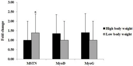
Considering the expression of selected genes MSTN, MyoD and MyoG in Cobb broiler at marketing age (37 days), the result showed that, the expression level of MSTN was significant higher in low body weight relative to high body weight. In contrast, the MyoD and MyoG expression were higher in high body weight relative to low body weight (Fig. 2).
Myostatin gene polymorphism
The PCR products of exon 1 (373 bp), exon 2 (374bp) and exon 3 (381bp) from the studied birds of Cobb broiler were illustrated in figure 3a and b.
Fig 3 PCR products of MSTN exons.(a) Showed exon 1 (373bp) and exon 3 (381bp) from high body weight (1-4) and low body weight (5-8) of exon 1 and (9-12) high body weight and (13-16) low body weight for exon 3 of Cobb broiler chickens. (b) Showed exon 2 (374bp) from high body weight (1-4) and (5-8) low body weight of Cobb broiler chickens.
DNA sequence alignment of exon1,2 and 3 of MSTN gene among high and low body weight Cobb broiler showing 100% similarity (Fig. 4). The comparison of Cobb MSTN with gene Table 1 List of primers used in this study
Amplicon size (bp) Annealing
temperature (ºC) Sequence (5`→3`)
Primers
119 59
F: GCAAAAGCTAGCAGTCTATG R: TCCGTCTTTTTCAGCGTTCT Myostatin
116 60
F:GATTTCCACAGACAACTCCACAT R: GAATCTGGGCTCCACTGTCACT Myo D
109 60
F: GTGGGATGGTGATGCTGGAA R: TTGGAGAGGAGTGGGAAAGGA Myo G
147 60
F: AACCCATTTTTAGAGGTCAGAG R: TCATGAGCACCCGCAACGAT GAPDH
373 55
F: ATGCAAAAGCTAGCAGTCTATG R: ACTCCGTAGGCATTGTGATAAT MSTN (Exon 1)
374 55
F:CTGATTTTCTTGTACAAATGGAG R: CAATCCATCTTCACCCGGTC MSTN (Exon 2)
381 55
F: AACCCATTTTTAGAGGTCAGAG R: TCATGAGCACCCGCAACGAT MSTN (Exon 3)
Fig 1 Expression level of selected genes in different embryonic stages E7, E13, E16 and 3 days old chick
Age of Cobb Broiler Chickens And Their Impact on The Marketing Weights
bank (Gallus gallus strain PB-1 breed Broiler myostatin (MSTN) mRNA, complete cds) accessions No. GU075927.1, revealed six SNPs (51 A>G), (195 C>G), (324 T>C) in exon 1, (408 G>A) in exon2, (966 G>T) and (1120 G>T) in exon 3. The translated amino acids of studied coding region of Cobb MSTN gene were it consists of 375amino acids (Fig.4).
Fig. 4 Cobb broiler Myostatin gene structure showing the amplified exons (gray color). The upper part shows the similarity percentage among four sequences for high and low body weights of Cobb broiler at the three exons. Lower parts show the detected SNPs at the three exons compared with gb|GU181322.1, gb|GU181325.1 and gb|GU181326.1 for exon 1, 2, and 3 respectively.
The amino acid alignment of MSTN gene of Cobb broilers as compared to gene bank accession number ACY68210.1showed in figure (5). The Two non-synonymous SNPs were detected in exon 3 lead to change in the amino acid where 321 Tyrosine (Y)> Histidin (H) and 374 Glycine (G)> Cysteine (C).
Fig 5 Amino acid alignment of Myostatin gene of Cobb broiler compared with broiler strain ACY68210.1.Two amino acid changes marked with gray color: 321 Tyrosine (Y)>Histidin (H) and 374 Glycine (G)> Cysteine (C).
DISCUSSION
Cobb broiler are the one of most effective broiler worldwide, has the best growth rate and lowest cost per kilogram or pound of live-weight (FAO 2013). Breast muscle is a valuable meat product of chicken, however, embryonic and fetal development are characterized by rapid growth and cell differentiation that affect later life (Miller and Stockdale 1986).
In this study, we investigate the expression level of myostatin and myogenic factors in different embryonic stages of Cobb broiler and the result revealed that the expression level of MSTN gene was significantly high at E7 relative to E16 (Fig. 1). Ban et al., (2013) studied expression level of MSTN gene in chicken and quail embryo from 7-17days and they found that
its expression level reaches its first peak at E7 in quail and E9 in chicken and this coincide with the period of primary and secondary fiber formation in chicken embryo and the difference between chicken and quail may be due to species difference. Down regulation of MSTN gene in our study at the embryonic days 13, 16 and three days post hatch were also reported by Gu
et al., (2013) in Pekin duck embryos, they studied its expression at embryonic days 11, 14, 17, 20, 23, and 26, they observed decline in its expression to reach its minimum level at E19 and then increase gradually to E26.
Regarding the expression of MSTN gene in broiler chickenat marketing age (37 days). The expression of MSTN was higher in low body weight relative to high body weight groups.
Bhattacharya et al., (2014) found thatthe lowest expressions of MSTN in broiler (CB- strain and PB_1 line) and layer (IWI line) were found at 6 weeks of age which allow muscle growth and get better body weight. On the other hand, no association between its expression and increased body weight was observed in Taihu and Wanxise geese at the marketing age (Tang et al., 2013).
In the present study, the expression of MyoD gene was prominent at early and late embryonic stages, its transcript increased to 42±2.03 at E7 and increased to 54.59±2.92 at E16, while its expression was not observed at E13. Supporting the role of this gene expression during primary muscle fiber formation which occurs atembryonic days (Ed) 4 and 7 and also secondary muscle fibers development from ED7-15 (Miller and Stockdale 1987) and also in muscle fiber hyperplasia (Al-Musawi et al., 2011).
The highest expression of MyoG was observed at E7 (25±0.09), and its transcripts are not prominent during the other embryonic days. Bentzinger et al., (2012) suggested that MyoG is more directly involved in the myoblasts differentiation process and trigger the expression of myotube-specific genes in the myogenesis.
The combined increased expression of MSTN, MyoD and MyoG genes in our study at the E7 supporting their role at early phase of myogenesis. However, combined down-regulation of the three genes was observed at 3 days posthatch compared with their levels at early embryonic stage. Lee et al., (2014) observed a low level of MyoG and MyoD expression from (0-3) old post hatch period compared with their transcripts at embryonic stages in two lines of Japanese quails, and the level of expression was lower in low weight lines compared with random breed control.
On the other hand, the expressions of both MyoD and MyoG were higher in high body weight group relative to low body weight group (Fig. 2). Similarly, Yin et al., (2014) reported that the relative expression level of MyoG and MyoD in pectorals muscle were greater in high weight selection than low weight selection at 28 days and 56days of adult broiler.
involves different lines or breeds of chickens (Ye et al., 2007). However, Zhang et al., (2010) identified only variant synonymous mutation at exon 1 in MSTN coding region which has the potential to be a genetic marker for body weight traits in female Bian chicken.
Comparison of Cobb broiler MSTN with Gallus gallus strain PB-1 breed broiler revealed presence of three synonymous SNPs at exon 1, one at exon 2 and two non-synonymous SNPs at exon 3 (966 G>T) and 1120 G>T altering 321Y> H and 374 G> C. Hubbard (2004) suggested that non-synonymous SNPs at highly conserved sites as MSTN gene is likely to affect function (intolerant effect). We suggested that the changes in Cobb MSTN protein sequence could modify its growth compared with other broiler because its role is regulation of skeletal muscle growth.
CONCLUSION
In conclusion, there are strong putative role for the expression of MSTN, MyoD and MyoG at embryonic day 7 in Cobb broiler chicks this role also found for these genes at the marketing age that influence bird's weights. Also, MyoD gene is expressed at early and late embryonic life, 3 days post hatch and at marketing age, suggesting its major role in muscle differentiation and develop.
Authors' contributions
SAH and AFE conceived and designed the experiments. AAMF and WSHA performed the experiments and AFE and WSHA analyzed the data. AAMF wrote the paper. SAH, AFE and WSHA authors read and approved the final manuscript.
Acknowledgment
We are thankful all members and assistants of the Department of Animal Husbandry and Animal Wealth Development, Faculty of Veterinary Medicine, Alexandria University, Egypt, for providing necessary facilities to achieve the present work.
Competing interests
The authors declare that they have no competing interests.
References
Ahad, W.A., Beigh, S.A., Mehraj, M., Hussain, M., Bhat, H.F., Shah, R.A. and Mudasir, A.S.(2016), Sequence Characterization of Coding Regions of the Myostatin Gene (GDF8) from Bakerwal Goats (Capra hircus) and Comparison with the Sheep (Ovisaries) Sequence. OJAS. 6:157-162
Al-Musawi, S.L., Lock., F., Simbi, B.H., Bayol, S.A.M. and Strickland, N.C.(2011), Muscle specific differences in the regulation of myogenic differentiation in chickens genetically selected for divergent growth rates. Dif. 82: (3), 127-135.
Amthor, H., Huang, R., McKinnell, I., Christ, B., Kambadur, R., Sharma, M. and Patel, K. (2002), The regulation and action of myostatin as a negative regulator of muscle development during avian embryogenesis. Dev. Biol. 251: (2), 241-257.
Ban, Q., Liang, Y., Zhao, Z., Liu, X. and Li, Q. (2013),
Differential expression levels of genes related to
myogenesis during embryogenesis of quail and chicken. Pak Vet J.33: (3), 317-320.
Bass, J., Oldham,J., Sharma, M. and Kambadur, R. (1999),
Growth factors controlling muscle development. Domest. Anim. Endocrinol. 17: 191-197.
Bhattacharya, T.K. and Chatterjee, R.N. (2013),
Polymorphism of myostatin gene and its association with growth traits in chicken. Dev Biol. 92: (4), 910-915. Bhattacharya,T.K., Chatterjee, R.N., Dushyanth, K. and
Shukla, R. (2014), Cloning, characterization and expression of myostatin (growth differentiating factor-8) gene in broiler and layer chicken (Gallus gallus). Mol. Biol. Rep. 42: (2), 319-327.
Bentzinger, C.F., Wang, Y.X. and Rudnicki, M.A. (2012),
Building muscle: molecular regulation of myogenesis. Cold Spring HarbPerspect Biol.4: (2).
Bergestorm, D.A. and Tapscott, S.J. (2001), Molecular distinction between specification and differentiation in the myogenic basic helix-loophelix transcription factor family. Mol. Cell. Biol. 21, 2404-12.
Berkes, C.A. and Tapscott, S.J. (2005), MyoD and the transcriptional control of myogenesis. Seminars Cell Dev. Biol.16: (4-5), 585-595.
Braun, T. and Gautel, M. (2011), Transcriptional mechanisms regulating skeletal muscle differentiation, growth and homeostasis. Nature Reviews Mol. Cell. Biol. 12: 349-361.
FAO (2013), Poultry Development Review. (www.fao.org/ publications)
Goddard, M.E. and Hayes, B.J. (2009), Mapping genes for complex traits in domestic animals and their use in breeding programmers. Nat Rev Genet.10: 381-391. Gu, L.H., Xu, T.S., Huang, W., Xie, M., Shi, W.B., Sun,
S.D. and Hou, S.S. (2013), Developmental characteristics of pectoralis muscle in Pekin duck embryos. Genet. Mol. Res. 12: (4), 6733-6742.
Hubbard, S. (2004), A genetic variation map for chicken with 2.8 million single-nucleotide polymorphisms.
Nature. 432: 717-722.
Lee, K., Min, C.Y., Suh, Y. and Shin, S. (2014), Skeletal Muscle Characterization of Japanese Quail Line Selectively Bred for Lower Body Weight as an Avian Model of Delayed Muscle Growth with Hypoplasia. PloS One. 9:(4), 95932.
Livak, J.K. and Schmittgen, D.T. (2001), Analysis of Relative Gene Expression Data Using Real Time Quantitative PCR and the 2-∆∆CT Method. Methods. 25:
402-408.
Massari, M.E. and Murre, C. (2000), Helix-loop-helix proteins. Regulators of transcription in eukaryotic organisms. Mol. Cell. Biol. 20: (2), 429-440.
McCroskery, S., Thomas, M., Maxwell, L., Sharma, M. and Kambadur, R. (2003), Myostatin negatively regulates satellite cell activation and self-renewal. J Cell BiolMol Sci. 162: (6), 1135-1147.
McPherron, A.C. and Lee, S. J. (1997), Double muscling in cattle due to mutations in the myostatin gene. PNAS. 94: (23), 12457-12461.
Age of Cobb Broiler Chickens And Their Impact on The Marketing Weights
Miller, J.B.and Stockdale, F.E. (1987), What muscle cells know that nerves don’t tell them. Trends Neuro sci. 10: 325-329.
Paswan, C., Bhattacharya, T.K., Nagaraj, C.S., Chaterjee, R.N.and Jayashankar, M.R. (2014), SNPs in minimal promoter of myostatin (GDF-8) gene and its association with body weight in broiler chicken. J. Appl. Anim. Res. 42: (3), 304-309.
Rehfeldt, C., Te Pas, M.F., Wimmer, K., Brameld, J.M., Nissen, P.M., Berri, C. et al. (2011), Advances in research on the prenatal development of skeletal muscle in animals in relation to the quality of muscle-based food. I. Regulation of myogenesis and environmental impact. Anim. 5: (5), 703-717.
Sato, F., Kurokawa,M., Yamauchi, N. and Hattori, M. (2006), Gene silencing of myostatin in differentiation of chicken embryonic myoblasts by small interfering RNA. Am. J. Cell Physiol. 291: (3), C538-C545.
Smith, J.H. (1963), Relation of body size to muscle size and number of chicken. Poult. Sci. 42:(2), 283-290.
Sun, W.H. (2008), Study on association between SNPs of chicken Myf5 and Myf6 gene and slaughter and meat quality traits. Dissertation. Sichuan Agricultural University.
Tanga, Q., Songa, C., Zhanga, S., Hua, Y., Zhaoa, D.and Zoua, J. (2013), Gene expression profile of IGF1 and MSTN mRNA and their correlation with carcass traits in different breeds of geese at 70 d of age. Br. Poult. Sci. 55: (1), 76-80.
Thompson, J.D., Higgins, D.G., Gibson, T.J. and Clustal, W. (1994), Improving sensitivity and progressive multiple sequence alignment through sequence weighting, position specific gap penalites and weight matrix choice. Nucleic Acids Res. 22: 4673-4680.
Thomas, M., Langley, B., Berry, C., Sharma, M., Kirk, S., Bass, J. and Kambadur, R. (2000), Myostatin, a negative regulator of muscle growth, functions by inhibiting myoblast proliferation. J. Biol. Chem. 275: 40235-40243.
Ye, X., Brown, S.R., Nones, K., Coutinho, L.L., Dekkers, J.C.M. and Lamont, S.J. (2007), Associations of myostatin gene polymorphisms with performance and mortality traits in broiler chickens. Genet. Sel. Evol.39: (1), 73-89.
Yin, H., Zhang, S., Gilbert, E.R., Siegel, P.B., Zhu, Q. and Wong, E.A. (2014), Expression profiles of muscle genes in postnatal skeletal muscle in lines of chickens divergently selected for high and low body weight. Poult Sci. 93: (1), 147-154.
Zhang, G., Ding, F., Wang, J., Dai, G., Xie, K., Zhang, L., et al. (2010), Polymorphism in Exons of the Myostatin
Gene and Its Relationship with Body Weight Traits in the Bian Chicken. Biochem.Genet.49: (1), 9-19.
Zhang, W., Behringer, R.R. and Olson, E.N. (1995),
Inactivation of the myogenic bHLH gene MRF4 results in up-regulation of myogenin and rib anomalies. Genes Dev. 9: 1388-1399.
Zhiliang, G.U., Dahai, Z., Ning, L., Hui, L., Xuemei, D. and Changxin, W. (2004), The single nucleotide polymorphisms of the chicken myostatin gene are associated with skeletal muscle and adipose growth Science in China Ser. C Life Sciences. 47: (1), 25-30. Zhu, Z., Wu, D.J.and Xu, N.Y. (2007), SNPs of myostatin
gene and its genetic effects on carcass traits in chicken. Yi Chuan. 29: (5), 593-598.
*******
How to cite this article: