To cite: Chung F, Wong J, Bellingham G, et al. Predictive factors for sleep apnoea in patients on opioids for chronic pain. BMJ Open Resp Res 2019;6:e000523. doi:10.1136/ bmjresp-2019-000523
►Additional material is published online only. To view please visit the journal online (http:// dx. doi. org/ 10. 1136/ bmjresp- 2019- 000523). FC and JW are joint first authors.
Received 4 November 2019 Revised 6 December 2019 Accepted 9 December 2019
For numbered affiliations see end of article.
Correspondence to Dr Frances Chung; Frances. Chung@ uhn. ca
Predictive factors for sleep apnoea in
patients on opioids for chronic pain
Frances Chung,1 Jean Wong,1,2 Geoff Bellingham,3 Gerald Lebovic,4 Mandeep Singh,1 Rida Waseem,1 Philip Peng,1 Charles F P George,5 Andrea Furlan,6 Anuj Bhatia,1 Hance Clarke,7 David N Juurlink,8 Muhammad M Mamdani,9,10 Richard Horner,11 Beverley A Orser,11,12 Clodagh M Ryan,13,14 on behalf of Op- Safe Investigators
© Author(s) (or their employer(s)) 2019. Re- use permitted under CC BY- NC. No commercial re- use. See rights and permissions. Published by BMJ.
AbstrAct
background The risk of death is elevated in patients taking opioids for chronic non- cancer pain. Respiratory depression is the main cause of death due to opioids and sleep apnoea is an important associated risk factor.
Methods In chronic pain clinics, we assessed the STOP- Bang questionnaire (a screening tool for sleep apnoea; Snoring, Tiredness, Observed apnoea, high blood Pressure, Body mass index, age, neck circumference and male gender), Epworth Sleepiness Scale, thyromental distance, Mallampati classification, daytime oxyhaemoglobin saturation (SpO2) and calculated daily morphine milligram equivalent (MME) approximations for each participant, and performed an inlaboratory polysomnogram. The primary objective was to determine the predictive factors for sleep apnoea in patients on chronic opioid therapy using multivariable logistic regression models.
results Of 332 consented participants, 204 underwent polysomnography, and 120 (58.8%) had sleep apnoea (AHI ≥5) (72% obstructive, 20% central and 8% indeterminate sleep apnoea), with a high prevalence of moderate (23.3%) and severe (30.8%) sleep apnoea. The STOP- Bang questionnaire and SpO2 are predictive factors for sleep apnoea (AHI ≥15) in patients on opioids for chronic pain. For each one- unit increase in the STOP- Bang score, the odds of moderate- to- severe sleep apnoea (AHI ≥15) increased by 70%, and for each 1% SpO2 decrease the odds increased by 33%. For each 10 mg MME increase, the odds of Central Apnoea Index ≥5 increased by 3%, and for each 1% SpO2 decrease the odds increased by 45%.
conclusion In patients on opioids for chronic pain, the STOP- Bang questionnaire and daytime SpO2 are predictive factors for sleep apnoea, and MME and daytime SpO2 are predictive factors for Central Apnoea Index ≥5.
trial registration number NCT02513836
IntroductIon
Over the past two decades, the prescription of opioids for chronic pain has increased dramat-ically, triggering an opioid crisis in North America, with heavy societal and economic impacts.1 The potential for acute respiratory
depression and death triggered by opioids is well known,2 but the synergism between
sleep apnoea and chronic opioid use was only
recently recognised.3 Although sleep apnoea
is highly prevalent in patients on opioids for chronic pain and is implicated as an impor-tant contributor to opioid- related deaths,4
patients are not routinely screened for the disease.3 5 6 The recent American Academy of
Sleep Medicine Position Statement indicated that opioids are associated with several types of sleep- disordered breathing, including obstructive sleep apnoea (OSA), central sleep apnoea (CSA) and sleep- related hypoventi-lation.7 Appropriate screening, diagnostic
testing and treatment of opioid- associated sleep- disordered breathing were recom-mended to improve patients’ health and quality of life.7
At present, no clinical tool allows the ready identification of sleep apnoea in patients on opioids for chronic pain. Polysomnog-raphy, the reference standard for diagnosing sleep apnoea, comes with high costs and restricted access. The STOP- Bang question-naire (a screening tool for sleep apnoea;
Snoring, Tiredness, Observed apnoea, high
Key messages
► In patients on opioids for chronic pain, we found that 58.8% had sleep apnea (72% obstructive, 20% cen-tral, and 8% indeterminate) with a high prevalence of moderate (23.3%) and severe sleep apnea (30.8%).
► We found that the STOP- Bang questionnaire (a screening tool for sleep apnoea; Snoring, Tiredness, Observed apnoea, high blood Pressure, Body mass index, age, neck circumference and male gender) and daytime oxygen saturation (SpO2) are predictive factors for moderate- to- severe sleep apnoea and morphine milligram equivalents and daytime SpO2 are predictive factors for central sleep apnoea. ► Chronic opioid use is associated with a high
preva-lence of sleep- disordered breathing, and we found novel screening tools for opioid- associated sleep- disordered breathing.
by copyright.
on September 18, 2020 by guest. Protected
blood Pressure, Body mass index, age, neck circumfer-ence and male gender) has been validated to screen for sleep apnoea in different populations.8–10 The primary
objective of this study was to determine whether the STOP- Bang questionnaire,8 9 Epworth Sleepiness Scale,11
Mallampati score,12 thyromental distance,13 resting
daytime oxyhaemoglobin saturation (SpO2) and calcu-lated daily morphine milligram equivalent (MME) approximations are predictive factors for sleep apnoea (AHI ≥15) in patients on opioids for chronic pain. The secondary objective was to identify predictive factors for Central Apnoea Index (CAI) ≥5 using the STOP- Bang questionnaire, daytime SpO2 and MME.
MAterIAls And Methods
This prospective cohort study ‘Development of an Inno-vative Opioid Safety Program in Pain Clinics (Op- Safe)’ was conducted at five university- affiliated tertiary care pain clinics. Adults ≥18 years taking opioid medications for chronic pain for >3 months with a stable daily dose for >4 weeks were eligible to participate. We excluded participants with a prior diagnosis of sleep apnoea, active neurological or psychiatric disorders, cancer and those in whom an urgent sleep evaluation was deemed neces-sary for safety reasons (detailed inclusion and exclusion criteria are found in online supplementary appendix).
In each pain clinic, eligible participants completed the STOP- Bang questionnaire8 9 and the Epworth Sleepiness
Scale (a self- reported measure of daytime sleepiness),11
and underwent a clinical assessment of the Mallampati score,12 thyromental distance13 and resting daytime
SpO2 (Pulsox-300, Konica Minolta). Patients were rested and seated for 5–10 min before SpO2 was obtained. The Pulsox- 300i oximeter has 1 Hz of sampling frequency, 3 s of averaging time and 0.1% SpO2 of resolution. The partic-ipants then underwent an inlaboratory polysomnogram.
The polysomnography was performed at the Univer-sity Health Network (Toronto) or the London Health Sciences (London, Canada) sleep study laboratories. A technologist was in attendance throughout the study. The polysomnography recordings were scored by expe-rienced technologists and reviewed by sleep physicians (CMR, CFPG). Both technologists and sleep physicians were blinded to the clinical information. The recording montage included electroencephalography, bilateral electrooculograms, a chin electromyography, single- lead electrocardiography, thoracic and abdominal respira-tory inductance plethysmography, airflow measured by thermocouple and nasal pressure cannula, finger pulse oximetry, and bilateral limb movements. All signals were recorded on a computerised sleep scoring system (Natus Medical Sandman, USA). Sleep stages and elec-troencephalogram (EEG) (cortical) arousals were scored according to published guidelines.14 Apnoeas were
scored when the nasal pressure signal flattened or nearly flattened for greater than 10 s. Hypopnoeas were scored when the amplitude of the sum of the abdominal and
thoracic inductance signals or the nasal pressure flow signal decreased by 30% or more for greater than or equal to 10 s in association with a ≥3% oxygen desatura-tion and/or an arousal. Events were classified as either ‘central’ or ‘obstructive’ according to the presence or absence of respiratory effort. An apnoea was scored as ‘mixed’ if there was absent inspiratory effort in the initial portion of the event, followed by resumption of inspira-tory effort in the second portion of the event. An oxygen desaturation index was quantified as the number of dips in SpO2 of 3% per hour of sleep.
Sleep apnoea was defined as an Apnoea- Hypopnoea
Index (AHI) of ≥5 events per hour of sleep. The
severity of sleep apnoea was classified as mild (AHI 5–<15 events/hour), moderate (15–<30 events/hour) and severe (≥30 events/hour).14 Participants were
strat-ified to have OSA if greater than or equal to 50% of events were obstructive in nature, and were stratified to have CSA if greater than or equal to 50% of events were central in nature.15 Participants were stratified to have
indeterminate sleep apnoea if they have a total AHI of >5 events per hour, but both the Obstructive Apnoea Hypopnoea Index (OAHI) and Central Apnoea Hypo-pnoea Index (CAHI) were <5 events per hour. Sleep- related hypoxaemia was defined as >30% of sleep time with SpO2 <90%.16 We documented participants’
demo-graphics, comorbidities and medications. Daily opioids doses were converted to approximate MME according to the US Centers for Disease Control and Prevention (CDC) (online supplementary e- Table 1).17
Patient involvement
The study did not involve patients in the development of the research question and the outcome measures. Patients were not involved in the design, recruitment and conduct of the study. The results of the study were not disseminated to study participants.
sample size
We calculated the sample size based on the STOP- Bang questionnaire, with 84% sensitivity,6 a clinically
mean-ingful precision of 0.09 for diagnosis, a conservative esti-mated prevalence of 50% for sleep apnoea in patients with chronic pain6 and a two- sided type I error of 0.05. A
total of 127 participants was required, and factoring in a 20% dropout rate the estimated sample size was 152; 304 subjects were to be recruited, with 50% each for deriva-tion and validaderiva-tion.
statistical analysis
Patient characteristics were described using descriptive statistics as appropriate. Univariate tests comparing cate-gorical variables were conducted using χ2 test or Fish-er’s exact test. The continuous variables were compared using t- tests, analysis of variance or non- parametric tests. The primary analysis used a multivariable logistic
by copyright.
on September 18, 2020 by guest. Protected
Figure 1 Study flow chart showing the number of participants involved in different phases. OSA, obstructive sleep apnoea; PSG, polysomnography.
Table 1 Characteristics of sleep apnoea in patients*
Variable n (%) 95% CI
AHI ≥5 n=120
AHI ≥15 65 (54.2) 45.3 to 62.8
AHI ≥30 37 (30.8) 23.3 to 39.6
CAI ≥5 33 (27.5) 20.3 to 36.1
CAI ≥15 18 (15) 9.7 to 22.5
CAI ≥30 12 (10) 5.8 to 16.7
Obstructive sleep
apnoea 86 (72) 63 to 79
Central sleep apnoea 24 (20) 13.8 to 28
Sleep apnoea:
indeterminate type 10 (8) 4.6 to 14.7
*All variables are expressed as events per hour. Participants were stratified to have obstructive sleep apnoea if greater than or equal to 50% of events were obstructive in nature, and were stratified to have central sleep apnoea if greater than or equal to 50% of events were central in nature.15 Participants were stratified to have
indeterminate sleep apnoea if they have AHI >5, but both the OAHI and CAHI were <5.
AHI, Apnoea- Hypopnoea Index; CAI, Central Apnoea Index.
regression model to examine the association between the STOP- Bang score,8 9 daytime SpO
2, daily MME, Epworth Sleepiness Scale,11 Mallampati score12 and thyromental
distance as predictor variables, and moderate- to- severe sleep apnoea (defined as AHI ≥15 events/hour) as the outcome. Similarly, modelling for all sleep apnoeas (AHI
≥5) was done. For the secondary analysis, due to the frequent presence of both obstructive and central events in participants, we chose CAI for modelling/analysis, in addition to those stratified as CSA. We developed a sepa-rate multivariable logistic regression model to examine the association between daytime SpO2, daily MME and STOP- Bang as predictor variables, and CAI ≥5 as the outcome.18 Variance inflation factors were <1.2 for
varia-bles entering the models, indicating multicollinearity was not a problem.
All variables in each model were a priori specified based on clinical reasoning, and model selection was not performed as this may lead to biased estimates of SE, CI and p values.18 To assess the linearity of the covariates in
the models, a likelihood ratio test was used to compare models with and without a restricted cubic spline.19
Due to the lower than expected recruitment, models were internally validated using bootstrap methods with n=1000.18 Statistical analysis was performed using STATA/
SE V.14.120 and R V.3.5.2.21
results
From 27 May 2015 to 28 February 2018, 332 participants consented for the study, 204 (61.4%) participants under-went polysomnography, and 128 (38.6%) dropped out due to failure to complete polysomnography (figure 1). The demographics of these dropouts were similar to the participants except for their lower Epworth Sleepi-ness Scale scores (online supplementary e- Table 2). The average age of participants who underwent polysomnog-raphy was 52 (SD 13.1) years and 41.7% were male. Eighty- four participants had no sleep apnoea, while 120 (58.5%) had newly diagnosed sleep apnoea (AHI ≥5) (72% (86 of 120) obstructive, 20% (24 of 120) central and 8% (10 of 120) indeterminate) (table 1). Sleep- related hypoxaemia
was present in 3.4% (7 of 120) of participants (AHI ≥5, n=5; AHI <5, n=2). Of 120 participants, 45.8% (55 of 120) had mild, 23.3% (28 of 120) had moderate and 30.8% (37 of 120) had severe sleep apnoea. Of 33 participants with CAI ≥5, 55% (18 of 33) had CAI ≥15 and 36% (12 of 33) CAI ≥30 (table 1). The participants of the study were prescribed opioids for different reasons: 28.9% were prescribed opioids for back pain, 16.7% for arthritis pain, 10.3% for neuropathic pain, 10.8% post- traumatic pain, 11.8% for fibromyalgia and the remaining for other reasons.
demographic data and sleep parameters
The demographic and anthropomorphic characteris-tics of participants with mild, moderate and severe sleep apnoea are shown in table 2 and online supplementary e- Table 3. Out of 204 patients undergoing polysomnog-raphy, 7 were classified to have sleep- related hypoxaemia. Five had AHI ≥5 and 2 had AHI <5. Participants with sleep- related hypoxaemia had a mean SpO2 of 88.4%±1.5, minimum SpO2 of 75%±4 and cumulative time SpO2 <90% of 79.5% (72.8–96.6) (table 3). Forty- four per cent of patients were taking more than one opioid medica-tion. Sixty- five per cent of participants were prescribed at least one other centrally acting medication (benzo-diazepines n=31, zopiclone n=12, antidepressants n=51, gabapentin n=40, pregabalin n=40 and/or muscle relax-ants n=14). Fifteen patients were using cannabis. There was no significant difference in doses of daily MME (72 (22.5–135) mg vs 68.8 (30–180) mg; p=0.544) and AHI (8 (3.2–17.8) vs 5.3 (1.5–19.4) events per hour; p=0.163) for participants taking opioids only versus opioids and other centrally acting medications.
by copyright.
on September 18, 2020 by guest. Protected
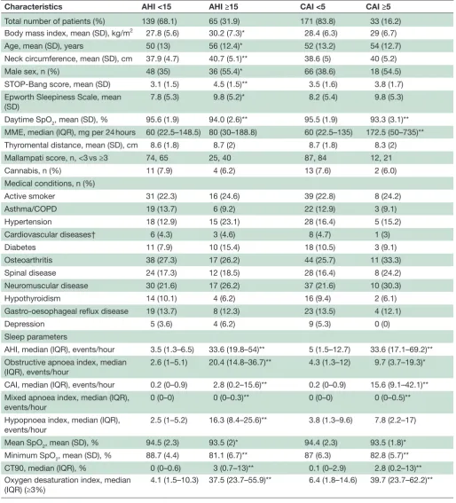
Table 2 Demographics and sleep parameters of patients grouped by moderate- to- severe sleep apnoea (AHI ≥15) and CAI ≥5
Characteristics AHI <15 AHI ≥15 CAI <5 CAI ≥5
Total number of patients (%) 139 (68.1) 65 (31.9) 171 (83.8) 33 (16.2)
Body mass index, mean (SD), kg/m2 27.8 (5.6) 30.2 (7.3)* 28.4 (6.3) 29 (6.7)
Age, mean (SD), years 50 (13) 56 (12.4)* 52 (13.2) 54 (12.7)
Neck circumference, mean (SD), cm 37.9 (4.7) 40.7 (5.1)** 38.6 (5) 40 (5.2)
Male sex, n (%) 48 (35) 36 (55.4)* 66 (38.6) 18 (54.5)
STOP- Bang score, mean (SD) 3.1 (1.5) 4.5 (1.5)** 3.5 (1.6) 3.8 (1.7)
Epworth Sleepiness Scale, mean
(SD) 7.8 (5.3) 9.8 (5.2)* 8.2 (5.4) 9.8 (5.3)
Daytime SpO2, mean (SD), % 95.6 (1.9) 94.0 (2.6)** 95.5 (1.9) 93.3 (3.1)**
MME, median (IQR), mg per 24 hours 60 (22.5–148.5) 80 (30–188.8) 60 (22.5–135) 172.5 (50–735)**
Thyromental distance, mean (SD), cm 8.6 (1.8) 8.7 (2) 8.7 (1.8) 8.3 (2)
Mallampati score, n, <3 vs ≥3 74, 65 25, 40 87, 84 12, 21
Cannabis, n (%) 11 (7.9) 4 (6.2) 13 (7.6) 2 (6.0)
Medical conditions, n (%)
Active smoker 31 (22.3) 16 (24.6) 39 (22.8) 8 (24.2)
Asthma/COPD 19 (13.7) 6 (9.2) 22 (12.9) 3 (9.1)
Hypertension 18 (12.9) 15 (23.1) 28 (16.4) 5 (15.2)
Cardiovascular diseases† 6 (4.3) 3 (4.6) 8 (4.7) 1 (3)
Diabetes 11 (7.9) 10 (15.4) 18 (10.5) 3 (9.1)
Osteoarthritis 38 (27.3) 17 (26.2) 44 (25.7) 11 (33.3)
Spinal disease 24 (17.3) 12 (18.5) 28 (16.4) 8 (24.2)
Neuromuscular disease 30 (21.6) 17 (26.2) 37 (21.6) 10 (30.3)
Hypothyroidism 14 (10.1) 4 (6.2) 16 (9.4) 2 (6.1)
Gastro- oesophageal reflux disease 19 (13.7) 8 (12.3) 23 (13.5) 4 (12.1)
Depression 5 (3.6) 4 (6.2) 9 (5.3) 0 (0)
Sleep parameters
AHI, median (IQR), events/hour 3.5 (1.3–6.5) 33.6 (19.8–54)** 5 (1.5–12.7) 33.6 (17.1–69.2)** Obstructive apnoea index, median
(IQR), events/hour 2.6 (1–5.1) 20.4 (14.8–36.7)** 4.3 (1.3–12) 9.7 (3.7–19.3)*
CAI, median (IQR), events/hour 0.2 (0–0.9) 2.8 (0.2–15.6)** 0.2 (0–0.9) 15.6 (9.1–42.1)** Mixed apnoea index, median (IQR),
events/hour 0 (0–0) 0 (0–0.3)** 0 (0–0) 0 (0–0.5)**
Hypopnoea index, median (IQR),
events/hour 2.5 (1–5.2) 16.3 (8.4–25.6)** 3.8 (1.3–9.6) 7.8 (2.2–17)
Mean SpO2, mean (SD), % 94.5 (2.3) 93.5 (2)* 94.4 (2.3) 93.5 (1.8)*
Minimum SpO2, mean (SD), % 88.7 (4.4) 81.1 (6.7)** 87 (6.3) 82.8 (5.7)**
CT90, median (IQR), % 0 (0–0.6) 3 (0.7–13)** 0.1 (0–2.9) 2.8 (0.2–13)**
Oxygen desaturation index, median (IQR) (≥3%)
4.1 (1.5–10.3) 37.5 (23.7–55.9)** 6.4 (1.8–14.6) 39.7 (23.7–62.2)**
Values are expressed as mean (SD) or median (IQR) as appropriate.
t- Test or Wilcoxon rank- sum test and χ2 analysis or Fisher’s exact test were conducted to examine differences in the characteristics of participants with sleep and central apnoea.
*P<0.05, **P<0.001.
†Cardiovascular diseases include angina, myocardial infarction, arrhythmia, peripheral vascular disease or stroke.
AHI, Apnoea- Hypopnoea Index; CAI, Central Apnoea Index; COPD, chronic obstructive pulmonary disease; CT90, cumulative time SpO2<90%; MME, morphine milligram equivalents; SpO2, oxyhaemoglobin saturation; STOP- Bang, a screening tool for sleep apnoea (Snoring, Tiredness, Observed apnoea, high blood Pressure, Body mass index, age, neck circumference and male gender).
by copyright.
on September 18, 2020 by guest. Protected
Table 3 Characteristics of patients with sleep- related hypoxaemia
Characteristics
Sleep- related hypoxaemia
Total number of patients 7 Body mass index, mean (SD), kg/m2 29.9 (7.2) Age, mean (SD), years 63.1 (11.1) Neck circumference, mean (SD), cm 37.9 (9.5)
Male sex, n (%) 1 (14.2)
STOP- Bang score, mean (SD) 3.9 (1.7) Epworth Sleepiness Scale, mean (SD) 8.3 (5.9) MME, median (IQR), mg per 24 hours 157.5 (70–476.4) Thyromental distance, mean (SD), cm 7.8 (1.5) Medical conditions, n (%)
Active smoker 3 (42.9)
Asthma/COPD 0
Sleep parameters
Apnoea- Hypopnoea Index, median (IQR), events/hour
15.8 (5.6–55.9)
Obstructive apnoea index, median (IQR), events/hour
9 (5.4–55.7)
Central Apnoea Index, median (IQR), events/ hour
1.3 (0.2–5.2)
Mixed apnoea index, median (IQR), events/ hour
0 (0–1.1)
Hypopnoea index, median (IQR), events/hour 7.7 (3.1–41.7) Mean SpO2, mean (SD), % 88.4 (1.5)
Minimum SpO2, mean (SD), % 75 (4)
CT90, median (IQR), % 79.5 (72.8–96.6)
Values are expressed as mean (SD) or median (IQR) as appropriate. COPD, chronic obstructive pulmonary disease; CT90, cumulative time SpO2 <90%; MME, morphine milligram equivalents; SpO2,
oxyhaemoglobin saturation; STOP- Bang, a screening tool for sleep apnoea (Snoring, Tiredness, Observed apnoea, high blood Pressure,
Body mass index, age, neck circumference and male gender).
Figure 2 Predicted probabilities for sleep apnoea with the different cut- offs of STOP- Bang score. Vertical bars represent SEM. AHI, Apnoea- Hypopnoea Index; STOP- Bang, a screening tool for sleep apnoea (Snoring, Tiredness, Observed apnoea, high blood Pressure, Body mass index, age, neck circumference and male gender).
Figure 3 Apnoea- Hypopnoea Index and Central Apnoea Index for the different categories of daytime oxyhaemoglobin saturation (SpO2). Lower and upper boundaries of boxplot indicate 25th and 75th percentile.
Association between moderate-to-severe sleep apnoea (AhI ≥15), stoP-bang score, cAI ≥5, spo2 and MMe
Figure 2 shows the probabilities for sleep apnoea with increasing STOP- Bang score. As the score increased from 3 to 7, the probabilities of moderate- to- severe sleep
apnoea (AHI ≥15) increased correspondingly from
23% to 75% in this population. Figure 3 shows the asso-ciation of SpO2 with AHI and CAI separately. AHI and CAI increased as SpO2 decreased. There was a signifi-cant difference in AHI (χ2(2)=21.9, p<0.0001) and CAI (χ2(2)=11.5, p=0.0032) for three categories of daytime SpO2: 96%–100%, 92%–95% and ≤91%. Figure 4 shows that increasing daily MME resulted in a corresponding percentage increase in the OR of CAI ≥5, adjusting for SpO2 and STOP- Bang score. The percentage increase in the odds of CAI ≥5 is almost 100% for 200 mg MME and 240% for 400 mg MME.
The percentage of patients with CAI ≥5 varied in the different types of opioids, which is likely a reflection
of MME (online supplementary e- Table 4). Among
participants on fentanyl patch with high MME (552.6 (90–1440) mg), significantly more participants had CAI
≥5 than CAI <5, and in those on tramadol with low MME
by copyright.
on September 18, 2020 by guest. Protected
Figure 4 Increase in the OR of patients with CAI ≥5 by increasing morphine milligram equivalents adjusting for oxyhaemoglobin saturation and STOP- Bang score. Vertical bars represent SE of OR. CAI, Central Apnoea Index; STOP- Bang, a screening tool for sleep apnoea (Snoring, Tiredness, Observed apnoea, high blood Pressure, Body mass index, age, neck circumference and male gender.
Figure 5 Cognitive aid model for sleep apnoea (AHI ≥15) and Central Apnoea Index (CAI ≥5). AHI, Apnoea- Hypopnoea Index; SpO2, oxyhaemoglobin saturation.
(22.9 (3.7–60) mg) significantly fewer patients had CAI
≥5 than CAI <5.
Modelling for moderate-to-severe sleep apnoea (AhI ≥15)
The primary analysis used a multivariable logistic regres-sion to examine the association of STOP- Bang score,8
daytime SpO2, MME, Epworth Sleepiness Scale,11
Mallam-pati score12 and thyromental distance as potential
predic-tors of sleep apnoea. The model showed that for each one- unit increase in the STOP- Bang score, the odds of moderate- to- severe sleep apnoea (AHI ≥15) increased by 70% (OR 1.70, 95% CI 1.34 to 2.16, p<0.0001), and
for each 1% SpO2 decrease the odds increased by 33% (OR 1.33, 95% CI 1.12 to 1.58, p=0.002) (figure 5). The c- statistics indicated good discrimination with a value of 0.79 (95% CI 0.72 to 0.85) (table 4). The Likelihood Ratio (LR) test confirmed a linear association between all numeric covariates and the odds of sleep apnoea.
Modelling for all sleep apnoeas (AhI ≥5)
Multivariable logistic regression was also performed for AHI ≥5 using the STOP- Bang score, daytime SpO2, MME, Epworth Sleepiness Scale, Mallampati score and thyromental distance as potential predictors of sleep
apnoea. The model showed that for each one- unit
increase in the STOP- Bang score, the odds of sleep apnoea (AHI ≥5) increased by 48% (OR 1.48, 95% CI 1.19 to 1.84, p=0.0004), and for each 1% SpO2 decrease the odds increased by 30% (OR 1.30, 95% CI 1.08 to 1.56, p=0.006). The c- statistics showed good discrimina-tion with a value of 0.75 (95% CI 0.67 to 0.82) (table 4). The LR test confirmed a linear association between all numeric covariates and the odds of sleep apnoea.
Modelling for cAI ≥5
Thirty- three patients had CAI ≥5. We performed a secondary analysis using a multivariable logistic regres-sion model to examine the relationship between CAI
≥5 and the STOP- Bang questionnaire, daytime SpO2 and MME (table 4). The LR test confirmed a linear associa-tion between covariates of interest and the odds of CSA. The model showed that for each 1% SpO2 decrease, the odds of CAI ≥5 increased by 45% (OR 1.45, 95% CI 1.19 to 1.78, p=0.0003), and for each 10 mg MME increase the odds of CAI ≥5 increased by 3% (OR 1.03, 95% CI 1.02 to 1.05, p<0.0001) (table 4 and figure 5). This model had good discriminatory properties with a c- statistics of 0.80 (95% CI 0.71 to 0.88).22
Modelling for csA
Using a more stringent criterion, 24 patients were strat-ified to have CSA if greater than or equal to 50% of the events are central in nature.16 Multivariable logistic
regres-sion was performed for CSA using MME and daytime SpO2 as predictors. The adjusted model was similar to CAI ≥5 described above. For each 1% SpO2 decrease, the odds of CSA increased by 47% (OR 1.47, 95% CI 1.17 to 1.84, p=0.001). For each 10 mg MME increase, the odds of CSA increased by 4% (OR 1.04, 95% CI 1.02 to 1.06, p<0.0001) (table 4). The c- statistics of the model of MME and daytime SpO2 suggested a good discrimination with a value of 0.85 (95% CI 0.76 to 0.94).
Internal validation of models
The internal validation of the four models (table 4) was performed using 1000 bootstrapped samples. All four models suggested minor to mild overfitting (online supplementary e- Table 5).
by copyright.
on September 18, 2020 by guest. Protected
Table 4 Models for moderate- to- severe sleep apnoea (AHI ≥15), CAI ≥5 and central sleep apnoea using different predictors
Predictors Unit of analysis OR (95% CI) P value OR (95% CI) P value
Moderate- to- severe sleep apnoea
(AHI ≥15) with 6 predictors Sleep apnoea (AHI ≥5) with 6 predictors
STOP- Bang score 1 1.70 (1.34 to 2.16) <0.0001 1.48 (1.19 to 1.84) 0.0004
Daytime SpO2 1% decrease 1.33 (1.12 to 1.58) 0.002 1.30 (1.08 to 1.56) 0.006
MME 10 mg 1.01 (0.99 to 1.02) 0.352 1.02 (0.999 to 1.034) 0.067
Epworth Sleepiness Scale*
11–24 vs 0–10 1.19 (0.56 to 2.51) 0.650 0.93 (0.46 to 1.89) 0.841
Mallampati score* ≥3 vs <3 1.23 (0.59 to 2.57) 0.580 1.16 (0.59 to 2.27) 0.666
Thyromental distance 1 cm 1.08 (0.89 to 1.30) 0.449 1.05 (0.88 to 1.25) 0.609
Model for CAI ≥5 with 3 predictors
(n=33 participants) Model for central sleep apnoea(n=24 participants)
Daytime SpO2 1% decrease 1.45 (1.19 to 1.78) 0.0003 1.47 (1.17 to 1.84) 0.001
MME 10 mg 1.03 (1.02 to 1.05) <0.0001 1.04 (1.02 to 1.06) <0.0001
STOP- Bang score 1 1.10 (0.85 to 1.42) 0.4563
An outlier value (74%) for SpO2 was removed from the analysis.
*Mallampati score and Epworth Sleepiness Scale expressed as dichotomous variables. Participants were stratified to have central sleep apnoea if greater than or equal to 50% of events were central in nature.15
AHI, Apnoea- Hypopnoea Index; CAI, Central Apnoea Index;MME, morphine milligram equivalents; SpO2, oxyhaemoglobin saturation; STOP- Bang, a screening tool for sleep apnoea (Snoring, Tiredness, Observed apnoea, high blood Pressure, Body mass index, age, neck circumference and male gender).
dIscussIon
To date, this is the largest prospective multicentre cohort study investigating the risks of sleep- disordered breathing in chronic opioid users. We found a high prevalence of both moderate (23.3%) and severe (30.8%) sleep apnoea and CSA (20%). Sleep- related hypoxaemia was present in 3.4% of participants. The predictive factors for moderate- to- severe sleep apnoea (AHI ≥15) in patients on opioids for chronic pain are STOP- Bang score and SpO2. The predictive factors for CAI ≥5 are MME and SpO2. For each one- unit increase in the STOP- Bang score, the odds of moderate- to- severe sleep apnoea (AHI ≥15) increased
by 70%, and for each 1% SpO2 decrease the odds
increased by 33%. Importantly, we have novel findings on the magnitude of risk and the additive effect by opioid dose. For each 10 mg MME increase, the odds of CAI
≥5 increased by 3%, and for each 1% SpO2 decrease the odds increased by 45%. Since STOP- Bang questionnaire, SpO2 and MME are simple to use in clinical settings, our findings are useful clinically.
sleep apnoea and opioids
The potential mechanisms by which chronic opioid use potentiates or causes an increased incidence of sleep apnoea include a reduction in the respiratory drive, depression of hypercapnic and hypoxic ventila-tory responses, and enhanced relaxation of the upper airway musculature.23–25 Our findings are consistent
with previous studies showing a high prevalence of sleep apnoea in patients on opioids for chronic pain.5 6 26–28
Sleep- related hypoxaemia has been described in 10% of opioid patients with/without sleep apnoea.5
A dose–response relationship has been shown between daily MME and adverse events.29 A daily opioid dose of
MME 100 mg or more increases the risk of fatal over-dose by sevenfold.30 The CDC guidelines recommend
90 mg as the limit that should not be exceeded unless for specific reasons.17 In our model for CAI ≥5, an
asso-ciation with SpO2 and daily MME exists. For each 10 mg MME increase, the odds of CAI ≥5 are elevated by 3%. These parameters are easily measured and can be used during a clinic visit. The addition of Epworth Sleepiness Scale,11 Mallampati score12 or thyromental distance was
poorly predictive in this population.
What is the potential pathophysiological link between awake spo2 and sleep apnoea?
Our finding of decreased awake SpO2 is consistent with a retrospective study by Walker et al,26 who showed a
signif-icantly lower SpO2 in patients on chronic opioids during wakefulness and non- rapid eye movement (REM) sleep than patients who were not taking opioids. The exact pathophysiological link between opioids and respiratory depression and/or sleep apnoea is unknown. Opioids exert their analgesic effects by binding to opioid recep-tors in the brain. The binding to mu opioid receprecep-tors may lead to respiratory depression during either wake or sleep, with a greater effect during sleep.31 It is postulated
that respiratory depression and sleep apnoea secondary to opioids may be due to a depression or an imbalance in the hypoxic and hypercapnic ventilatory responses during wake and sleep, predisposing to unstable breathing, an increase in the arousal threshold and reduced upper airway activity in vivo.23 32–35 In those on chronic opioids,
by copyright.
on September 18, 2020 by guest. Protected
studies have shown augmented hypoxic ventilatory responses (peripheral chemosensitivity) and depressed hypercapnic ventilatory responses (central chemosensi-tivity).34 35 The presence of augmented daytime hypoxic
ventilatory responses suggests prior exposure to inter-mittent hypoxia either during daytime or at night,33 and
has been shown to be a predisposing factor for sleep apnoea.34
Hence, we suggest that the presence of hypoxia during wake in those on opioids is a marker of respira-tory depression (hypoventilation) by opioids which may predispose individuals to more marked effects when sleep. Indirect evidence to support this theory is found from a study by Waters et al35 in children undergoing
tonsillectomy both with and without OSA. The admin-istration of fentanyl led to an elevation in end- tidal carbon dioxide in association with central events,35 with
increased frequency in those with OSA, suggesting that those with a propensity to hypoventilation may prog-ress to central apnoeas.35 The presence of increased
central events in those on opioids may also be attribut-able to the REM- suppressing effects of opioids. This was demonstrated in a small study in which there was a shift from obstructive to central events with an intravenous infusion of a short- acting opioid.36
recommendation for a stepped care approach
While the evidence relating to the presence of CSA and death due to opioids is limited, there is some evidence from the paediatric population which suggests that CSA/ periodic breathing secondary to opioids may lead to unexpected death.37 38 Therefore, identifying patients at
risk for sleep apnoea can lead to strategies to mitigate the risk of opioid- related complications and deaths, such as dose reduction of opioids and use of non- opioid medi-cations.17 29 39 Predicting the risk of sleep apnoea will
identify patients who require possible polysomnography and other management strategies to mitigate adverse outcomes.40 The American Academy of Sleep Medicine
recommends recently appropriate screening and diag-nostic testing to identify OSA, CSA and sleep- related hypoventilation in people treated with chronic opioid therapy, as the treatment of opioid- associated sleep- disordered breathing can improve patients’ health and well- being.7
Although CDC stratifies the risk of opioid- induced respiratory depression based on opioid dose alone,17
based on our results other predictive factors such as STOP- Bang score and SpO2 should be considered. We recommend a stepped care approach, with the initial evaluation of the resting SpO2 and the STOP- Bang score as a simple approach to identify patients with possible sleep apnoea. As a second step, those who are deemed to be at high risk for moderate- to- severe sleep apnoea should proceed to have a sleep study for confirmation and clarification of the diagnosis. This proposed approach would enable the exclusion of
low- risk patients and the identification of those with a high likelihood of sleep apnoea. It would also facil-itate the efficient allocation of healthcare resources and expedite inlaboratory testing and/or treatment of those with previously unrecognised sleep apnoea.
limitations of the study
Our study has a few limitations. Only 61% of consented patients completed the polysomnography, leading to possible selection bias, as those with sleep complaints are more likely to complete a polysomnogram, resulting in the prevalence of sleep apnoea possibly being over-estimated. Besides opioids, other non- opioid centrally acting medications may contribute to sleep apnoea. Since compliance with medications or screening for substance use disorder was not assessed, documented opioid use may not be reflective of actual consump-tion. As we did not assess arterial blood gas, nocturnal transcutaneous or end- tidal carbon dioxide, we cannot determine whether these participants had hypercapnic or hypocapnic CSA and/or sleep- related hypoventila-tion. Although logistic regression is a valuable tool in observational studies to assess the association between the covariates and the binary outcome, this method cannot examine causality, and therefore definitive prediction of diagnosis based on these factors is not possible. However, external validation showing a good model fit would lend credence regarding the associa-tion of the predictors and the outcome.
conclusIon
In patients on chronic opioid therapy, we found a high prevalence of moderate- to- severe sleep apnoea together with CSA. For each one- unit increase in the STOP- Bang score, the odds of moderate- to- severe sleep apnoea (AHI ≥15) increased by 70% and for each 1% SpO2 decrease the odds increased by 33%. For each 10 mg MME increase, the odds of CAI ≥5 increased by 3% and for each 1% SpO2 decrease the odds increased by 45%. We suggest that these factors should be incorporated to identify chronic opioid users most at risk for sleep apnoea and possible adverse health consequences.
Author affiliations
1Department of Anesthesia and Pain Medicine, Toronto Western Hospital,
University Health Network, University of Toronto, Toronto, Ontario, Canada
2Department of Anesthesia, Women's College Hospital, University of Toronto,
Toronto, On, Canada
3Department of Anesthesia and Perioperative Medicine, St. Joseph's Health
Care, Western University, London, Ontario, Canada
4Applied Health Research Centre, St Michael's Hospital, Institute for Health
Policy Management and Evaluation, University of Toronto, Toronto, On, Canada
5Department of Medicine, University of Western Ontario, London, Ontario,
Canada
6Toronto Rehabilitation Institute, Faculty of Medicine, University of Toronto,
Toronto, Ontario, Canada
7Department of Anesthesia and Pain Medicine, Toronto General Hospital,
University Health Network, University of Toronto, Toronto, On, Canada
by copyright.
on September 18, 2020 by guest. Protected
8Department of Medicine, Sunnybrook Hospital, University of Toronto,
Toronto, Ontario, Canada
9Applied Health Research Centre, St Michael's Hospital, Toronto, Ontario,
Canada
10Applied Health Research Center, Department of Medicine, St. Michael's
Hospital, University of Toronto, Toronto, Ontario, Canada
11Department of Physiology, University of Toronto, Toronto, Ontario, Canada 12Department of Anesthesia and Pain Medicine, Sunnybrook Research
Institute, University of Toronto, Toronto, On, Canada
13Sleep Research Laboratory, University Health Network, Toronto
Rehabilitation Institute, Toronto, On, Canada
14Department of Medicine, University of Toronto, Toronto, On, Canada
Acknowledgements We acknowledge the help of the research staff in collecting data (Emad Al Azazi, Asmita Bhoite, Rabia Jogezai, Halema Khan, Fatiha Mim and Sazzadul Islam).
collaborators Op- Safe investigators: Neilesh Soneji MD, Paul Tumber MD (University Health Network- Mount Sinai Hospital, Toronto, Ontario); John Flannery MD, Dinesh Kumbhare MD, PhD (Toronto Rehabilitation Institute, Toronto, Ontario); Arsenio Avila MD (Sunnybrook Hospital and Women’s College Hospital, Toronto, Ontario); and Sylvia Hyland (Institute for Safe Medication Practices Canada, Toronto, Ontario).
contributors Study concept and design: FC, JW, MS, GL, CMR. Acquisition of data: FC, JW, GB, PP, AF. Statistical analysis and interpretation of data: FC, GL, JW, RW, CMR. Drafting of the manuscript: FC, JW, CMR, GL. Critical revision of the manuscript for intellectual content: GB, MS, PP, CFPG, AF, AB, HC, DNJ, MMM, RH, BO. Study supervision: FC, JW, GB, CMR, CFPG. Obtained funding: FC, JW, GB. Had full access to all of the data in the study and take responsibility for the integrity of the data and the accuracy of the data analysis: FC, JW, CMR, GL. All authors edited the manuscript for important intellectual content and approved the final draft. Funding Funded by the Ontario Ministry of Health and Long- Term Care Innovation Fund, University Health Network Foundation, and the Department of Anesthesia and Pain Medicine, University Health Network- Mount Sinai Hospital, University of Toronto, Toronto, Ontario, Canada.
competing interests FC reports research support from the Ontario Ministry of Health and Long- Term Care and the University Health Network Foundation; and UpToDate royalties and STOP- Bang proprietary to the University Health Network. JW reports grants from the Ontario Ministry of Health and Long- Term Care, Anesthesia Patient Safety Foundation and the University of Toronto Merit Research Award. MMM: reports support from Novo Nordisk, Allergan and Amgen. DNJ reports personal fees from universities, colleges and professional medical associations. He is an unpaid member of the Physicians for Responsible Opioid Prescribing (PROP), which is sometimes cast as an 'anti- opioid' group, but in actuality is invested in prescribing that is safer and more evidence- based. He is also a member of the American College of Medical Toxicology. Both groups have publicly available positions on this issue. Other coauthors have no conflict of interest reported. Patient consent for publication Not required.
ethics approval The research ethics board of each participating institution approved the research protocol. All participants provided written informed consent (research ethics board approval numbers: 14-8611- AE, 15-0004- A, 2014-0122 and 106620).
Provenance and peer review Not commissioned; externally peer reviewed. data availability statement Data are available upon reasonable request. open access This is an open access article distributed in accordance with the Creative Commons Attribution Non Commercial (CC BY- NC 4.0) license, which permits others to distribute, remix, adapt, build upon this work non- commercially, and license their derivative works on different terms, provided the original work is properly cited, appropriate credit is given, any changes made indicated, and the use is non- commercial. See: http:// creativecommons. org/ licenses/ by- nc/ 4. 0/.
RefeRenCes
1 Han B, Compton WM, Blanco C, et al. Prescription opioid use, misuse, and use disorders in U.S. adults: 2015 national survey on drug use and health. Ann Intern Med 2017;167:293–301. 2 Gomes T, Mamdani MM, Dhalla IA, et al. Opioid dose and drug-
related mortality in patients with nonmalignant pain. Arch Intern Med 2011;171:686–91.
3 Correa D, Farney RJ, Chung F, et al. Chronic opioid use and central sleep apnea: a review of the prevalence, mechanisms, and perioperative considerations. Anesth Analg 2015;120:1273–85.
4 Webster LR, Cochella S, Dasgupta N, et al. An analysis of the root causes for opioid- related overdose deaths in the United States. Pain Med 2011;12:S26–35.
5 Mogri M, Desai H, Webster L, et al. Hypoxemia in patients on chronic opiate therapy with and without sleep apnea. Sleep Breath 2009;13:49–57.
6 Webster LR, Choi Y, Desai H, et al. Sleep- Disordered breathing and chronic opioid therapy. Pain Med 2008;9:425–32.
7 Rosen IM, Aurora RN, Kirsch DB, et al. Chronic opioid therapy and sleep: an American Academy of sleep medicine position statement. JCSM 2019;15:1671–3.
8 Chung F, Subramanyam R, Liao P, et al. High Stop- Bang score indicates a high probability of obstructive sleep apnoea. Br J Anaesth 2012;108:768–75.
9 Chung F, Yegneswaran B, Liao P, et al. Stop questionnaire: a tool to screen patients for obstructive sleep apnea. Anesthesiology 2008;108:812–21.
10 Nagappa M, Liao P, Wong J, et al. Validation of the Stop- Bang questionnaire as a screening tool for obstructive sleep apnea among different populations: a systematic review and meta- analysis. PLoS One 2015;10:e0143697.
11 Johns MW. A new method for measuring daytime sleepiness: the Epworth Sleepiness scale. Sleep 1991;14:540–5.
12 Shiga T, Zi W, Inoue T, et al. Predicting difficult intubation in apparently normal patients: a meta- analysis of bedside screening test performance. Anesthesiology 2005;103:429–37.
13 Butler PJ, Dhara SS. Prediction of difficult laryngoscopy: an assessment of the thyromental distance and Mallampati predictive tests. Anaesth Intensive Care 1992;20:139–42.
14 Berry RB, Brooks R, Gamaldo C, et al. AASM scoring manual version 2.2 updates: new Chapters for scoring infant sleep staging and home sleep apnea testing. J Clin Sleep Med 2012;11:1253–4. 15 Sin DOND, Fitzgerald F, Parker JD, et al. Risk factors for
central and obstructive sleep apnea in 450 men and women with congestive heart failure. Am J Respir Crit Care Med 1999;160:1101–6.
16 AASM. International classification of sleep disorders. diagnostic and coding manual 2005:51–5.
17 Dowell D, Haegerich TM, Chou R. CDC Guideline for Prescribing Opioids for Chronic Pain - United States, 2016. MMWR Recomm Rep 2016;65:1–49.
18 Harrell FE, LEE KL, MARK DB. Multivariable prognostic models: issues in developing models, evaluating assumptions and adequacy, and measuring and reducing errors. Stat Med 1996;15:361–87. 19 Sullivan LM, Massaro JM, D'Agostino RB. Presentation of
multivariate data for clinical use: the Framingham study risk score functions. Stat Med 2004;23:1631–60.
20 STATA. Stata statistical software: release 15. 2015.
21 Team RC. A language and environment for statistical computing. R foundation for statistical computing 2018.
22 Merkow RP, Hall BL, Cohen ME, et al. Relevance of the c- statistic when evaluating risk- adjustment models in surgery. J Am Coll Surg 2012;214:822–30.
23 Hajiha M, DuBord M- A, Liu H, et al. Opioid receptor mechanisms at the hypoglossal motor pool and effects on tongue muscle activity in vivo. J Physiol 2009;587:2677–92.
24 Montandon G, Qin W, Liu H, et al. PreBotzinger complex neurokinin-1 receptor- expressing neurons mediate opioid- induced respiratory depression. J Neurosci 2011;31:1292–301.
25 Weil JV, McCullough RE, Kline JS, et al. Diminished ventilatory response to hypoxia and hypercapnia after morphine in normal man. N Engl J Med 1975;292:1103–6.
26 Walker JM, Farney RJ, Rhondeau SM, et al. Chronic opioid use is a risk factor for the development of central sleep apnea and ataxic breathing. J Clin Sleep Med 2007;3:455–61.
27 Farney RJ, McDonald AM, Boyle KM, et al. Sleep disordered breathing in patients receiving therapy with buprenorphine/naloxone. Eur Respir J 2013;42:394–403.
28 Jungquist CR, Flannery M, Perlis ML, et al. Relationship of chronic pain and opioid use with respiratory disturbance during sleep. Pain Manag Nurs 2012;13:70–9.
29 Busse JW, Craigie S, Juurlink DN, et al. Guideline for opioid therapy and chronic noncancer pain. Can Med Assoc J 2017;189:E659–66. 30 Bohnert ASBet al. Association between opioid prescribing patterns
and opioid overdose- related deaths. JAMA 2011;305:1315–21. 31 Nagappa M, Weingarten TN, Montandon G, et al. Opioids,
respiratory depression, and sleep- disordered breathing. Best Pract Res Clin Anaesthesiol 2017;31:469–85.
32 Levitt ES, Abdala AP, Paton JFR, et al. μ opioid receptor activation hyperpolarizes respiratory- controlling Kölliker- Fuse neurons and suppresses post- inspiratory drive. J Physiol 2015;593:4453–69.
by copyright.
on September 18, 2020 by guest. Protected
33 Fatemian M, Kim DY, Poulin MJ, et al. Very mild exposure to hypoxia for 8 H can induce ventilatory acclimatization in humans. Pflugers Arch 2001;441:840–3.
34 Dunai J, Kleiman J, Trinder J. Ventilatory instability during sleep onset in individuals with high peripheral chemosensitivity. J Appl Physiol 1999;87:661–72.
35 Waters KA, McBrien F, Stewart P, et al. Effects of OSA, inhalational anesthesia, and fentanyl on the airway and ventilation of children. J Appl Physiol 2002;92:1987–94.
36 Bernards CM, Knowlton SL, Schmidt DF, et al. Respiratory and sleep effects of remifentanil in volunteers with moderate obstructive sleep apnea. Anesthesiology 2009;110:41–9.
37 Ward SL, Bautista D, Chan L, et al. Sudden infant death syndrome in infants of substance- abusing mothers. J Pediatr 1990;117:876–81.
38 Davidson Ward SL, Bautista DB, Woo MS, et al. Responses to hypoxia and hypercapnia in infants of substance- abusing mothers. J Pediatr 1992;121:704–9.
39 Javaheri S, Patel S. Opioids cause central and complex sleep apnea in humans and reversal with discontinuation: a plea for detoxification. J Clin Sleep Med 2017;13:829–33.
40 Davis MJ, Livingston M, Scharf SM. Reversal of central sleep apnea following discontinuation of opioids. J Clin Sleep Med 2012;8:579–80.
by copyright.
on September 18, 2020 by guest. Protected