International Journal of
http://www.arjournals.org/index.php/ijpm/index
Bone regenerative effect of aqueous
activator of nuclear factor
estrogen deficiency
Woojin Jun
1*, Chul
*Corresponding authors:
Woojin JunChul-yung Choi
1Division of Food and Nutrition, Chonnam
National University, Gwangju61186, Korea
2Jeollanamdo Institute of Natural Resources
Research, Jangheung-gun, Jeollanamdo 59338, South Korea
Received: 27 December 2016 Accepted: 10 April 2017 Published: 02 September 2017
A b s t r a c t
Osteoporosis increases with age, most frequently in postmenopausal women because of reduced ovarian hormone levels. Furthermore, estrogen deficiency impairs trabecular metaphyseal bone. Although
estrogen
herbal therapies maybe safer alternatives. Therefore, the aim of this study the effects of aqueous extracts
factor
mediated osteoporosis tartrate
cells. We investigated the osteoprotective effect of CWW in an ovariectomized (OVX) Sprague Dawley rat model
doses (100, 200, and 400 mg/kg). After a 24
were not affected except in the OVX/E2 group. Additionally, bone mineral density (BMD) and histological analyses
significantly higher group rats. collagen type I C results show that compar
the OVX model. Taken together, CWW exhibited inhibitory effects on osteoclastogenesis
vitro,
Key
induced osteoporosis, osteoclast differentiation
Introduction
Osteoporosis is an age-dependent multifunctional skeletal disease characterized by decreased bone mineral density (BMD), deterioration of bone microarchitecture, and increased risk of fragility fractures [1]. Bone mass is balanced by bone remodeling through bone-forming osteoblasts and bone-resorbing osteoclasts [2]. Consequently, bone homeostasis maintains continuous remodeling of osteoblasts and osteoclasts, which
determined by the proliferation and differentiation of precursors of these two bone-associated cells. Especially, osteoclasts, which are bone-resorbing multinucleated giant cells, are differentiated from hemopoietic progenitors of monocyte-macrophage lineage precursor cells following their activation by crucial
DOI:10.5138/09750185.1979
This article is distributed under the terms of the
and redistribution provided that the original author and source are credited.
International Journal of Phytomedicine 9 (2017) 4
http://www.arjournals.org/index.php/ijpm/index
Original Research Article
Bone regenerative effect of aqueous
Cynanchum wilfordii
extract in receptor
activator of nuclear factor-
κ
B ligand-induced osteoclast differentiation and
estrogen deficiency-induced osteoporosis
Chul-yung Choi
2*, Gyuok Lee
1,2, SangO Pan
2, Jaeyong Kim
A b s t r a c t
Osteoporosis increases with age, most frequently in postmenopausal women because of reduced ovarian hormone levels. Furthermore, estrogen deficiency impairs trabecular metaphyseal bone. Although efficacious, long-term hormone replacement therapy (HRT) has estrogen-like side effects including breast and endometrial cancers, and non
herbal therapies maybe safer alternatives. Therefore, the aim of this study the effects of aqueous extracts of Cynanchum wilfordii (CWW) on receptor ac factor-κ B (NF-κ B) ligand (RANKL)-induced osteoclast differentiation
mediated osteoporosis in vivo. CWW inhibited RANKL-induced osteoclast formation and tartrate-resistant acid phosphatase (TRAP) activity in primary mouse bone marrow cells. We investigated the osteoprotective effect of CWW in an ovariectomized (OVX) Sprague Dawley rat model treated with vehicle (OVX/vehicle), 17β-estradiol (OVX/E2), or three CWW doses (100, 200, and 400 mg/kg). After a 24-week treatment, the body and uterus weights were not affected except in the OVX/E2 group. Additionally, bone mineral density (BMD) and histological analyses showed that the BMD of the femurs of CWW400
significantly higher than that of the OVX/vehicle rats, and comparable to that of the OVX/E2 group rats. Serum levels of bone turnover markers alkaline phosphatase (ALP), osteocalcin, collagen type I C-telopeptide, and TRAP significantly decreased in the CWW400 group. results show that compared to the vehicle, CWW had a significant anti
the OVX model. Taken together, CWW exhibited inhibitory effects on osteoclastogenesis
vitro, and we confirmed its in vivo efficacy in the prevention of osteoporosis.
Keywords : Bone mineral density, Cynanchum wilfordii aqueous extract, estrogen deficiency induced osteoporosis, osteoclast differentiation
dependent multifunctional skeletal disease ased bone mineral density (BMD), deterioration of bone microarchitecture, and increased risk of [1]. Bone mass is balanced by bone remodeling resorbing osteoclasts [2]. Consequently, bone homeostasis maintains continuous remodeling of osteoblasts and osteoclasts, which is ultimately by the proliferation and differentiation of precursors of associated cells. Especially, osteoclasts, which are resorbing multinucleated giant cells, are differentiated from macrophage lineage crucial cytokines:
macrophage colony-stimulating factor (M activator of nuclear factor-κ B
(NF-plays a crucial role in the proliferation and survival of osteoclast precursors and the constitutive expression
Osteoclasts secrete tartrate-resistant acid phosphatase(TRAP), a biomarker of osteoclast differentiation, during bone resorption and the secretion is found to correlate positively with resorptive behavior [5]. Excessive osteoclastogenesis
pathogenesis of osteoporosis [6]. In preclinical and clinical research,
categorized into bone formation and resorption markers formation markers areosteoblastic enzymes or by
active osteoblasts produced during the various phases of bone development. Most bone resorption marker
products such as collagen type I,
This article is distributed under the terms of the Creative Commons Attribution License, which permits unrestricted use redistribution provided that the original author and source are credited.
461-470
extract in receptor
induced osteoclast differentiation and
induced osteoporosis
, Jaeyong Kim
2Osteoporosis increases with age, most frequently in postmenopausal women because of reduced ovarian hormone levels. Furthermore, estrogen deficiency impairs trabecular term hormone replacement therapy (HRT) has like side effects including breast and endometrial cancers, and non-hormonal or herbal therapies maybe safer alternatives. Therefore, the aim of this study was to investigate (CWW) on receptor activator of nuclear induced osteoclast differentiation in vitro and
ovariectomy-induced osteoclast formation and mary mouse bone marrow-derived cells. We investigated the osteoprotective effect of CWW in an ovariectomized (OVX)
Sprague-estradiol (OVX/E2), or three CWW week treatment, the body and uterus weights were not affected except in the OVX/E2 group. Additionally, bone mineral density (BMD) and of CWW400-treated rats was /vehicle rats, and comparable to that of the OVX/E2 Serum levels of bone turnover markers alkaline phosphatase (ALP), osteocalcin, telopeptide, and TRAP significantly decreased in the CWW400 group. Our ed to the vehicle, CWW had a significant anti-osteoporotic effect in the OVX model. Taken together, CWW exhibited inhibitory effects on osteoclastogenesis in
and we confirmed its in vivo efficacy in the prevention of osteoporosis.
aqueous extract, estrogen
deficiency-stimulating factor (M-CSF) and receptor -κ B) ligand (RANKL) [3]. M-CSF plays a crucial role in the proliferation and survival of osteoclast precursors and the constitutive expression of RANK [4]. resistant acid phosphatase(TRAP), a osteoclast differentiation, during bone resorption and the secretion is found to correlate positively with resorptive behavior [5]. Excessive osteoclastogenesis is associated with the
In preclinical and clinical research, bone turnover biomarkers are into bone formation and resorption markers [7]. Bone formation markers areosteoblastic enzymes or by-products of active osteoblasts produced during the various phases of bone development. Most bone resorption markers are degradation products such as collagen type I, non collagenous bone matrix ISSN: 0975-0185
PAGE |
462
|
proteins, and osteoclastic enzymes. In addition, several regulators of bone cell activity and, therefore, bone turnover may also be used as biomarkers. Bone turnover biomarkers used in preclinical and clinical research of bone metastasis are serum bone-specific alkaline phosphatase (BALP), osteocalcin (OCN), and procollagen type I N propeptide (P1NP), as well as pyridinoline (PYD), deoxypyridinoline (DPD), aminoterminal crosslinked telopeptide of type I collagen (NTX-I), and carboxy-terminal crosslinked telopeptides of collagen type I (CTx-I and ICTP) [8].
A menopause-triggered decrease in BMD and high turnover bone metabolism is associated with the occurrence of post-menopausal osteoporosis [9]. Estrogens are also the main factors regulating bone mass in postmenopausal women by maintaining the balance of osteoblastic bone formation and osteoclastic bone resorption [10]. Ovariectomy-induced estrogen deficiency triggers similar bone imbalances in experimental animals while estrogen supplementation was shown to recover the normal bone mass and turnover in post-menopausal women and ovariectomized animals [11].
The occurrence of osteoporosis increases with age, and most frequently in postmenopausal women owing to the menopausal decline in ovarian hormones [12]. In addition, estrogen deficiency impairs trabecular metaphyseal bone, which reduces bone mass in humans and animals [13]. Hormone replacement therapy (HRT) has proven efficacious in preventing osteoporosis and reducing the incidence of bone fractures in postmenopausal women[14]. However, long-term HRT has estrogen-like side effects such as breast and endometrial cancers [15]. Therefore, non-hormonal or herbal medicine therapies have gained attention as alternative therapies from natural sources [16].
Cynanchum wilfordii components have been reported to show anti-inflammatory, hepatoprotective, neuroprotective, and gastroprotective effects [17]. In the treatment of menopausal symptoms, herbal mixtures containing C. wilfordii have been used to improve symptoms including hot flashes, vaginal dryness, night sweats, and insomnia[18]. However, the anti-osteoporotic effect of
C. wilfordii in an ovariectomized (OVX)-induced rat model and on osteoclastic bone resorption has not been reported. Therefore, in the present study, we investigated the anti-osteoporotic effect of C. wilfordii on RANKL-induced osteoclastogenesis in vitro and ovariectomy-induced bone loss in vivo.
Material and Methods
Chemicals
α-Modified minimal essential medium (α-MEM), fetal bovine serum (FBS), sodium pyruvate, L-glutamine, antibiotic-antimycotic solution, and trypsin-ethylenediaminetetraacetic acid (EDTA)were purchased from Invitrogen Co., (Grand Island, NY, USA). RANKL and M-CSF were purchased from Peprotech (Rocky Hill, NJ,USA).
The rat Gla-OCNhigh sensitive enzyme immunosorbent assay (EIA), TRACP and ALP assay (Takara), TRAP staining(Cosmotic), and rat CTx enzyme-linked immunosorbent assay (ELISA, MyBiosource) kits, as well as eosin Y solution, hematoxylin solution, 3-(4,5-dimethylthiazol-2-yl)-2,5-diphenyltetrazolium bromide (MTT), and other chemicals were obtained from Sigma-Aldrich (St. Louis, MO, USA) unless otherwise indicated.
Preparation of aqueous extract of
C. wilfordii
(CWW)
C. wilfordii root (2 kg) was extracted with distilled water at 100°C for 4h at 0.7–0.75 kgf/cm2,filtered through Whatman No. 4 filter
paper (Maidstone, UK), and then concentrated using a rotary evaporator (R-210, Buch, Flawil, Switzerland). The concentrated extract was then lyophilized using a freezedryer and stored at -20°C until it was used. The aqueous extract of C. wilfordii was designated as CWW, which was used as the test sample.
Murine bone marrow-derived osteoclasts
Bone marrow cells were isolated from 5-week-old ICR mouse as previously described [19]. In brief, bone marrow from the tibia and femur of each mouse was flushed with Hank’s balanced salt solution (HBSS) and the cells were plated in 24-well plates in 30 ng/mL M-CSF for 24 h. Next, the cells were treated with the indicated concentrations of CWW in the osteoclastogenic medium (30 and 200 ng/mL M-CSF and RANKL, respectively) for 5 days. The medium was replaced with a 50% volume with and without CWW every 2 days.
TRAP staining
Osteoclast differentiation was assessed by analyzing the TRAP activity. Five days after stimulating the cells with M-CSF and RANKL (30 and 200 ng/mL, respectively), they were washed with phosphate-buffered saline (PBS) and fixed with 4% paraformaldehyde for 5 min. Then, the cells were rinsed in deionized water, incubated in tartrate-staining solution in the dark for 1 h at 37°C, rinsed in deionized water, and allowed to air dry. Images of the TRAP-positive multinucleated cells were captured using a microscope (Foculus, Leica, Germany). To measure the TRAP activity, the reaction mixture was transferred to new plates containing an equal volume of 0.1M sodium hydroxide (NaOH), the absorbance was measured at 410nm, and TRAP was expressed as a percentage (%) of the control.
Animals
PAGE |
463
|
access to food and water for 1week before the experiment. The temperature was thermostatically regulated to 23± 2°C and a 12-h light-dark schedule was maintained. Prior to their use, the rats were allowed a 1-week acclimatization to the experimental environment, and all the animal experimental protocols were approved by the Animal Ethical Committee of Jeollanamdo Institute for Natural Resources Research (JINR1422). At 11weeks old, 50and 10 Sprague-Dawley rats were bilaterally OVX, and sham-operated (Sham/vehicle), respectively.
After a 1-week recovery from the surgery, the OVX rats were randomly divided into the following five groups of 10 ratseach and treated as indicated: OVX administered vehicle (OVX/vehicle);OVX administered 17β-estradiol 91 μg/kg (OVX/E2); and OVX administered CWW100, 200, or 400 mg/kg (OVX/CWW100, 200, or 400, respectively). CWW was orally administered in distilled water (0.3 mL) for 24 weeks, and the Sham/vehicle and OVX/vehicle groups were administered the same volume of distilled water. After 24 weeks of treatment, the animals were euthanized, and blood samples were collected for serum isolation. The femur bones were dissected, and the soft tissue removed to enable the analysis of the trabecular microarchitecture.
Dual-energy
X-ray
absorptiometry
(DEXA)
measurement
All dual-energy X-ray absorptiometry (DEXA) measurements were performed using the Norlandp DEXA Sabre (Fort Atkinson, WI, USA) equipped with the Sabre Research software (v3.6). The inter-assay coefficient of variation (CV) for the BMD and BMC was 1.7%. The scanner was calibrated daily to a dual-material standard according to the manufacturer’s recommendations, and the scanner performance was controlled using a quality assurance protocol from our laboratory. The rats were scanned using DEXA to determine the BMC and BMD and the DEXA measurements were collected using a special collimator (0.8-mm diameter).The scan length, width, and speed were 5 cm, 2 cm, and 10 mm/s, respectively at a resolution of 0.2 mm × 0.2 mm [20]. The deltoid tuberosity was positioned upward to avoid an irregular projecting shape. The scan started above the distal condyle of the femur and ended proximal to the femoral end so that the scanner arm moved along the long axis of the femoral shaft, which allowed us to evaluate the femur length. The baseline point was on the cotton [21], and the BMD was calculated using the BMC of the measured area.
Preparation of histological specimens
One day following the final treatment, the animals were euthanized by exsanguination, the uteri were dissected out, and then weighed
immediately [22]. The right femurs were fixed in 4% paraformaldehyde for 24 h and then decalcified in 10% EDTA at 4°C for 4 weeks. Then, the decalcified samples were dehydrated in an ethanol gradient of 80%, 90%, and 100% for 2 days at each step, defatted in xylene for 2 days, and embedded in plastic polymer. Then decalcified sections (5-μm) were cut using a microtome (Reichert-Jung 2040, Leica, Germany), and stained with hematoxylin and eosin (H&E).
Analysis of serum bone turnover biomarkers
The serum concentrations of OCN and ALP activity were assayed using an ELISA kit (Biomedical Technologies Inc., Stoughton, MA, USA) and QuantiChrome ALP assay kit (DALP-250, BioAssay Systems, CA, USA), respectively according to the manufacturers’ instructions. The serum levels of CTx bone resorption biomarkers that are indicative of osteoclastic activity were also analyzed using commercial ELISA kits (Serum Cross Laps, Nordic Bioscience, Herlev, Denmark). The TRAP concentration was determined using a rat TRAP assay kit (Suomen BioanalytikkaOy, Turku, Finland).
Statistical analysis
The results are expressed as mean ± standard error of the mean (SEM) and the group data were compared using an analysis of variance (ANOVA), followed by Dunnet’sposthoctest. All statistical analyses were performed using the Graph Pad Prism 5 for Windows (Graph Pad Software, San Diego, CA, USA) and ap-value < 0.05 was considered statistically significant.
Results
Effect of CWW on osteoclast differentiation
PAGE |
464
|
-10 50 100 200 400 600 800 1000
0
50
100
150
CWW(ug/mL)
A
C
e
ll
v
ia
b
il
it
y
(
%
)
- +
10 50 100 200 10-8
0
50
100
150
***
CWW(ug/mL) E(M)
RANKL + M-CSF
B
T
R
A
P
a
c
ti
v
it
y
(
%
o
f
C
o
n
tr
o
l)
Figure1. Effect of aqueous extract of Cynanchum wilfordii (CWW) on osteoclast differentiation. (A) Cell viability was measured using3-(4,5-dimethylthiazol-2-yl)-2,5-diphenyltetrazolium bromide (MTT) assay. (B) Bone marrow cells (BMC, 1 × 10⁴ cells/mL) were exposed receptor
activator of nuclear factor kappa-B ligand (RANKL, 200ng/mL) and macrophage-colony stimulating factor (M-CSF, 30 ng/mL) for 5 days with or without different CWW concentrations. Tartrate-resistant acid phosphatase (TRAP) activity was measured at 410 nm. (C) Cells were fixed and stained for TRAP. 40×. Data are expressed as a percentage of TRAP activity of untreated cells. Mean ± standard deviation (SD, n = 4);***p< 0.005 vs.control group.
+ 10
50 100 200 10-8
PAGE |
465
|
Body and uterine weights
The effects of CWW on the body and uterine weight are presented in Table 1, which shows that the initial body weights were similar among groups. After 24-week treatments, the body weight of the OVX/vehicle group was significantly higher than that of the Sham/vehicle was. The OVX-induced body weight increase was
significantly inhibited in the OVX/E2 group, but CWW did not significantly affect the body weight at all three doses. Additionally, ovariectomy caused atrophy, which was mediated by estrogen deficiency in the OVX/vehicle rats. E2 administration increased the uterus index significantly compared to that of the OVX/vehicle group, but the uterine weight was still lower than that of the Sham/vehicle group while CWW showed nouterotrophic effects.
Table 1. Comparison of body weight and uterine index before and after 24-week treatment Group Body weight (g) Uterine index
(g/100 g BW) Initial Final
Sham/vehicle 229.2 ± 4.72 319.4 ± 14.77 0.37 ± 0.07 OVX/vehicle 231.0 ± 4.61 414.5 ± 8.83### 0.05 ± 0.01###
OVX/E2 230.5 ± 4.14 348.9 ± 6.93** 0.12 ± 0.01*** OVX/CWW100 231.6 ± 3.92 365.4 ± 20.09 0.07 ± 0.01 OVX/CWW200 228.9 ± 3.63 385.9 ± 18.20 0.04 ± 0.01 OVX/CWW400 229.1 ± 3.49 377.1 ± 9.76 0.05 ± 0.14
Values are mean ± standard error of the mean (SEM), n = 8 rats. In columns, superscripts show significant differences; ### p< 0.005 vs
Sham/vehicle, **p < 0.01 and ***p < 0.001 vs.OVX/vehicle. Groups and treatments: Sham/vehicle, sham-operated, vehicle; OVX/vehicle. Ovariectomized, vehicle; OVX/E2 ovariectomized, 17β-estradiol 91 μg/kg; and OVX/CWW100, 200, or 400, ovariectomized, CWW100, 200, or 400 mg/kg, respectively.CWW, aqueous extract of Cynanchum wilfordii.
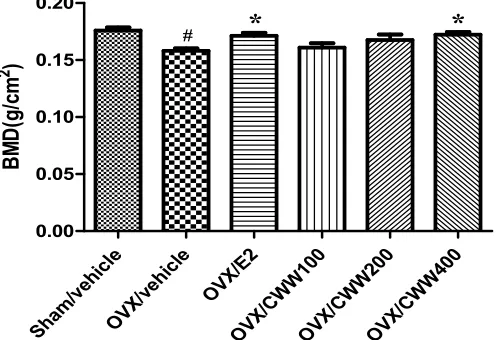
Effects of CWW on BMD, BMC, and projected bone
area
To investigate the anti-osteoporotic effects of CWW, the total femur BMD, BMC, and projected bone area (AREA) were measured using DEXA. Figure 2 shows that no differences occurred in the total
femur BMC and AREA between the OVX groups by the end of the treatment. However, ovariectomy significantly decreased the total femur BMD compared with that of the Sham/vehicle (p<0.05).After 24-week treatments, the final femur BMD of the OVX/E2 and OVX/CWW400 groups increased more significantly than that of the OVX/vehicle group did.
Sham /veh
icle
OVX /veh
icle OVX
/E2
OVX /CW
W10 0
OVX /CW
W20 0
OVX /CW
W40 0
0.00 0.05 0.10 0.15 0.20
*
*
#
B
M
D
(g
/c
m
2
)
PAGE |
466
|
Effect of CWW on histological morphology of femur
bone
Bone turnover is a life-long process involving two remodeling processes (bone resorption and formation). In this study, the histomorphology of the femur bone region of OVX rats was investigated using H&E staining. The femur region of the OVX group was small, thin, and sparse compared with that of the sham group. Furthermore, compared with the OVX group, the CWW
group showed less trabecular loss in the femurs. Figure 3 shows a representative histological section of the femur bone region of the Sham/vehicle and OVX/vehicle rats and those treated with E2 and CWW. Sections from sham-operated rats showed thick trabecular bone with relatively scant marrow space (Figure 3A). Ovariectomy markedly reduced the trabecular bone volume and increased the marrow space (Figure 3B) while24-week treatment with E2 and CWW markedly increased trabecular bone volume (Figure 3E and F), although E2 was more potent than CWW was (Figure 3C).
A. Sham/vehicle B. OVX/vehicle C. OVX/E2
D. OVX/CWW100 E. OVX/CWW200 F. OVX/CWW400
Figure 3. Histology of trabecular bone region of femur in rats. Sections were hematoxylin and eosin (H&E)-stained; scale bar = 100μm; original magnification, 40×.Red arrow indicates trabecular bone. Groups and treatments: Sham/vehicle, sham-operated, vehicle; OVX/vehicle. ovariectomized, vehicle; OVX/E2 ovariectomized, 17β-estradiol 91 μg/kg; and OVX/CWW100, 200, or 400, ovariectomized, aqueous extract of
Cynanchum wilfordii 100, 200, or 400 mg/kg, respectively.
Effect of CWW on serum biochemical markers in OVX
rats
PAGE |
467
|
by high bone turnover in the rats, as evidenced by high serum ALP, OCN, CTx, and TRAP levels. E2 treatment reversed the OVX-induced increase in ALP concentration. OCN markedly increased in OVX rats, but CWW and E2 treatment inhibited this effect. The serum levels of bone formation markers (ALP and OCN) significantly increased in the OVX/vehicle group compared to levels in the Sham/vehicle group. However, E2 and CWW decreased the
elevated serum ALP and OCN levels in the OVX rats (p< 0.05). Additionally, the serum TRAP level, which is responsible for enhanced osteoclastogenesis and activation of mature osteoclasts for bone resorption, was increased in the OVX/vehicle group, but CWW significantly reduced TRAP activity (p< 0.05). The increase in another bone resorption marker, CTx, in the OVX/vehicle group was also inhibited by CWW (p< 0.05).
Sham /veh icle OVX /veh icle OVX /E2 OVX /CW W10 0 OVX /CW W20 0 OVX /CW W40 0 0 100 200 300 400
*
*
## A L P (n g /m L ) Sha m/v ehic le OVX /veh icle OV X/E2 OVX /CW W10 0 OVX /CW W20 0 OVX /CW W40 0 0 50 100 150***
*
# R a t G la -O s te o c a lc in (n g /m L )Figure 4. Effect of aqueous Cynanchum wilfordii extract (CWW) on serum alkaline (ALP) and osteocalcin (OCN). Rats were treated for 24 weeks;# p< 0.05 and ## p< 0.01 vs Sham/vehicle, *p < 0.05and *** p< 0.005 vs. OVX/vehicle group. Groups and treatments: Sham/vehicle, sham-operated, vehicle; OVX/vehicle, ovariectomized, vehicle; OVX/E2 ovariectomized, 17β-estradiol 91 μg/kg; and OVX/CWW100, 200, or 400, ovariectomized, CWW 100, 200, or 400 mg/kg, respectively.
Sha m/v ehic le OV X/ve hicl e OV X/E 2 OV X/C WW 100 OV X/C WW 200 OV X/C WW 400 0 50 100 150
*
*
## T ra p a c ti v it y (% o f c o n tr o l) Sha m/v ehic le OV X/v ehic le OV X/E 2 OV X/C WW 100 OV X/C WW 200 OV X/C WW 400 0 20 40 60 80 100*
*
# C T x (n g /m L )Figure-5. Effect of aqueous extract of Cynanchum wilfordii (CWW) on bone metabolic markers. Effects of CWW on C-terminal cross-linked telopeptides of type I collagen (CTx) and tartrate-resistant acid phosphatase (TRAP) in serum after 24-week treatment;# p< 0.05 and ## p< 0.01
PAGE |
468
|
Discussion
Bone remodeling is regulated by a balance between the formation of new bone by osteoblasts and resorption of old bone by osteoclasts, and an imbalance in osteoclastogenesis causes the bone loss in osteoporosis [23]. M-CSF and RANKL are important cytokines that induce the differentiation of osteoclast precursors into activated osteoclasts [24]. Using in vitro and in vivo studies, we demonstrated the anti-osteoclastogenic activity of CWW, which exhibited anti-osteoporotic effects. In vitro, CWW effectively inhibited RANKL-induced osteoclast differentiation. Osteoporosis is a disease characterized by bone loss and deterioration of bone tissue, and patients exhibit lower bone density and bone mass than healthy individuals do [25]. Bone loss triggered by estrogen decline in experimental animals and humans is due to an increase in osteoclastic bone resorption [26]. OVX rats, which exhibit most of the characteristics of human postmenopausal osteoporosis [27], are widely used to evaluate potential osteoporosis treatments [28]. Although the exact mechanisms by which OVX increases body weight are not clear, a recent study reported that an estrogen-deficient state induced body fat accumulation and increased body weight [29]. OVX-rats show increased body weight compared to sham-operated rats while E2 treatment prevents body weight gain [30]. Estrogen deficiency induces body fat accumulation and subsequently increases body weight [29].Estrogen acts by binding to different ERs (ERα and ERβ). ERβ is more abundant than ERα is in bone tissue, while ERα, which, is the dominant receptor mediating the effects of E2 on the breast and uterus [31].
Selective estrogen receptor modulators (SERMs) are synthetic non-steroidal agents that exhibit ER agonistic or antagonistic activities depending on the target tissue. A novel approach to postmenopausal therapy is tissue-selective estrogen complex (TSEC) treatment, which pairs a SERM with one or more estrogens. The goals of TSEC are to provide the benefits of estrogens such as reduced hot flashes and vulvar-vaginal atrophy, thereby preventing menopausal osteoporosis events, and protecting the endometrium and breast tissue from estrogen stimulation [32]. In our experiments, oral administration of CWW did not affect body weight gain and uterotrophic activity in OVX rats. These results suggest that CWW might have anti-osteoporotic effects in OVX rats without the hormonal effects of estrogen. These results are in accordance with previous studies[33]. Therefore, it would be useful to study the interaction between C. wilfordii and ERs.
Bone loss is a major factor jeopardizing bone integrity, resulting in reduced bone strength and increased susceptibility to fracturing [34]. BMD is the gold standard for evaluating individuals who are at risk for osteoporosis because it best predicts the fracture risk in people without previous fractures [35]. Following ovariectomy, BMD markedly decreases owing to increased bone turnover in OVX rats
compared to the Sham rats. The current results reveal that OVX significantly decreased the total femur BMD compared with that of the sham group. CWW prevented OVX-induced bone loss of the total femur.
Bone turnover biochemical markers have been determined previously, and allow evaluation of the bone remodeling status [36]. ALP is a ubiquitous ectoenzyme, which catalyzes the hydrolysis of monophosphate ester groups. Four genes encode the isoenzymes, which are the intestinal, placental, germ cell, and tissue non-specific (bone/osteoblast, liver, and kidney isoforms) [37]. OCN, a critical non collagenous protein synthesized by osteoblasts, is primarily deposited in the bone extracellular matrix and considered a specific marker of osteoblast function[38]. The crosslinked telopeptides are released from type Icollagen degradation by proteases during bone resorption generate neoepitopes (CTX-I, ICTP, NTX-I). Serum ALP and OCN are phenotypic biomarkers of osteoblastic activity, and CTx and TRAP are widely accepted biomarkers of bone resorption. Serum ALP, OCN, CTx, and TRAP concentrations in OVX rat were significantly higher than those in the Sham group were while 24-week E2 or CWW treatments lowered the increased bone turnover. This was evidenced by the significant decreases in ALP, OCN, CTx and TRAP. In this study, we found that CWW prevented bone loss, suggesting it most likely prevented bone loss by decreasing bone turnover. Finally, we showed that CWW inhibited osteoclast differentiation, prevented bone mass reduction, and improve trabecular bone structure and biochemical markers in OVX-induced osteoporosis.
Conclusion
In conclusion, C. wilfordii inhibited osteoclast differentiation, prevented bone mass reduction, and improved trabecular bone structure and biochemical markers in OVX-induced osteoporosis. These results indicate that C. wilfordii has ameliorative effects on osteoporosis. Inaddition further studies are needed to clarify the molecular mechanisms.
Acknowledgements
This research was supported by the Support Program For Creative Industry Institutes (Commercial Biotechnology Sophistication Platform Construction Program, R0003950) funded by the Ministry of Trade, Industry & Energy (MOTIE, Korea).
Conflict of interest
All the authors state that there are no conflicts of interest within this article.
PAGE |
469
|
References
[1]. Raisz L. Pathogenesis of osteoporosis: concepts, conflicts, and prospects. J
Clin Invest. 2005; 115: 3318-3325. [2]. Papachroni K, Karatzas D,
Papavassiliou K, Basdra E, Papavassiliou A. Mechanotransduction in osteoblast regulation and bone disease. trends Mol Med. 2009; 15: 208-216.
[3]. Asagiri M, Takayanagi H. The molecular understanding of osteoclast differentiation. Bone. 2007;40: 250-64.
[4]. Arai, Miyamoto T, Ohneda O, Inada T, Sudo T, Brasel K, Miyata T, Anderson DM, Suda T. Commitment and differentiation of osteoclast precursor cells by the sequential expression of c-fms and receptor activator of nuclear factor kappa B (RANK) receptors. J Exp Med. 1999;19: 1741-1754.
[5]. Kirstein B, Chambers TJ, Fuller K. Secretion of tartrate resistant acid phosphatase by osteoclasts with resorptive behavior. J cell biochem. 2006; 98:1085-1094.
[6]. Hadjidakis D, Androulakis I. Bone remodeling. Ann N Y Acad Sci. 2006;1092:385-396.
[7]. Jung K, Lein M. Bone turnover markers in serum and urine as diagnostic, prognastic and monitoring biomarkers of bone metastasis. Biochim Biophys Acta. 2014;1846: 425-438.
[8]. Ferreira A, Alho I, Casimiro S, Costa L. Bone remodeling markers and bone metastases: From cancer research to clinical implications. Bone KEy Reports.2015;4:1-9.
[9]. Syed F, Khosla S. Mechanisms of sex steroid effects on bone. Biochem Biophys Res Commum. 2005;328:688-696.
[10].Turner R, Riggs B, Spelsberg T. Skeletal effects of estrogen. Endocr Rev. 1994;15:275-300.
[11].Hukkanen M, Platts LA, Lawes T, Girgis SI, Konttinen YT, Goodship AE, Maclntyre I, Polak JM. Effect of nitric oxide donor nitroglycerin on bone mineral density in a rat model of estrogen deficiency-induced osteopenia. Bone. 2003;32:142-149.
[12].Shin C, Wu Y, Lin W. Ameliorative effects of Anoectochilus formosanus
extract on osteopenia in ovariectomized rats. J Ethnopharmacol. 2001;77: 233-238.
[13].Ahldorg H, Johnell O, Turner CH, Rannevik G, Karlsson M. Bone loss and bone size after menopause. N Engl J Med. 2006;349: 327-334.
[14].Stevenson J. Justification for the use of HRT in the long-term prevention of osteoporosis. Maturitas. 2005; 51:113-126.
[15].prelevic G, Kocjan T, Markou A. Hormone replacement therapy in postmeno-pausal women. Minerva Endocrinol. 2005;30: 27-36.
[16].Banu J, Varela E, Fernandes G. Alternative therapies for the prevention and treatment of osteoporosis. Nutrition. 2012; 70: 22-40.
[17].Yang SB, Lee SM, Park J, Lee TH, Baek N, Park H, Lee H, Kim J. Cynandione A from Cynanchum wilfordii Attenuates the Production of Inflammatory Mediators in LPS-Induced BV-2 Microglial Cells via
NF-κB Inactivation. Bio Pharm Bull. 2014; 37:1390-1396.
[18].Chang A, Kwak B, Yi K, Kim J. The Effects of Herbal Extract (EstroG-100) on pre-, peri-and post-Menopausal Women: A Randomized Double-blind, Placebo-controlled study. Phytother Res. 2012;26: 510-516.
[19].Takahashi N, Yamada H, Yosiki S, Roodman G, Mundy G, Jones S, Boyde A, Suda T. Osteoclast-like cell formation and its regulation by osteotropic hormones in mouse bone marrow cultures. Endocrinology. 1998; 122: 1373-1382.
[20].Jarvinen T, Sievanen H, Kannus P, Jarvinen M. Dual-energy X-ray absorptiometry in predicting mechanical characteristics of rat femur. Bone. 1998; 22: 551-558.
[21].Kastl S, Sommer T, Klein P, Hohenberger W, Engelke K. Accuracy and precision of bone mineral density and bone mineral content in excised rat
humeri using fan beam dual-energy X-ray absorptiometry. Bone. 2002; 30: 243-246.
[22].Hidaka S, Okamoto Y, Yamada Y, Kon Y, Kimura T. A Japanese herbal medicine, Chujoto, has a beneficial effect on osteoporosis in rats. Phytother. Res. 1999; 13: 14-19.
[23].Ross F, Teitelbaum S. Alphavbeta and macrophage colony-stimulating factor: partnersin osteoclast biology. Immunol Rev. 2005; 208:88-105.
[24].Khosla S. Minireview: the OPG/RANKL/RANK system. Endocrinology, 2001; 142:5050-5055. [25].Levine J. Effective strategies to identify
postmenopausal women at risk for osteoporosis. Geriatrics. 2007; 62:22– 30.
[26].Hoegh-Andersen P, Tanko L, Andersen T, Lundberg C, Mo J, Heegaard A, Delaisse J, Christgau S. Ovariectomized rats as a model of postmenopausal osteoarthritis: Validation and application. Arthritis REs Ther. 2004;6: R169-R180.
[27].Jee W, Yao W. overview: animal models of osteopenia and osteoporosis. J
Musculoskel Neuron Interact. 2001;1: 193-207.
[28].Lelovas P, Xanthos T, Thoma S, Lyritis G, Dontas I. The laboratory rat as an animal model for osteoporosis research. Comp Med. 2008;58: 424-430.
[29].Dang Z, van Bezooijen R, Karperien M, Papapoulos S, Lowik C. Exposure of KS483 cells to estrogen enhances osteogenesis and inhibits adipogenesis. J Bone Miner Res. 2002;17: 394-405.
[30].Devareddy L, Khalil D, Smith B, Lucas E, Soung D, Marlow D, Arjmandi B. Soy moderately improves microstructural properties without affecting bone mass in an ovariectomized rat model of osteoporosis. Bone. 2006; 38:686-693. [31].Hewitt S, Korach K. Oestrogen receptor
PAGE |
470
|
[32].Kim J, Cho H, Kim Y. The role of estrogen in adipose tissue metabolism: insights into glucose homeostasis regulation. Endocr J. 2014;61: 1055-1067.
[33].Han S, Lee T, Jang J, Song H, Hong S, Kim Y, Han B. Mixture of extracts of
Cynanchum wilfordii and Phlomis umbrosa Turcz. does not have an estrogenic effect in ovariectomized rats. Korean J food SCitechnol. 2015;47:
667-675.
[34].Park J, Ha S, Kang T, Oh M, Cho M, Lee S, Park J, Kim S. Protective effect of apigenin on ovariectomy-induced bone loss in rats. Life Sci. 2008;82: 1217-1223.
[35].Cummings S, Bates D, Black D. Clinical use of bone densitometry: scientific review. JAMA. 2002;288:1889-1897. [36].Bahlous A, Kalai E, Hadj Salah M,
Bouzid K, Zerelli L. Biochemical
markers of bone remodeling: recent data of their applications in managing postmenopausal osteoporosis. Tunis Med. 2006; 84:751–757.
[37].McComb R, Posen S. Alkaline phosphatase. New York: Plenum Press.1979