ORIGINAL ARTICLE
Mortality outcomes of low-dose
computed tomography screening for lung
cancer in urban China: a decision analysis
and implications for practice
Zixing Wang
1†, Wei Han
1†, Weiwei Zhang
1, Fang Xue
1, Yuyan Wang
1, Yaoda Hu
1, Lei Wang
1, Chunwu Zhou
2,
Yao Huang
2, Shijun Zhao
2, Wei Song
3, Xin Sui
3, Ruihong Shi
4and Jingmei Jiang
1*Abstract
Background: Mortality outcomes in trials of low-dose computed tomography (CT) screening for lung cancer are inconsistent. This study aimed to evaluate whether CT screening in urban areas of China could reduce lung cancer mortality and to investigate the factors that associate with the screening effect.
Methods: A decision tree model with three scenarios (low-dose CT screening, chest X-ray screening, and no screen-ing) was developed to compare screening results in a simulated Chinese urban cohort (100,000 smokers aged 45–80 years). Data of participant characteristics were obtained from national registries and epidemiological surveys for estimating lung cancer prevalence. The selection of other tree variables such as sensitivities and specificities of low-dose CT and chest X-ray screening were based on literature research. Differences in lung cancer mortality (pri-mary outcome), false diagnoses, and deaths due to false diagnosis were calculated. Sensitivity analyses were per-formed to identify the factors that associate with the screening results and to ascertain worst and optimal screening effects considering possible ranges of the variables.
Results: Among the 100,000 subjects, there were 448, 541, and 591 lung cancer deaths in the low-dose CT, chest X-ray, and no screening scenarios, respectively (17.2% reduction in low-dose CT screening over chest X-ray screening and 24.2% over no screening). The costs of the two screening scenarios were 9387 and 2497 false diagnoses and 7 and 2 deaths due to false diagnosis among the 100,000 persons, respectively. The factors that most influenced death reduction with dose CT screening over no screening were lung cancer prevalence in the screened cohort, low-dose CT sensitivity, and proportion of early-stage cancers among low-low-dose CT detected lung cancers. Considering all possibilities, reduction in deaths (relative numbers) with low-dose CT screening in the worst and optimal cases were 16 (5.4%) and 288 (40.2%) over no screening, respectively.
Conclusions: In terms of mortality outcomes, our findings favor conducting low-dose CT screening in urban China. However, approaches to reducing false diagnoses and optimizing important screening conditions such as enrollment criteria for screening are highly needed.
Keywords: Lung cancer, Low-dose CT, Screening, Mortality outcome, Decision analysis
© The Author(s) 2017. This article is distributed under the terms of the Creative Commons Attribution 4.0 International License (http://creativecommons.org/licenses/by/4.0/), which permits unrestricted use, distribution, and reproduction in any medium, provided you give appropriate credit to the original author(s) and the source, provide a link to the Creative Commons license, and indicate if changes were made. The Creative Commons Public Domain Dedication waiver (http://creativecommons.org/ publicdomain/zero/1.0/) applies to the data made available in this article, unless otherwise stated.
Open Access
*Correspondence: [email protected] †Zixing Wang and Wei Han contributed equally to this work 1 Institute of Basic Medical Sciences, Chinese Academy of Medical Sciences, Beijing 100005, P. R. China
Background
Lung cancer is the most common malignant tumor and the dominant cause of cancer-related deaths in China [1]. The 5-year overall survival rate of patients with lung can-cer remains approximately 15%–18% even in developed countries [2], whereas for patients who undergo surgi-cal resection of stage I cancer this rate is well above 70% [3], highlighting the urgent need for early detection and treatment. First introduced in the 1990s, low-dose com-puted tomography (CT) has become the most promis-ing approach for lung cancer screenpromis-ing [4]. Increasing number of screening programs in North America [5], Japan [6], and Europe [7–14] have greatly augmented the volume of the evidence base concerning low-dose CT screening practice. However, in China, a country with 36% of all lung cancers worldwide [15], mortality out-come, which is the most important measure for assess-ing screenassess-ing effects, has not yet been evaluated despite numerous preliminary studies on diagnostic accuracy [16].
The United States National Lung Screening Trial (NLST) has reported an encouraging 20% reduction in lung cancer mortality with low-dose CT screening over chest X-ray screening [5]. However, uncertainties remain regarding the mortality outcomes of screening programs because of controversial results from other countries [7–14]. Two randomized controlled trials (RCTs) from Italy [7, 8] and one from Denmark [9] reported equal or slightly increased mortalities (not statistically significant) for low-dose CT screening compared with no screening. Five ongoing RCTs (from Germany [10], Italy [11], France [12], the Netherlands, Belgium and Hungary [13], and the United Kingdom [14]) have not yet released their mor-tality outcomes. Outcomes of screening programs may vary between settings because of diversity in participants’ characteristics and healthcare service conditions [17]. Therefore, there is a need to investigate whether early detection by low-dose CT screening could reduce lung cancer mortality in China and to investigate the varia-tions in screening effects before introducing such screen-ing countrywide. This study aimed to analyze differences in lung cancer mortality between three scenarios using available data from China and to identify factors that most strongly influence the outcomes in low-dose CT screening.
Methods
Study population
In this study, we simulated a cohort of 100,000 urban residents and offered them one-off lung cancer screening (baseline screening, i.e., prevalence screening). The age and sex structures of this cohort were based on data from the China Population & Employment Statistics Yearbook
2014 [18]. According to data on lung cancer incidence reported by the National Cancer Registry 2012 [19], age criterion for this cohort was set at 45–80 years. As there is currently no high-quality evidence-based recommen-dation on inclusion criteria for lung cancer screening in China [20], we adopted the relatively relaxed criterion of “smokers” as a requirement of the 100,000 participants, regardless of daily smoking dose, smoking years, and cumulative pack-years (this choice of criterion was made also because it was used in national surveys on smoking in China [21]). No other restrictions were imposed such as family history of lung cancer or individual history of pulmonary disease. However, we did exclude lung can-cer screening for special categories, such as patients with tuberculosis or human immunodeficiency virus infection and individuals with work-place exposure to asbestos, coal dust, nuclear radiation, organic solvents, and fuels.
Decision tree model
Decision analysis is widely used to make choices between different paths in Chaotics and uncertain conditions [22], especially in healthcare studies where medical out-comes such as mortality depend on a great number of potentially influencing factors [23]. To identify the best approach to detecting lung cancer so as to improve out-comes and to test the degree of uncertainty, we consid-ered and compared the following three possible paths for lung cancer detection and treatment (Fig. 1).
Low‑dose CT screening scenario
Individuals with findings suggestive of lung cancer on low-dose CT images were asked to undergo further investigation, such as bronchoscopy and percutaneous biopsy, to determine the diagnosis and then either accept or refuse surgical treatment. Additionally, we set up branches in the path corresponding with “missed diag-nosis” caused by false negative imaging results (merging into the no screening path described below), “false diag-nosis” caused by false positive imaging results, and the “over diagnosis” issue (individuals with indolent cancer who live for a long time though untreated) recently raised by researchers [24].
Chest X‑ray screening scenario
The overall path was similar to that in the low-dose CT screening scenario, with the only difference being that the variables were adjusted to chest X-ray screening.
No screening scenario
All the three scenarios were stratified by early (stage I for non-small cell lung cancers and limited stage for small cell lung cancers) and non-early stage cancers, consider-ing the impact of disease stage on the acceptance rate of surgery and survival possibility after surgery.
Additionally, the following assumptions were made for the decision tree. First, surgery is a determinant for lung cancer mortality (except for indolent cancers), and although non-surgical treatments could prolong patient survival, their small impact on the final outcomes was not taken into account [25]. Second, a 5-year time span was adopted. As the screened cohort had a high compet-ing risk from other diseases, deaths beyond the 5-year range were not considered in the outcome evaluation.
Parameter setting
Table 1 lists all the variables and the estimated values of parameters used in the decision tree. Specifically, the selec-tion of screening parameters of low-dose CT and chest X-ray and the clinical parameters of diagnosis and treat-ment path were based on a systematic search of literatures (mainly from studies in China, together with a few studies in other countries, all extracted from MEDLINE, EMBASE, CNKI, and ChinaInfo databases and published between January 1990 and February 2016). Quality-weighted meta-analyses were performed to obtain the average estimate of parameter for each tree variable for base-case analysis (analysis based on the most likely estimates of parameters) and a reliable range for sensitivity analysis.
Lung cancer prevalence of the screened cohort was cal-culated as follows.
First, age- and sex-specific lung cancer incidences in the general Chinese urban population (IG) were extracted from the China Cancer Registry 2012 (urban data) at age intervals of 5 years [19]. Next, the incidence of the screened cohort (IS) for each age and sex group was cal-culated using the formula below:
where R is the age- and sex-specific rate of smoking reported in the Global Adult Tobacco Survey (GATS) China 2010 Country Report [26] and OR (2.85 for men
IS=
OR×IG
1+(OR−1)×R
and 2.33 for women) is the odds ratio according to meta-analysis results based on five case–control studies in China that used newly incident lung cancers to estimate the degree of association between smoking and lung can-cer between 2001 and 2014 [27–31].
Further, because lung cancer prevalence is relatively stable in China, the following formula was used to calcu-late age- and sex-specific lung cancer prevalence in the screened cohort (PS):
where t = 3.0 years (sensitivity range 1.5–3.5 years) is the average course of lung cancer [32].
Finally, the overall prevalence of lung cancer in the screened cohort aged 45–80 years was calculated by standardizing the age- and sex-specific prevalence using the actual demographical structure reported in the China Population & Employment Statistics Yearbook 2014 [18] and the smoking rates in the GATS China 2010 Country Report [26].
Quality-adjusted life year (QALY) [33] was analyzed for the three scenarios over a 5-year time span. Qual-ity utilities that were incorporated into the model were 0.76 for individuals without lung cancer [34], 0.62 for the time from detection of lung cancer to surgical treatment [35], 0.67 for early- and 0.55 for non-early-stage cancer patients after surgical treatment [35], and 0.56 for those who refused surgical treatment after detection, regard-less of cancer stage and presence of other treatments [35]. We used an average delay of 0.5 years to lung can-cer detection in the no screening scenario compared with the two screening scenarios, and incorporated an interval of 0.25 years for all the individuals with lung cancer from detection to surgical treatment (by expert consultation). The mean survival within the 5-year time span was esti-mated as 4.31 and 2.99 years for individuals with resected early- and non-early-stage cancers, respectively [3], and as 2.78 and 0.71 years for individuals with non-resected early- and non-early-stage cancers, respectively [36].
Additionally, to explore the influences of criteria of age, smoking, and sex on the prevalence of lung cancer in the screened cohort (and therefore on screening outcomes), with the same procedures described above, we further
PS=IS×t (See figure on previous page.)
Fig. 1 Decision tree model for the analysis of lung cancer mortality, false positive diagnosis, and death due to false diagnosis with low-dose computed tomography (CT) screening and no screening. Chest X-ray screening is similar to low-dose CT screening and is not shown. prev lung cancer prevalence in the screened cohort, CTse sensitivity of low-dose CT, CTsp specificity of low-dose CT, CTerl proportion of early-stage cancers among lung cancers detected with low-dose CT, NSerl proportion of early-stage cancers among lung cancers detected with no screening, fpMt
death possibility due to false diagnosis and invasive treatment, trt_Er acceptance rate of surgery for individuals with early-stage lung cancers, trt_Ne
calculated different PS for 12 plausible sub-intervals within the 45–80-year age range to determine the best lower and upper age limits in terms of mortality, and PS for men and women separately, each with 11 different proportions of smoking among the enrolled individuals if screening was not restricted to smokers.
Statistical analysis
Lung cancer death was the primary outcome measure in this study. Using base-case analysis, we calculated the absolute and relative reductions in lung cancer deaths with low-dose CT screening over chest X-ray screen-ing and no screenscreen-ing. False diagnosis, death due to false diagnosis, and QALY as secondary outcome measures were simultaneously calculated and compared. Univari-ate sensitivity analysis (by separUnivari-ately letting each variable in the model fluctuate within its lower and upper ranges for sensitivity analysis while keeping the others at their
base-case analysis values) was performed to investigate the influences of screening conditions on outcomes in different scenarios, and a tornado diagram was plotted to vividly display the magnitude of the influence of each variable (relative importance of the variables was ranked by percentages of variations in the outcomes due to their own influence over the variation summary due to influ-ence of all variables). Optimal and worst cases of reduc-tions in lung cancer deaths with low-dose CT screening over other scenarios were determined by considering combinations of all possible ranges of the variables. All the analyses were performed with Treeage Pro 2011 soft-ware (TreeAge Softsoft-ware, Inc, Williamstown, MA, USA).
Results
Base‑case analysis
Age and sex distribution of the simulated cohort in the base-case analysis is displayed in Table 2. Among these
Table 1 Parameter settings in the decision tree for lung cancer screening in urban China
CT computed tomography
a Stage I for non-small cell lung cancers and limited stage for small cell lung cancers were defined as early-stage cancers, and others as non-early-stage cancers b Lung cancers such as bronchioloalveolar carcinoma that did not affect survival for a long time if left untreated were defined as indolent cancers
c Based on 5-year estimation
d Seven studies that were conducted among the Chinese population with complete stage information [85–91] in this meta-analysis were included and reanalyzed in
this study
e Eight studies with complete stage information [92–99] in this meta-analysis were included and reanalyzed in this study Variable Definition Base‑case
analysis (%) Sensitivity analysis (%) Reference source Lower limit Upper limit
prev Lung cancer prevalence in the screened cohort 0.7666 0.3833 0.8943 Calculated from [18, 22, 26, 32, 53]
CTse Sensitivity of low-dose CT 87.7 71.8 100.0 Meta of [21, 54–66]
CTsp Specificity of low-dose CT 90.6 86.3 91.1 Meta of [55, 57–60, 63, 66–69]
XRse Sensitivity of chest X-ray 65.1 61.4 69.4 Meta of [21, 55–57, 60, 62–65, 70]
XRsp Specificity of chest X-ray 97.5 89.5 98.4 Meta of [55, 57, 60, 63, 69, 71, 72]
CTerl Proportion of early-stage cancera among lung cancers
detected with low-dose CT 70.1 63.9 76.0 [73]
XRerl Proportion of early-stage cancera among lung cancers
detected with chest X-ray 46.6 38.1 55.0 [73, 74]
NSerl Proportion of early-stage cancersa among lung cancers
detected with no screening 27.9 23.2 32.7 Meta of [24, 55, 76–7952
d, 75e, ]
CTidl Proportion of indolent cancersb among lung cancers
detected with low-dose CT 25.0 15.0 35.0 [20]
XRidl Proportion of indolent cancersb among lung cancers
detected with chest X-ray 20.0 15.0 25.0 [20]
trt_Er Acceptance rate of surgery for early-stage lung cancersa 72.5 68.2 76.0 Meta of [80–82]
trt_Ne Acceptance rate of surgery for non-early-stage lung
cancersa 28.6 28.2 30.6 Meta of [80–82]
srv_Er Survival possibility of individuals with resected
early-stage lung cancerc 72.7 62.3 75.7 Meta of [3, 35, 83]
srv_Ne Survival possibility of individuals with resected
non-early-stage lung cancerc 39.7 30.2 42.4 Meta of [3, 35, 83]
fpMt Death possibility due to false diagnosis and invasive
100,000 urban smokers (94,012 men and 5988 women) aged 45–80 years, 721 men and 46 women had lung can-cers. As to the outcomes in the three scenarios for the same cohort, the number of lung cancer deaths in the low-dose CT screening scenario was 448, a reduction of 143 (24.2%) over the no screening scenario (591 lung can-cer deaths); meanwhile, low-dose CT screening resulted in 9387 false diagnoses and 7 deaths due to false diagno-sis. In the chest X-ray screening scenario, there were 541 lung cancer deaths, a reduction of 50 (8.5%) over the no screening scenario, and chest X-ray screening resulted in 2497 false diagnoses and 2 deaths due to false diag-nosis. There were 93 fewer lung cancer deaths (17.2%) in the low-dose CT screening scenario than in the chest X-ray screening scenario; however, false diagnoses and deaths due to false diagnosis were 3.76 and 3.50 times higher, respectively, in the former scenario. Additionally, QALYs were 378,427 years in low-dose CT screening, 378,280 years in chest X-ray screening, and 378,177 years in no screening scenarios, that is, low-dose CT screen-ing resulted in slightly more QALYs: 147 years over chest X-ray screening and 250 years over no screening.
Univariate sensitivity analysis
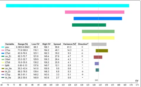
Lung cancer prevalence in the screened cohort was the factor that most strongly influenced the death reduction with low-dose CT screening over no screening (Fig. 2). The variation in prevalence (from 383.3 to 894.3 per 100,000) led to a great variation in the absolute reduction in lung cancer deaths (from 68 to 168), being responsi-ble for 61.3% of the all-variaresponsi-ble influence. Other six fac-tors besides the lung cancer prevalence contributed positively to the death reduction benefit in the following
order: low-dose CT sensitivity (14.3% of the all-variable influence), proportion of early-stage cancers among lung cancers detected with low-dose CT (9.5%), survival possibility of individuals with resected early-stage lung cancer (4.7%), proportion of indolent cancers among lung cancers detected with low-dose CT (4.1%), accept-ance rate of surgery for early-stage lung caccept-ancers (0.3%), and specificity of low-dose CT (0.1%). These six factors together contributed 33.0% to the variation in reduc-tion in lung cancer deaths. Four factors were negatively related with reduction in lung cancer deaths with low-dose CT screening over no screening, namely proportion of early-stage cancers among lung cancers detected with no screening (4.3%), possibility of death due to false diag-nosis and invasive treatment (0.9%), survival possibility of patients with resected non-early-stage lung cancer (0.6%), and acceptance rate of surgery for non-early-stage lung cancers (0.01%); however, their cumulative influence was subtle (5.8%).
For the secondary outcome measures (not shown in figures), the factor that most influenced the number of false diagnosis was specificity of low-dose CT (nega-tively contributing 99.9% to variation in increased false diagnoses); lung cancer prevalence had a tiny negative influence (0.01%). Three factors were related to num-ber of deaths due to false diagnosis with low-dose CT screening, namely, death possibility due to false diag-nosis (93.0%, positive), low-dose CT specificity (7.0%, negative), and lung cancer prevalence (0.01%, negative). The factor that most influenced DALY was lung cancer prevalence (58.7%), followed by low-dose CT sensitivity (13.5%), low-dose CT specificity (11.0%), and proportion of early-stage cancers among lung cancers detected with
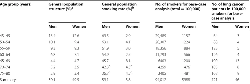
Table 2 Age and sex distribution of the simulated smoking cohort for base-case analysis
a Obtained from Sampling Survey Data of the National Population Change in 2013 in China Population & Employment Statistics Yearbook [18] b Obtained from Global Adult Tobacco Survey (GATS) China 2010 Country Report [26]
c Predicted using linear regression
Age group (years) General population
structure (%)a General population smoking rate (%)b No. of smokers for base‑case analysis (total
= 100,000) No. of lung cancer patients in 100,000 smokers for base‑ case analysis
Men Women Men Women Men Women Men Women
45–49 13.4 12.6 69.5 2.9 29,489 1157 64 3
50–54 10.1 9.4 63.1 4.1 20,307 1224 88 4
55–59 9.3 9.3 61.9 3.0 18,356 884 123 5
60–64 6.8 7.1 54.9 2.5 11,793 566 126 4
65–69 4.4 4.7 45.7 8.1 6403 1200 109 13
70–74 3.2 3.5 42.3c 4.3c 4259 476 103 8
75–80 2.9 3.4 36.7c 4.5c 3405 481 108 9
low-dose CT (10.0%; all positive). Notably, a threshold of QALY was detected in lung cancer prevalence (436 per 100,000); the gain of QALY in low-dose CT screening over no screening would diminish if screening was per-formed among populations with a low prevalence of lung cancer.
Worst and optimal case analysis
Considering the possible ranges of all influential fac-tors, reductions in lung cancer deaths with low-dose CT screening over no screening were 281 versus 297 (16 absolute reduction, 5.4% relative reduction) in the worst case and 428 versus 716 (288, 40.2% reduction) in the optimal case, and those over chest X-ray screening were 281 versus 270 (−11, −4.1% reduction) in the worst case and 428 versus 669 (241, 36.0% reduction) in the optimal case.
Influence of age criteria
The influences of upper and lower age limits on preva-lence of lung cancer in the screened cohort and on dif-ferences in numbers of lung cancer deaths between the three scenarios are shown in Table 3. Relative to no screening, more than 200 fewer lung cancer deaths (24.5% relative reduction) could be achieved by setting the age criteria for low-dose CT screening at 55–75, 55–80, 60–70, 60–75, or 60–80 years. Similarly, these intervals for age criteria could lead to more than 130 fewer deaths (17.5% relative reduction) with low-dose CT screening over chest X-ray screening. However, some of these age intervals covered relatively small portions of the total lung cancer patients among the whole popu-lation of 45–80 years old, e.g., only 32.9% and 47.3% of patients with lung cancer would be covered if age crite-ria of 60–70 and 60–75 years were adopted, respectively. Fig. 2 Tornado histogram of lung cancer deaths in the low-dose CT screening scenario over the no screening scenario. EV effect value, horizontal line represents the relative difference in lung cancer deaths with low-dose CT screening over no screening (the higher the EV, the higher the screen-ing benefits), and vertical dash line indicates the location of estimated average EV, prev lung cancer prevalence in the screened cohort, CTse sensitiv-ity of low-dose CT, CTerl proportion of early-stage cancers among lung cancers detected with low-dose CT, srv_Er survival possibility of patients with resected early-stage lung cancers, NSerl proportion of early-stage cancers among lung cancers detected with no screening, CTidl proportion of indolent cancers among lung cancers detected with low-dose CT, fpMt death possibility due to false diagnosis and invasive treatment, srv_Ne
survival possibility of individuals with resected non-early-stage lung cancer, trt_Er acceptance rate of surgery for early-stage lung cancers, CTsp
The age interval of 50–75 years currently recommended by the Chinese expert consensus for lung cancer screen-ing [20] covered 76.0% of all lung cancer patients aged 45–80 years, but the reduction in lung cancer deaths with low-dose CT screening was relatively low, 109 (17.4%) and 164 (24.3%), respectively, as compared with chest X-ray screening and no screening.
Influence of smoking and sex criteria
Figure 3 shows that when not restricted to smokers, the reduction in lung cancer deaths with low-dose CT screening over chest X-ray screening and no screen-ing increased in parallel with the proportion of smokers in the screened cohort, both among men and women.
For example, compared with no screening, the relative reduction in lung cancer deaths among men undergo-ing low-dose CT screenundergo-ing increased from 23.2% for non-smokers to 24.2% for smokers, and the absolute screening benefit doubled from 69 to 143 fewer deaths. Similarly, the absolute benefit gained by low-dose CT screening also doubled compared with chest X-ray screening (from 45 to 93 fewer deaths) for men. Nota-bly, among women the relative reduction in lung cancer deaths with low-dose CT screening was as great as that for men when screening 100% smokers (24.2% over no screening and 17.2% over chest X-ray screening), and the absolute reduction in lung cancer deaths with low-dose CT screening almost tripled both compared with no
Table 3 Prevalence of lung cancer among 100,000 smokers and predicted lung cancer deaths in low-dose CT, chest X-ray, and no screening scenarios with different age inclusion criteria
a Proportion of lung cancer patients within the listed age interval among all lung cancer patients aged 45–80 years in urban China Age interval
(years) Lung cancer prevalence (per 100,000)
Target coverage
(%)a Lung cancer deaths (cases) Death reduction [cases (%)]
Low‑dose CT Chest X‑ray No screening Low‑dose CT
over chest X‑ray Low‑dose CT over no screening
45–70 589.2 70.2 346 417 454 71 (17.0) 108 (23.8)
45–75 675.4 84.7 395 477 521 82 (17.2) 126 (24.2)
45–80 766.6 100.0 448 541 591 93 (17.2) 143 (24.2)
50–70 777.0 61.6 454 549 599 95 (17.3) 145 (24.2)
50–75 890.0 76.0 519 628 686 109 (17.4) 167 (24.3)
50–80 1009.4 91.3 588 712 778 124 (17.4) 190 (24.4)
55–70 967.8 49.5 564 683 746 119 (17.4) 182 (24.4)
55–75 1115.6 63.9 649 787 860 138 (17.5) 211 (24.5)
55–80 1270.4 79.3 738 896 980 158 (17.6) 242 (24.7)
60–70 1263.0 32.9 734 891 974 157 (17.6) 240 (24.6)
60–75 1469.2 47.3 852 1036 1133 184 (17.8) 281 (24.8)
60–80 1680.2 62.7 974 1185 1296 211 (17.8) 322 (24.8)
screening (from 49 in non-smokers to 143 in smokers) and chest X-ray screening (from 32 to 93).
Discussion
This study evaluated the outcomes of lung cancer screen-ing in China, includscreen-ing the influences of several factors on screening effects. According to the base-case analysis of 100,000 urban smokers aged 45–80 years, low-dose CT screening decreased lung cancer deaths by 24.2% over no screening and by 17.2% over chest X-ray screening. There were fewer deaths even in the worst case analysis (lung cancer deaths of 245 vs 269 for low-dose CT screening and no screening, respectively; 8.9% relative reduction), and the reduction was as high as 43.6% (lung cancer deaths of 364 vs 645 in low-dose CT screening and no screening, respectively) in an optimal situation. Thus, in terms of mortality outcome, our findings indicate that screening old smokers in urban China with low-dose CT would reduce lung cancer deaths. However, the effect of low-dose CT screening on lung cancer death reduc-tion was inferior to that of chest X-ray screening in the worst situation (lung cancer deaths of 281 vs 270 for low-dose CT and chest X-ray screenings, respectively; −4.1% relative reduction). On average, low-dose CT screening resulted in more than three-fold false diagnoses and even deaths due to false diagnosis and over-treatment. Addi-tionally, the gain in QALYs with low-dose CT screening (250 years over no screening and 147 years over chest X-ray screening) was not as pronounced as that for mor-tality. Thus, better screening strategies are required to optimize the outcomes of low-dose CT screening.
Being a country with a large lung cancer burden (with more than 3,500,000 new cases and more than 2,000,000 fatal cases each year [37]), China has a considerable interest in the detection of early-stage lung cancer and prevention of lung cancer deaths via screening [16, 17]. Several studies have published preliminary results for the comparative ability of low-dose CT screening over chest X-ray screening to detect lung cancer and for other screening variables such as screening-detected cancer stage, pathological, and individual characteristics [16, 38]. In 2010, a demonstration program was initiated at three centers to test the feasibility of conducting popu-lation-based screening [39]. Two years later, the govern-ment incorporated urban lung cancer screening into a key national public health program [16]. Since 2013, approximately 8000 Beijing citizens have been screened in another program for 3 years using low-dose CT to optimize the screening protocol [16]. Most studies have reported rates of detection, early diagnosis, and early treatment as the main outcomes; however, none have yet reported the endpoint of lung cancer mortality, both because it takes so long to obtain this important but
time-consuming endpoint of screening and because a great many participants are needed to identify mortality differences at a low cancer prevalence. Thus, this analysis of currently available domestic data can provide infor-mation to the current screening practice before mortal-ity outcome studies are available in China (which will not be very soon). The failure of small trials in Europe to detect any reduction in mortality has usually been attrib-uted to insufficient in subject number (fewer than 2000 in low-dose CT arms) [7–9]. Only the statistically powered NLST study, with a sample size of 53,454, has detected a 20% mortality reduction with low-dose CT screen-ing over chest X-ray screenscreen-ing [5], which was why we simulated 100,000 participants to obtain robust results. The relatively smaller reduction in lung cancer deaths of 17.2% with low-dose CT screening over chest X-ray screening in the present study compared with the 20% in the NLST may be attributable to differences in enroll-ment criteria [40] (such as smokers in the present study and heavy smokers in the NLST), other population char-acteristics, and quality of healthcare services.
in lung cancer deaths was not quite pronounced in this study with the relaxed inclusion criteria. It is noteworthy that when the emphasis is on heavy smokers and other criteria for screening are imposed (such as those in the Chinese Consensus on Early Diagnosis of Primary Lung Cancer [20]), a greater screening benefit can be expected compared with nonselective criteria [40]. Considering the important contribution of lung cancer prevalence to screening benefit, a combination of other methods such as biomarkers to identify high-risk individuals would also likely to increase screening benefits [41].
Regarding other factors that influenced screening effects, the sensitivity of low-dose CT had a relatively important positive influence on the reduction in lung cancer deaths; so was the proportion of early-stage can-cer among lung cancan-cers detected with low-dose CT screening, reinforcing the need to develop highly sensi-tive screening techniques [42]. The positive relation-ship between survival possibility after early treatment of cancer and reduction in deaths emphasizes the need to improve the effectiveness of early treatment to achieve better prognoses and screening benefits. Given the effect of acceptance of treatment on outcomes, screening pro-grams should also consider incorporating approaches to enhancing willingness and financial ability of patients with lung cancer to undergo recommended treatment, particularly in developing countries. Additionally, our findings indicate that survival rates of non-early-stage cancer patients have a very limited impact on mortality outcomes.
For individuals who do not have lung cancers, false pos-itive results remain the most critical issue of low-dose CT screening [43–45]. This is reinforced by the higher num-bers of false diagnoses and deaths due to false diagnosis with low-dose CT screening compared with chest X-ray screening in the present study. Because the specificity of low-dose CT directly influences such outcomes, further efforts should be put into increasing the discriminative ability of imaging techniques and their computer-aided diagnostic systems [46], developing optimal diagnostic thresholds [44, 45], and employing other imaging tech-niques (such as positron emission tomography) [47] or other non-imaging approaches (such as highly specific biomarkers) [41, 48, 49]. Additionally, standardization of workflow for diagnosis and treatment, in addition to screening, is indispensable to eliminating unnecessary cost and morbidities associated with lung cancer screen-ing programs [50].
In our study, gains in QALYs of low-dose CT screen-ing over chest X-ray screenscreen-ing and no screenscreen-ing were less impressive than reduction in lung cancer deaths. This was mainly because unscreened individuals with lung cancer can enjoy a higher quality of life during the
symptom-free interval before diagnosis of their cancers, whereas individuals with false-positive diagnoses experi-ence a decreased quality of life as a result of anxiety and unnecessary diagnosis or treatment. For example, in the base-case analysis, these two sources resulted in loss of 562 QALYs in the low-dose CT screening scenario than in the no screening scenario (counterbalanced primarily by the stage-shift benefit of 812 QALYs, which led to the final 250-year gain). Thus, close attention should also be paid to physiological reactions if we wish to provide par-ticipants in the low-dose CT screening scenario with bet-ter quality of life [51].
Limitations remain in this study. First, the decision tree we used is a simplified representation of complex clinical paths and individuals’ behaviors. For example, we dichotomized the distribution of cancer stages into early and non-early; full I–IV staging (especially with associ-ated differences in survival) would have made the model more accurate. This was mainly because of lack of large domestic studies from which we could draw robust esti-mates of screening and clinical parameters. Additionally, the absence of transition possibilities in lung cancer stage among the extremely heterogeneous histological types of lung cancer prevented us from employing stage tran-sition Markov models. Second, for the same reasons, we only considered baseline screening in this study, whereas some programs in China already adopted a baseline and annual repeat screening fashion [52], in which higher proportion of early-stage lung cancers in repeat rounds could be expected. Thus, longitudinal screening param-eters from such programs and their impact on mortality reduction are highly needed for further decision analyses. Third, although false diagnoses, missed diagnoses, and over-diagnoses of indolent cancers were considered, we did not consider complex issues such as passive smoking and competing risk from other diseases in old individu-als, but left these for future studies. Finally, the lack of expenditure data at the national level and differences in costs of lung cancer treatment between different regions thus far prevents evaluation of the cost-effectiveness of lung cancer screening. Thus, the balance between input and yield requires further in-depth evaluation.
Conclusions
methods, and adapting screening protocols according to availability of funding and other local conditions.
Authors’ contributions
JJ and CZ conceptualized this study. FX and WS formulated the overall study design. YW, YH, and LW did the literature review and participated in the meta-analysis. YH, SZ, and XS participated in constructing the decision tree and provided considerable input to the discussion of practical implication. ZW and WH performed the formal analysis and wrote the manuscript. WZ contributed greatly to writing the final draft. RS validated the analysis results and refined the manuscript. All authors read and approved the final manuscript.
Author details
1 Institute of Basic Medical Sciences, Chinese Academy of Medical Sciences, Beijing 100005, P. R. China. 2 Cancer Hospital, Chinese Academy of Medical Sciences, Beijing 100021, P. R. China. 3 Peking Union Medical College Hospital, Chinese Academy of Medical Sciences, Beijing 100730, P. R. China. 4 National Institutes for Food and Drug Control, State Food and Drug Administration, Beijing 100050, P. R. China.
Acknowledgements Not applicable.
Competing interests
The authors declare that they have no competing interests.
Funding
This study was supported by Peking Union Medical College Youth Fund and the Fundamental Research Funds for the Central Universities (No. 2017310049).
Received: 29 August 2016 Accepted: 16 January 2017
References
1. Chen W, Zheng R, Baade PD, Zhang S, Zeng H, Bray F, et al. Cancer statistics in China, 2015. CA Cancer J Clin. 2016;66(2):115–32. doi:10.3322/ caac.21338.
2. American Cancer Society. Cancer facts & figures 2014. http://www.cancer. org/research/cancerfactsstatistics/cancerfactsfigures2014/. Accessed 28 Aug 2016.
3. Liang W, Shao W, Jiang G, Wang Q, Liu L, Liu D, et al. Chinese multi-insti-tutional registry (CMIR) for resected non-small cell lung cancer: survival analysis of 5,853 cases. J Thorac Dis. 2013;5(6):726–9. doi:10.3978/j. issn.2072-1439.2013.12.32.
4. Wood DE, Eapen GA, Ettinger DS, Hou L, Jackman D, Kazerooni E, et al. Lung cancer screening. J Natl Compr Canc Netw. 2012;10(2):240–65. 5. National Lung Screening Trial Research Team, Aberle DR, Adams AM, Berg
CD, Black WC, Clapp JD, et al. Reduced lung-cancer mortality with low-dose computed tomographic screening. N Engl J Med. 2011;365(5):395– 409. doi:10.1056/NEJMoa1102873.
6. Nawa T, Nakagawa T, Mizoue T, Kusano S, Chonan T, Hayashihara K, et al. A decrease in lung cancer mortality following the introduction of low-dose chest CT screening in Hitachi, Japan. Lung Cancer. 2012;78(3):225–8. doi:10.1016/j.lungcan.2012.09.012.
7. Infante M, Cavuto S, Lutman FR, Brambilla G, Chiesa G, Ceresoli G, et al. A randomized study of lung cancer screening with spiral computed tomography. Am J Respir Care Med. 2009;180(5):445–53. doi:10.1164/ rccm.200901-0076OC.
8. Pastorino U, Rossi M, Rosato V, Marchianò A, Sverzellati N, Morosi C, et al. Annual or biennial CT screening versus observation in heavy smokers: 5-year results of the MILD trial. Eur J Cancer Prev. 2012;21(3):308–15. doi:10.1097/CEJ.0b013e328351e1b6.
9. Saghir Z, Dirksen A, Ashraf H, Bach KS, Brodersen J, Clementsen PF, et al. CT screening for lung cancer brings forward early disease. The randomised Danish Lung Cancer Screening Trial: status after five annual
screening rounds with low-dose CT. Thorax. 2012;67(4):296–301. doi:10.1136/thoraxjnl-2011-200736.
10. Becker N, Motsch E, Gross ML, Eigentopf A, Heussel CP, Dienemann H, et al. Randomized study on early detection of lung cancer with MSCT in Germany: results of the first 3 years of follow-up after randomization. J Thorac Oncol. 2015;10(6):890–6. doi:10.1097/JTO.0000000000000530. 11. Lopes Pegna A, Picozzi G, Falaschi F, Carrozzi L, Falchini M, Carozzi FM, et al. Four-year results of low-dose CT screening and nodule manage-ment in the ITALUNG trial. J Thorac Oncol. 2013;8(7):866–75. doi:10.1097/ JTO.0b013e31828f68d6.
12. Blanchon T, Bréchot JM, Grenier PA, Ferretti GR, Lemarié E, Milleron B, et al. Baseline results of the Depiscan study: a French randomized pilot trial of lung cancer screening comparing low-dose CT scan (LDCT) and chest X-ray (CXR). Lung Cancer. 2007;58(1):50–8.
13. Horeweg N, Scholten ET, de Jong PA, van der Aalst CM, Weenink C, Lam-mers JW, et al. Detection of lung cancer through low-dose CT screening (NELSON): a prespecified analysis of screening test performance and interval cancers. Lancet Oncol. 2014;15(12):1342–50. doi:10.1016/ S1470-2045(14)70387-0.
14. McRonald FE, Yadegarfar G, Baldwin DR, Devaraj A, Brain KE, Eisen T, et al. The UK lung screen (UKLS): demographic profile of first 88,897 approaches provides recommendations for population screening. Cancer Prev Res (Phila). 2014;7(3):362–71. doi:10.1158/1940-6207.CAPR-13-0206. 15. Globocan 2012: Estimated cancer incidence, mortality and prevalence
world-wide in 2012. http://globocan.iarc.fr/Default.aspx. Accessed 28 Aug 2016. 16. Zhao SJ, Wu N. Early detection of lung cancer: low-dose computed
tomography screening in China. Thorac Cancer. 2015;6(4):385–9. doi:10.1111/1759-7714.12253.
17. World Health Organization. Global heath observatory (GHO) data. http:// www.who.int/gho/health_workforce/en/. Accessed 28 Aug 2016. 18. Sampling survey data of the national population change in 2013. In:
China Population & Employment Statistics Yearbook 2014. China Eco-nomic and social development statistics database. http://tongji.cnki.net/ kns55/Navi/result.aspx?id=N2016010131&file=N2016010131000039&fl oor=1. Accessed 11 Dec 2016.
19. Chen W, He J. Annual report of cancer registration in China 2012. Beijing: Military Medical Science Press; 2012.
20. Zhou QH, Fan YG, Bu H, Wang Y, Wu N, et al. China national lung cancer screening guideline with low-dose computed tomography (2015 ver-sion). Thorac Cancer. 2015;6(6):812–8.
21. Zhang M, Wang LM, Li YC, Li XY, Jiang Y, Hu N, et al. Cross-sectional survey on smoking and smoking cessation behaviors among Chinese adults in 2010. Chin J Prev Med. 2012;46(5):404–8. doi:10.3760/cma.j.i ssn.0253-9624.2012.05.006(in Chinese).
22. Moussa M, Ruwanpura JY, Jergeas G. Decision tree module within deci-sion support simulation system. In: Proceedings of the IEEE simulation conference. 2004. p. 1268–76.
23. Burton A, Altman DG, Royston P, Holder RL. The design of simulation studies in medical statistics. Stat Med. 2006;25(24):4279–92.
24. Reich JM. A critical appraisal of overdiagnosis: estimates of its magnitude and implications for lung cancer screening. Thorax. 2008;63(4):377–83. doi:10.1136/thx.2007.079673.
25. Hurria A, Kris MG. Management of lung cancer in older adults. CA Cancer J Clin. 2003;53(6):325–41.
26. CDC China. Global adult tobacco survey (GATS) China 2010 country report. Shanghai: China Three Gorges Press; 2011 (in Chinese). 27. Zhong L, Goldberg MS, Gao YT, Hanley JA, Parent ME, Jin F. A
population-based case–control study of lung cancer and green tea consumption among women living in Shanghai, China. Epidemiology. 2001;12(6):695–700. 28. Hu J, Galeone C, Lui R, Pelucchi C, La Vecchia C, Negri E. Smoking and
lung cancer in Harbin, Northeast China. Ann Oncol. 2005;16(10):1605–8. 29. Wang H. Association of human aryl hydrocarbon receptor gene
polymor-phisms with risk of lung cancer among cigarette smokers in a Chinese population. Pharmacogenet Genom. 2008;19(1):25–34.
30. Li Y, Chang SC, Niu R, Liu L, Crabtree-Ide CR, Zhao B, et al. TP53 genetic polymorphisms, interactions with lifestyle factors and lung can-cer risk: a case control study in a Chinese population. BMC Cancan-cer. 2013;251(1):22–32.
lung cancer risk: a case–control study in the han population from North-west China. Medicine. 2014;93(28):e289.
32. Goldstraw P, Chansky K, Crowley J, Rami-Porta R, Asamura H, Eberhardt WE, et al. The IASLC lung cancer staging project: proposals for revision of the TNM stage groupings in the forthcoming (Eighth) edition of the TNM classification for lung cancer. J Thorac Oncol. 2016;11(1):39–51. doi:10.1016/j.jtho.2015.09.009.
33. Drummond MF, Sculpher MJ, Claxton K, et al. Methods for the economic evaluation of health care programmes. 4th ed. Oxford: OUP; 2015. 34. Zhao Y, Lu RZ. Study on quality adjusted life years of old people in
Chinese communities. Proc Clin Med. 2006;15(12):948–9. doi:10.3969/j. issn.1671-8631.2006.12.052(in Chinese).
35. Manser RL, Wright G, Byrnes G, Hart D, Conron M, Carter R. Validity of the assessment of quality of life (AQoL) utility instrument in patients with operable and inoperable lung cancer. Lung Cancer. 2006;53(2):217–29. 36. Fang MY. Clinical investigation of lung cancer: a retrospective analysis of
3751 in Zhejiang Province Cancer Hospital. Zhejiang: Zhejiang University; 2007 (in Chinese).
37. Chen W, Zheng R, Zeng H, Zhang S. The incidence and mortality of major cancers in China, 2012. Chin J Cancer. 2016;35(1):430–4.
38. Tang W, Wu N, Huang Y, Zhao S, Xu Z, et al. Results of low-dose computed tomography (LDCT) screening for early lung cancer: prevalence in 4690 asymptomatic participants. Chin J Oncol. 2014;36(7):549–54 (in Chinese).
39. Zhou Q, Fan Y, Wu N, Huang Y, Wang Y, Li L, et al. Demonstration program of population-based lung cancer screening in China: rationale and study design. Thorac Cancer. 2014;5(3):197–203. doi:10.1111/1759-7714.12078. 40. Tammemägi MC, Katki HA, Hocking WG, Church TR, Caporaso N, Kvale
PA, et al. Selection criteria for lung-cancer screening. N Engl J Med. 2013;368(8):728–36. doi:10.1056/NEJMoa1211776.
41. Xiang D, Zhang B, Doll D, Shen K, Kloecker G, Freter C. Lung cancer screening: from imaging to biomarker. Biomark Res. 2013;1(1):4. doi:10.1186/2050-7771-1-4.
42. Nunzio GD, Tommasi E, Agrusti A, Cataldo R, Mitri ID, Favetta M, et al. An innovative lung segmentation algorithm in CT images with accurate delimitation of the hilus pulmonis. In: IEEE nuclear science symposium conference. 2008. p. 5359–61.
43. Kauczor HU, Bonomo L, Gaga M, Nackaerts K, Peled N, Prokop M, et al. ESR/ERS white paper on lung cancer screening. Eur Radiol. 2015;25(9):2519–31. doi:10.1007/s00330-015-3697-0.
44. Gierada DS, Pinsky P, Nath H, Chiles C, Duan F, Aberle DR. Projected outcomes using different nodule sizes to define a positive CT lung cancer screening examination. J Natl Cancer Inst. 2014. doi:10.1093/ jnci/dju284.
45. Heuvelmans MA, Oudkerk M, de Bock GH, de Koning HJ, Xie X, van Ooijen PM, et al. Optimisation of volume-doubling time cutoff for fast-growing lung nodules in CT lung cancer screening reduces false-positive referrals. Eur Radiol. 2013;23(7):1836–45. doi:10.1007/s00330-013-2799-9. 46. Brown MS, Lo P, Goldin JG, Barnoy E, Kim GH, McNitt-Gray MF, et al.
Toward clinically usable CAD for lung cancer screening with com-puted tomography. Eur Radiol. 2014;24(11):2719–28. doi:10.1007/ s00330-014-3329-0.
47. Ashraf H, Dirksen A, Loft A, Bertelsen AK, Bach KS, Hansen H, et al. Combined use of positron emission tomography and volume doubling time in lung cancer screening with low-dose CT scanning. Thorax. 2011;66(4):315–9. doi:10.1136/thx.2010.136747.
48. Hasan N, Kumar R, Kavuru MS. Lung cancer screening beyond low-dose computed tomography: the role of novel biomarkers. Lung. 2014;192(5):639–48. doi:10.1007/s00408-014-9636-z.
49. Li Y, Li X. Global efforts in conquering lung cancer in China. Chin J Cancer. 2015;34(3):320–2.
50. Mazzone P, Powell CA, Arenberg D, Bach P, Detterbeck F, Gould MK, et al. Components necessary for high-quality lung cancer screening: American College of Chest Physicians and American Thoracic Society policy state-ment. Chest. 2015;147(2):295–303. doi:10.1378/chest.14-2500. 51. Quaife SL, Janes SM. Lung cancer screening: improving understanding
of the psychological impact. Thorax. 2016;71(11):971–2. doi:10.1136/ thoraxjnl-2016-208966.
52. Zhao F, Xu M, Lei H, Zhou Z, Wang L, Li P, et al. Clinicopathological characteristics of patients with non-small-cell lung cancer who harbor EML4-ALK fusion gene: a meta-analysis. PLoS ONE. 2014;10(2):e0117333.
53. He RX, Zhou YH. Application value of 64 slice spiral CT low-dose lung examination for early lung cancer screening. Health Way. 2015;14(8):15
(in Chinese).
54. Yin RG, Wang DQ, Zhao L, Gan Q, Sun WB, Peng WB, et al. The analysis of feasibility of low-dose thin section multi-slice helical CT in screening for lung cancer. J Pract Radiol. 2005;20(4):370–3 (in Chinese).
55. Zhan X. Comparative study of diagnostic value of X-ray and CT in lung cancer. J Mod Clin Med. 2005;31(4):238–9 (in Chinese).
56. Yang M, Wang J, Meng LJ, Wang Y, Xu L, Liu FY, et al. Analysis of feasibility of lung cancer screening with low-radiation-dose spiral CT scan plus detection of p16 gene methylation in serum. Chin J Cancer Prev Treat. 2008;15(1):8–10. doi:10.16073/j.cnki.cjcpt.2008.01.010(in Chinese). 57. Huang MG, Wang Q, Qi M, Wu XH. Diagnostic value on lung cancer
screening using low-dose spiral CT. J Pract Radiol. 2008;24(8):1030–3 (in Chinese).
58. Qiu Y, Je JX, Cheng XY, Chen HZ, Ge LH, Wei B, et al. Low-dose spiral CT screening for lung cancer: Guangzhou experience. Acad J Guangzhou Med Coll. 2007;35(1):39–42. doi:10.3969/j.issn.1008-1836.2007.01.011(in Chinese).
59. Li J. Analysis of 433 cases of lung cancer with digital chest radiography. Suzhou Univ J Med Sci. 2010;30(4):846–7 (in Chinese).
60. Wang GW. Application value of chest low-dose spiral CT for screen-ing pulmonary nodules in healthy population. Clin Educ Gen Pract. 2014;12(6):633–5. doi:10.13558/j.cnki.issn1672-3686.2014.06.010(in Chinese).
61. Yan Z, Wang YJ, Xu HY, Ma LH. Application value of low dose spiral CT scanning in the diagnosis of early lung cancer. Chin J Clin Ration Drug Use. 2014;7(13):122–3 (in Chinese).
62. Dong L, Zhang XH, Han LL, Liu Z. Study of diagnosis of chest X ray, CT and bronchoscopy in lung cancer. China Foreign Med Treat. 2015;34(27):179– 80. doi:10.3969/j.issn.1674-0742.2015.27.083(in Chinese).
63. Gong GZ. Comparative analysis of the effect of X-ray and CT scanning in diagnosis of central lung cancer. Guide China Med. 2015;13(31):136 (in Chinese).
64. Li FZ, Zou NA, Hu HM, Wu QH, Ou YL. Low-dose spiral CT combined tumor markers for lung cancer screening high-risk groups. Jiangxi Med J. 2015;50(5):384–7. doi:10.3969/j.issn.1006-2238.2015.05.004(in Chinese). 65. Liu J. Value of different imaging methods in diagnosis of primary
lung cancer. China Prac Med. 2016;11(7):55–6. doi: 10.14163/j.cnki.11-5547/r.2016.07.038(in Chinese).
66. Lei Y, Chen BJ, Zeng L, Li WM. Application value of chest digital radiogra-phy in screening lung-cancer among high-risk group. Chinese general practice.
67. Shi LX. Comparison of the diagnostic value of spiral CT and chest X-ray in lung cancer. J Bethune Mil Med Coll. 2014;12(3):292–4 (in Chinese).
68. Wang X, Hong N, Sun C. Chest X-ray digital radiography in screening for heart and lung disease for middle-aged and elderly population in Beijing area. Chin J Med Imag Technol. 2013;29(2):213–6 (in Chinese). 69. Li YZ. Application value of chest digital radiography and low-dose
spiral CT in screening of lung cancer. Chin J Prev Contr Chron Dis. 2015;23(6):463–4. doi:10.16386/j.cjpccd.issn.1004-6194.2015.06.019(in Chinese).
70. Lei Y, Chen BJ, Zeng L, Li WM. Application value of low-dose computed tomography for the screening of lung-cancer in high-risk group. Sichuan Da Xue Xue Bao Yi Xue Ban. 2012;43(4):584–7 (in Chinese).
71. Yan Z, Wang YJ, Xu HY, Ma LH. The value of low-dose spiral CT in the diagnosis of lung cancer in high risk population. Chin J Clin Ration Drug Use. 2014;7(10):151 (in Chinese).
72. Wu JM. Clinical value of low-dose and conventional-dose 6-slice spiral CT in the diagnosis of patients with lung cancer. J Clin Pulm Med. 2013;18(5):796–7. doi:10.3969/j.issn.1009-6663.2013.05.010.
73. Wang Z, Hu Y, Wang Y, Han W, Wang L, Xue F, et al. Can CT screening give rise to a beneficial stage shift in lung cancer patients? Systematic review and meta-analysis. PLoS ONE. 2016;11(10):e0164416.
74. National Lung Screening Trial Research Team. Results of initial low-dose computed tomographic screening for lung cancer. N Engl J Med. 2013;368(21):1980–91.
• We accept pre-submission inquiries
• Our selector tool helps you to find the most relevant journal • We provide round the clock customer support
• Convenient online submission • Thorough peer review
• Inclusion in PubMed and all major indexing services • Maximum visibility for your research
Submit your manuscript at www.biomedcentral.com/submit
Submit your next manuscript to BioMed Central
and we will help you at every step:
76. Shao Q, Jianbin L, Fengxiang L, Wang S, Wang W, Liu S, et al. Clinical investigation into the initial diagnosis and treatment of 1,168 lung cancer patients. Oncol Lett. 2015;9(2):563–8.
77. Fan H, Shao ZY, Xiao YY, Xie ZH, Chen W, Xie H, et al. Incidence and survival of non-small cell lung cancer in Shanghai: a population-based cohort study. BMJ Open. 2015;5(12):e009419.
78. Wang N, Yang L, Yuan XM, Sun TT, Yuan YN. Investigation on the diag-nosis of lung cancer inpatients in Beijing city in 2008. Chin J Prev Med. 2013;47(3):278–9 (in Chinese).
79. Zhao S, Li L, Qiu ZX, Chen Y, Jing Y, Zhou Y, et al. Clinical epidemiology and histological characteristics of 3,663 lung cancer patients in sichuan province from 2008 to 2013. Chin J Lung Cancer. 2016;19(2):70–6. 80. Zhang HB, Zhao JH, Bai L, Su HX, Zhao YX. An analysis on
clinicopatho-logical data of surgery and non-surgery treatment for patients with primary lung carcinoma. J Mod Oncol. 2012;20(7):1378–80. doi:10.3969/j. issn.1672-4992.2012.07.21(in Chinese).
81. Yuan LZ, Zhang J, Tan H, Zhou W, Li XZ, Yang X. An analysis of 1657 inpatients with lung cancer from 2001 to 2008. J Ningxia Med Univ. 2010;32(3):374–7. doi:10.3969/j.issn.1674-6309.2010.03.018(in Chinese). 82. Wu Y. Clinical features and prognostic analysis of the primary lung cancer
patients experience at Fujian from 2009–2012. Fujian Med Univ. 2013. (in Chinese).
83. Wei WD, Wen ZS, Su XD, Lin P, Rong TH, Chen LK. Multivariate sur-vival analysis of 899 patients with non-small cell lung cancer after complete resection. Chin J Cancer. 2007;26(11):1231–6. doi:10.3321/j. issn:1000-467x.2007.11.015.
84. Xiao YH, Zhang WJ, Han XW. Prognostic analysis of 1091 patients with lung cancer. J Pract Cancer. 2000;15(6):626–8 (in Chinese).
85. Zhang X, Zhang S, Yang X, Yang J, Zhou Q, Yin L, et al. Fusion of EML4, and ALK, is associated with development of lung adenocarcinomas lacking EGFR, and KRAS, mutations and is correlated with ALK expression. Mol Cancer. 2010;9(1):1–12.
86. Lin XM, Mo JM, Zou M, Zeng AP, Yu QT, Zhou SZ, et al. Detection of EML4-ALK fusion gene and analysis of its clinical features in NSCLC patients with EGFR mutation. Chin J Pathophysiol. 2012;28:1135–9 (in Chinese). 87. Zhang YG, Jin ML, Li L, Zhao HY, Zeng X, Jiang L, et al. Evaluation of
ALK rearrangement in Chinese non-small cell lung cancer using FISH, immunohistochemistry, and real-time quantitative RT-PCR on paraffin-embedded tissues. PLoS ONE. 2013;8(5):e64821.
88. Zhong S, Zhang HP, Zheng J. Detection of EMIA-ALK fusion gene in non-small cell lung cancer and its clinicopathologic correlation. Chin J Pathol. 2013;42:252–6 (in Chinese).
89. Wang M, Yang JY, Li JC, Chen YX. Expression of EML4-ALK in non-small cell lung cancer and its clinical significance. Chin Clin Oncol. 2013;18:688–90
(in Chinese).
90. Li Y, Li YG, Yang T, Wei S, Wang J, Wang M, et al. Clinical significance of EML4-ALK fusion gene and association with EGFR and KRAS gene muta-tions in 208 Chinese patients with non-small cell lung cancer. PLoS ONE. 2013;8(1):e52093.
91. Yang JJ, Zhang XC, Su J, Xu CR, Zhou Q, Tian HX, et al. Lung cancers with concomitant EGFR mutations and ALK rearrangements: diverse responses to EGFR-TKI and crizotinib in relation to diverse receptors phosphoryla-tion. Clin Cancer Res. 2014;20:1383–92.
92. Wu Y, Lin J, Wang K. EGFR mutations in lung cancers and sensitivity to gefitinib in Chinese (Abstract). J Clin Oncol. 2005;23:7089.
93. Zhou C, Su B, Zhao Y. Epidermal growth factor receptor mutations in Chinese patients with non-small cell lung cancer. Lung Cancer. 2005;49(Suppl2):S152–3.
94. Zhang J, Liang ZY, Zeng X, Wu SF, Gao J, Liu TH. Detection of epidermal growth factor receptor gene mutations in non-small cell lung cancers by real-time polymerase chain reaction using scorpion amplification refrac-tory mutation system. Zhonghua Bing Li Xue Za Zhi. 2008;37:294–9 (in Chinese).
95. Yin XW, Jiang XT, Yuan YT, Du YP. Influence of mutations in epidermal growth factor receptor gene on growth, metastasis and survival rate of non-small cell lung carcinoma. Zhonghua Yi Xue Za Zhi. 2010;90:1808–12
(in Chinese).
96. An SJ, Chen ZH, Su J, Zhang XC, Zhong WZ, Yang JJ, et al. Identification of enriched driver gene alterations in subgroups of non-small cell lung cancer patients based on histology and smoking status. PLoS ONE. 2012;7:e40109.
97. Li Y, Li Y, Wang T, Wei S, Wang J, Wong M, et al. Clinical significance of EML4-ALK fusion gene and association with EGFR and KRAS gene muta-tions in 208 Chinese patients with non-small cell lung cancer. PLoS ONE. 2013;8:e52093.
98. Zhang Y, Wang Q, Han ZG, Shan L. Differences in epidermal growth fac-tor recepfac-tor gene mutations and relationship with clinicopathological features in NSCLC between Uygur and Han ethnic groups. Asian Pac J Cancer Prev. 2013;14:2879–83.