R E S E A R C H
Open Access
Characterization of the TnsD-attTn7
complex that
promotes site-specific insertion of
Tn7
Rupak Mitra
1†, Gregory J McKenzie
1,2†, Liang Yi
1,3, Cherline A Lee
1,4, Nancy L Craig
1*Abstract
The bacterial transposonTn7 is distinguished by its ability to recognize a specific site calledattTn7, and insert just downstream of the highly conserved chromosomalglmSgene. TnsD is one of four transposon-encoded polypep-tides (TnsABC+D) required for site-specific insertion of Tn7into attTn7, and is the target site-selector that binds to a highly conserved sequence in the end of theglmSprotein coding region. In this study, we identified important nucleotides within this region that are crucial for TnsD-attTn7 interaction. We also probed the regions of TnsD that interact withattTn7and found that there are important DNA-binding determinants throughout the entire length of the protein, including an amino-terminal CCCH zinc-finger motif. A key role of TnsD is to recruit the non-sequence specific DNA-binding protein TnsC toattTn7; TnsC also interacts with and controls both the TnsA and TnsB subunits of the Tn7 transposase. TnsC stimulates the binding of TnsD toattTn7 in vivo, and TnsCD and TnsD can also interact in the absence of DNA and localize their interaction domains to the N-terminal region of each protein.
Background
Tn7 is a very distinctive bacterial transposon that
encodes five transposition proteins: Tns A, B, C, D and E [1]. Strikingly, whereas most transposons insert
rela-tively randomly into many different sites, Tn7
transposi-tion is quite specific. TnsD and TnsE are alternative
target site-selectors that direct Tn7 transposition into
either of two different target DNAs [2]: a very specific chromosomal attachment site or DNAs undergoing DNA replication [3].
When TnsD is the target selector, Tn7inserts at high
frequency into a specific chromosomal site called an
attachment site, attTn7 [4]. Insertion occurs directly
downstream of the essentialglmSgene, ensuring that it
does not disrupt theglmSopen reading frame andglmS
expression is preserved [5]. Thus, Tn7 can access this
highly conserved ‘safe haven’ insertion site with no
obvious fitness costs to the host. Tn7 inserts into
attTn7because TnsD specifically recognizes highly con-served sequences within the protein coding region of
glmS [4], and recruits the rest of the transposition
machinery to this site.
The TnsD binding site inEscherichia coli glmS
occu-pies the last 36 bp of theglmSORF [6]. TnsD also binds
the human glmShomologsgfpt-1andgfpt-2[7]. GlmS
(L-glucosamine–fructose-6-phosphate aminotransferase)
is highly conserved and found in a wide variety of organ-isms from bacteria to humans [7]. The TnsD binding
region ofglmSencodes the active site region of GlmS,
and this amino acid sequence is nearly completely (100% conserved) in all organisms [8]. Indeed, most of the DNA sequence divergence results from variation at the wobble position of each codon (see below). Intriguingly, no parti-cular DNA sequence other than the TnsD binding site is
apparently required forattTn7function, even though the
actual point of Tn7 insertion is about 25 bp downstream
of the TnsD binding site. Changing this region inE. coli
attTn7[9] does not changeTn7insertion frequency, and
the sequences at the point of Tn7 insertion inattTn7::
Tn7sites in other bacteria are also distinct [10].
Further-more, the humanglmShomologsgfpt-1and gfpt-2are
efficient targets forTn7insertion despite their different
sequences downstream of the GlmS ORF [7]. Thus, all
the sequence information necessary forTn7insertion in
attTn7is apparently conferred by TnsD binding to the
end ofglmS.
* Correspondence: [email protected] †Contributed equally
1
Howard Hughes Medical Institute, Department of Molecular Biology and Genetics, Johns Hopkins University School of Medicine, Baltimore MD 21205, USA
protein. It has no homologs outside of Tn7-type trans-posons, about 130 of which are now reported in
Gen-bank. When TnsD binds to attTn7, another Tns
protein, TnsC, is recruited [6], forming a TnsCD-attTn7
complex. DNA footprinting reveals that the TnsCD
complex on attTn7extends from the TnsD binding site
to the point of Tn7 insertion that lies about 20 bp
downstream of the stop codon ofglmSin an intergenic
region. Understanding how TnsD works is key to a detailed understanding of how the unique transposon
Tn7functions.
In this paper, we present results that provide insight
into TnsD interactions with its attTn7 DNA-binding
site and how TnsD functions to recruit its partner
attTn7 binding protein TnsC. We determined the
nucleotides that are important for TnsD binding in the
glmS gene using both in vitro and in vivo assays. Our
studies also revealed that in addition to binding to DNA, TnsD interacts with TnsC independently from
interactions withattTn7. We also identified key amino
acids in TnsD for DNA binding and important regions in TnsD for protein-protein interactions with TnsC. Finally, we characterized dominant-negative mutants of TnsD, which suggest that important interaction domains are distributed throughout the protein. The data show TnsD as a highly complex DNA-binding
protein that regulates TnsC activity to activate Tn7
transposition.
Defining important base pairs in the TnsD binding site
Tn7insertion occurs by the attack of the 3’OH ends of
Tn7 on staggered positions on the top and bottom
strands inattTn7, and DNA repair of the resulting gaps,
which results in 5 bpattTn7 duplications flanking the
newly inserted Tn7. Throughout this paper, the middle
base pair of this duplication sequence is designated ‘0’,
sequences that lie to the right towardsglmS as‘+’, and
those to the left as‘-’; thus the Tn7target site
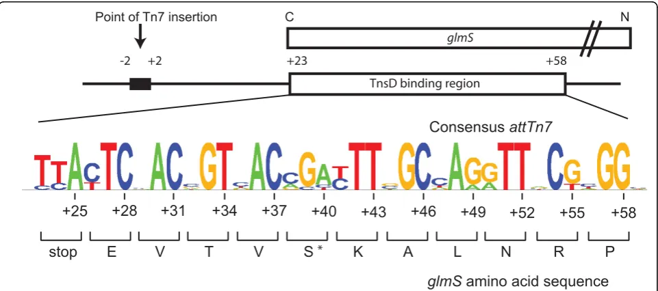
duplica-tion is attTn7 -2 to +2. The minimal E. coli 36 bp
TnsD-binding site that has been defined by footprinting
studies extends fromattTn7 +23 to +58, and can
pro-mote maximum insertion activity in vivo and in vitro
(Figure 1) [6]. TnsD also binds to the human glmS
homologs gfpt-1andgfpt-2[7], theDrosophilahomologs
gfat-1 and gfat-2, and the zebrafish gfpt-1 (see Addi-tional file 1).
The 36 bp TnsD-binding site lies within the 3’end of
glmS, which encodes the C-terminus of the protein.
This region includes the GlmS active site and is highly conserved at the protein level. We aligned this 36 bp
sequence from 25 bacterial glmS genes that have a
downstreamTn7family transposon (GenBank) and the
homologous Drosophila, zebrafish and human genes
using the Sequence Logo algorithm [11] to generate a consensus for the attachment site (Figure 1). Not sur-prisingly for a region that encodes a highly conserved protein, the first two positions at each codon are highly
+23 +58
TnsD binding region
P
E
V
T
V
S
K
A
L
N
R
stop
glmS
amino acid sequence
Consensus
attTn7
Point of Tn7 insertion
-2 +2
+25
+28
+31
+34
+37
+40
+43
+46
+49
+52
+55
+58
glmS
N C
*
conserved, and variation is seen only at the third‘
wob-ble’position.
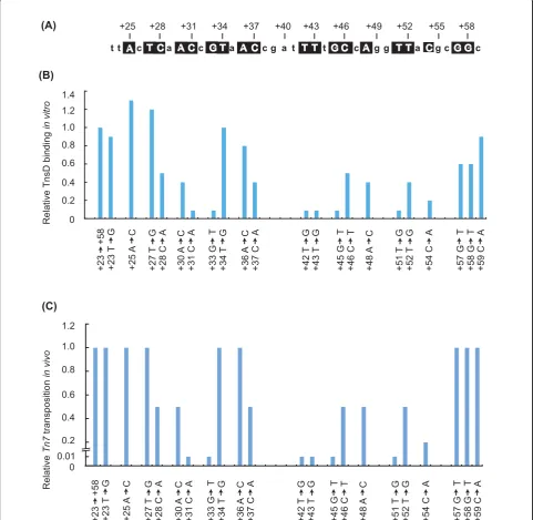
Reasoning that TnsD would contact with the most highly conserved bases within this region, we mutated
each of the most conserved bases in theE. coli attTn7
sequence (+23 ® +58) (Figure 1, Figure 2A) to the
opposite type (that is, pyrimidines to purines (e.g., cyto-sine to adenine) and purines to pyrimidines (e.g., gua-nine to thymine)). We performed gel-shift assays using purified TnsD and radiolabeled attachment-site oligonu-cleotides, and compared the relative amount of
TnsD-attTn7 complex formation of the mutants with that of
the wild-type attachment site (Figure 2A). Mutations at
positions attTn7+31, +33, +42, +43, +45, +51 and +54
all had strong negative effects; that is, they resulted in a reduction in binding of at least five-fold, indicating that those positions are very important for TnsD binding to
attTn7. More modest (about two-fold) reductions in
attTn7 binding activity resulted from mutation at
attTn7+30, +37, +48, +51 and +52.
We also examined the binding of TnsD to wild-type
and representative mutantattTn7sitesin vivo(Table 1).
We used the P22 challenge phage assay for DNA
bind-ing inSalmonella, which has been used to identify and
characterize DNA-binding sites and protein-DNA inter-actions in a wide variety of bacterial and eukaryotic DNA-binding proteins [12]. Briefly, the anti-repressor of
lysogeny (ant) gene of phage P22 controls the choice
between P22 lysis and lysogeny upon infection. Ifantis
expressed, the phage grows lytically, whereas if
expres-sion ofantis blocked, lysogeny can be established. The
expression of antcan be blocked by engineering the
binding site for a heterologous DNA-binding protein
(here attTn7), upstream of ant and providing the
cog-nate binding protein (here TnsD), intrans. If binding of
the tested protein to the introduced site occurs,
expres-sion of antis blocked and lysogeny ensues, which can
be scored by the number of kanamycin-resistant colo-nies, as the P22 genome imparts kanamycin resistance to the cell. Thus, P22 lysogeny can be a measure of pro-tein binding; lysogeny reflects binding of TnsD, whereas
if TnsD does not bind, antis expressed and the phage
grows lytically.
We know from previous work that Tn7transposition
functions well in Salmonella [13]. For the challenge
phage experiment, we constructed a P22 phage with
the E. coli attTn7 attachment site (+23 ® +58)
upstream of ant, thus putting the choice of lysogenic
vs lytic growth under the control of TnsD binding to
attTn7: if TnsD binds, lysogeny should occur. We
infected a Salmonella enterica Typhimurium strain
containing a plasmid-borne isopropyl b
-D-1-thiogalac-topyranoside (IPTG)-inducible tnsD gene with the
challenge phage carrying the E. coli attachment site
upstream of ant. The frequency of lysogenization of
the attTn7phage increased more than 100-fold in the
presence of TnsD (Table 1). Thus the challenge phage assay can be successfully used to evaluate TnsD
bind-ing to attTn7 in vivo. When assayed, the frequency of
lysogeny of a phage containing a wild-type attachment
site (+23 ® +58) was several orders of magnitude
higher than the frequency of lysogeny of phages con-taining any of four mutant attachment sites that bind
TnsD poorlyin vitro (Table 1). These assays show that
TnsD also binds poorly to these mutant sites in vivo,
supporting the view that the identity of these positions is important for the site-specific binding activity of TnsD.
attTn7mutations that block TnsD binding also blockTn7
transpositionin vivo
As described above, mutations in attTn7 can block
TnsD binding both in vitroandin vivo. We also
deter-mined the effect of attTn7mutations on Tn7insertion
intoattTn7 in vivo inE. coli. In these assays,Tn7
trans-position into a target plasmid containing attTn7 was
evaluated using a‘l-hop’assay [14], in which cells
con-taining a plasmid(s) expressing TnsABC+D were infected with a replication- and integration-defective
lambda phage derivative carrying a miniTn7kanamycin
resistance cassette (miniTn7-KanR), and the frequency
of miniTn7insertion into theattTn7plasmid was
mea-sured as the fraction of infected cells that became kana-mycin-resistant.
We found that mutations that significantly (> 10-fold)
decreased TnsD binding to attTn7also decreased Tn7
transposition in vivoby at least 100-fold (Figure 2C).
Thus, attTn7+31, +33, +42, +43, +45 and +51 are key
positions for TnsD-attTn7interaction. attTn7sequence
changes that had more modest effects on TnsD binding, (reductions of two- to five-fold), also reduced
transposi-tion by two- to five-fold. Thus, attTn7+28, +30, +37,
+46, +48 and +54 also make contributions to TnsD-attTn7interaction.
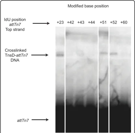
Crosslinking ofattTn7to TnsD
Crosslinking studies with radiolabeledattTn7DNA were
also used to further probe TnsD-attTn7 interaction.
Each ‘T’ on both strands of the attachment site was
individually replaced with iodouracil, then the attTn7
derivatives were tested for binding to TnsD by bandshift assay and crosslinking was performed in the presence of UV light. None of these iodouracil substitutions had a
detectable effect on TnsD binding in vitro (data not
shown). Of all of the positions tested, only IdUs at
posi-tions at attTn7 +23, +51 and +52 were in sufficiently
the crosslinkable base was on the top strand of attTn7. As described above, mutation alterations of the
attach-ment site at both attTn7+51 and +52 had a negative
effect on TnsD binding toattTn7. These results suggest
that there are important base-specific contacts between
attTn7and TnsD at these positions. By contrast,
muta-tion of the crosslinkable posimuta-tion attTn7 +23 had no
effect on TnsD binding or target activity in vivo. It
should be noted that in such an assay, crosslinking requires several different amino acids to be juxtaposed, thus a failure to observe crosslinking does not mean that TnsD is not immediately adjacent to DNA at these positions, but rather that no crosslinkable amino acids are nearby. t 0 1.4 1.2 1.0 0.8 0.6 0.4 0.2 Relative TnsD binding in vitro
t Ac T Ca A Cc G T a A Ccg a t TT t G C c A g g TTa Cg c GGc
+25 +58 Relative Tn7 transposition in vivo 0.4 0.6 0.8 1.0
+28 +31 +34 +37 +40 +43 +46 +49 +52 +55
(A)
(C) (B)
+23 +58 +23
T G +25 A C +27 T G +28 C A +30 A C +31 C A +33 G T +34 T G +36 A C +37 C A +43 T G +42 T G +45 G T +46 C T +48 A C +52 T G +51 T G +54 C A +59 C A +58 G T +57 G T 0 0.2 1.2
+23 +58 +23
T G +25 A C +27 T G +28 C A +30 A C +31 C A +33 G T +34 T G +36 A C +37 C A +43 T G +42 T G +45 G T +46 C T +48 A C +52 T G +51 T G +54 C A +59 C A +58 G T +57 G T 0.01
Figure 2attTn7target activity.(A)TheE. coli attTn7sequence from +23 to +59 is shown; nucleotides that are highly conserved and subsequently analyzed by mutation are boxed.(B)Graphical representation of the relative target DNA binding activity of TnsD on the mutant
attTn7sitein vitrois shown. Double stranded oligonucleotides with indicated mutations listed were used in gel-shift assays with TnsD.(C) Graphical representation of the relativein vitrotransposition activity on the mutantattTn7sitein vitrois shown. The activity of mutantattTn7
TnsD contains a zinc-finger motif essential for DNA binding
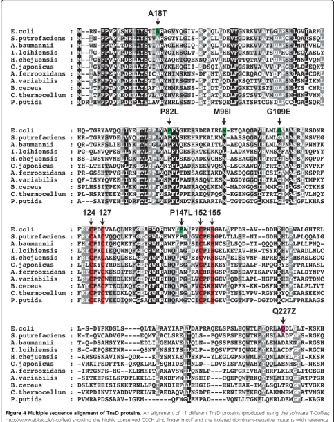
Before the modern era of extensive bacterial genome
sequencing,Tn7, which was identified inE. coli[15,16],
was unique. Now there are many examples in GenBank
ofTn7relatives in a variety of different bacteria that
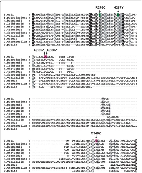
con-tain obvious TnsD homologs (Figure 4, Figure 5, Figure 6). The most notable feature of this alignment is a highly conserved N-terminal region of about 170 amino acids that contains a CCCH motif characteristic of zinc-finger
proteins [17]. InE. coli Tn7TnsD, the motif consists of
Cys124, Cys127, Cys152and His155(Figure 4).
To determine directly whether TnsD binding to
attTn7 is dependent on zinc, gel-shift assays were
performed using wild-type TnsD and attTn7 in the
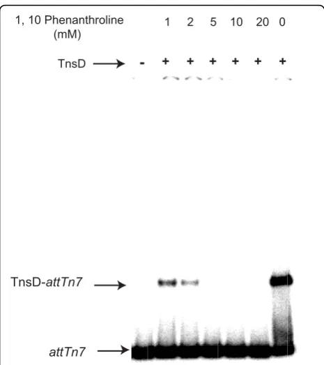
presence of increasing amounts of the zinc chelator 1,10-phenanthroline. Complex formation was severely diminished by 2 mM 1,10-phenanthroline, indicating that TnsD DNA binding is zinc-dependent (Figure 7). We also individually mutated Cys124, Cys127, Cys152 and His155 to serine, and analyzed the ability of these
mutants to bindattTn7 in vitro and to promote
trans-position in vivo (Table 2). When purified, none of the
mutant TnsD proteins were able to bindattTn7(Table
2). We again used the ‘l-hop’assay to assay
transposi-tion of a miniTn7-KanR [14] from the infecting phage
into an attTn7 plasmid, by measuring the fraction of
infected cells that become kanamycin-resistant. The fre-quency of transposition frefre-quency promoted by the TnsD zinc-finger mutants was reduced 1000-fold from that of wild-type cells. Sequencing of junctions of independent
insertions from thein vivotransposition assay showed that
Tn7insertions were present inattTn7in a sequence- and
orientation-specific manner, with the 5-bp target
duplica-tion characteristic ofTn7 transposition, as occurs with
wild-type TnsD (data not shown). Thus, the insertions recovered, although occurring at very low frequency, were probably TnsD-mediated transposition events. As described in more detail below, we were unable to observe
DNA bindingin vitrowith various N-terminal fragments
of TnsD, thus could not identify a discrete zinc finger motif-containing DNA-binding domain.
TnsD has important DNA-binding determinants throughout the protein
In an attempt to identify directly the domains of TnsD involved in DNA binding, we generated a number of TnsD deletion derivatives, and assessed DNA binding of
these mutantsin vivoand in vitro. All tested C-terminal
deletion derivatives, (TnsD1 to 350, 1 to 399, 1 to 480, 1 to 490 and 1 to 498) were unable to bind DNA as
evaluated by in vitro band-shift assays and by in vivo
challenge phage assays (data not shown). However, these truncated proteins did retain the ability to interact with TnsC, indicating that they were not simply unfolded (we discuss interaction of TnsD with TnsC below). These observations suggest that in addition to the zinc finger, TnsD contains DNA-binding determi-nants spread across the entire length of the protein and
that several of these are required for attTn7 binding.
We were unable to detect interaction of N-terminal TnsD truncations with TnsC or to purify these proteins (data not shown).
Isolation of TnsD dominant-negative mutations
In another approach to identify the functional regions of TnsD, we conducted a screen for mutations in TnsD that have dominant-negative effects on TnsD-dependent Table 1 TnsD binding toattTn7 in vivo
attTn7 position
Lysogeny frequencya
TnsD induced
TnsD uninduced
+23®+58(wt) 7.4 × 10⁻⁵ 5.4 × 10⁻⁷
+31 C®A 5.2 × 10⁻⁹
-+42 T®G 3.6 × 10⁻⁸
-+43 T®G 1.6 × 10⁻⁸
-+51 T®G 5.2 × 10⁻⁹
-a
Challenge assay phages carrying the indicated mutations inattTn7were infected into cells expressing TnsD and the frequency of lysogeny measured; values are the average of three independent infections performed in parallel and relative to the wild type.
Modified base position
Crosslinked TnsD-attTn7
DNA
attTn7
IdU position
attTn7
Top strand
+52 +60 +51
+44 +43 +42 +23
transposition. Expected classes of TnsD
dominant-nega-tive mutants include those able to bind attTn7 but
unable to interact with TnsC, and also mutants unable to
bindattTn7but able to interact with TnsC. We used a
promoter capture (papillation) assay as described by
Stellwagen and Craig[18], in which aminiTn7containing
a promoterless lac gene is mobilized from a plasmid
donor to chromosomal pseudo-attTn7sites;attTn7itself
was blocked by anotherTn7element. TnsABC+D were
expressed from a pACYC plasmid. Productive hops
pro-duce Lac+cells that form papillae on Lac-colonies on
MacConkey lactose plates. To look for dominant-nega-tives, we used hydroxylamine to mutate a pUC-based
TnsDgene, transformed it into the assay strain
contain-ing wild-type TnsABC+D, and looked for transformants on MacConkey lactose plates within decreased numbers
of papillae. After hydroxylamine mutagenesis of TnsD
and screening of 15,000 transformants, we isolated 16 TnsD mutants, 11 of which were missense mutants and five of which were nonsense mutants yielding truncated
TnsD; only two mutants (TnsDP147Land TnsDR276C)
were isolated twice (Figure 4, Figure 5, Figure 6).
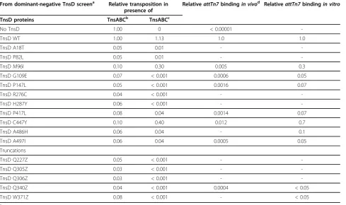
We also quantitatively evaluated the effect of the domi-nant-negative TnsD mutants on transposition. Using the
lhop assay, we assayed miniTn7-KanRinsertion into
chromosomalattTn7in the presence of pACYC TnsABC
+D and a pUC-based plasmid containing the TnsD mutants (Table 3). In this assay, all of the dominant-negative mutants identified by the papillation screen with the exception of TnsD C447Y inhibited TnsABC+D transposition at least 10-fold, confirming that they were indeed negatives. Although the dominant-negative effect of TnsD C447Y was more modest, this may reflect the fact that the insertion targets are different in these assays: the papillation assay measures insertion
into pseudo-attTn7sites, whereas thel-hop assay
mea-sures insertion into chromosomalattTn7.
We also examined the ability of the dominant-negative TnsD mutants to promote transposition in the presence of TnsABC alone to determine if they possess residual TnsD activity. All had substantial defects in TnsD
activity, with reductions ranging from at least 2.5-fold to 100-fold compared with wild-type cells.
Using the challenge phage assay, we also evaluated the ability of most of the dominant-negative TnsD missense
mutants to bind to attTn7 in vivo, and found that all
were significantly reduced in their ability to bindattTn7
(Table 3). Using purified TnsD, we also found that all the missense mutants except C447Y had significantly
impairedattTn7bindingin vitro(Table 3).
TnsC increases the binding of TnsD toattTn7
TnsC and TnsD form a novel complex with attTn7 in
vitro, and the presence of TnsC increases the amount of protein-DNA complex formed [6]. We also used the challenge phage assay to examine the TnsC-TnsD-attTn7interactionin vivo(Table 4). TnsC does not pro-mote the formation of lysogens on its own, as expected given its non-specific DNA-binding activity [19]. How-ever, when TnsC and TnsD were expressed simulta-neously in the host cells, we observed a enhancement of lysogeny of nearly 100-fold relative to cells expressing only TnsD. Thus, TnsC considerably enhances TnsD
binding toattTn7 both in vivo andin vitro.
Although the binding of wild-type TnsD toattTn7is
stimulated by TnsC, incubation with TnsC did not ‘
res-cue’the formation of TnsCD-attTn7complexes with the
dominant-negative missense mutant TnsDin vitro(data
not shown).
TnsC and TnsD can interact in the absence ofattTn7 Previous studies revealed that a key determinant in the
recruitment of TnsC to TnsD-attTn7is a distortion
intro-duced by TnsD intoattTn7, which provides a binding site
for TnsC [6]. We have now found, using a yeast two-hybrid assay, that TnsC and TnsD also interact directly in
the absence ofattTn7[20,21] (Table 5). The interaction
between full-length TnsC and TnsD is only slightly less than that of the robust interaction displayed by the Fos-Jun control, but equivalent to that of RB and E2F1 [22].
As determined by analysis of a series of C-terminal deletion derivatives, the amino-terminal amino acid sequence (1 to 309) of TnsD retain full binding to TnsC in this assay and thus contains the determinants for TnsD binding to TnsC. The isolation of the TnsD trun-cation sequence (1 to 340; full-length TnsD is 507 aa) as a dominant negative is consistent with this hypothesis (see below). However, if as few as seven amino acids are removed from the N-terminus of TnsD, the TnsC-TnsD interaction is disrupted (data not shown). The lack of activity of TnsD sequence 1 to 436, even though both longer and shorter constructs are active, may be due to inappropriate folding or to the unmasking of an inhibi-tory domain.
TnsD-attTn7
attTn7
1 2 5 10 20 0
TnsD 1, 10 Phenanthroline (mM)
-
++ + + + +
Figure 7 Chelation of zinc using 1,10-phenanthroline eliminates TnsD binding toattTn7DNA.
Table 2 Targeted mutations to the conserved CCCH residues in TnsD eliminatesTn7transposition and
TnsD-attTn7binding
Protein RelativeattTn7 bindingin vitro
Relative transposition frequencyin vivo
TnsD wt 1.0 1.0a
TnsD C124S 0.05 < 0.001
TnsD C127S 0.05 < 0.001
TnsD C152S 0.05 < 0.001
TnsD C155S 0.05 < 0.001
a
We also performed deletion analysis of TnsC using the yeast two-hybrid system (Table 5). TnsC-TnsD interaction with TnsD is maintained with deletion of 75 or 171 amino acids from the C terminus of TnsC (full-length is 555 aa), but a deletion of 223 amino acids leads to loss of the interaction with TnsD (Table 5); thus interaction of TnsC with TnsD occurs within the 1 to 293 region of TnsC. The finding that Tn7 inserts at
high-frequency inattTn7 using TnsC 86 to 555
(Spen-cer J and NLC, unpublished observation) suggests that the TnsD interaction domain lies within TnsC 1 to 293.
Discussion
The sequence-specific binding of TnsD to the 3’end of
the coding region of the glmS gene is sufficient to
prompt the site - and orientation-specific insertion of
Tn7to a position about 20 bp downstream ofglmS, that
is, to produce a functional attTn7 site [23]. Previous
work in vitro identified the 36 bp span of the TnsD
binding site, and demonstrated that this sequence alone is sufficient to direct transposition [6]. In this study, we have further probed the interaction of TnsD with
attTn7 and deepened our understanding of Tn7
site-specific insertion. Our results have revealed that
deter-minants of the interaction of TnsD withattTn7extend
throughout the TnsD protein and the TnsD binding site. The amino acid sequence of the C-terminus of vir-tually all GlmS proteins is identical. Comparison of the
TnsD binding site inattTn7sites from a variety of
bac-teria and several eukaryotes revealed that 19 of the 35 bp were completely conserved, and correspond to
non-wobble positions for each codon at the glmSterminus.
Analysis of TnsD binding and transposition to these mutant sites with substitutions at each of these
con-served positions revealed that changes at seven (attTn7
+31, +33, +42, +43, +45, +51 and +54) had significant
effects on TnsD binding and Tn7transposition.
Model-ing of the attachment site as B-form DNA (Figure 8) indicates that the important nucleotides are on one face From dominant-negative TnsD screen Relative transposition in
presence of
RelativeattTn7bindingin vivo RelativeattTn7bindingin vitro
TnsD proteins TnsABCb TnsABCc
No TnsD 1.00 0 < 0.00001
-TnsD WT 1.00 1.13 1.0 1.0
TnsD A18T 0.05 0.01 -
-TnsD P82L 0.05 0.01 -
-TnsD M96I 0.10 0.30 0.005 0.3
TnsD G109E 0.07 < 0.001 0.0006 0.05
TnsD P147L 0.05 < 0.001 0.0016 0.07
TnsD R276C 0.04 < 0.001 -
-TnsD H287Y 0.06 < 0.001 -
-TnsD P417L 0.08 0.04 0.0014 0.07
TnsD C447Y 0.10 0.40 0.012 0.7
TnsD A486H 0.06 0.04 - 0.1
TnsD A497I 0.06 0.04 0.0005 0.05
Truncations
TnsD Q227Z 0.05 < 0.001 -
-TnsD Q305Z 0.03 < 0.001 -
-TnsD Q306Z 0.03 < 0.001 -
-TnsD Q340Z 0.04 < 0.001 0.0004 < 0.05
TnsD W371Z 0.08 < 0.001 - < 0.05
a
These substitutions and truncations were isolated as dominant negatives and then analysed for transpositionin vivoand DNA bindingin vivoandin vitro.
b
Tn7 transposition frequencyin vivowas measured using the lambda hop assay and carried out in presence of TnsABCD + TnsD (dominant-negative mutants).
c
Tn7 transposition frequencyin vivowas measured using the lambda hop assay and was carried out in presence of TnsABC + TnsD (dominant-negative mutants).
d
The binding of mutant TnsDs toattTn7 in vivowas measured using the challenge phage assay.
Table 4 TnsC increases TnsD binding toattTn7 in vivo
Tns protein Lysogeny frequencya
TnsC < 8.0 × 10⁻⁷b
TnsD 2.4 × 10⁻³
TnsC + TnsD 2.4 × 10⁻¹
a
Using thein vivochallenge phage assay, adding TnsC to TnsD increased lysogeny, also indicating that TnsC is stabilizing TnsD on the DNA. TnsC alone did not increase lysogeny.
b
of the DNA. This is consistent with the crosslinking data presented here, in which contacts with the major groove were detected (Figure 3), and with previous DNA footprinting studies showing that TnsD makes major groove contacts [6].
The activity of attTn7 is strongly directional: Tn7
always inserts downstream of the glmS termination
codon, mediated by activation of a TnsCD-attTn7
com-plex that promotes orientation-specific insertion ofTn7.
Consistent with this asymmetric pattern of insertion, attTn7does not contain inverted repeats for the binding of an oligomeric TnsD.
Previous studies have shown that a key step inTn7
insertion intoattTn7is the ability of TnsD to introduce
a DNA distortion(s) into attTn7, which we have
pro-posed prompts the binding of TnsC, the regulator of the TnsAB transposase [6,24]. In addition to providing a
higher resolution view of TnsD-attTn7 interaction, we
also identified a previously unknown interaction pro-tein-protein interaction between TnsC and TnsD. Deter-minants for this interaction are located within the N-terminal region of TnsD covering positions 1 to 309 and the N-terminal region of TnsC covering positions 1 to 293. It seems likely that the DNA-mediated and pro-tein-protein interactions are both important to TnsC-mediated activation of transposition.
TnsC also interacts with and activates both subunits
of the Tn7transposase (TnsA and TnsB), which mediate
breakage and joining of the ends of Tn7 [25]. TnsA, the
transposase subunit that cleaves at the 5’ends ofTn7,
and TnsC form a TnsA2C2 complex in solution,
mediated by interactions between the N-terminal region of TnsA and the 50 carboxy-terminal amino acids of TnsC [26]. A distinct TnsACD complex has also been
observed [27], which we suggested is the‘target’DNA
complex, with which the TnsB transposase subunit
med-iates breakage and joining at the 3’ends of the
transpo-son [28]. The other transposase subunit TnsB binds
specifically to the ends of Tn7, and mediates breakage
and joining at the 3’ ends [29]. Interactions between
TnsC and the C-terminus of TnsB have also been
detected [30]. The apposition of the TnsACD-attTn7
and TnsB-Tn7 end complexes results in the assembly
Table 5 Yeast 2 hybrid assay shows that TnsC and TnsD can interactin vivo
Protein fragment b-galactosidase activity
Fos Jun 5.0
E2F1 RB 1.0
Empty vector Gal4-AD
Empty vector Gal4-DB
0.075
TnsC (1 to 555) wt TnsD (1 to 507) wt 1.0
TnsC (1 to 555) wt TnsD (1 to 436) 0.075
TnsC (1 to 555) wt TnsD (1 to 399) 4.5
TnsC (1 to 555) wt TnsD (1 to 375) 5.0
TnsC (1 to 555) wt TnsD (1 to 350) 4.5
TnsC (1 to 555) wt TnsD (1 to 309)a 4.5
TnsC (1 to 555) wt TnsD (1 to 293) 0.075
TnsC (1 to 555) wt TnsD (8 to 507) 0.075
TnsC (1 to 555) wt TnsD (12 to 507) 0.075
TnsC (1 to 555) wt TnsD (16 to 507) 0.075
TnsC (1 to 555) wt TnsD (22 to 507) 0.075
TnsC (1 to 555) wt TnsD (1 to 507) wt 1.0
TnsC (1 to 480) TnsD (1 to 507) wt 1.0
TnsC (1 to 384) TnsD (1 to 507) wt 1.0
TnsC (1 to 332) TnsD (1 to 507) wt 0.20
TnsC (1 to 293)a TnsD (1 to 507) wt 0.20
TnsC (1 to 280) TnsD (1 to 507) wt 0.014
a
Deletion analysis of TnsD and TnsC revealed that this protein-protein interaction activity is contained within TnsC 1 to 293 and TnsD 1 to 309.
Tn7
insertion +31 +33 +42 +43 +45 +51
-2 +2 +25
distortion
TnsD attachment site
attTn7
to learned about the mechanism by which TnsD-attTn7 activates TnsC and hence TnsAB.
Functional domains of TnsD
TnsD is a multifunctional protein: it binds specifically to attTn7and, as we have shown here, interacts with its partner in transpositon, TnsC. In this study, we have made considerable progress in understanding the unique TnsD family of DNA-binding proteins by defin-ing important protein determinants for DNA binddefin-ing and transposition, and key nucleotides for the TnsD-attTn7 interaction. Although we identified a zinc finger motif in the N-terminal region of TnsD, we were unable to use deletion analysis to identify a functional discrete DNA-binding domain(s). It is possible there are multi-ple DNA-binding domains throughout the protein, a model consistent with the fact that the region of TnsD interaction with DNA spans about 35 bp. However, we were able to use deletion analysis to localize the TnsC-interaction region of TnsD to its N-terminal amino acids 1 to 309, and the TnsD-interaction region of TnsC to its amino acids 1 to 293. Although our ability to isolate TnsD dominant negatives is consistent with this protein being multifunctional, our inability to further identify crucial subregions within TnsD pre-cludes definitive conclusions of the physical basis of the dominant negatives.
As previously proposed, the ability of TnsD to direct transposition to a defined sequence makes it an attrac-tive candidate for use in targeted delivery of DNA sequences for genetic manipulation in organisms from bacteria to human [7,13]. Further structure-function analyses of TnsD will not only facilitate a deeper
under-standing of Tn7 transposition but also the use ofTn7
and TnsD as tools for genomic engineering.
Methods
Bacterial strains, plasmids and phages
CAG456 is E. coli (SC122 htpR165) [32]. NLC51 is E.
coliF-araD139Δ(argF-lac)U169 rpsL150 relA1 flbB5301
deoC1 ptsF25 rbsR valR recA56 [33]. BD409 is a
deriva-tive of E. coli CW51 with att::promoterless lacZY
flanked byTn7 transposon end sequences sufficient for
transposition: 166 bp from the left end of Tn7(Tn7L)
and 90 bp from the right end of (Tn7R) [4,34]. Plasmid
pMR1 was constructed by PCR amplifying the tnsD
gene from pCW4 [4] and cloning it into pCYB1 digested
withNcoI andSapI (New England Biolabs, Ipswich, MA,
USA.). pCW4 was used as a source of TnsABCD and
pCW15 as a source of TnsABC in thein vivo
transposi-tion assays [4]. pCW23 was used for mutatingtnsD[4].
Phage KK1, used as the source of Tn7transposon in the
l-hop assay, is a derivative of 780 (b2::hisOGD b522
attTn7(-342 to +165)::miniTn7kan [35].
Affinity purification of intein-TnsD
The full length TnsD wild-type and mutant proteins were purified as intein fusions from CAG456 contain-ing plasmid pRM1 or mutant TnsD derivative
plas-mids. Site-directed mutations in tnsDwere generated
by PCR (QuickChange Site Directed Mutagenesis Sys-tem; Agilent Technologies) and verified by direct DNA sequencing. Representative cells were grown at 30°C to
an OD600 = 0.5 in Luria broth supplemented with 100
mg/ml carbenicillin, IPTG was added to give 0.4 mM final concentration, and the cells were allowed to grow for an additional 4 h. All subsequent steps were per-formed at 4°C unless otherwise stated. The cells were separated by centrifugation, and resuspended in the buffer supplied with the kit (Buffer A; 50 mM HEPES pH 8, 500 mM NaCl, 10% v/v glycerol). The cells were then lysed by sonication and separated by centrifuga-tion at 26,000 g for 30 min, the resulting supernatant
was then filtered through a 0.45 μm syringe filter
(Nal-gene, Rochester, NY, USA). The filtrate was applied to pre-equilibrated chitin beads (New England Biolabs) in a 10 ml column and the beads then washed several times (5×) in Buffer A. The washed chitin beads were then treated with Buffer B (50 mM HEPES pH 8, 500
mM NaCl, 10% v/v glycerol, 10 mM MgCl2, 10 mM
ATP) for 1 h at room temperature to remove residual GroEL protein. The beads were then washed with sev-eral volumes of buffer B at 4°C (2×), and incubated overnight with Buffer C (50 mM HEPES pH 8, 500 mM NaCl, 10% v/v glycerol, 50 mM dithiothreitol (DTT)), which promotes cleavage of TnsD from the intein tag [36,37]. Full-length TnsD was then eluted from the column using Buffer C without DTT, and peak fractions were pooled, dialyzed against another buffer (500 mM KCl, 50 mM Tris-HCl (pH 8.0), 1 mM EDTA, 2 mM DTT and 25% v/v glycerol), then stored at -80°C.
Isolation of dominant-negativetnsDmutants
pCW23 (TnsD) [4] was treated with 1 M hydroxylamine hydrochloride in NaOH at 37°C for 24 h. The mutagen was dialyzed out of the DNA in Tris-EDTA buffer. The DNA was recovered by ethanol precipitation, and
trans-formed into BD409 carrying wild-type tnsABCD on a
compatible plasmid, pCW4 [4]. Transformants were pla-ted onto MacConkey lactose plates containing appropri-ate antibiotics. The plappropri-ates were incubappropri-ated at 30°C for 5 days, and transformants were screened for decreased
papillation (to Lac+). Plasmid DNA was extracted from
DNA was sequenced to identify the mutations, and recloned for expression.
lhop assay
The transposition frequency of a miniTn7kanRelement
from the integration- and replication-defective phage
KK1 into the chromosomal attTn7site ofE. colistrain
NLC51 and a plasmid containing the attTn7 site ofE.
coli strain LA3 was evaluated when Tns proteins were
supplied in trans[35,38] The transposition frequency
was the number of kanamycin-resistant colonies per plaque-forming unit.
DNA-binding reactions
Binding reactions were performed with wild-type and mutant versions of TnsD proteins and wild-type TnsC protein as described previously [6].
Protein-DNA UV photo-crosslinking reaction
Protein-DNA crosslinking was performed as described
previously [39].. Oligonucleotides were synthesized
(Eurofins MWG Operon, Huntsville, AL, USA) with dT replaced by IdU. Double-stranded (ds)DNAs were then formed by gradual annealing with a complemen-tary oligonucleotide, and then radio-labeled with 5' phosphorylation.
For crosslinking reactions, 25 pmol TnsD and 0.5
pmol attTn7 dsDNA were incubated in solution (25
mM HEPES, 2.5 mM TrisCl, 40 mM DTT, 0.005% BSA and 50 ng/ul herring sperm DNA, pH 7.5) for 20 min at 30°C. Photo-crosslinking was then carried out with UV irradiation at 312 nm (StrataLinker; Agilent Technolo-gies, La Jolla, CA, USA) for 30 min. The reaction was set up so that the top of the open 1.5 ml Eppendorf tube touched the UV tube. After UV crosslinking, the reaction was analyzed by SDS-PAGE.
Yeast two-hybrid assay
The yeast two-hybrid assay was performed according to
the manufacturer’s protocol (Proquest Two-Hybrid
Sys-tem; Life Technologies Inc., Carlsbad, CA, USA). TnsD was cloned into pDBLeu (Life Technologies Inc) and
TnsC into pPC86 (Life Technologies Inc). b
-galactosi-dase assays were performed as described previously [40].
In vitrotransposition reactions
In vitro transposition reactions were performed essen-tially as described previously [19]. The donor plasmid
pEMΔ(5.9 kb) contains a 1.6 kb miniTn7kanRelement.
The 3.2 kb target plasmid pRM2 [35] contains a 555 bp
attTn7segment (-342 to +165). Reaction mixtures (100
μl final volume) contained 0.25 nM pEM donor DNA,
2.5 nM pRM2 target plasmid, 28 mM HEPES pH 8.0, 2.2 mM DTT, 4.4 mM Tris pH 7.5, 100 mg/ml tRNA,
50 mg/ml bovine serum albumin (BSA), 0.16 mM
EDTA, 0.1 mM MgCl2, 0.1 mM CHAPS detergent, 30
mM NaCl, 21 mM KCl, 1.8% v/v glycerol, 2.0 mM ATP and 15 mM MgOAc unless otherwise indicated. Tns proteins were added as follows: 40 ng TnsA, 25 ng TnsB, 30 ng TnsC and 22 ng TnsD. Reaction mixtures containing all components except donor DNA, TnsA, TnsB and MgOAc were assembled on ice. The assembly reaction mixtures were incubated for 20 min at 30°C; donor DNA, TnsA, TnsB and MgOAc were then added, and the incubation was continued for an additional 20 min. Reactions were stopped by adjusting to 25 mM EDTA, followed by extraction with phenol:chloroform (1:1). The DNA was then precipitated with ethanol,
digested with NdeI and separated in a 0.6% agarose gel,
with electrophoresis carried out at 50 V for 16 h. The DNAs were transferred to a hybridization membrane (Gene Screen Plus; PerkinElmer, Foster City, CA, USA)
and hybridized with a probe specific for miniTn7kanR.
The probe was labeled by random priming with 32
P-dCTP and the Klenow fragment of DNA polymerase I (Boehringer Mannheim Biochemicals (BMB), Indianapo-lis, IN, USA). All blots were analyzed using a phosphor-escent imager (PhosphorImager; Molecular Dynamics, Sunnyvale, CA, USA).
Challenge phage assays
Challenge phages were constructed as described in [12].
The wild-typeattTn7attachment siteattTn7(+23®+58)
was cloned in as oligonucleotides (5’
-CCGCGTA-ACCTGGCAAAATCGGTTACGGTTGAGTAA-3’and
the complementary oligonucleotide into theSmaI site of
pPY190, creating pGRG60). All mutant attachment sites were variants of that sequence (as described in the text). The challenge phage assay was carried out as described previously [12], using plasmid pCYB1-TnsD or mutant variants of that plasmid to express TnsD, selecting for kanamycin-resistant P22 lysogens. Expression of TnsD proteins was induced with 1 mM IPTG. Results are expressed in lysogens/cell. TnsC was expressed constitu-tively from pGRG63, a plasmid constructed by cutting
pCW15[4] withSapI andBstXI, blunting and religating
the plasmid, to eliminate the TnsAB genes.
Additional material
Additional file 1: TnsD protein binds toattTn7-like sequences within theglmSanalog ofDrosophila gfat-1, and thegfat-2and glmSanalog of zebrafishgfpt-1.
Acknowledgements
Howard Hughes Medical Institute.
Author details
1Howard Hughes Medical Institute, Department of Molecular Biology and Genetics, Johns Hopkins University School of Medicine, Baltimore MD 21205, USA.2Current Address:Verenium Corporation. 4955 Directors Place, San Diego, CA 92121, USA.3Current Address: Laboratory of Host Defense, NIAID/ NIH, Bethesda, MD 20892, USA.4Current Address: Mayo Clinic, 417 Guggenheim Bldg, 200 First St. SW, Rochester, MN 55905, USA.
Authors’contributions
RM, GJM, LY and NLC designed the experiments. RM, GJM, LY and CL performed the experiments. RM, GJM and NLC wrote the manuscript.
Competing interests
The authors declare that they have no competing interests.
Received: 7 May 2010 Accepted: 23 July 2010 Published: 23 July 2010
References
1. Peters JE, Craig NL:Tn7: smarter than we thought.Nat Rev Mol Cell Biol
2001,2:806-814.
2. Peters JE, Craig NL:Tn7 recognizes transposition target structures associated with DNA replication using the DNA-binding protein TnsE. Genes Dev2001,15:737-747.
3. Parks AR, Li Z, Shi Q, Owens RM, Jin MM, Peters JE:Transposition into replicating DNA occurs through interaction with the processivity factor. Cell2009,138:685-695.
4. Waddell CS, Craig NL:Tn7transposition: two transposition pathways directed by five Tn7-encoded genes.Genes Dev1988,2:137-149. 5. Gay NJ, Tybulewicz VL, Walker JE:Insertion of transposon Tn7into the
Escherichia coli glmStranscriptional terminator.Biochem J1986,
234:111-117.
6. Kuduvalli PN, Rao JE, Craig NL:Target DNA structure plays a critical role in Tn7 transposition.EMBO J2001,20:924-932.
7. Kuduvalli PN, Mitra R, Craig NL:Site-specific Tn7transposition into the human genome.Nucleic Acids Res2005,33:857-863.
8. Milewski S:Glucosamine-6-phosphate synthase–the multi-facets enzyme. Biochem Biophys Acta2002,1597:173-192.
9. Gringauz E, Orle KA, Waddell CS, Craig NL:Recognition ofEscherichia coli
attTn7by transposon Tn7: lack of specific sequence requirements at the point of Tn7insertion.J Bacteriol1988,170:2832-2840.
10. Oppon JC, Sarnovsky RJ, Craig NL, Rawlings DE:A Tn7-like transposon is present in the glmUS region of the obligately
chemoautolithotrophic bacterium Thiobacillus ferrooxidans. J Bacteriol1998,180:3007-3012.
11. Schneider TD, Stephens RM:Sequence logos: a new way to display consensus sequences.Nucleic Acids Res1990,18:6097-6100.
12. Maloy SR, Gardner J:Dissecting nucleic acid-protein interactions using challenge phage.Methods Enzymol2007,421:227-249.
13. McKenzie GJ, Craig NL:Fast, easy and efficient: site-specific insertion of transgenes into Enterobacterial chromosomes using Tn7without need for selection of the insertion event.BMC Microbiol2006,6:39. 14. Peters JE, Craig NL:Tn7 transposes proximal to DNA double-strand
breaks and into regions where chromosomal DNA replication terminates.Mol Cell2000,6:573-582.
15. Barth PT, Datta N, Hedges RW, Grinter NJ:Transposition of a deoxyribonucleic acid sequence encoding trimethoprim and streptomycin resistances from R483 to other replicons.J Bacteriol1976,
125:800-810.
16. Lichtenstein C, Brenner S:Unique insertion site of Tn7 in the E. coli chromosome.Nature1982,297:601-603.
17. Laity JH, Lee BM, Wright PE:Zinc finger proteins: new insights into structural and functional diversity.Curr Opin Struct Biol2001,11:39-46. 18. Stellwagen AE, Craig NL:Gain-of-function mutations in TnsC, an
ATP-dependent transposition protein that activates the bacterial transposon Tn7.Genetics1997,145:573-585.
in vitro system.Cell1993,72:931-943.
20. Fields S, Song O:A novel genetic system to detect protein-protein interactions.Nature1989,340:245-246.
21. Vidal M, Legrain P:Yeast forward and reverse‘n’-hybrid systems.Nucleic Acids Res1999,27:919-929.
22. Shan B, Durfee T, Lee WH:Disruption of RB/E2F-1 interaction by single point mutations in E2F-1 enhances S-phase entry and apoptosis.Proc Natl Acad Sci USA1996,93:679-684.
23. Craig N:Tn7.Mobile DNA IIASM PressCraig N, Craigie R, Gellert M, Lambowitz A 2002, 423-456.
24. Rao JE, Miller PS, Craig NL:Recognition of triple-helical DNA structures by transposon Tn7.Proc Natl Acad Sci USA2000,97:3936-3941.
25. Ronning DR, Li Y, Perez ZN, Ross PD, Hickman AB, Craig NL, Dyda F:The carboxy-terminal portion of TnsC activates the Tn7 transposase through a specific interaction with TnsA.Embo J2004,23:2972-2981.
26. Skelding Z, Sarnovsky R, Craig NL:Formation of a nucleoprotein complex containing Tn7 and its target DNA regulates transposition initiation. EMBO J2002,21:3494-3504.
27. Lu F, Craig NL:Isolation and characterization of Tn7transposase gain-of-function mutants: a model for transposase activation.Embo J2000,
19:3446-3457.
28. Holder JW:Assembly and architecture of Tn7 transpososomes.PhD dissertation2006.
29. Sarnovsky RJ, May EW, Craig NL:The Tn7 transposase is a heteromeric complex in which DNA breakage and joining activities are distributed between different gene products.EMBO J1996,15:6348-6361.
30. Skelding Z, Queen-Baker J, Craig NL:Alternative interactions between the Tn7 transposase and the Tn7 target DNA binding protein regulate target immunity and transposition.EMBO J2003,22:5904-5917. 31. May EW, Craig NL:Switching from cut-and-paste to replicative Tn7
transposition.Science1996,272:401-404.
32. Baker TA, Grossman AD, Gross CA:A gene regulating the heat shock response inEscherichia colialso affects proteolysis.Proc Natl Acad Sci, USA1984,81:6779-6783.
33. McKown RL, Waddell CS, Arciszewska LA, Craig NL:Identification of a transposon Tn7-dependent DNA-binding activity that recognizes the ends of Tn7.Proc Natl Acad Sci, USA1987,84:7807-7811.
34. DeBoy RT, Craig NL:Target site selection by Tn7: attTn7 transcription and target activity.J Bacteriol2000,182:3310-3313.
35. McKown RL, Orle KA, Chen T, Craig NL:Sequence requirements of
Escherichia coli attTn7, a specific site of transposon Tn7 insertion. J Bacteriol1988,170:352-358.
36. Chong S, Mersha FB, Comb DG, Scott ME, Landry D, Vence LM, Perler FB, Benner J, Kucera RB, Hirvonen CA, Pelletier JJ, Paulus H, Xu MQ: Single-column purification of free recombinant proteins using a self-cleavable affinity tag derived from a protein splicing element.Gene1997,
192:271-281.
37. Chong S, Xu MQ:Protein splicing of the Saccharomyces cerevisiae VMA intein without the endonuclease motifs.J Biol Chem1997,
272:15587-15590.
38. Waddell CS, Craig NL:Tn7 transposition: recognition of the attTn7 target sequence.Proc Natl Acad Sci USA1989,86:3958-3962.
39. Steen H, Jensen ON:Analysis of protein-nucleic acid interactions by photochemical cross-linking and mass spectrometry.Mass Spectrom Rev
2002,21:163-182.
40. Miller JH:A Short Course in Bacterial GeneticsCold Spring Harbor Laboratory Press 1992, 76-77.
doi:10.1186/1759-8753-1-18
Cite this article as:Mitraet al.:Characterization of the TnsD-attTn7 complex that promotes site-specific insertion ofTn7.Mobile DNA2010