lymphocyte differentiation, NHL is usually incur-able. The early distal dissemination frequently seen in NHL patients suggests that NHL may originate from multiple centers. In some cases, NHL has al-ready spread to all parts of the body by the time the clinical diagnosis is confirmed [2–4].
It has been found in many studies [5] that B-cell activating factor (BAFF), as a member of the tumor necrosis factor (TNF) superfamily, could specifically activate B lymphocytes and pro-mote their proliferation. However, more recent studies [6–9]found that excessive expression of Lymphoma is a malignant tumor of the
im-mune system originating from lymph nodes and extralymphatic tissues. Its occurrence is believed to be associated with various immune cells due to the proliferation and differentiation of lym-phocytes during the immune response [1]. Histo-pathologically, lymphoma is classified as Hodg-kin’s disease (HD) and non-Hodgkin lymphoma (NHL). NHL is seen in extralymphatic tissues and does not spread along lymph nodes in sequence; rather, it often disseminates by skipping across ad-jacent lymph nodes to distal ones. Despite good
*Xianjuan Shen
1, A, F, *Mei Wang
2, B, C, Yuehua Guo
2, C, E, F, Shaoqing Ju
1, A–DThe Correlation Between Non-Hodgkin Lymphoma
and Expression Levels of B-Cell Activating Factor
and Its Receptors**
1 Surgical Comprehensive Laboratory, Affiliated Hospital of Nantong University, China 2 Medical Office, Nantong University, China
A – research concept and design; B – collection and/or assembly of data; C – data analysis and interpretation;
D – writing the article; E – critical revision of the article; F – final approval of article
Abstract
Background. Lymphoma is a malignant tumor of the immune system originating from lymph nodes and extra-lymphatic tissues. Its occurrence is believed to be associated with various immune cells due to the proliferation and differentiation of lymphocytes during the immune response. It has been found in many studies that B-cell activating factor (BAFF), as a member of the tumor necrosis factor (TNF) superfamily, could specifically activate B lymphocytes and promote their proliferation.
Objectives. To explore correlations between non-Hodgkin lymphoma (NHL) and the expression of BAFF and its receptors in NHL patients.
Material and Methods. The protein expression of BAFF and its receptors in serum and BAFF mRNA expression in peripheral blood mononuclear cells (PBMCs) of 47 NHL patients and 20 healthy subjects were detected by ELISA and RFQ-PCR and compared with LDH and β2M levels.
Results. BAFF mRNA expression in the PBMCs of NHL patients was significantly higher than in healthy controls. The expression levels of serum BAFF and the three receptors (TACI, BCMA and BAFF-R) in NHL patients were significantly higher than in healthy controls, and were not significantly correlated with β2M and LDH levels.
Conclusions. The serum protein concentration of BAFF and the expression level of BAFF mRNA in PBMCs of NHL patients underwent abnormal changes, indicating that BAFF and its receptors may play some role in the pathogenesis of NHL (Adv Clin Exp Med 2016, 25, 5, 837–844).
Key words: BAFF, RFQ-PCR, NHL.
ORIGINAL PAPERS
Adv Clin Exp Med 2016, 25, 5, 837–844
DOI: 10.17219/acem/29182 © Copyright by Wroclaw Medical University ISSN 1899–5276
* These authors contributed equally to this work.
BAFF can trigger the occurrence of some malig-nant tumors, including NHL. BAFF plays its bio-logical roles by binding with its receptors. Three BAFF receptors have been identified: B-cell matu-ration antigen (BCMA), transmembrane activator and CAML-interactor (TACI) and BAFF receptor (BAFF-R, or BR3) [10].
The purpose of the present study was to explore how BAFF and its receptors correlate with the oc-currence and prognosis of NHL by detecting BAFF levels in NHL patients and analyzing correlations between serum concentrations of BAFF and its re-ceptors with clinical parameters in NHL patients.
Material and Methods
Specimen Collection
Twenty healthy subjects were selected from among blood donors who underwent physical exam-inations at the Affiliated Hospital of Nantong Uni-versity (Nantong, China). They included 13 females and 7 males, and ranged in age from 50 to 65 years, with a mean age of 59 ± 5 years. Forty-seven NHL patients were selected from among hospitalized NHL patients who received treatment at the same hospital between August 2010 and August 2013. They includ-ed 27 males and 20 females, and ranginclud-ed in age from 48 to 73 years, with a mean age of 62 ± 7 years. From each patient and healthy subject, 3 mL of peripher-al venous blood was withdrawn and centrifuged at 2000 rpm for about 10 min to separate peripher-al blood mononuclear cells (PBMCs). Cell depos-its were collected by centrifuge and stored at –70°C until use. At the same time, an additional 2 mL of peripheral venous blood was drawn, centrifuged at 2000 rpm for about 20 min to collect 0.5 mL serum in a 0.5 mL EP tube, and stored at –70°C until use.
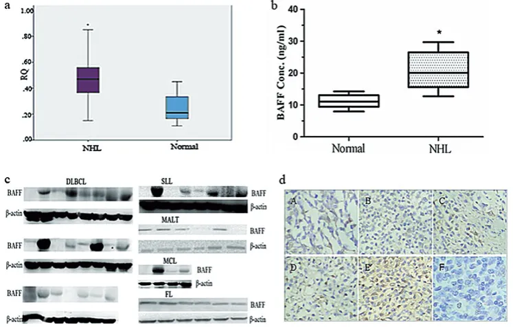
Pathological NHL specimens were selected from the paraffin wax-embedded tissue sections. The 47 cases of pathological NHL specimens in-cluded 23 cases of diffuse large B cell lymphoma (DLBCL), 6 cases of mucosa-associated lymphoid tissue (MALT), four cases of mantle cell lympho-ma (MCL), eight cases of slympho-mall lymphocytic lym-phoma (SLL) and six cases of follicular lymlym-phoma (FL). Clinical staging was done according to the Union for International Cancer Control (UICC) NHL TNM classification. All the histological spec-imens were pathohistologically confirmed as NHL.
Main Reagents and Instruments
The reagents and instruments used in this study were SuperScript® III Kit (Life Technolo-gies, USA); BAFF, BAFF-R, TACI and
BCMA ELI-SA Kits (Aquatic Diagnostics Ltd., USA); PTC-200 PCR Amplifier (MJ Research, USA); LightCycler Quantitative PCR Amplifier (Roche, Germany); U-0080D Nucleic Acid UV detector (Hitachi, Ja-pan); Dolphin-Doc Gel Imaging System (Wealtec, USA); Alisei Automatic ELISA Instrument (Seac, Italy); Hitachi 7600-020 Automated Biochemical Analyzer (Hitachi, Japan); and Image Auto Immu-nological Analyzer (Beckman-Coulter, USA).
SYBR Green I Real Time PCR
Primers of BAFF (registration number AY129225) and the internal reference GAPDH (registration number NC_000012.10) were de-signed by Shanghai Sangon Biological Engineer-ing Co., Ltd. Real-time quantification was per-formed in triplicate with a FastStart Universal SYBR Green Master (Rox) kit (Roche, Germa-ny). The BAFF primer sequences were 5’-CAC-GCCTTACTTCTTGCC-3’ (forward primer), and 5’-CTTGGAGGATCGGACAG-3’ (reverse prim-er), which yield products of 102 bp. The GAP-DH primer sequences were 5’-CGGAGTCAAC-GGATTGGTCGTAT-3’ (forward primer), and 5’-AGCCTTCTCCATGGTGGTGAAGAC-3’ (re-verse primer), which yield products of 193 bp. Each reaction was performed in a final volume of 20 μL, containing 10 μL SYBR Green I mix (Rox), 3 μL cDNA, 0.5 μL forward primer, 0.5 μL reverse primer and RNase-free H2O. The mix was incubat-ed at 94°C for 3 min, followincubat-ed by 33 cycles of 94°C for 30 s, 55°C for 40 s, and 72°C for 31 s.
Detection of BAFF Receptor
Protein Expression by ELISA
BAFF, BAFF-R, TACI and BCMA receptor protein levels were detected by ELISA according to the kit manufacturer’s instructions (Aquatic Di-agnostics Ltd., USA).
Detection of Serum LDH and
β
2M Concentrations
Serum LDH concentrations were measured continuously using the Hitachi-7600-020 auto-matic biochemical analyzer, and β2M concentra-tions were measured using the AxSYM automatic immune analyzer.
Immunoblot Analysis
the protein was transferred from the SDS-poly-acrylamide gel electrophoresis to a polyvinylidene fluoride (PVDF) membrane (300 mA, 120 min). After blocking with 5% nonfat milk in Tris-buff-ered saline containing 0.1% Tween 20 (TBST), the PVDF membrane was incubated with the prima-ry antibody in 5% bovine serum albumin in TBST overnight at 4°C, washed three times with TBST, and incubated with the secondary antibody in 5% milk in TBST. After three washes with TBST, the membrane was developed with enhanced chemi-luminescence (ECL, Amersham Pharmacia, UK).
Statistical Analysis
Using SPSS 18.0 statistical software, the mean (x–) and standard deviation (SD) were calculated in
both the healthy and patient groups. The results were compared by t-test. P < 0.05 was considered to be statistically significant.
Results
BAFF Expression in Peripheral
Blood and Tumor Tissue of NHL
Patients
Peripheral PBMCs in the 47 NHL patients and 20 healthy subjects were detected by real-time fluorescence quantitative polymerase chain
re-action (RFQ-PCR). The results showed that the amount of BAFF mRNA expression in PBMCs was 0.48 ± 0.023 in the NHL patients and 0.25 ± 0.023 in the healthy subjects, and the difference was sta-tistically significant (t = 17.75, p = 0.0001, Fig. 1 a). BAFF protein expression in the serum and PBMCs of the NHL patients were detected by ELISA and the Western blot technique. The results are shown in Figures 1b and 1c, and showed that BAFF pro-tein expression was higher in the NHL patients than in the healthy subjects. Among the 47 NHL patients, BAFF was elevated in DLBCL (20/23), SLL (6/8), MALT (4/6), MCL (3/4) and FL (6/6). BAFF expression levels were also detected by im-munohistochemistry in tumor tissue specimens of the NHL patients (Fig. 1 d). The results showed that BAFF mRNA and protein expression were elevated in the NHL patients.
Protein Expression of Soluble
BAFF and Its Receptors in the
Serum of NHL Patients
To ensure the consistency of the specimen sources, the protein expression of soluble BAFF and its receptors – TACI, BCMA and BAFF-R – were assessed in the serum of the NHL patients and the healthy subjects, to check whether these levels had any correlation with the clinical parameters. ELISA was used to detect the protein expression of the
three BAFF receptors in the serum of the NHL pa-tients and the healthy subjects (Table 1). The re-sults showed that the levels of serum BAFF and its receptors in the NHL patients (BAFF 19.18 ± 1.29, TACI 212.74 ± 7.98, BCMA 102.98 ± 6.18, BAFF-R 2.95 ± 0.19) were significantly higher than those in the healthy controls (BAFF 10.94 ± 1.56, TACI 139.00 ± 12.51, BCMA 59.66 ± 7.06, BAFF-R 1.34 ± 0.26; t-values were 7.05, 8.61, 7.99 and 8.66, respectively; p < 0.05). These results suggest that the expression of BAFF and its receptors may be helpful in the auxiliary diagnosis of NHL.
Correlations Between Serum
BAFF Protein Expression Levels
and Clinical Parameters
The association between serum BAFF protein concentration and various clinicopathological pa-rameters such as age, sex, histological grading and
the concentration of LDH and β2M were inves-tigated. As can be seen from the data in Table 2, no significant association was found between se-rum BAFF protein concentration and the patients’ age, gender or the concentration of β2M (p > 0.05). However, the association between different TNM stages (I–II vs. III–IV) and the serum BAFF pro-tein concentration was statistically significant. The LDH group also had similar results (p < 0.05).
Correlations Between Serum
Protein Expression of BAFF
and Its Receptors and BAFF
mRNA Expression
Correlations between serum BAFF/receptor protein expression and BAFF mRNA expression in the NHL patients were analyzed. The results showed that there were no significant correlations between serum BAFF, BAFF-R and TACI protein expres-Table 1. Serum protein expression of soluble BAFF and its receptors in NHL patients (± S)
n BAFF (ng/mL) BAFF-R (ng/mL) TACI (pg/mL) BCMA(pg/mL)
Healthy controls 20 10.94 ± 1.56 1.34 ± 0.26 139.00 ± 12.51 59.66 ± 7.06
NHL patients 47 19.18 ± 1.29 2.95 ± 0.19 212.74 ± 7.98 102.98 ± 6.18
t-value 7.05 8.66 8.61 7.99
p-value 0.001 0.000 0.000 0.002
Table 2. Serum BAFF protein expression levels and the relationship with the clinical parameters
Clinical characteristics n Median (25th–75th percentiles) p-value
Sex
Male 27 19.87 (14.27–25.18) 0.4710
Female 20 17.35 (11.13–25.15)
Age group (years)
≤ 60 21 21.51 (16.40–25.02) 0.1642
> 60 26 17.74 (11.02–25.17)
TNM stage
I–II 20 15.89 (8.09–21.09) 0.0079
III–IV 27 21.08 (14.38–27.98)
LDH
Normal LDH group 28 16.03 (7.78–17.35) 0.0262
Elevated LDH group 19 20.93 (13.98–26.41)
β2M
Normal β2M group 22 18.25 (10.51–22.83) 0.2163
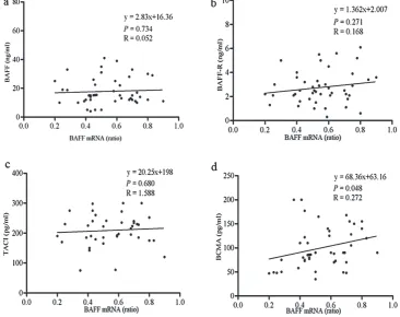
sion and BAFF mRNA expression in the NHL pa-tients (R values were 0.052, 0.168, 1.588, p > 0.05) (Fig. 2 a–c), while there was a correlation be-tween serum BCMA protein expression and BAFF mRNA (r = 0.272, p = 0.048) (Fig. 2 d). These results show that BCMA might play a major role in NHL.
Correlations Between Serum
Protein Expression of BAFF and
Its Receptors and Serum β
2M
Concentration
Knowing that β2M is of some value in predict-ing disease prognosis, the β2M concentration in the NHL patients was assessed and its correlation with BAFF/receptor expression was analyzed. The re-sults showed that there was no significant corre-lation between them (BAFF and β2M, r = 0.014, p > 0.05; BAFF-R and β2M, r = 0.127, p > 0.05;
TACI and β2M, r = 0.003, p > 0.05; BCMA and β2M, r = 0.0005 p > 0.05) (Fig. 3). These results suggest that serum BAFF/receptor protein expres-sion may not be related to the prognosis of NHL.
Correlations Between Serum
Protein Expression of BAFF
and Its Receptors and LDH
Concentration
Knowing that LDH is often used as an indica-tor of disease activity, the LDH concentration in the serum of the NHL patients was assessed. Based on the concentration of LDH, the NHL patients were divided into two groups: The elevated LDH group (310.96 ± 108.00 U/L) and the normal LDH group (42.65 ± 36.00 U/L). BAFF mRNA and se-rum BAFF/receptors protein expression were compared between the two groups (Table 3).
Fig. 2. Correlation between serum BAFF/receptor protein concentration and BAFF mRNA expression
Table 3. Expression levels of serum BAFF/receptors protein and BAFF mRNA in elevated the LDH group and normal LDH group
n LDH
(U/L) BAFF mRNA (ratio) BAFF(ng/mL) BCMA(pg/mL) TACI(pg/mL) BAFF-R(ng/mL)
Normal LDH group 28 142.65 ± 36 0.35 ± 0.02 13.48 ± 0.69 91 ± 6.24 203 ± 31 2.98 ± 0.13 Elevated LDH group 19 310.96 ± 108* 0.68 ± 0.16* 22.45 ± 0.50* 124.36 ± 25* 220 ± 37 3.99 ± 1.07
It was found that there were significant differ-ences in BAFF mRNA and the expression of BAFF and BCMA serum protein between the two groups (t-values were 6.658, 21.06, 7.254, p < 0.05). The results showed that BAFF and BCMA expression might be related to NHL
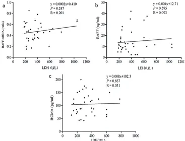
dis-ease activity. However, when linear correlation analyses of LDH concentration with the expres-sions of BAFF mRNA, BAFF and BCMA serum protein were used, there was no significant corre-lation between them (R values were 0.201, 0.093, 0.031, p > 0.05) (Fig. 4).
Fig. 4. Correlation between serum BAFF/receptor protein concentration and LDH concentration
Correlation of Serum BAFF
Protein Concentration
with the Clinicopathological
Stage of NHL
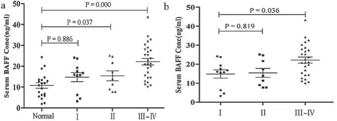
Staging of the NHL patients was performed according to UICC NHL TNM classification, and differences in BAFF expression at different stag-es were compared. It was found that serum BAFF concentration in NHL patients at stages II and III–IV was significantly higher than in the healthy subjects (p < 0.05) (Fig. 5 a). In addition, serum BAFF protein levels in NHL patients at stage III–IV were significantly higher than those in stage I NHL patients (p < 0.05) (Fig. 5 b), indicating that high serum BAFF protein levels in NHL patients may be indicative of an adverse prognosis.
Discussion
B-cell activating factor regulates the survival, proliferation and differentiation of B cells main-ly through binding with the three cell-surface re-ceptors (TACI, BCMA and BAFF-R). In addition, BAFF also promotes the generation of immuno-globulin and regulates the immune response [11]. Studies in recent years have shown that BAFF/re-ceptor protein and/or mRNA expression under-went abnormal changes in patients with systemic lupus erythematosus (SLE), Sjögren’s syndrome (SS), multiple myeloma (MM) and NHL [12, 13]. Therefore, it is of clinical significance to accurately quantify the level of BAFF expression.
The mortality of lymphoma in China is 1.5/100,000, ranking 11th–13th among all malig-nant tumor deaths. NHL accounts for more than 90% of lymphomas. Most NHL originates from extralymphatic tissues and disseminates by skip-ping across adjacent lymph nodes to distal ones, which is the reason there are many cases in which NHL has already spread throughout the body
at the time clinical diagnosis is confirmed. It is therefore necessary to explore the role of BAFF in NHL. It was found in some studies [14] that BAFF mRNA was expressed in the peripheral blood and tumor tissue of NHL patients. Ju et al. [15] found that the expression level of BAFF mRNA in the peripheral blood of NHL patients was no higher than that of normal controls; however, their blood specimens were selected from NHL patients who had received medical treatment. The true relation-ship between BAFF and clinical parameters, and whether BAFF could be used as an accessory clin-ical marker for the diagnosis of NHL, remains un-clear. In the present study, the blood specimens were taken from NHL patients who had not used any medication before admission. RNA was ex-tracted from PBMCs and assessed for BAFF mRNA level by reverse transcription. The results showed that BAFF mRNA was expressed in the peripheral blood of NHL patients, and that the ex-pression level was significantly different from that in the healthy controls, indicating that BAFF may play some role in the development and progres-sion of NHL.
The present study also showed that soluble BAFF/receptor levels were elevated in the serum of NHL patients, but the elevation of receptor lev-els was not linearly correlated with the elevation of the BAFF level, probably because the expres-sion levels varied among the receptors. Serum BAFF/receptor protein concentration was elevat-ed in the NHL patients, indicating that BAFF/re-ceptors play a role in the pathogenesis of NHL. LDH and β2M, two clinical indexes related to NHL, were also assessed. As the LDH level may reflect tumor-bearing status, it could be used as an index for monitoring the therapeutic effect and prognosis. β2M is a variable for predicting the sur-vival of NHL patients. The results of the present study showed that there was no significant corre-lation between BAFF, TACI, BAFF-R protein lev-els and serum LDH and β2M levels in the NHL patients. However, there was a correlation
tween serum BCMA protein expression and BAFF mRNA, showing that BCMA might play a major role in NHL. In addition, the BAFF serum protein concentration of stage II and III–IV NHL patients was significantly higher than in the healthy sub-jects, indicating that increased serum BAFF pro-tein concentration might be indicative of an ad-verse prognosis, and could be used as a marker of clinical NHL staging. In short, BAFF/receptors may have significant impact on the auxiliary diag-nosis and progdiag-nosis of NHL.
In summary, the results of this study showed that serum BAFF/receptor protein concentration and BAFF/mRNA expression level in the PBMCs of NHL patients underwent abnormal changes, in-dicating that BAFF/receptors may play some role in the pathogenesis of NHL. In the study, the BAFF/ receptor protein level and BAFF mRNA level in the PBMCs and serum of NHL patients were as-sessed. More experimental and clinical studies are needed to clarify their actual roles in the progres-sion, prognosis and medical treatment of NHL.
References
[1] Campo E, Swerdlow SH, Harris NL, Pileri S, Stein H, Jaffe ES: The 2008 WHO classification of lymphoid neo-plasms and beyond: Evolving concepts and practical applications. Blood 2011, 117, 5019–5032.
[2] Freeman MD, Kohles SS: Plasma levels of polychlorinated biphenyls, non-Hodgkin lymphoma, and causation. J Environ Public Health 2012, 258981.
[3] Cabanillas F: Non-Hodgkin’s lymphoma: The old and the new. Clin Lymphoma Myeloma Leuk 2011, 11, 87–90.
[4] Nogai H, Dorken B, Lenz G: Pathogenesis of non-Hodgkin’s lymphoma. J Clin Oncol 2011, 29, 1803–1811.
[5] Moore PA, Belvedere O, Orr A, Pieri K, LaFleur DW, Feng P, Soppet D, Charters M, Gentz R, Parmelee D, Li Y, Galperina O, Giri J, Roschke V, Nardelli B, Carrell J, Sosnovtseva S, Greenfield W, Ruben SM, Olsen HS, Fikes J, Hilbert DM: BLyS: Member of the tumor necrosis factor family and B lymphocyte stimulator. Science 1999, 285, 260–263.
[6] Hong SD, Reiff A, Yang HT, Migone TS, Ward CD, Marzan K, Shaham B, Phei WC, Garza J, Bernstein B, Stohl W: B lymphocyte stimulator expression in pediatric systemic lupus erythematosus and juvenile idiopathic arthritis patients. Arthritis Rheum 2009, 60, 3400–3409.
[7] Ju S, Wang Y, Ni H, Wang X, Jiang P, Kong X, Zhong R: Correlation of expression levels of BLyS and its receptors with multiple myeloma. Clin Biochem 2009, 42, 387–399.
[8] Mariette X, Roux S, Zhang J, Bengoufa D, Lavie F, Zhou T, Kimberly R: The level of BLyS (BAFF) correlates with the titre of autoantibodies in human Sjogren’s syndrome. Ann Rheum Dis 2003, 62, 168–171.
[9] Sun J, Lin Z, Li Y, Shen BF: B lymphocyte stimulator: A new target for treating B cell malignancies. Chin Med J (Engl) 2008, 121, 1319–1323.
[10] Lied GA, Berstad A: Functional and clinical aspects of the B-cell-activating factor (BAFF): A narrative review. Scand J Immunol 2011, 73, 1–7.
[11] Mackay F, Schneider P, Rennert P, Browning J: BAFF and APRIL: A tutorial on B cell survival. Annu Rev Immunol 2003, 21, 231–264.
[12] Pers JO, Daridon C, Devauchelle V, Jousse S, Saraux A, Jamin C, Youinou P: BAFF overexpression is associated with autoantibody production in autoimmune diseases. Ann N Y Acad Sci 2005, 1050, 34–39.
[13] Jiang P, Yueguo W, Huiming H, Hongxiang Y, Mei W, Ju S: B-Lymphocyte stimulator: A new biomarker for multiple myeloma. Eur J Haematol 2009, 82, 267–276.
[14] Novak AJ, Grote DM, Stenson M, Ziesmer SC, Witzig TE, Habermann TM, Harder B, Ristow KM, Bram RJ, Jelinek DF, Gross JA, Ansell SM: Expression of BLyS and its receptors in B-cell non-Hodgkin lymphoma: Correlation with disease activity and patient outcome. Blood 2004, 104, 2247–2253.
[15] Shaoqing J, Hongbing N, Yueguo W, Qin Z, Yihong H, Jinhong Z, Caiwang L, Xiantao K, Renqian Z:
Correlation of expression levels of serum BLyS and APRIL with non-Hodgkin lymphoma. Chinese J Immunol 2007, 23, 563–566.
Address for correspondence:
Shaoqing Ju
Laboratory Medicine Center
Affiliated Hospital of Nantong University Nantong 226001
China
E-mail: jsq814@hotmail.com Tel: +86 0513 850 521 03
Conflict of interest: None declared Received: 26.04.2014